AN IN VIVO BLOOD MICROSAMPLING DEVICE FOR PHARMACOKINETIC...
4
AN IN VIVO BLOOD MICROSAMPLING DEVICE FOR PHARMACOKINETIC APPLICATIONS Tao Li *1 , Adam Barnett 1 , Deqing Xiao 2 , Min Zhong 2 , and Yogesh B. Gianchandani 1 1 Engineering Research Center for Wireless Integrated Microsystems, University of Michigan, Ann Arbor, USA 2 Pharmacokinetics, Dynamics, and Metabolism Department, Pfizer Corporation, Ann Arbor, USA Abstract: This paper describes an in vivo microsystem for automated blood microsampling from a lab mouse for pharmacokinetic (PK) studies. Intended to be mounted as a “backpack” on a mouse, it uses a micro-needle, reservoir, and an actuator. It is designed to suddenly prick the animal for a time-point sample, minimizing the potential impact of stress-related endocrine changes. The blood is collected by the 33-gauge micro-needle (210 μm OD) into the ≈1 μL micro-electro-discharge machined steel reservoir. The small volume enables repeated sampling, permitting a full PK profile to be obtained from a single mouse. The voice coil actuator provides a peak force of ≈170 mN, which can easily pierce the needle through mouse skin. The device was tested for blood vessel sampling using a mock vessel with adjustable pressure, and the reservoir was filled in <0.15 sec by a combination of the capillary effect and blood pressure. For interstitial fluid sampling, the capillary effect of the needle inner surface was enhanced by electropolishing. Blood from fresh bovine tissue was collected into the reservoir to simulate interstitial fluid sampling. Keyword: Blood Microsampling, Pharmacokinetic Study, Capillary Effect, Micro Electro-Discharge Machining * Corresponding author: 1301 Beal Ave., Ann Arbor, MI, 48109, USA; Tel: (734) 647-2040, Fax: (734) 763-9324. E-mail: [email protected] I. INTRODUCTION Pharmacokinetics (PK) is the study of how the body transports and eliminates drugs, and is essential to pharmaceutical research. PK studies involve administering a drug to a lab animal and withdrawing blood samples over time. The samples are then analyzed to quantify the drug and metabolite concentration. Commercially-available in vivo sampling systems (e.g. AccuSampler ® ) use surgically-implanted catheters to obtain blood samples from a lab animal. The catheter is connected to a processing system that automatically withdraws blood at set time points. Such systems require the animal to be tethered, and are known to cause stress-related endocrine changes [1]. Further, the need for sample transfers and tube flushing set a lower limit of ≈100 μL on the sample size that must be withdrawn. This relatively large minimum sample size substantially increases the number of mice that must be sacrificed during a PK study. A 20 g transgenic mouse has a total blood volume of only 1.7 mL. A single 100 μL sample is nearly 6% of the total blood volume, making such a sample very detrimental to the mouse. This has a multiplicative effect: at least 3 mice are sacrificed for each time sample to account for variability between specimens. Thus, an eight-time- point study typically consumes 24 mice. A technology that provides isolated 1-10 μL samples at each time point would enable obtaining a full PK profile from a single mouse, which is desirable scientifically and saves animal resources. This paper proposes a device that can sample blood from a lab mouse in the ≈1 μL range. The device is designed to be worn as a “backpack” that suddenly pricks the animal with a micro-needle. The blood sample is drawn through the needle and into a biocompatible steel reservoir made by micro-electro- discharge machining (μEDM). A voice coil actuator is triggered to provide the piercing action into the skin. The force that the actuator can provide is evaluated by experiments. The device is tested for blood vessel sampling using a mock vessel with adjustable internal pressure. Interstitial fluid sampling by the enhanced capillary effect is also tested using fresh bovine tissue. II. DESIGN & FABRICATION The scheme for in-vivo microsampling of mouse blood is shown in Fig. 1. A miniaturized “backpack” positions the microsampling device at a target spot on a lab mouse. The sampling device consists of a micro- needle, reservoir, and an actuator. The actuator can be triggered to suddenly prick the animal for a single time- point sample without noticeable stress to the mouse. The structure of the prototype device is shown in Figs. 2 and 3. A steel micro-needle is mounted on a miniature blood reservoir with a target volume of 1-10 μL. An opening channel formed on the wall of the reservoir permits air to leave the cavity during sampling, or interface with additional components for Fig. 1 : System diagram showing application scheme of the blood microsampling device for pharmacokinetic studies.
Transcript of AN IN VIVO BLOOD MICROSAMPLING DEVICE FOR PHARMACOKINETIC...

AN IN VIVO BLOOD MICROSAMPLING DEVICE FOR PHARMACOKINETIC APPLICATIONS
Tao Li*1, Adam Barnett1, Deqing Xiao2, Min Zhong2, and Yogesh B. Gianchandani1 1Engineering Research Center for Wireless Integrated Microsystems, University of Michigan, Ann Arbor, USA
2Pharmacokinetics, Dynamics, and Metabolism Department, Pfizer Corporation, Ann Arbor, USA Abstract: This paper describes an in vivo microsystem for automated blood microsampling from a lab mouse for pharmacokinetic (PK) studies. Intended to be mounted as a “backpack” on a mouse, it uses a micro-needle, reservoir, and an actuator. It is designed to suddenly prick the animal for a time-point sample, minimizing the potential impact of stress-related endocrine changes. The blood is collected by the 33-gauge micro-needle (210 µm OD) into the ≈1 µL micro-electro-discharge machined steel reservoir. The small volume enables repeated sampling, permitting a full PK profile to be obtained from a single mouse. The voice coil actuator provides a peak force of ≈170 mN, which can easily pierce the needle through mouse skin. The device was tested for blood vessel sampling using a mock vessel with adjustable pressure, and the reservoir was filled in <0.15 sec by a combination of the capillary effect and blood pressure. For interstitial fluid sampling, the capillary effect of the needle inner surface was enhanced by electropolishing. Blood from fresh bovine tissue was collected into the reservoir to simulate interstitial fluid sampling. Keyword: Blood Microsampling, Pharmacokinetic Study, Capillary Effect, Micro Electro-Discharge Machining
* Corresponding author: 1301 Beal Ave., Ann Arbor, MI, 48109, USA; Tel: (734) 647-2040, Fax: (734) 763-9324. E-mail: [email protected]
I. INTRODUCTION Pharmacokinetics (PK) is the study of how the
body transports and eliminates drugs, and is essential to pharmaceutical research. PK studies involve administering a drug to a lab animal and withdrawing blood samples over time. The samples are then analyzed to quantify the drug and metabolite concentration.
Commercially-available in vivo sampling systems (e.g. AccuSampler®) use surgically-implanted catheters to obtain blood samples from a lab animal. The catheter is connected to a processing system that automatically withdraws blood at set time points. Such systems require the animal to be tethered, and are known to cause stress-related endocrine changes [1]. Further, the need for sample transfers and tube flushing set a lower limit of ≈100 µL on the sample size that must be withdrawn. This relatively large minimum sample size substantially increases the number of mice that must be sacrificed during a PK study. A 20 g transgenic mouse has a total blood volume of only 1.7 mL. A single 100 μL sample is nearly 6% of the total blood volume, making such a sample very detrimental to the mouse. This has a multiplicative effect: at least 3 mice are sacrificed for each time sample to account for variability between specimens. Thus, an eight-time-point study typically consumes 24 mice. A technology that provides isolated 1-10 µL samples at each time point would enable obtaining a full PK profile from a single mouse, which is desirable scientifically and saves animal resources.
This paper proposes a device that can sample blood from a lab mouse in the ≈1 µL range. The device is designed to be worn as a “backpack” that suddenly
pricks the animal with a micro-needle. The blood sample is drawn through the needle and into a biocompatible steel reservoir made by micro-electro-discharge machining (µEDM). A voice coil actuator is triggered to provide the piercing action into the skin. The force that the actuator can provide is evaluated by experiments. The device is tested for blood vessel sampling using a mock vessel with adjustable internal pressure. Interstitial fluid sampling by the enhanced capillary effect is also tested using fresh bovine tissue.
II. DESIGN & FABRICATION The scheme for in-vivo microsampling of mouse
blood is shown in Fig. 1. A miniaturized “backpack” positions the microsampling device at a target spot on a lab mouse. The sampling device consists of a micro-needle, reservoir, and an actuator. The actuator can be triggered to suddenly prick the animal for a single time-point sample without noticeable stress to the mouse.
The structure of the prototype device is shown in Figs. 2 and 3. A steel micro-needle is mounted on a miniature blood reservoir with a target volume of 1-10 µL. An opening channel formed on the wall of the reservoir permits air to leave the cavity during sampling, or interface with additional components for
����������� �����������
���������
�
Fig. 1: System diagram showing application scheme of the blood microsampling device for pharmacokinetic studies.
in-situ sample analysis during PK studies. The reservoir is covered with a glass lid that enables visual observation of the sample. A rare earth magnet is bonded to the glass for easy attachment or removal of the reservoir from a commercial electromagnetic voice coil actuator (H2W Technologies, Inc., USA). This magnet also enhances the actuator performance by increasing the strength of its magnetic field. The coil of the actuator is attached by an adjustment screw to a Macor® ceramic housing, which also provides a movement guide for the actuator magnet. With the proper needle and housing dimensions, a 2 mm deflection of the actuator can drive the needle 1 mm into the mouse through a hole in the ceramic housing. This depth can reach the mouse jugular in certain locations for vessel blood sampling [2]. The blood is subsequently transported into the reservoir by a combination of the capillary effect and blood pressure.
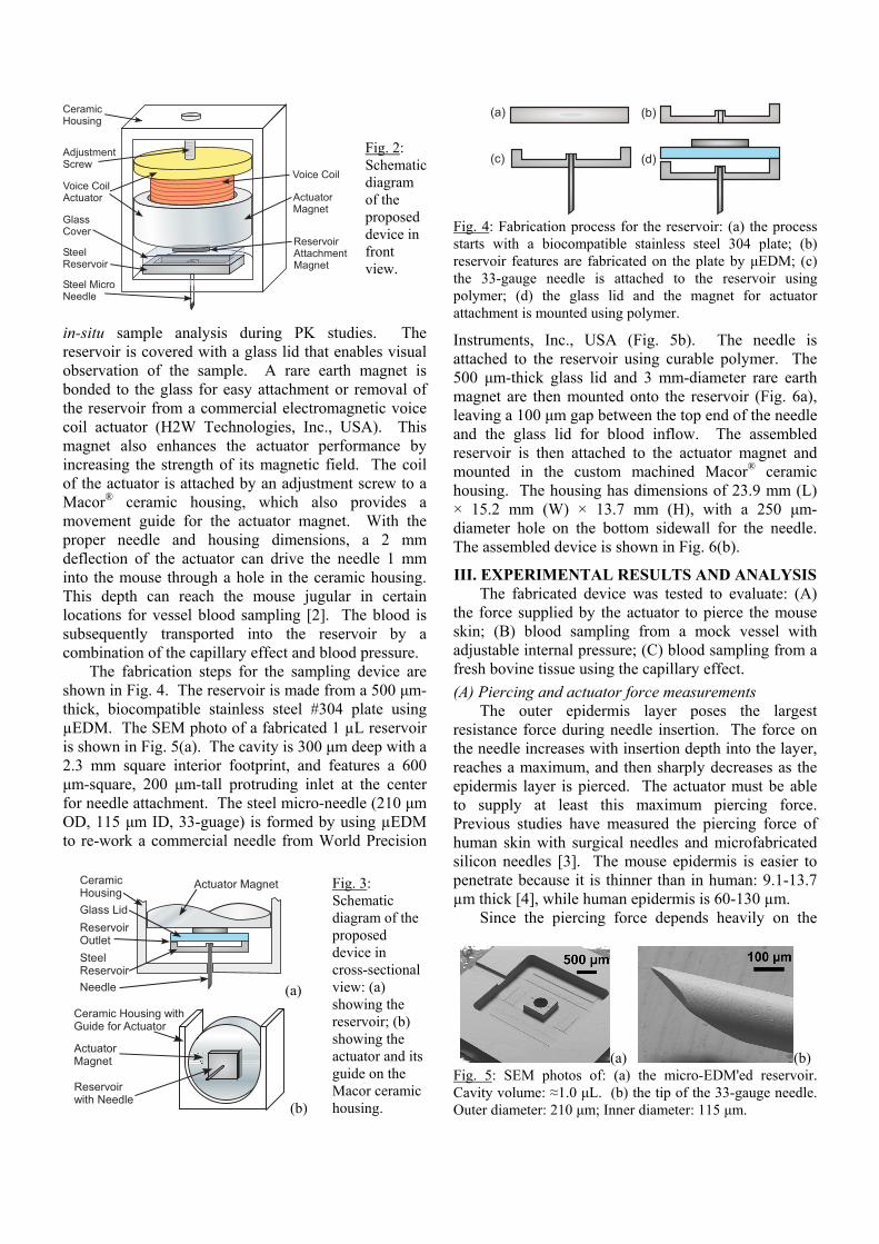
The fabrication steps for the sampling device are shown in Fig. 4. The reservoir is made from a 500 μm-thick, biocompatible stainless steel #304 plate using µEDM. The SEM photo of a fabricated 1 µL reservoir is shown in Fig. 5(a). The cavity is 300 μm deep with a 2.3 mm square interior footprint, and features a 600 μm-square, 200 μm-tall protruding inlet at the center for needle attachment. The steel micro-needle (210 μm OD, 115 μm ID, 33-guage) is formed by using µEDM to re-work a commercial needle from World Precision
Instruments, Inc., USA (Fig. 5b). The needle is attached to the reservoir using curable polymer. The 500 μm-thick glass lid and 3 mm-diameter rare earth magnet are then mounted onto the reservoir (Fig. 6a), leaving a 100 μm gap between the top end of the needle and the glass lid for blood inflow. The assembled reservoir is then attached to the actuator magnet and mounted in the custom machined Macor® ceramic housing. The housing has dimensions of 23.9 mm (L) × 15.2 mm (W) × 13.7 mm (H), with a 250 μm-diameter hole on the bottom sidewall for the needle. The assembled device is shown in Fig. 6(b).
III. EXPERIMENTAL RESULTS AND ANALYSIS The fabricated device was tested to evaluate: (A)
the force supplied by the actuator to pierce the mouse skin; (B) blood sampling from a mock vessel with adjustable internal pressure; (C) blood sampling from a fresh bovine tissue using the capillary effect. (A) Piercing and actuator force measurements
The outer epidermis layer poses the largest resistance force during needle insertion. The force on the needle increases with insertion depth into the layer, reaches a maximum, and then sharply decreases as the epidermis layer is pierced. The actuator must be able to supply at least this maximum piercing force. Previous studies have measured the piercing force of human skin with surgical needles and microfabricated silicon needles [3]. The mouse epidermis is easier to penetrate because it is thinner than in human: 9.1-13.7 µm thick [4], while human epidermis is 60-130 µm.
Since the piercing force depends heavily on the
����������������
�������������
����������������
��� ���������
����������
�� ��� ������!
�������������
���������
������������" ����������
Fig. 2: Schematic diagram of the proposed device in front view.
�������������
�������������
��������#�����
������
���������
��� ���������
(a)
Fig. 3: Schematic diagram of the proposed device in cross-sectional view: (a) showing the reservoir; (b) showing the actuator and its guide on the Macor ceramic housing.
��� ����������!��"�������$��������
�������������
��������!��"������� (b)
(a) (b) Fig. 5: SEM photos of: (a) the micro-EDM'ed reservoir. Cavity volume: ≈1.0 μL. (b) the tip of the 33-gauge needle. Outer diameter: 210 μm; Inner diameter: 115 μm.
%�& %�&
%& %�&
Fig. 4: Fabrication process for the reservoir: (a) the process starts with a biocompatible stainless steel 304 plate; (b) reservoir features are fabricated on the plate by μEDM; (c) the 33-gauge needle is attached to the reservoir using polymer; (d) the glass lid and the magnet for actuator attachment is mounted using polymer.

needle tip size and bevel shape, an experiment was carried out to characterize the piercing force of the 33-gauge micro-needle with 25° bevel into silicon rubber phantoms. Silicon rubber is an artificial skin material with mechanical properties similar to human skin [5]. It can be used to establish an upper bound on the piercing force. The insertion force into silicone rubber was measured with a load cell and found to be ≈7.23 mN for a piercing depth of 15 µm (the thickness of the mouse epidermis). This value is comparable to that estimated using empirical equations from [3], and can be used as a design guide for the actuator.
The force supplied by the commercial voice coil actuator is also measured using a load cell. The results shown in Fig. 7 indicate that the actuator amply exceeds the piercing force requirement. The additional reservoir attachment magnet provides a ≈30% enhancement, reaching ≈170 mN with 360 mA applied. (B) Blood sampling experiment with mock vessel
The experimental setup shown in Fig. 8(a) is built to evaluate the capability of the device to sample blood from a vessel, e.g. the jugular vein. The needle is inserted into a mock vessel with adjustable internal pressure controlled by a hand pump. Due to the weak hydrophilic nature of the inner surface of the needle, the capillary effect cannot work by itself when the vessel pressure is zero, though it has previously been used to sample interstitial fluid using an array of silicon microneedles [6]. Measurements show that a pressure threshold of ≈29.0 mmHg is necessary to permit water
flow through the needle. Once the water reaches the highly hydrophilic glass lid, the reservoir quickly fills by a combination of pressure and the capillary effect. This pressure threshold is much smaller than mouse blood pressure (125 mmHg systolic, 90 mmHg diastolic), so additional surface treatment to enhance the capillary effect is unnecessary for vessel sampling. At mouse blood pressure, it takes <0.15 sec to fill the reservoir. The flow rate through the needle, reservoir, and a pipe of 600 μm ID attached at the reservoir outlet was measured to study the capability of the device to drive other microfluidic components for in-situ sample analysis. The measured flow rate through the whole device is plotted as a function of “vascular” pressure in the mock vessel in Fig. 8(b). The resulting flow rate is adequate for many microfluidic applications. (C) Blood sampling experiment with bovine tissue
Although the device works with the presence of vascular pressure, the pressure threshold is too high for interstitial fluid sampling. The capillary effect of the needle must be enhanced to draw interstitial fluid into the reservoir without blood pressure. One approach is to enhance the hydrophilic property of the inner surface of the needle. This can be done by improving the wettability of the steel through surface treatments [7-9] or SiO2 coating [10]. The electropolishing method discussed in [9] using commercially-available EPS4000 (a solution based on a mixture of phosphoric and sulfuric acids) was tested on a bare stainless steel 304 sample. The water contact angle improved from ≈94° before polishing to ≈34° after polishing, indicating the improved wettability of the electropolished surface. The 33-gauge micro-needle was then electropolished by EPS4000, with the outer surface protected from the
'(
)(
*((
*+(
*,(
*'(
*)(
*-( *.( +*( +/( +0( --( -.(
������% �&
1�
�
�%
�&
+ �������� �������������* �������� �������������
* �������� ����2��"�������+ �������� ����2��"�������
Fig. 7: Measurement results of the supplied force from the voice coil actuator. The actuator force is enhanced by ≈30% with the magnet for reservoir attachment in place.
�����3� ������
3����������
��4�������
����� �����������
(a)
/
*(
*/
+(
+/
(( +/ /( ./ *(( *+/
3������% ��&
1��
!��
���
�%�
�5�
&
������������3�����
�6�����
��������
2"���������7������"���������
3����"���������
(b) Fig. 8: (a) Schematic of testing setup for blood sampling from mock vessel. (b) Results showing measured flow rate vs. applied pressure through the whole device, a polished and an unpolished needle.
(a) (b) Fig. 6: Photos of: (a) the assembled reservoir with glass lid and attached magnet; (b) the full microsampling device. A glass cover can be used to seal the ceramic housing.

solution by a polymer coating. The polishing result is shown in the SEM photos in Fig. 9. The capillary effect was tested and is compared in Table 1. After polishing, the capillary effect raises the water level in a vertical needle by ≈4 mm and fills a 10 mm horizontal needle in ≈1 seconds, the difference being caused by gravity. The 4 mm height is enough to fill the needle in the present design. To evaluate the effect of polishing on flow rate, a 25 mm-long needle was tested with the setup in Fig. 8(a) before and after electropolishing. The results shown in Fig. 8(b) have been scaled to match the 4 mm length of the needle in the sampling device, and indicate a clear increase in flow rate with the polished inner surface and increased inner diameter of the needle.
Table 1: Capillary effect of the 33-guage needle Unpolished PolishedWater height in vertical needle ≈0 ≈4 mm Time to fill a 10 mm-long horizontal needle >3 sec ≈1 sec
To explore the possibility of sampling interstitial fluid rather than blood from vasculature, the device was also tested with a bovine tissue sample. Fig. 10 shows the reservoir is almost completely flooded, leaving a small bubble of air.
IV. CONCLUSIONS An in vivo device for automated blood
microsampling from a lab mouse has been proposed for PK studies. The device can be triggered to suddenly
prick the animal, minimizing the potential impact of stress-related endocrine changes. The steel reservoir made by µEDM has a small volume of ≈1 µL, reducing the minimum sample size and the number of mice required during a PK study. The device successfully performed sampling from a mock vessel with internal pressure, and also collected fluid from fresh bovine tissue by the capillary effect enhanced by electropolishing, suggesting its capability to perform interstitial fluid sampling. Further experiments with live lab animals will be pursued in a future effort.
ACKNOWLEDGEMENT This work was supported primarily by the Engineering
Research Centers Program of the National Science Foundation under Award Number EEC-9986866.
REFERENCES [1] F. Royo, N. Björk, H.-E. Carlsson, S. Mayo, and J. Hau, “Impact of chronic catheterization and automated blood sampling (Accusampler) on serum corticosterone and fecal immunoreactive corticosterone metabolites and immunoglobulin A in male rats,” J. Endocrinology, vol. 180, pp. 145-153, 2004 [2] J. Hoff, “Methods of blood collection in the mouse,” Lab Animal, 29(10), pp. 47-53, 2000 [3] S.P. Davis, B.J. Landis, Z.H. Adams, M.G. Allen, and M.R. Prausnitz, “Insertion of microneedles into skin: measurement and prediction of insertion force and needle fracture force", J. Biomechanics, vol. 37, pp. 1155-63, 2004 [4] L. Azzi, M. El-Alfy, C. Martel, and F. Labrie, “Gender differences in mouse skin morphology and specific effects of sex steroids and Dehydroepiandrosterone,” J. Investigative Dermatology, 124(1), pp. 22-27, January 2005 [5] O.A. Shergold and N.A. Fleck, “Experimental investigation into the deep penetration of soft solids by sharp and blunt punches, with application to the piercing of skin,” J. Biomech. Eng., 127(5), pp. 838-848, 2005 [6] E.V. Mukerjee, S.D. Collins, R.R. Isseroff, and R.L. Smith, “Microneedle array for transdermal biological fluid extraction and in situ analysis,” Sensors and Actuators A (Physical), A114(2-3), pp. 267-75, Sept. 2004 [7] M. Mantel and J.P. Wightman, “Influence of the surface chemistry on the wettability of stainless steel,” Surface and Interface Analysis, vol. 21, pp. 595-605, 1994 [8] H. P. Jennissen, “Ultra-hydrophilic transition metals as histophilic biomaterials,” Macromol. Symp., vol. 225, pp. 43-69, 2005 [9] A. Bhuyan, B. Gregory, H. Lei, S.Y. Yee, and Y.B. Gianchandani, “Pulsed and DC electropolishing of stainless steel for stents and other other devices,” in Proc. IEEE Conf. Sensors, Irvine, CA, 2005, pp. 314-7 [10] O. Santos, et al., "Modified stainless steel surfaces targeted to reduce fouling - surface characterization," J. Food Eng., 64(1), pp. 63-79, Sept. 2004
(a)
Fig. 10: Experiments with bovine tissue to explore the possibility of sampling interstitial fluid. (a) Photo of the device placed on the tissue sample for experiment. (b) Photo of the obtained blood sample in the reservoir with a small air bubble.
(b)
(a) (b) Fig. 9: SEM photos of the needle inner surface: (a) before electropolishing; (b) after electropolishing using EPS4000. A slot opening was cut in the needle tube using µEDM before taking the SEM photos.