Alexis Collins, DO October 15, 2011. cc: Weakness bilateral lower extremities with multiple areas of...
33
Alexis Collins, DO October 15, 2011
-
Upload
lisbeth-stackhouse -
Category
Documents
-
view
214 -
download
1
Transcript of Alexis Collins, DO October 15, 2011. cc: Weakness bilateral lower extremities with multiple areas of...
Alexis Collins, DOOctober 15, 2011

cc: Weakness bilateral lower extremities with multiple areas of skin necrosis of both lower extremities
G.C. is a 50 y/o WM with end stage renal disease and history of nephrotic syndrome of unknown etiology, who was changed from peritoneal to hemodialysis.
G.C. gained a significant amount of weight while on peritoneal dialysis secondary to anasarca, especially of lower extremities
G.C. has been experiencing leg weakness on standing up with unsteadiness of gait for last 3 months and is now unable to ambulate, requiring assistance with transferring from bed to chair and is now confined to a wheelchair

At first presentation of patient’s lower extremity weakness it was felt that symptoms were related to his history of painful diabetic peripheral neuropathy which was being managed with Neurontin and degenerative disk disease with mild bulging of the disk and mild to moderate spinal stenosis at L4 and L5
Evaluated by neurologist in Grundy, VA for the leg weakness who added Cymbalta, but symptoms did not improve
2nd Consultation with another neurologist in Bristol, TN believed symptoms may be due to Neurontin toxicity, Neurontin was discontinued but still no improvement

To further investigate the leg weakness and pain, patient underwent muscle biopsy that was negative
Consultation by 3rd neurologist was concerned about muscular etiology such as mild myositis versus myopathy, with superimposed diabetic polyneuropathy
Patient went through inpatient rehabilitation that resulted in only minimal improvement

On peritoneal dialysis G.C. Also developed severe hyperphosphatemia with
serum phosphorous levels >11 due to poor compliance with diet, phosphate binder therapy, and lower efficiency of peritoneal dialysis
Calcium phosphorous product significantly elevated and serum intact PTH >500
G.C. was converted from peritoneal dialysis to hemodialysis

In the days following G.C. developed areas of skin necrosis with eschar in the folds of the skin of lower extremities and an ulcerating lesion with secondary infection over medial aspect of his calf
Patient developed hard edema of his lower extremities, especially medial aspects of his thighs and calvesFeet were warm and not cyanoticAreas of skin necrosis with black eschar were noted
in Right thigh skin folds and Left popliteal areaUlcerated area of skin necrosis medial aspect Left
calf Area of erythema developing over the Right thigh
medial aspect

PMH: ESRD, undefined etiology
Assumed secondary to diabetic nephropathy &/or secondary to focal glomerular sclerosis due to morbid obesity
Nephrotic range proteinuria as high as 11 gRecently converted from peritoneal dialysis to
hemodialysisSecondary hyperparathyroidism
Severe hyperphosphatemia and markedly increased calcium, phosphorous products while on peritoneal dialysis
Arterial calcifications noted on radiographic examination of knees

PMH:Anemia of chronic renal failure
Refractory to erythropoietin stimulating therapyInsulin-requiring type 2 DM, diagnosed in 1997
Peripheral neuropathyNephrotic range proteinuria
Hypertension, long-standingDyslipidemiaGERDSuspected OSABilateral lower extremity weakness believed to
be due to severe myopathy, most likely diabetic

PMH:Febrile nonhemolytic transfusion reaction,
February 2010Streptococcal pharyngitis, April 2010Morbid obesity with BMI greater than 40

PSH:Incision and drainage of abscess, Left groinPlacement of tunneled hemodialysis catheter,
Right internal jugular vein by Dr Ajkay in November 2010
Placement of peritoneal dialysis catheter by laparoscopic assistance, by Dr. Grady Stephens on October 14, 2009
Right quadriceps muscle biopsy by Dr Oon May 18, 2010
Placement of Tunneled hemodialysis catheter, June 2010

Allergies: None KnownMeds:
Amlodipine 10 mg dailyApidra 15 U SQ qacLantus 40 U SQ at bedtimeAtenolol 100 mg daily Colace 100 mg BIDDiovan 320 mg dailyLipitor 80 mg bedtimeLortab 10/500 q6h PRNNephro-Vite 1 tab dailyPhosLo 667 mg 3 tablets with each mealProstat 30 mL PO TIDProtonix 40 mg dailyTekturna 300 mg daily

FH: HTN and heart disease in his parents. Father was also diabetic
SH: Married and lives at home with his spouse, who is a LPN. He chews tobacco occasionally but does not smoke. No history of alcohol or drug abuse. He worked in the mines for ~24 years. He was a high school football player

ROS: Chronically overweight, increased weight while
on peritoneal dialysis, however has decreased by over 50 lbs since the conversion to hemodialysis
Chronic parasthesias in lower extremities distally, now significant weakness of his lower extremities with limited functioning, able to stand up only for few minutes
Minimal urine output
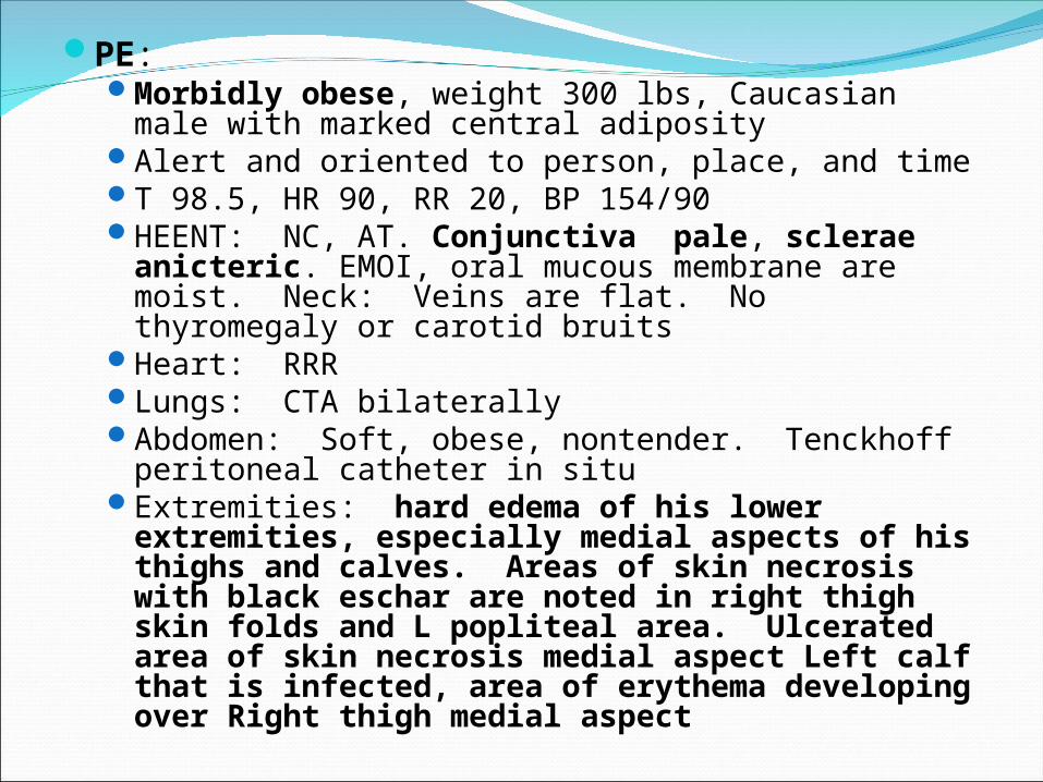
PE:Morbidly obese, weight 300 lbs, Caucasian male
with marked central adiposityAlert and oriented to person, place, and timeT 98.5, HR 90, RR 20, BP 154/90HEENT: NC, AT. Conjunctiva pale, sclerae
anicteric. EMOI, oral mucous membrane are moist. Neck: Veins are flat. No thyromegaly or carotid bruits
Heart: RRRLungs: CTA bilaterallyAbdomen: Soft, obese, nontender. Tenckhoff
peritoneal catheter in situExtremities: hard edema of his lower
extremities, especially medial aspects of his thighs and calves. Areas of skin necrosis with black eschar are noted in right thigh skin folds and L popliteal area. Ulcerated area of skin necrosis medial aspect Left calf that is infected, area of erythema developing over Right thigh medial aspect


Labs: BMP: Sodium 140, potassium 4.0, Chloride
97, CO2 34, BUN 19, Creatinine 4.1, Glucose 114, Calcium 8.7, GFR 16.5
Phosphorous: 3.9CBC: WBC 11.4, Hgb 8.9, Hct 27.4, Plt 239Aerobic culture Wound L leg—Morganella
morganii, PTH 333

Radiology:CXR: Resolution of CHFCT A&P: Cholelithiasis, Atheroscleortic vascular
disease, mild periaortic adenopathyNM Bone Imaging: Foci of increased activity
within the posterior and posterolateral left ribs as described likely representing old fractures.
X-ray Right Knee: Mild osteoarthritic changes involving the lateral joint compartment of the knee. Extensive arterial calcification noted.
X-ray Left knee: Mild osteoarthritic changes involving the lateral joint compartment of the knee. Extensive arterial calcification noted.
Assessment:Necrotizing skin lesion and hard edema of
lower extremities secondary to CalciphylaxisAnemia of CRF, on Epogen
Refractory to Epogen therapy, most likely due to chronic inflammation
Secondary hyperparathyroidismRecent intact PTH 333 and serum phosphorous
levels improvedESRD, on hemodialysisType 2 DM, insulin requiring

CalciphylaxisRare and serious disorder Systemic medial calcification of the arterioles
that leads to ischemia and subcutaneous necrosisOne of several types of extra-osseous
calcification (which also include intimal, medial, and valvular calcifications) may occur in ESRD patients
Passive mineralization of serum calcium and phosphate crystals
Syndrome of vascular calcification, thrombosis, and skin necrosis
Almost exclusively in patients with Stage 5 chronic kidney disease (occurs in 1 to 4% of affected patients)
Pathogenesis:Systemic anaphylactic reaction Pathogenesis is complex and yet to be
determinedAbnormalities in mineral metabolism are
thought to be a passive precipitation that predispose ESRD patients to vascular and soft tissue calcifications
Reduction in the arteriolar blood flow is a consequence of intimal fibrosis associated with the calcifications

Pathogenesis:Vascular endothelial injury and dysfunction
result in cutaneous arterioler narrowing and hypercoagulable state producing frank tissue infarction
Hyperparathyroidism, active vitamin D administration, hyperphosphatemia, and an elevated plasma calcium x phosphate product are implicated in the genesis of calciphylaxis

Patients at higher risk:Females undergoing dialysis have higher risk of
developing calciphylaxisESRD patients with obesity (BMI>30) may lead to
excess stress on dermal and hypodermal arterioles, resulting in focally dystrophic calcification on arterioles
Increase in phosphorous levels among those with ESRD Administration of certain medicines to ESRD patients
including: warfarin, calcium based binders and vitamin D analogues, and systemic corticosteroids
Presence of AI disorders in ESRD patientsNon-uremic calciphylaxis is noted in patients with
primary hyperthyroidism, breast cancer (treated with chemotherapy), liver cirrhosis (due to alcohol abuse), cholangiocarcinoma, Crohn's disease, rheumatoid arthritis (RA), and systemic lupus erythematosus (SLE).

Clinical Manifestations:Areas of excruciatingly painful ischemic necrosis
that usually develop on areas with greatest adiposity including abdomen, buttock, and thigh
Violaceous, painful, plaque-like subcutaneous nodules
Initial purpuric plaques and nodules subsequently progress to ischemic/necrotic ulcers with eschars that often become superinfected
Ischemic changes are reflection of medial arteriolar calcification and subintimal fibrosis leading to luminal narrowing, diminished blood flow, hypoxia, and procoagulant state
When vascular thrombosis is advanced, the result is necrotic ulcer with eschar that is clinically evident

Laboratory and radiographic manifestations:No specific laboratory findingsSome patients will have elevated PTH,
phosphorous, calcium, and calcium-phosphorous product
Plain radiographs, high resolution CT scans, and mammographic techniques can distinguish calcium from surrounding non-calcium containing radiolucent tissues

Evaluation and Diagnosis:Suspect in patients with skin lesions characterized
by painful, nonulcerating subcutaneous nodules or plaques, nonhealing ulcers, and/or necrosis, which are most commonly present in the thigh and areas of increased adiposity
Also may be increasing Ca X P product and PTH levels in ESRD patients especially in setting of medication noncompliance &/or concurrent warfarin use
Sensitivity and specificity of biopsy has not been determined
3-phase bone scan may be useful in diagnosis: positive scan suggests presence of calcifications in subcutaneous nodules or non-ulcerating lesions in viable tissue

Prevention and Treatment:HIGH mortality No controlled prospective studies that compare
different treatment strategiesPlan should include:
Aggressive program of wound care and adequate pain control
Avoidance of local tissue trauma, including subcutaneous injections
Correction of underlying abnormalities in plasma calcium & phosphorous concentrations, lowering Ca X P product below 55 mg2/dL2 in ESRD patients

Treatment:Mainly supportive1st step: normalize serum calcium and
phosphate concentrations with dietary modification and phosphate and calcium binders
If calcium and phosphate levels remain high, consider parathyroidectomy, especially if parathyroidism in present

Treatment:Increase sessions of renal replacement therapy
in ESRD patients with inadequate clearanceRole of surgical debridement controversial
because removal of necrotic tissue exposes vital subcutaneous tissue to bacteria and increases risk of sepsis
Sodium Thiosulfate: showed reduction of pain and inflammation and improved healing of ulcerative lesions

Prognosis:Lethal disease that carries high morbidity and
mortalitySporadic so prospective studies regarding exact
outcomes associated with calciphylaxis do not exist
Response to any therapeutic regimen appears to be poor and there remains high mortality regardless of efforts to treat the underlying disordered mineral metabolism
Infection primary cause of high mortality (up to 58% in one report)
Ulceration has high mortality >80%Estimated one-year survival 45.8%

Resources:Wikipedia, Calciphylaxis http://
en.wikipedia.org/wiki/CalciphylaxisNoah S. Scheinfield, MD, JD Skin Disorders in
Older Adults: Cutaneous Ulcers, Part 1 Counsultant Vol. 51 No. 8 August 2011, Pages 565-570
Peter W Santos, DO, J Edward Hartle, II, MD, L Darryl Quarles, MD Calciphylaxis http://www.uptodate.com/contents/calciphylaxis?source=search_result&search=calciphylaxis&selectedTitle=1%7E14