Core Muscle Activation During Swiss Ball and Traditional Abdominal ...

DIFFERENCES IN MUSCLE ACTIVATION IN THE LOWER EXTREMITIES
WHILE PERFORMING TRADITIONAL SQUATS
AND NON-TRADITIONAL SQUATS
by
Christopher M. Scotten
A thesis
submitted in partial fulfillment
of the requirements for the degree of
Master of Science in Exercise and Sport Studies, Biophysical Studies
Boise State University
August 2010

BOISE STATE UNIVERSITY GRADUATE COLLEGE
DEFENSE COMMITTEE AND FINAL READING APPROVALS
of the thesis submitted by
Christopher Michael Scotten
Thesis Title: Differences in Muscle Activation in the Lower Extremities While
Performing Traditional Squats and Non-Traditional Squats Date of Final Oral Examination: 12 May 2010
The following individuals read and discussed the thesis submitted by student Christopher Michael Scotten, and they also evaluated his presentation and response to questions during the final oral examination. They found that the student passed the final oral examination, and that the thesis was satisfactory for a master’s degree.
Shawn R. Simonson, Ed.D. Chair, Supervisory Committee Lynda Ransdell, Ph.D. Member, Supervisory Committee James R. Moore, MS Member, Supervisory Committee The final reading approval of the thesis was granted by Shawn R. Simonson, Ed.D., Chair of the Supervisory Committee. The thesis was approved for the Graduate College by John R. Pelton, Ph.D., Dean of the Graduate College.

ACKNOWLEDGEMENTS
First, I want to thank my parents for supporting me and continuing to push me to
greater accomplishments, which I would not be able to achieve without them. I would
also like to thank the rest of my family and friends for being there when I needed them.
Next, I want to thank Seth Kuhlman and the GA’s in the Biomechanics lab for thr
use of their space, equipment, and personal time which they sacrificed to help collect,
process, and analyze the data for this study. I would also like to thank Dr. Simonson, Dr.
Ransdell, and Jim Moore for their patience and assistance in editing my thesis. Lastly, I
would like to thank all the participants who volunteered for my study, without them this
could not have been accomplished.
iii

ABSTRACT
Differences in Muscle Activation in the Lower Extremities While Performing Traditional Squats and Non-Traditional Squats
Christopher M Scotten
Purpose: To determine if muscle activation in the lower back and lower extremities differ when performing traditional squats compared to non-traditional (forward center of pressure on foot) squats. The erector spinae, hamstrings, quadriceps, adductor longus, gastrocnemius, and gluteus maximus muscles were monitored for differences in this study. There are several variations of the back squat and each variation may possibly target muscles differently. Determining if non-traditional squats leads to larger erector spinae muscle activation, which in turn may lead to more lower back fatigue and possible lower back injury is a major aim of this study. Participants: Thirteen healthy males (age = 25.15 ± 2.38 yrs, height = 70.35 ± 3.2 in, weight = 174.45 ± 18.35 lbs and body fat = 10.31% ± 2.97%), which have participated in a steady exercise program for at least a year and included a version of the squat exercise in their routine at least once a week, were the participants in this study. Participants could not have sustained a serious knee, back, or ankle injury in order to qualify for this study. Participants were recruited from Boise State University via flyers and word of mouth. Methods: This study consisted of individuals performing traditional squats for one set of ten reps and non-traditional squats for one set of ten reps. Prior to testing, each subject performed maximum voluntary isometric contraction tests for each muscle being monitored (vastus medialis, vastus lateralis, gluteus maximus, bicep femoris, semitendinosus, adductor longus, gastrocnemius, and erector spinae) in order to normalize data collected during the two squatting variations. All testing took place at the biomechanics lab in the Micron Engineering Center at BSU. Statistical Analysis: Data was analyzed using the SPSS statistical software package. An ANOVA with a post hoc test consisting of paired t-tests were used to compare differences in activity between the two squatting techniques. Hypothesis: The gluteus maximus, biceps femoris, and semitendinosus muscle activation will be significantly larger during the traditional squats. The erector spinae and gastrocnemius muscle activation will be significantly larger during the non-traditional squats. The vastus medialis, vastus lateralis, and adductor longus muscle activation will not be significantly different between the two squat variations. Results: The semitendinosus and gastrocnemius muscle activation was significantly larger during the non-traditional squat. The vastus medialis and vastus lateralis muscle activation was significantly larger during the traditional squats. Conclusions: When performing back squats, keeping one’s center of pressure on the heels of their feet will activate the quadriceps to a larger degree than if performing squats while the center of pressure is on one’s toes. Participants claimed their lower back felt more activated during the non-traditional squats; however, the quantitative data did not support this claim.
iv

TABLE OF CONTENTS
ACKNOWLEDGEMENTS…………………………………………………………… iii
ABSTRACT…………………………………………………………………………… iv
LIST OF TABLES…………………………………………………………………….. viii
LIST OF FIGURES……………………………………………………………………. ix
CHAPTER ONE: INTRODUCTION ….……………………...……………………... 1
Hypotheses ……………………………………………………………………. 4
Limitations ……………………………………………………………………. 5
Delimitations ………………………………………………………………….. 6
Definitions …………………………………………………………………….. 6
CHAPTER TWO: REVIEW OF LITERATURE …………………………..………... 8
Analysis of Joint and Ligament Forces ……………………………………….. 8
EMG Muscle Activity ………………………………………………………... 11
Lower Back Load and Activity ………………………………………………. 14
CHAPTER THREE: METHODS ………………………………..………………….. 17
Subjects ………………………………………………………………………. 18
Procedures …………………………………………………………………… 18
Isometric Tests ………………………………………………………………. 19
Techniques …………………………………………………………………... 20
Traditional Technique ……………………………………………….. 21
v
Non-Traditional Technique …………………………………….…… 22
Data Collection ……………………………………………………………… 22
Data Analysis ………………………………………………………………... 23
Statistics ……………………………………………………………………... 24
CHAPTER FOUR: RESULTS ……………………………..……………..………… 26
CHAPTER FIVE: DISCUSSION ……………………………………..……………. 36
Conclusion ………………………...………………………………………… 43
BIBLIOGRAPHY ………………………...…………………………………...……. 45
APPENDIX A ……………………...……………………………………………….. 48
IRB Approval
APPENDIX B …………………...………………………………………………….. 50
Consent Form
APPENDIX C ………………………………………………...…………………….. 54
Questionnaire
APPENDIX D …………………...………………………………………………….. 57
Recruitment Flyer
APPENDIX E ………………………………………………...…...………………... 59
Isometric Motions for MVIC Data Collection
APPENDIX F ……………………………………….....…………………………… 65
Noraxon EMG Electrode Placements
APPENDIX G ………………………………………………….…………………... 67
Retro-Reflective Spherical Marker Placements
vi

APPENDIX H …………………...………………………………………………... 69
Raw Data
APPENDIX I ………..………………………………………………….…………. 73
Statistics
APPENDIX J ……..…………………………………………………..…………… 82
Charts
vii

LIST OF TABLES Table 4.1 Averages and standard deviations for peak EMG data……...…………… 27
Table 4.2 Averages and standard deviations for total EMG area data………….…... 27
Table 4.3 Averages and standard deviations for % contribution total EMG area ...... 28
Table 4.4 Averages and standard deviations for % contribution of peak EMG ……. 29
Table 4.5 Averages and standard deviations for kinetic and kinematic data...……... 31
Table H.1 Raw data for normalized peak EMG activity…..……………………...... 70
Table H.2 Raw data for total EMG area ………………………………………….... 70
Table H.3 Raw data for percent contribution of normalized peak EMG activity....... 71
Table H.4 Raw data for percent contribution of total EMG area………………..….. 71
Table H.5 Raw data for COP and joint range of motions……….………………….. 72
viii

LIST OF FIGURES
Figure 3.1 Example of lowest point during traditional squat…………………………. 21
Figure 3.2 Example of lowest point during the nontraditional squat…………….…… 22 Figure 4.1 Averages and standard deviations for total EMG area data…………….… 28
Figure 4.2 Average % contribution of muscle based on total EMG area…………..… 29
Figure 4.3 Average % contribution of muscle based on peak normalized EMG amplitude…………………………………………………………………. 30
Figure 4.4 Average center of pressure………………………………………………....31
Figure 4.5 Average ankle flexion…………………………………………………….. 32
Figure 4.6 Average ankle power…………………………………………………...… 32
Figure 4.7 Average knee range of motion during the squatting variations……….….. 33
Figure 4.8 Average knee power………………………………….……………….…... 34
Figure 4.9 Average hip power……………………………………………………..…. 35
Figure E.1 MVIC gluteus maximus position…………………………………………. 60
Figure E.2 MVIC adductor longus position……………………………………….…. 61
Figure E.3 MVIC for vastus medialis, vastus lateralis, biceps femoris and semitendinosus............................................................................................ 62
Figure E.4 MVIC gastrocnemius position………………………………...…………. 63
Figure E.5 MVIC erector spinae position……………………………………...…….. 64
Figure F.1 EMG electrode placement……………………………………...………… 66
Figure G.1 Retro reflective spherical marker placement………………………...…... 68
Figure J.1 Average hip flexion/extension…………………...……………………….. 83
ix

x
Figure J.2 Average hip moment……………………………………………...……...... 83
Figure J.3 Average knee moment………………………………………………......… 84
Figure J.4 Average ankle moment………………………………..……………..…..... 84

1
CHAPTER ONE: INTRODUCTION
Determining the activation of specific muscles during resistance training is
important in designing a workout that effectively utilizes targeted muscles so that time
and effort are not wasted when performing exercises that may not provide the desired
benefits. Knowing what muscles are activated during specific exercises can also lower
the risk of injury during a workout. In addition, many untrained and trained individuals
are not aware of what muscles they are activating during certain exercises due to either
lack of proper instruction or lack of available material that explains how to properly
perform a specific exercise. A lack of knowledge about certain exercise techniques in the
general population demands studies that clarify why certain exercise techniques are more
effective than others.
The squat exercise is a frequently used exercise because it activates several lower
extremity muscles as well as core stabilizer muscles (2, 6, 19). The squat is a resistance
training exercise with many variations and each may provide a different benefit by
altering the joint angles and range of motion. In addition, it is a closed-chain kinetic
exercise and can be a heavy load-bearing exercise that can be used to increase strength
and power, while also being used to rehabilitate individuals with various knee injuries (6,
12). Differing the placement of the squat bar, varying squat depth, changing stance width
and foot rotation, or performing the squat on a stable or labile surface are all examples of
technique variations. Researchers have utilized electromyography (EMG) and video

2
analysis to compare different versions of the squat exercise and how they effect joint
forces and muscle activation throughout the lower extremities (5, 9, 10, 12, 17, 18, 19,
20, 21, 22, 23, 29, 30).
The traditional back squat consists of placing a weighted squat bar across the
upper trapezius muscles of the back with feet shoulder width apart and in a natural foot
placement (whatever is comfortable for the lifter, usually feet slightly abducted). The
lifter begins in a standing erect position and then descends bending the knees and
lowering the hips, keeping the heels planted, head up and preventing the upper body from
leaning forward. The lifter should focus on keeping the center of pressure (COP) over
the center or heel of their foot the entire repetition. When the lifter has reached the
desired point of knee flexion (usually 70-100°), the ascending motion is started and
continued until returning to the beginning position (6).
When monitoring the activation of the muscles in the lower body during
resistance exercises, the quadriceps and hamstrings receive much of the attention, but an
increasing number of studies have recently focused on the activation of the gluteus
maximus (7, 9, 17, 18, 19, 20), calves (7, 20, 21), and erector spinae (2, 17, 18). This is
valuable as determining how all of the lower body and core muscles are activated during
the back squat and its variations will help trainers, coaches, and athletes apply the proper
technique safely into their exercise regimen. For example, Caterisano et al. studied the
effects of back squat depth (knee angles of 135°, 90°, and 45°) and determined that as
squat depth increased, the activation of the gluteus maximus significantly increased,
whereas the activation of the quadriceps and hamstrings did not (5).

3
There have been studies that compare lower body muscle activation in utilizing
different squatting stances (narrow, shoulder width, and wide), foot positions (straight
ahead and 30° abducted), bar positions (upper trapezius and mid-trapezius), and loads (9,
10, 19). Escamilla et al. found that there were no significant differences in knee forces or
muscle activation when different foot angles were implemented in a narrow or wide
stance position, although the gastrocnemius was significantly more active during the
narrow stance squat (10). Gullett determined that back squats had higher compressive
knee forces compared to front squats but relative muscle activity was the same for the
quadriceps, hamstrings, and erector spinae. The higher compressive knee force was
determined to be due to the heavier load being used during the more common back squat
and not to differences in technique (15).
Of the reviewed studies, few have monitored more than four muscle groups that
may have different activation during the squat exercise. This may be due to the lack of
equipment (only being able to monitor 2 or 3 muscle groups) or an assumption that the
muscles monitored were the only muscles that may have differed significantly during the
comparison of the techniques being studied. Experience confirms that when comparing
squat techniques, monitoring as many muscle groups in the lower body as possible will
help explain muscle activity more thoroughly when using different techniques.
Although several studies have evaluated muscle activation while performing
squats, it has not been documented how performing squats with significantly different
COP’s hinder an individual’s goals and thus activate their muscles differently. The COP
is the location of the resultant force vector of the load acting through the CG and at a
single point, in this case on the foot (34). Determining if a COP over the toe instead of

4
the heel or arch of the foot alters the muscle activity in limbs of the lower body, as well
as in the erector spinae muscles, can help trainers, coaches, and therapists teach proper
squatting technique to their clients and players. No studies investigating the difference in
muscle activation of the lower body between squats with varying COP’s on the foot have
been located in the literature review, although one study did monitor the participants’
COP change while performing a body weight squat of a single technique (7). Given the
lack of studies involving COP as a measured variable, this study will examine the muscle
activation of the major muscle groups (gluteus maximus, vastus lateralis, vastus medialis,
biceps femoris, semitendinosus, gastrocnemius, adductor longus, and erector spinae) in
the lower body while performing squats traditionally (COP over heel) and non-
traditionally (COP over toe).
Hypotheses
Eight hypotheses will be investigated during this study:
1. Compared to the traditional squat, knee flexion should be less and hip flexion
should be more during the non-traditional squat; therefore, gluteus maximus
muscle activation will be significantly higher during the traditional squat
compared to the non-traditional squat.
2. Because squat stance width does not deviate between techniques, the adductor
longus muscle activation will not be significantly different during the
traditional squat compared to the non-tradtional squat.
3. Due to the longer moment arm caused at the erector spinae by the anterior
motion of the upper body during the non-traditional squat, the erector spinae

5
muscle activation will be significantly higher during the non-traditional squat
compared to the traditional squat.
4. Due to the forward lean in the non-traditional squat and the COP being on the
toes, the gastrocnemius muscle activation will be significantly higher during
the non-traditional squat compared to the traditional squat.
5. Since the vastus medialis is a large contributor during both techniques, the
muscle activation will not be significantly different during the traditional squat
compared to the non-traditional squat.
6. Since the vastus lateralis is a large contributor during both techniques, the
muscle activation will not be significantly different during the traditional squat
compared to the non-traditional squat.
7. Since knee flexion should be less and hip flexion should be more during the
non-traditional squat, the bicep femoris muscle activation will be significantly
higher during the traditional squat compared to the non-traditional squat.
8. Since knee flexion should be less and hip flexion should be more during the
non-traditional squat, the semitendinosus muscle activation will be
significantly higher during the traditional squat compared to the non-traditional
squat.
Limitations
Due to ease of recruitment and interest in this population, this study will consist
of young male adults who are trained in the squatting technique. Untrained individuals
may have different muscle recruitment and fatigue that causes the results to be skewed;

6
therefore, results are limited to individuals with at least one year of trained lifting
experience.
Delimitations
Studying men and women separately may help determine exercise programs
specifically designed for each gender, since biomechanically and physiologically men
and women are different. Compared to males, females typically have a significantly
lower hamstring/quadriceps strength ratio (0.62 and 2.25, respectively) (29); therefore,
the participants of this study consisted of the same gender to avoid any discrepancies that
may occur. This study consisted of men since they were more readily available as squat-
knowledgeable participants.
Definitions
Electromyography (EMG) - An instrument used in the diagnosis of neuromuscular
disorders that produces an audio or visual record of the electrical activity of a skeletal
muscle by means of an electrode inserted into the muscle or placed on the skin (1).
Center of pressure (COP) – The moment in the y-coordinate divided by the vertical force.
COP is reported as a % of the longitudinal foot length of the participants from the farthest
back part of the heel to the tip of the toe (7). COP is the projection on the ground plane
of the centroid of the vertical force distribution. The COP is the location where the
resultant force vector would act if it could be considered to have a single point of
application (34).
Center of gravity (CG) – The point at which the total body mass can be assumed to be
concentrated without altering the body’s translational inertia properties. Forces applied
7
through the CG of an unrestrained body generate zero moment and result in translation
but no rotation of the body (34).
Closed-chain kinetic exercises (CCK) – Closed linkages in which a movement in one
joint simultaneously produces movements in other joints of the extremity (24). During
CCK, the end of the limb is in contact with a surface (foot on the floor during the squat)
and the adjacent joints (ankle, knee, hip) accompany the movement (35).
Stoop technique – When the knee joints fully extended and the hip joints and vertebral
column are flexed to reach the load on the ground (3).

8
CHAPTER TWO: REVIEW OF LITERATURE
The squat is an exercise implemented in almost every athlete’s and serious lifter’s
regimen because it can effectively strengthen and increase power in the lower body (6).
The squat movement has both biomechanical and neuromuscular similarities to several
athletic motions, which makes it a useful and popular exercise in the sporting world (6).
However, performing the squat is not always easy for everyone and proper form can be
an issue in causing injury or decreasing the efficiency and effectiveness of the exercise.
Squatting exercises are closed-chain kinetic exercises, which recruit several joints
and muscles in order to perform the lift properly. Many variables need to be considered
when performing a proper squat. Neglecting any one of them may cause harm or result
in different muscle activation than desired. Studying muscle activity and joint forces
during different variations of the squat can help determine the proper technique for each
squat and clarify which benefits are gained by performing different variations of the
squat.
Analysis of Joint and Ligament Forces
Toutoungi et al. performed a study analyzing the forces placed on the anterior and
posterior cruciate ligaments while performing typical rehabilitation exercises, including
two types of squats (26). This study used 16 subjects separated into two groups (n = 8),
with each group performing isometric exercise, isokinetic exercise, or two types of
squats. The subjects performing squats were instructed to keep a shoulder-width stance
and bend at the knees to a point that was no further than comfortable. One set of squats

9
was performed while keeping the heels in contact with the ground, while a second set of
squats was performed while raising the heels off the ground. For the subjects who could
perform one-legged squats, data were also collected during this action and compared to
the two-legged squats. The difference in the peak posterior cruciate ligament (PCL) force
between the three squats was significant (p<0.05) during the descending phase but not
during the ascending phase. The heels on the ground squat had the largest peak PCL
force of the three types of squats (26). Since the PCL prevents the femur from moving
too far forward over the tibia and works in conjunction with the quadriceps muscle, it can
be concluded that the quadriceps may also have a large peak activity during squats where
the heels are on the ground. They also determined that the forces placed on the PCL are
significantly larger than the forces placed on the anterior cruciate ligament (ACL), which
is in agreement with a study by Frohm et al. (12). Although ACL forces were not
significantly different between squats, the heel off the ground squats did tend to have a
larger peak ACL force (although not significantly larger) compared to the heel on the
ground squats (26). This finding supports the theory that heels coming off the ground,
which is characteristic in the non-traditional squat, will have an effect on muscle
activation in the lower body, since hamstrings work in conjunction with the ACL to
prevent the forward movement of the tibia from underneath the femur.
In the study performed by Frohm et al., loading of the patellar tendon was
measured while subjects performed four different types of eccentric squats: submaximal
and maximal efforts (using a device designed for eccentric overloading) on a decline
board and a horizontal surface. Fourteen healthy habitually active males volunteered
with ten subjects completing both parts of the study. In the submaximal free weight

10
condition, the patellar tendon forces were 25-30% higher during the eccentric squat on a
decline board compared to the horizontal surface condition (12). The biomechanics of
the squat performed on the decline board could be comparable to a squat performed with
a forward COP, since the heels of the participant are elevated compared to the toes in
both techniques. Although both techniques share this similarity, a factor that may be
more significant in determining COP is the forward lean of the upper body. Forward lean
in the upper body is characteristic of a non-traditional squat, but not necessarily in a squat
performed on a decline board. A patellar tendon force that is larger than optimal (optimal
load was not determined by the reviewed study) may lead to an increased risk for injury,
especially if the lifter is performing squats when recovering from tendinopathy (12).
When writing an exercise program, correcting muscle imbalances is important to
a well rounded exercise routine. Bilateral difference between legs during the squat is an
issue that may cause muscle imbalances. An investigation comparing bilateral
differences in net joint torques during the squat exercise found that it should not be
assumed that net joint torques are equal between legs during a squat (11). This study
measured average net joint moment, maximum flexion angle, average vertical ground
reaction force, and average distance from the ankle joint center to the COP for 18
subjects (men, n = 9; women, n = 9) while they performed squats under four loading
conditions (25, 50, 75, and 100% of their three repetition maximum). The investigators
discovered that the average net joint moment for the hip, knee, and ankle were
significantly different between legs for the group but few subjects exhibited the pattern
identified by the group average. Also, no subject exhibited insignificant bilateral

11
differences for all three joints (11). In order to verify equal dispersion of work between
the left and right leg during the squat, force plates were used during the current study.
EMG Muscle Activation
Isear et al. determined the lower extremity muscle recruitment patterns during an
unloaded squat, as well as the amount of hamstring-quadriceps co-contraction (16). After
41 healthy subjects performed three series of four complete squats for data collection, the
results revealed minimal hamstring activity. The conclusion was that the minimal
hamstring activity was due to the low demand placed on the hamstring muscles to counter
the anterior shear forces acting at the proximal tibia (16). It was suggested that further
research needed to be performed in order to support or refute the co-contraction
hypothesis of the hamstrings and quadriceps during a squat. Including external weights
(beside own body weight) during squatting exercises to induce a significant reaction from
the hamstring muscles is a study that would increase support of the authors’ theory,
therefore the current study used 75% of the participants’ 10 repetition maximum of the
squat as an external load.
In a study by McCaw and Melrose, the effect of stance width and bar load on the
leg muscle activity during the parallel squat was investigated (19). EMG data was
collected for 9 male lifters who performed five non-consecutive reps of the squat using a
shoulder width stance, narrow stance (75% shoulder width), wide stance (140% shoulder
width), and two loads (60% and 75% of 1 RM, respectively). It was determined that the
rectus femoris, vastus medialis, and vastus lateralis all had an increase in muscle activity
with the higher load, while the bicep femoris demonstrated no increase in muscle activity.
The adductor longus exhibited higher activity in the narrow stance during the descent

12
phase, the gluteus maximus exhibited higher activity in the wide stance (compared to the
narrow stance) during the ascent phase, and the biceps femoris had higher activity during
the ascent phase during all three stances (19). The findings of this study suggest that
quadriceps muscles do not increase or decrease activity significantly with varying stance
width. The authors also concluded that the gluteus maximus and biceps femoris are more
active during the ascent phase of the squat to contribute to the large hip torque needed to
return to the upright position, as well as stabilizing the knee joint (19). A traditional
squat should have more hip flexion compared to a non-traditional squat, which should
lead to larger gluteus maximus and biceps femoris muscle activity as determined by the
previous study.
Escamilla et al. investigated the effects of the back squat of different foot
positions and foot angles on lower extremity muscle activity (10). Ten experienced male
lifters performed squats while employing a wide stance, narrow stance, and two foot
angle positions (0° or 30° abducted). The investigators discovered that there were no
differences in muscle activity between the two foot angle positions (straight ahead and
30° abductied). However, it was determined that significant differences in muscle
activity were evident between the narrow stance squat and wide stance squat. The
narrow stance squat showed higher gastrocnemius activity than the wide stance squat
(10). The biomechanics of the narrow stance squat cause the CG of the lifter to shift to
anterior region of the frontal plane, which is similar to the non-traditional squat using a
shoulder width stance. When the CG shifts forward, the COP concurrently shifts forward
(toward the toes) because the vertical force that runs through the CG of the participant
and remains perpendicular to the floor intersects the floor at the COP. From this

13
observation, both narrow stance squats and non-traditional squats should induce a higher
gastrocnemius activity than the traditional squat.
Another investigation analyzing squat depth, conducted by Caterisano et al. (5),
revealed significant differences in muscle activation for the gluteus maximus. For this
study, EMG surface electrodes were placed on the vastus medialis, vastus lateralis, biceps
femoris, and gluteus maximus. Ten experienced lifters performed squats at three
different depths and it was discovered that as depth increased, so did the activity of the
gluteus maximus during the ascent phase of the lift. The biceps femoris, vastus medialis,
and vastus lateralis, however, did not show a significant difference in activity between
the three squat depths (5). The findings of this study suggest lifters increase squat depth
if the goal of the lifter is to induce muscle activity in the gluteus maximus. Since squat
depth is not a variable of this study, any differences in the gluteus maximus muscle
activity will be attributed to a factor other than squat depth.
A study by Manabe et al. (17) had ten male athletes squatting at three different
speeds (slow, normal, and quick), all stances shoulder width apart and all loads at 30% of
the participants one rep maximum for the normal squat speed. Eight muscles of the lower
extremities were monitored using EMG surface electrodes. The result was that seven
muscles (erector spinae, gluteus maximus, gluteus medius, rectus femoris, biceps
femoris, adductor longus, and vastus lateralis) had significantly higher activity during the
quick squat compared to both the normal squat and the slow squat. The conclusion was
that during the quick squat, a stretch-shortening cycle increased the activity of these
muscles, especially the gluteus maximus, but the slow squat posed a lower risk of injury
(17). The current study will employ verbal cues for the up and down phases of the squat

14
to maintain uniformity between participants and to decrease variability of muscle activity
due to squat speed.
Lower Back Load and Activity
A recent study in 2008 by Sasaki et al. analyzed the effects of fatigue in the
quadriceps femoris and load placed on the lower back due to this fatigue (22). An
isometric muscle force analyzer (Musculator GT-30; OG Giken, Okayama, Japan) was
used to determine quadriceps muscle fatigue of 18 male students. Joint angles, EMG,
and ground forces were measured while the participants lifted a heavy load for 3 different
levels of muscle fatigue determined by the isometric muscle force analyzer (0%, 25%,
and 50%). It was discovered that at 25% fatigue of the quadriceps femoris, the subjects
changed their mode of lifting from squat to stoop technique and at 50% fatigue the
lumbar muscle activity increased. The load being placed at 3 different distances from the
participants toes (5 cm, 15 cm, and 25 cm) was also a variable that was measured in the
study. They found that as the object being lifted moved farther from the participants’
feet, the anterior load also increased. The investigators concluded that during relatively
low levels of quadriceps femoris fatigue, altering the mode of lifting somewhat lessens
low back load, but during high quadriceps femoris fatigue, changing lifting technique
does not decrease the low back load. The authors also theorized that an increase in low
back load can increase the risk of lumbar injury (22). The load during the non-traditional
squat is similar to moving a load on the ground farther from the toes, since it creates a
shift of the upper body forward. This may increase the force felt on the lower back due
to an increase in the moment arm created due to the forward shift of the upper body.

15
The study by Anderson and Behm measured the muscle activity of the major
muscles in the lower extremities including the trunk muscles and limb muscles, while
squats were performed on a Smith machine, using free weights and on an unstable disc
(2). EMG was used to measure the muscle activity of the soleus, vastus lateralis, biceps
femoris, abdominal stabilizers, upper lumbar erector spinae, and lumbo-sacral erector
spinae in 14 male participants. The investigators found that all of the trunk muscles had
higher activity while squats were being performed on the unstable discs. They also
discovered that the vastus lateralis muscle activity was significantly higher when squats
were performed on the Smith machine compared to the free weight squat. Free weight
squats did show the second highest trunk muscle activity and the highest bicep femoris
muscle activity. The soleus had significantly higher activity on the unstable discs than
either of the stable squats. This increase in activity may be due to the soleus being an
important muscle in controlling the ankle and maintaining posture (2). Traditional and
non-traditional squats should illicit erector spinae muscle activity, therefore experienced
lifters will be used to avoid the possibility of injury due to the unstable nature of free
weight squats compared to Smith machine squats.
A study comprised of 10 male athletes performing three different types of squats
(normal squats, knee push squats, and hip drive squats) used EMG to monitor the muscle
activity of eight lower extremity muscles (18). Knee push squats emphasize knee joint
movement without moving the hip joint position back and forth, which would shift the
weight farther over the toes, causing a forward lean compared to normal squats or hip
drive squats. Hip drive squats emphasize hip joint movement, while keeping the knee
joint position fixed. The investigators found that erector spinae muscle activity was

16
significantly higher during hip drive squats compared to knee push squats and that hip
drive squats were effective for training hip extensor muscles, while knee push squats
were effective for training rectus femoris muscles (18). Hip drive squats and traditional
squats both de-emphasize knee movement anteriorly, therefore the study supports the
hypotheses that gluteus maximus and hamstring muscle activity should increase when
performing traditional squats.
From this review of literature, it is supported that different squatting techniques
can cause significant differences in lower body muscle activity. However, there was no
study found that investigated the differences in muscle activity between squats with an
anterior COP or a posterior COP. As a certified personal trainer, I believe many
individuals perform non-traditional squats when they should be performing traditional
squats. Therefore, the purpose of this study was to measure muscle activity of the lower
back and the lower extremities during a traditional squat and a non-traditional squat to
determine if there was a significant difference in muscle activity between the two
variations. This will help identify significant differences in muscle activity due to
alterations of the COP, which will induce other researchers to delve further into
comparing squats using different COP’s as a main variable.

17
CHAPTER THREE: METHODS
The squat exercise has been utilized extensively due to its success in activating
the lower extremities (5, 7, 10, 17, 18, 19, 20) and core muscle groups (2). Athletes and
the everyday exercisers alike perform all several different techniques, including back
squats, front squats, hack squats, and single leg squats. Therefore, determining which
technique targets which muscle groups differently than another technique may be useful
in designing an exercise program for specific individuals or purposes. Personal
observance of several gym patrons and athletes who believe they are properly performing
a traditional back squat, while they are actually performing a back squat that includes
excess forward lean (non-traditional squat) leads me to the conclusion that a study needs
to be performed to compare the muscular activation between these two variations of the
squat. EMG was used to monitor the traditional squat and non-traditional squat
techniques to verify which variation activated the hamstrings, quadriceps, gastrocnemius,
gluteus maximus, adductor longus, and erector spinae more.
The traditional back squat activates lower body muscles including the vastus
medialis (VM), vastus lateralis (VL), gluteus maximus (GM), bicep femoris (BF),
semitendinosus (ST), adductor longus (AL), gastrocnemius (GT), and erector spinae (ES)
muscles (2, 5, 6, 7). The previously listed muscles were monitored by electromyography
(EMG) during this study.
The information gathered during this study will be helpful in determining if a
forward COP during the back squat results in differences in muscle activation of the

18
lower extremeties compared to traditional back squats. Thus, the purpose of this study
was to clarify if the VM, VL, GM, BF, ST, AL, GT, and ES muscles are activated
significantly differently (p < 0.05) when performing traditional squats and non-traditional
squats.
Subjects
After performing a power analysis using the G-power 3.1.2 statistical software
package (33), it was determined that 16 participants were needed to obtain enough power
to have a 95% confidence interval with the statistical calculations. The participants for
this study consisted of 13 healthy males (age = 25.15 ± 2.38 yrs, height = 1.79 ± 0.08 m,
weight = 79.3 ± 8.3 kg, and body fat = 10.31% ± 2.97%) with at least a year of
participating in a workout program that utilized a version of the weighted squat exercise
once a week (or more). The 10 repetition maximum squat average for the group was
101.4 ± 13.8 kg, therefore the average weight each participant squatted (75% of 10
repetition maximum) was 76.1 ± 10.4 kg. All participants were volunteers from the BSU
campus. All subjects signed an informed consent form and filled out a questionnaire to
determine if they qualified for the study.
Procedures
The first meeting session consisted of filling out the consent form (Appendix B)
and questionnaire (Appendix C), conducting a Jackson & Pollock skinfold body
composition test, and determining the participants 10 repetition maximum for the back
squat. The second meeting session took place 3-14 days after the initial session. All
testing for the second session took place in the biomechanics laboratory in the Micron
Engineering Center at Boise State University. At the beginning of the second session, the

19
participant was familiarized with each of the squatting techniques being used by visually
watching the techniques being performed and then the participant simulated the
techniques using just their body weight. After practicing the techniques using just their
body weight they performed weighted squats with an Olympic barbell. The weight used
during each technique was 75% of the subject’s 10 repetition maximum, which was
determined in the first session. In the Anderson and Behm study, the subjects lifted 60%
of their body weight while standing on unstable discs and no injuries occurred, so it was
correctly anticipated that lifting 75% of one’s 10 repetition maximum on a steady surface
would not lead to injuries (2). There were no problems due to excessive weight being
used while performing either variation of the back squat during this study.
The second session include the main data collection and isometric tests that were
performed on each muscle group being monitored in order to determine the maximum
voluntary isometric contraction (MVIC). The MVIC data were used for comparison of
the muscle activity during the two different squatting techniques. The isometric tests
were performed on the same day as the data collection of the two squat variations
because the participant’s hydration level could affect EMG output readings, so
performing EMG tests on different days could skew results due to different hydration
levels. Also, with multiple sessions, placement of EMG electrodes may be slightly
different (e.g., on different areas of the participant’s muscle), which may result in
different muscle output readings.
Isometric Tests
All but one of the isometric tests (Appendix E) were performed on the Biodex
machine (Shirley, NY). The EMG device was a Telemyo 900 unit (Noraxon, Scottsdale,

20
AZ) with a capture rate of 1000 Hz and silver-silver chloride surface electrodes. The
participant had eight electrodes connected to the belly of each muscle on the surface per
the Noraxon EMG & Sensor Systems diagram (Appendix F) (31).
The following were performed to measure the MVIC for each muscle group (32):
• BF and ST – isometric knee flexion on Biodex with knee at a 90° angle for three
seconds. Three trials were performed.
• VM and VL – isometric knee extension on Biodex with knee at a 90° angle for
three seconds. Three trials were performed.
• GT – isometric plantar flexion on Biodex foot pad for three seconds. Three trials
were performed.
• AL – isometric hip adductor motion (lying on side) on Biodex for three seconds.
Three trials were performed.
• GM – isometric hip extension (supine) on the Biodex for three seconds. Three
trials were performed.
• ES – superman isometric exercise on the trainer table in the prone position, while
lower body was resisted behind the knees and upper body was resisted mid-back
by tester, for three seconds. Three trials were performed.
Techniques
Both techniques were used by each lifter. Half of the participants performed the
traditional technique first while the other half performed the non-traditional technique
first. A randomizer found on the Google website (www.random.org/lists/) was used to
determine which subjects were to perform which squats first. Before the traditional or
non-traditional squat was performed, each subject performed five reps of squats in the

21
manner in which they usually perform this exercise in order to warm up and also make
sure the EMG and motion capture was functioning properly.
Figure 3.1 Example of lowest point during traditional squat
Traditional Technique
The traditional technique has the feet slightly wider than shoulder width apart at a
comfortable foot angle. The lifter descends until the upper thigh is parallel to the floor
while the heels remain in contact with floor the entire time. The shanks need to be as
close to vertical as possible (less than 30° from vertical) and the knees crossing the
vertical plane of the toes as little as possible, if at all. The upper body remains as still as
possible with chest out and the eyes looking forward or slightly up. The hips are lowered
as if sitting in a chair and at the lowest point the COP is over the heel or arch of the foot
(ideally, 45-60% of length of the foot). The lifter then begins to ascend, extending the
knees, hips, and ankle until they are again standing erect in the starting position. The
weight bar needs to be maintained over the fulcrum point in the ankle.

22
Figure 3.2 Example of lowest point during the non-traditional squat.
Non-Traditional Technique
The non-traditional technique has the feet slightly wider than shoulder width apart
at a comfortable foot angle. The lifter descends until the upper thigh is parallel to the
floor. The knees cross the vertical plane of the toes due to forward lean. The upper body
leans forward and slightly lowers during descent, while the hips are lowered, but not as
dramatically as in the traditional squat. At the lowest point of the squat, the COP is over
the toes or balls of the feet (greater than 60% of the length of the foot). The lifter then
begins to ascend extending their knees and hips, and returning their torso to the beginning
position. The weight bar is ahead of the fulcrum point in the ankle.
Data Collection
Along with EMG monitoring, retro-reflective markers were placed on sites of the
hip, thigh, knee, shank, ankle, foot, and torso (Appendix G) to analyze squatting
kinematics and kinetics using the Vicon motion system (Nexus, Los Angeles, CA) with a
capture rate of 250 Hz. Prior to executing the squatting techniques, each participant

23
performed a series of motions that allowed the Vicon motion system to identify the
participants’ hips and knees.
Kistler Force plates (Model 9281CA, Switzerland), with a capture rate of 1000
Hz, were used to collect the force displacement throughout the participant’s foot. The
force plate data also allowed for the calculation of the power output at the knee, hip, and
ankle. Each participant performed 10 reps for each squatting technique and the
measurements for the middle 6 reps of each technique were averaged and used for the
statistical analysis. The middle 6 reps were used for analysis because the first 2 reps are
considered learning reps and the last 2 reps may be affected by fatigue. The participants
were allowed a five minute rest between the two squat techniques.
Data Analysis
The data collected via the Vicon Nexus motion capture were displayed as a 3D
model through which kinematic and kinetic data were calculated. The EMG and
kinematic data, which were statistically analyzed, were first post processed
(normalization, area under the curve, peak amplitudes, joint moment, joint power, and
joint range of motion) via a custom Matlab program (Math Works Inc., version 6.0,
Natick, MA).
• Kinematics/Kinetics
o Trajectories filtered
6 Hz zero-lag low pass Butterworth (4th order)
o Time normalized to 100% of squat rep based on vertical movement of the
Center of Mass
o Subjects average calculated from reps 3-8
24
First two reps discarded because participant may be adjusting to
verbal cues given to properly perform the technique.
Last two reps discarded due to possibility of fatigue becoming a
factor.
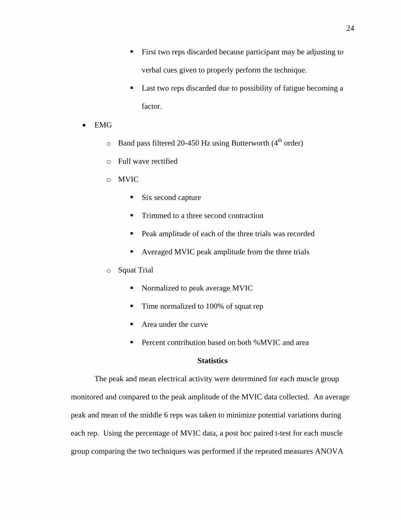
• EMG
o Band pass filtered 20-450 Hz using Butterworth (4th order)
o Full wave rectified
o MVIC
Six second capture
Trimmed to a three second contraction
Peak amplitude of each of the three trials was recorded
Averaged MVIC peak amplitude from the three trials
o Squat Trial
Normalized to peak average MVIC
Time normalized to 100% of squat rep
Area under the curve
Percent contribution based on both %MVIC and area
Statistics
The peak and mean electrical activity were determined for each muscle group
monitored and compared to the peak amplitude of the MVIC data collected. An average
peak and mean of the middle 6 reps was taken to minimize potential variations during
each rep. Using the percentage of MVIC data, a post hoc paired t-test for each muscle
group comparing the two techniques was performed if the repeated measures ANOVA

25
determined any significant (p < 0.05) differences in muscle activity. Kinematic and
kinetic data were also analyzed using repeated measures ANOVA to determine
significant (p < 0.05) differences in joint range of motion (hip, knee, and ankle) and squat
variation COPs. All data were analyzed using the PAWS statistical package (Winwrap
Basic, 1993-2007 Polar Engineering and Consulting) and data were stored on a computer
in the biomechanics laboratory and a flash drive, which was kept in the biomechanics
laboratory. Paired t-tests were used for post hoc analyses to determine significant
differences (p < 0.05) between each of the eight muscles monitored and to determine
significant differences (p < 0.05) in the kinematic data collected.

26
CHAPTER FOUR: RESULTS
The purpose of this study was to analyze the muscle activity, kinetic and
kinematic differences between a traditional and non-traditional squat. The main factor
that determined the difference between the two squat variations was the COP on the
participant’s foot during the execution of the exercise. Sixteen healthy males participated
in this study but only 13 participants’ data were usable for statistical analysis due to EMG
and video equipment issues during three of the participants’ trials. The results of this
study will be presented in tables and graphs that illustrate the averages of all the
participants’ data collected during repetitions 3-8 of the set of both the traditional and
non-traditional squat techniques. The average peak normalized EMG amplitude data are
presented as a ratio of the average peak amplitude (mV) during each repetition and the
average of the MVIC (mV) data for each muscle. The average total EMG area (mV·s)
data are presented as the average of the total volume of muscle activity occurring during
each repetition of the squatting exercise for each muscle.
No significant difference existed in the normalized peak EMG activities between
the traditional (Trad) and non-traditional (Non) squat.

27
Table 4.1 Averages and standard deviations for peak EMG data.
Average Peak Normalized EMG Amplitude (Peak Amplitude[mV]/ MVIC[mV])
Muscle Gluteus Maximus Adductor Gastrocnemius Vastus Medialis
Squat Trad Non Trad Non Trad Non Trad Non Average
± SD 4.76 ± 4.48
4.47 ± 4.47
1.63 ± 1.38
1.68 ± 1.59
0.43 ± 0.26
0.67 ± 0.23
2.71 ± 2.92
1.86 ± 1.25
Muscle Vastus Lateralis Biceps Femoris Semitendinosus Erector Spinae
Squat Trad Non Trad Non Trad Non Trad Non Average
± SD 1.98 ± 1.06
1.73 ± 0.93
1.37 ± 0.83
1.67 ± 1.22
0.47 ± 0.33
0.64 ± 0.37
1.46 ± 0.72
1.49 ± 0.68
Overall average total area in the EMG output between traditional and non-
traditional squats returned an F(1,7) = 4.359, p < 0.05, and the GS, VM, VL, and ST
muscles post hoc results gave a p < 0.05. Figure 3 displays the averages and standard
deviations for the eight muscles monitored. Muscles for which activation was
significantly different between the two squats are marked with an asterisk. Significantly
higher muscle activity readings were measured for the gastrocnemius and
semitendinousus during the non-traditional squat. The vastus lateralis and vastus
medialis muscles had a significantly higher muscle activity reading during the traditional
squat compared to the non-traditional squat.
Table 4.2 Averages and standard deviations for total EMG area data. *- Significant difference between Trad and Non technique (p < 0.05)
Average Total EMG Area (mV·s)
Muscle Gluteus Maximus Adductor Gastrocnemius* Vastus Medialis*
Squat Trad Non Trad Non Trad Non Trad Non Average
± SD 1048 ± 910.48
893.38 ± 762.0
310.11 ± 236.7
284.80 ± 270.0
66.76 ± 43.7
103.16 ± 33.9
506.26 ± 338.6
371.76 ± 261.5
Muscle Vastus Lateralis* Biceps Femoris Semitendinosus* Erector Spinae
Squat Trad Non Trad Non Trad Non Trad Non Average
± SD 463.67 ± 265.4
353.82 ± 219.2
330.68 ± 206.6
344.37 ± 235.3
80.78 ± 55.5
111.90 ± 68.1
349.75 ± 184.8
336.88 ± 182.9

28
Figure 4.1 Averages and standard deviations for total EMG area data.
Significant differences based on paired t-tests. ( = p < 0.05)
Overall percent contribution for the average total area in the EMG output between
traditional and non-traditional squats returned an F(1,7) =4.192, p < 0.05, and the GS, VM,
VL, and ST muscles post hoc results gave a p < 0.05. Figure 4 displays the averages and
standard deviations for the eight muscles monitored as well as identifying the muscles
that were significantly different between the two squats. The gastrocnemius and
semitendinousus reported significantly higher muscle activity readings during the non-
traditional squat. The vastus lateralis and vastus medialis muscles had significantly
higher muscle activity during the traditional squat compared to the non-traditional squat.
Table 4.3 Averages and standard deviations for % contribution total EMG area *- Significant difference between Trad and Non technique (p < 0.05)
Average Percent Contribution of Each Muscle Based on Total EMG Area
Muscle Gluteus Maximus Adductor Gastrocnemius* Vastus Medialis*
Squat Trad Non Trad Non Trad Non Trad Non Average
± SD 29.43% ± 17%
28.74% ± 17%
9.57% ± 5.7%
9.26% ± 6.2%
2.58% ± 1.8%
4.39% ± 2.4%
16.53% ± 6.9%
13.63% ± 6.8%
Muscle Vastus Lateralis* Biceps Femoris Semitendinosus* Erector Spinae
Squat Trad Non Trad Non Trad Non Trad Non Average
± SD 14.86% ± 4.3%
12.63% ± 4.5%
11.14% ± 5.6%
12.64% ± 6.3%
2.67% ± 1.7%
4.07% ± 2.4%
13.23% ± 8.1%
14.65% ±9.99%

29
Figure 3.2 Average % Contribution of Muscle Based on Total EMG
Significant differences based on paired t-tests. ( = p < 0.05)
Overall percent contribution for the average peak normalized EMG values
between traditional and non-traditional squats returned an F(1,7) = 2.785, p < 0.05, and the
GS and ST muscles post hoc results gave a p < 0.05. Figure 5 displays the averages and
standard deviations for the eight muscles monitored as well as identifying the muscles
that were significantly different between the two squats. The gastrocnemius and
semitendinousus reported significantly higher muscle activity readings during the non-
traditional squat.
Table 4.4 Averages and standard deviations for % contribution of peak EMG. *- Significant difference between Trad and Non technique (p < 0.05)
Average Percent Contribution of Each Muscle Based on Peak Normalized EMG Amplitude
Muscle Gluteus Maximus Adductor Gastrocnemius* Vastus Medialis
Squat Trad Non Trad Non Trad Non Trad Non Average
± SD 29.15% ±18.6%
27.58% ± 17%
10.32% ± 5.8%
10.91% ± 7.7%
3.67% ± 2.6%
5.51% ± 2.6%
17.46% ± 10%
13.97% ± 7.5%
Muscle Vastus Lateralis Biceps Femoris Semitendinosus* Erector Spinae
Squat Trad Non Trad Non Trad Non Trad Non Average
± SD 13.98% ± 4.64%
12.41% ± 4.1%
10.05% ± 5.3%
11.98% ± 6.5%
3.35% ± 2.2%
4.70% ± 2.8%
12.03% ± 7.7%
12.92% ± 8.3%

30
Figure 4.3 Average % contribution of muscle based on peak normalized EMG amplitude.
Significant differences based on paired T-tests. ( = p < 0.05) ANOVA results for the overall kinematic and kinetic data analysis between
traditional and non-traditional squats returned an F(1,5) = 4.138, p < 0.05, and post hoc
results for the COP and range of motion for the ankle and knee gave a p < 0.05. In Table
5, the average COP and ROM for the knee, hip, and ankle are displayed. COP is
measured as a percentage of the longitudinal length of the participant’s foot with the heel
= 0 and the toe = 100. The ranges of motion are measured from the beginning of the
squat to the lowest decent point. Post-hoc t-tests revealed that the COP was significantly
closer to the heels during the traditional squat compared to the non-traditional squat. T-
tests also revealed that the ROM knee and ROM ankle were significantly larger in the
traditional squats compared to the non-traditional squats. The ROM hip was not
significantly different but the data revealed a trend that the traditional squat elicits a
larger range of motion compared to the non-traditional squat.

31
Table 4.5 Averages and standard deviations for kinetic and kinematic data. : *- Significant difference between Trad and Non technique (p < 0.05)
Kinematic and Kinetic Average Data Average COPy* ROM Knee (degrees)* ROM Hip (degrees) ROM Ankle (degrees)*
Squat Trad Non Trad Non Trad Non Trad Non Average
± SD 52 ± 9.0
70 ± 4.0
101.56 ± 6.68
93.30 ± 14.52
110.96 ± 25.76
101.50 ± 19.24
26.50 ± 4.23
20.62 ± 6.09
The % Squat in Figures 6, 7, 8, 9, 10, and 11 refer to the time it took the center of
mass of the participant to cycle through one repetition. One repetition begins when the
center of mass begins to descend and ends when the center of mass returns to the
beginning position. The average COP for the traditional squat was significantly closer to
the heel during the entire downward and upward phase of the motion as seen in Figure 6
and determined by the paired t-test for COP between the two squats giving a p < 0.05.
Figure 4.4 Average center of pressure. (0 = heel, 100 = toe).
The range of motion for the ankle was significantly less (p < 0.05) in the non-
traditional squat; however, both squat types follow a similar range of motion through the
entire squatting technique as shown in Figure 7.

32
Figure 4.5 Average ankle flexion.
T-test gave p < 0.05.
In Figure 8, the average ankle power is displayed during each squat and it can be
seen that the lowest and highest points recorded were during the non-traditional squat.
The EMG average total area, percent contribution to the total area, and percent
contribution to the peak EMG data for the GT was significantly higher in non-traditional
variation of the squat, and Figure 8 complements these results by showing that the ankle
power output is larger during the non-traditional squat.
Figure 4.6 Average ankle power.

33
The paired t-test for knee range of motion gave a p < 0.05, with the traditional
squat having a significantly larger range of motion compared to the non-traditional squat.
Figure 9 does show that both squatting techniques averaged over 90 º of knee flexion, and
although the peak for both variations are close, almost every participants’ knee range of
motion was larger during the traditional squat.
Figure 4.7 Average knee range of motion during the squatting variations.
In Figure 10, it can be seen that the traditional squat has a lower minimum and
higher maximum power output. From the EMG data, the VL and VM were significantly
more active during the traditional squat. Figure 9 complements the results from the EMG
data by showing that knee power output is larger during the traditional squat.

34
Figure 4.8 Average knee power.
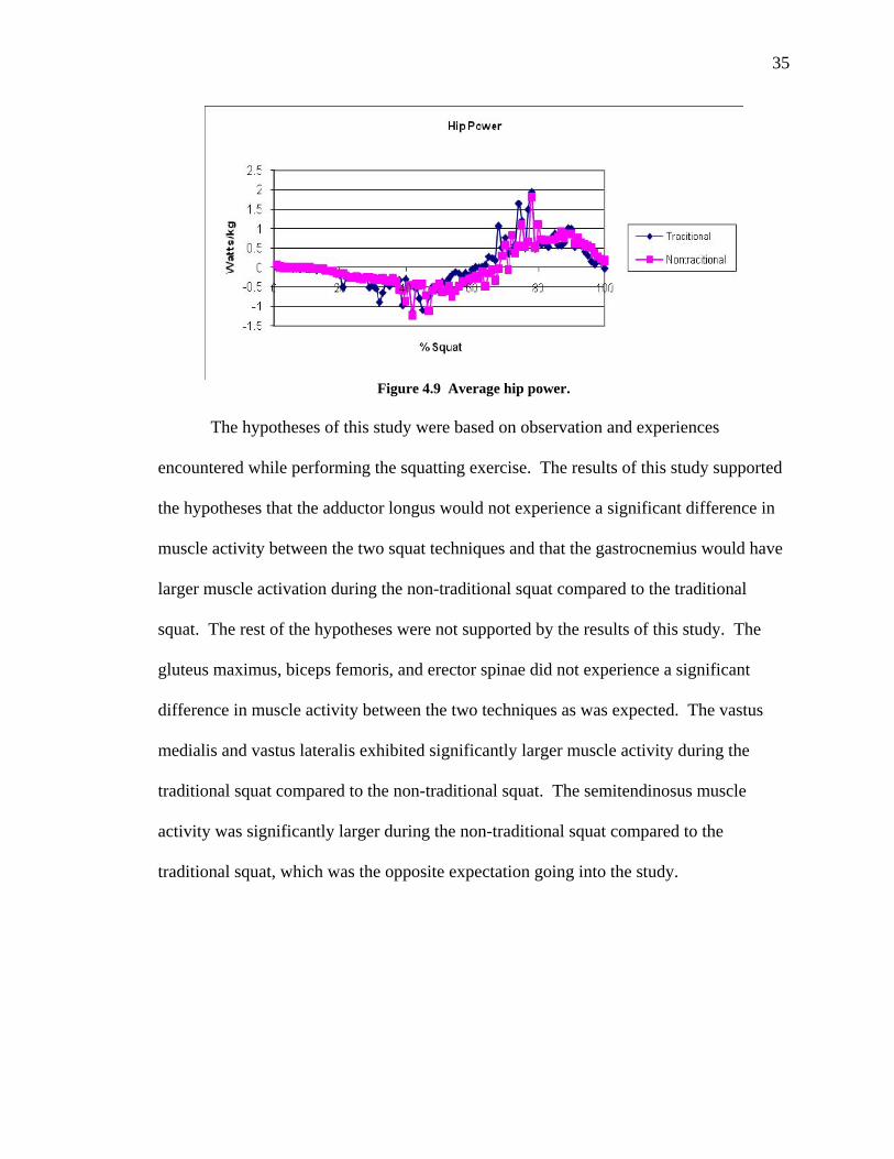
In Figure 4.9, the average hip power is illustrated during the percent squat. The
data used in creating this chart were not always consistent due to an interruption of the
monitoring of the hip reflectors in several of the participants. This interruption was due
in part to the front hip reflectors being covered inadvertently by either clothing during the
lowest part of the squat or by the cameras losing tracking due to the height of the squat
rack safety bar being around hip level at the bottom of the squat. The majority of
tracking was lost between 40-80% of the squat as can be seen by the erratic data points in
that range in Figure 11. Due to processing of the video taking place after half of the
participants completed the study, this interruption was not noticed until midway through
the study. Although this is an artifact of the study, some results can be drawn from the
data. The data points that were identified as legitimate were not significantly different
between the two techniques, which correspond to the gluteus maximus muscle activity
not being significantly different between the two squats.
35
Figure 4.9 Average hip power.
The hypotheses of this study were based on observation and experiences
encountered while performing the squatting exercise. The results of this study supported
the hypotheses that the adductor longus would not experience a significant difference in
muscle activity between the two squat techniques and that the gastrocnemius would have
larger muscle activation during the non-traditional squat compared to the traditional
squat. The rest of the hypotheses were not supported by the results of this study. The
gluteus maximus, biceps femoris, and erector spinae did not experience a significant
difference in muscle activity between the two techniques as was expected. The vastus
medialis and vastus lateralis exhibited significantly larger muscle activity during the
traditional squat compared to the non-traditional squat. The semitendinosus muscle
activity was significantly larger during the non-traditional squat compared to the
traditional squat, which was the opposite expectation going into the study.

36
CHAPTER FIVE: DISCUSSION
Performing exercises with proper form increases efficiency, effectiveness, and
safety. The squat exercise is a strength exercise that is implemented in workout routines
in order to activate the quadriceps, hamstrings, calves, gluteus, and core musculature.
Several variations of the squat exercise have been compared in laboratory settings in
order to determine specific muscle activation differences between the various techniques
and discover the most effective technique to train a specific muscle group (2, 5, 9, 10, 12,
19, 21).
The aim of this study was to determine significant differences between activation
of the lower body musculature while performing two variations of the squatting exercise.
The two squatting techniques were labeled traditional and non-traditional, and were
described in detail in previous chapters. Statistical analysis of the eight muscles
monitored during the squatting variations indicated significant differences between the
two techniques.
The gluteus maximus showed no difference in muscle activity between the two
techniques. The GM is typically more active when squat depth is increased (5) and when
stance width is increased from 75% of shoulder width to 140% of shoulder width (19).
However, this study did not use differing squat depth or stance width as variables, so the
hypothesis that the GM activity would be significantly different between the two squats
was based on the COP being either more toward the heel (traditional squat) or more
toward the toe (non-traditional squat) of the foot, which was not supported in this study.

37
Thus, the position of the COP does not appear to be a factor that would cause a
significant difference in muscle activity of the GM.
The adductor longus has been shown to increase in muscle activity as stance
width increases by a previous study (19), however stance width was maintained at
shoulder width during both squat variations in this study and the results were as expected.
There were no previous studies using COP or squat depth as a variable measuring
adductor longus muscle activity, therefore comparison of results is limited. The adductor
longus does not appear to be affected by COP positioning but is affected by stance width.
A surprising finding of this study was that the erector spinae musculature did not
show a significant difference in activation between the two squat variations. Sparto et al.
determined that repetitive lifting caused forward tilt angle of the upper body, which in
turn increased the demand on the trunk extensors (36). Therefore, it was hypothesized
that the erector spinae would increase in activity during the non-traditional squat because
of the anterior shift of the upper body, causing a larger moment arm for the erector spinae
muscle; however, the results of this study do not support this. Interestingly, several of the
participants communicated that their lower back felt more strain during the non-
traditional squats compared to the traditional squats. This “feeling” may be attributed to
stressors or forces being applied to tissues (e.g., tendons, ligaments, bone, or muscles)
that were not monitored during this study. Further research should be conducted in order
verify this speculation.
Another possibility that needs to be researched further is the increase in fatigue
during repetitive lifting being the main contributor to the increase in erector spinae
muscle activation. Since fatigue was not a measured variable in this study, future work

38
may include fatigue as a factor and compare it to previous studies in which lower back
musculature was prone to increased activity as muscle fatigue increased (22, 36).
The total area of EMG activity, percent contribution of total area of EMG activity,
and percent contribution of peak EMG activity of the gastrocnemius showed a significant
increase in muscle activity during the non-traditional squat. Figure 7 displays the ankle
power during both traditional and non-traditional squats and it can be seen that ankle
power is stronger during the non-traditional squat. This complements the results of more
muscle activity in the gastrocnemius during the non-traditional squat, since the insertion
point of this muscle is at the ankle. A study by Roelants et al. discovered that the
gastrocnemius was significantly more active when squats were performed while
experiencing whole body vibration compared to no vibration stimulus (21). Both non-
traditional squats and squats performed during whole body vibration can be considered
unstable conditions. These studies reported that unstable squatting conditions will
produce more muscle activation from the gastrocnemius, and that the gastrocnemius
appears to be more active when an individual is off balance. The gastrocnemius is a
muscle that contributes largely to the balancing of an individual when performing lifting
maneuvers. Another speculation is that if the heel comes off the ground during the non-
traditional squat, the gastrocnemius and other calf muscles may be responsible for this
action eliciting further muscle activity, although the heel coming off the ground may be
due to lack of flexibility in the gluteus, hamstring, and calf musculature. In the study by
Dionisio et al., the ankle torque, COP, and gastrocnemius muscle activity was monitored
during the descent and ascent of a body weight squat. As the COP shifted toward the toe,
the ankle torque and the gastronemius muscle activity increased, which is in agreement

39
with the current study (7). If the goal of an athlete is to increase gastrocnemius strength,
then performing squats in which the COP is directly over the toes will help accomplish
that goal more completely than performing traditional squats.
Total EMG area activation and percent contribution of total EMG area activation
for the quadriceps were significantly (p < 0.05) larger for the traditional squat. The
participants again stated that after performing the non-traditional squats that they felt
their quadriceps were “worked” more compared to the traditional squats. However, after
evaluating power output of the knee from Figure 9 and realizing the moment arm at the
knee joint would be shortened due to the forward shifting of the COP in the non-
traditional squat, it can be expected that the quadriceps muscle activity would be larger
during the traditional squat. This complements the study by Toutoungi et al., which
found PCL peak forces to be larger during squats where the participants’ heels remained
in contact with the ground compared to squats where the participants’ heels came off the
ground (26). The PCL and quadriceps work together to stabilize the femur from sliding
forward over the tibia or prevent the tibia from moving posterior, so when measuring just
the PCL or just the quadriceps, it may be assumed that when a large force is placed on
one, a large force will also be placed on the other. This may also be a reason why certain
individuals perform squats where they lean forward and their COP shifts over their toes.
If the PCL is injured or weak, shifting the COP over the toes would place less force on
the PCL. Conversely, a decrease of force on the PCL would mean an increase of force
placed on the ACL and hamstrings.
After observing the results of the study, rationalizing the data, and further
reviewing previous studies, the statement made about the hamstring musculature was

40
determined to be an incorrect hypothesis. From the results, it was determined that the
biceps femoris did not show any significant difference in muscle activation between the
two squats. The results did show that the semitendinosus exhibited significantly more
muscle activity during the non-traditional squat compared to the traditional squat,
although this was not the difference that was hypothesized. The total area of EMG
activity, percent contribution of total area of EMG activity, and percent contribution of
peak EMG activity of the semitendinosus showed a significant increase in muscle activity
during the non-traditional squat compared to the traditional squat. One explanation for
the increased muscle activity during the non-traditional squat is that the forward lean
experienced during this technique needs to be countered in order to return the participant
back to the original position. The semitendinosus is a major muscle being recruited in
order to accomplish this counter balancing force.
The biceps femoris muscle is also part of the hamstring musculature that is
responsible for returning the lifter to the original position while performing the non-
traditional squat. However, the findings of this study did not indicate a significant
difference in muscle activity between the two techniques, although all the EMG data for
the BF were larger in the non-traditional squat compared to the traditional squat. De
Looze et al. noted that the biceps femoris activated to a greater degree during the ascent
phase of the squat in order to contribute to the large hip extensor torque required to return
the lifter to the upright position and also stablilize the knee joint, which agrees with the
higher muscle activation of the hamstrings in the non-traditional squat compared to the
traditional squat (37). This trend may also suggest that a larger participant pool might
41
lead to finding significantly higher muscle activity in the biceps femoris during the non-
traditional squat compared to the traditional squat in later studies.
Wright et al. determined that compared to back squats, stiff-leg deadlifts elicited
nearly double the EMG muscle activity from both the biceps femoris and semitendinosus
(28). The non-traditional squat is a version of the back squat but has some attributes of
the stiff-leg deadlift, mainly a forward COP. The anterior motion of the upper body
during descent is also a feature seen in both exercises, which shifts the COP forward and
also causes the hamstrings to activate in order to return the upper body to the beginning
position. Similar findings of increased hamstring activity as trunk flexion increased were
observed during a study by Ohkoshi et al. and discussed in the study by Wright et al.(28,
38). Lack of knee flexion in the stiff-leg deadlifts and less knee flexion in the non-
traditional squat increased the lengthening of the hamstrings compared to the traditional
squat, therefore more contraction of the hamstrings takes place during the ascent phase of
the stiff-leg deadlift and non-traditional squat compared to the traditional squat.
In the study by Toutoungi et al., the ACL peak forces were larger during the heel
off the ground squats compared to the heel on the ground squats (26). Since the ACL and
hamstrings work together to stabilize the tibia from sliding too far forward under the
femur, an increase of force on the ACL would lead one to believe that hamstrings muscle
activity would increase in male athletes as well. These findings concur with the results
that semitendinosus muscle activity increases when the COP is focused over the toes
compared to the heels during the squat.
McLaughlin et al. found that inexperienced lifters tended to lean forward with the
trunk more than skilled lifters and that this forward lean increased trunk torque, which

42
stretches the hamstrings and increases their muscle activation during the ascent of the
squatting motion (39). The observation of McLaughlin et al. concurs with the findings of
this study that forward trunk motion, as seen in the non-traditional squat, increases
hamstring activation. Since the more skilled lifters in McLaughlin et al.’s study had
lower trunk torque due to less forward trunk lean, which is similar to the traditional squat;
this leads one to determine that traditional squats may be considered a more proper form
of the squat technique compared to the non-traditional squat.
The major findings of this study were that there is a difference in muscle activity,
kinetics, and kinematics when the COP is shifted from the heel/arch of the foot to the toe.
These findings will help trainers and coaches explain why they prefer their clients or
athletes to stay back on their heels when squatting or why they might want them to lean
forward on their toes. Although this study was able to determine muscle activation
differences in the squat variations, it was not determined if COP over the toes during the
weighted back squat is unsafe compared to a squat that focuses on keeping the COP over
the heels. Participant feedback did reveal that during the non-traditional squat, they felt
more tension in the lower back; however, the measured variable (ES EMG) did not reveal
a significant difference between the traditional and non-traditional squat. Participant
feedback points to the need for further studies designed to determine the risk of possible
injury during a non-traditional squat; however, with a light load or body weight,
performing squats where the COP is over the toes will safely help strengthen the
gastrocnemius and semitendinosus muscles more compared to squats where the COP is
over the heels.

43
Conclusion
Of the several hypotheses made prior to this study, only two were accepted while
six were rejected. Major findings of this study were that COP on the heel of the foot
would elicit different muscle activation for variations of the same lift compared to COP
on the toes or ball of the foot. In comparing the traditional squat and non-traditional
squat, it can be determined that traditional squats (COP on the heel) will elicit more
muscle activation in the quadriceps and non-traditional squats (COP on the toes) will
activate the hamstrings and gastrocnemius more effectively. Another observation in this
study was that participants had a “feeling” of muscle activation in the lower back and
quadriceps after performing non-traditional squats, but the EMG readings were not
significantly different for the erector spinae and actually lower in the non-traditional
squats compared to the traditional squats for the quadriceps.
Overall power between the squats displayed larger output in the knees for the
traditional squat, larger output in the ankles for the non-traditional squat, and no
difference in hip power. It appears that when the COP is over the toes, the calf muscles
compensate for the loss of power in the quadriceps in order to move the same load.
However, non-traditional squats may also cause unwanted stressors in the lower back,
which was communicated by the participants after performing non-traditional squats.
Further studies, which are more focused on the lower back, spine, and core musculature,
comparing these two variations of the squat, could help determine if there is a spinal
safety discrepancy between the traditional and non-traditional squat. Studies that use
fatigue of different muscle groups as a factor will also help determine safety procedures
that should be followed when performing squats, since fatigue was not a measured factor

44
in this study but previous work found fatigue to be a factor that changed muscle activity
and biomechanical motion significantly (22, 36). The study was successful in showing
that COP shifting from the heels to the toes will elicit different muscle activation,
although it was not successful in determining if the traditional technique was safer due to
lower back stressors compared to the non-traditional squat. In conclusion, each technique
is valuable in strengthening the lower extremities and simply shifting the COP will elicit
significant differences in quadriceps, hamstrings, and gastrocnemius muscle activity.

45
BIBLIOGRAPHY
1. The American Heritage Dictionary English Language 4th ed: Boston, MA: Houghton Mifflin Company; 2000.
2. Anderson K, Behm DG. Trunk muscle activity increases with unstable squat movements. Can. J. Appl. Physiol. 2005;30(1):33-45.
3. Bazrgari B, Shirazi-Adl A, Arjmand N. Analysis of squat and stoop dynamic lifting: muscle forces and internal spinal loads. Eur. Spine J. 2007;16(5):687-699.
4. Burgess-Limerick R. Squat, stoop, or something in between?. Int. J. of Ind. Ergonom. 2003; 31: 143-148.
5. Caterisano A, Moss RF, Pellinger TK, Woodruff K, Lewis VC, Booth W, Khadra T. The effect of back squat depth on the EMG activity of 4 superficial hip and thigh muscles. J. Strength Cond. Res. 2002;16(3):428-432.
6. Comfort P, Kasim P. Optimizing squat technique. Strength Cond. J. 2007;29(6):10-13.
7. Dionisio VC, Almeida GL, Duarte M, Hirata RP. Kinematic, kinetic and EMG patterns during downward squatting. J. Electromyogr. Kines.. 2008;18:134-143.
8. Escamilla RF. Knee biomechanics of the dynamic squat exercise. Med. Sci. Sports Exer. 2001;33(1):127-141.
9. Escamilla RF, Fleisig GS, Lowry TM, Barrentine SW, Andrews JR. A three-dimensional biomechanical analysis of the squat during varying stance widths. Med. Sci. Sports Exer. 2001;33(6):984-998.
10. Escamilla RF, Fleisig GS, Zheng NQ, Lander JE, Barrentine SW, Andrews JR, Bergemann BW, Moorman, III CT. Effects of technique variations on knee biomechanics during the squat and leg press. Med. Sci. Sports Exer. 2001;33(9):1552-1566.
11. Flanagan SP, Salem GJ. Bilateral differences in the net joint torques during the squat exercise. J. Strength Cond. Res. 2007;21(4):1220-1226.
12. Frohm A, Halvorsen K, Thorstensson A. Patellar tendon load in different types of eccentric squats. Clin. Biomech. 2007;22(6):704-711.
13. Fry AC, Smith JC, Schilling BK. Effect of knee position on hip and knee torques during the barbell squat. J. Strength Cond. Res. 2003;17(4):629-633.
14. Gallo RA, Reitman RD, Altman DT, Altman GT, Jones CB, Chapman JR. Flexion-distraction injury of the thoracolumbar spine during squat exercise with the Smith machine. Am. J. Sport. Med. 2004;32(8):1962-1967.
15. Gullett JC, Tillman MD, Gutierrez GM, Chow JW. A biomechanical comparison of back and front squats in healthy trained individuals. J. Strength Cond. Res. 2009;23(1):284-292.
16. Isear JA, Erickson JC, Worrell TW. EMG analysis of lower extremity muscle recruitment patterns during an unloaded squat. Med. Sci. Sports Exer. 1997;29(4):532-539.

46
17. Manabe Y, Shimada K, Ogata M. Effect of slow movement and stretch-shortening cycle on lower extremity muscle activity and joint moments during squat. J. Sport. Med. Phys. Fit. 2007;47(1):1-12.
18. Manabe Y, Yokozawa T, Ogata M. Effect of movement variation on lower limb muscle activity and joint torque during squat. Jpn. J. Phys. Fit. Sport. 2004;53(3):321-336.
19. McCaw ST, Melrose DR. Stance width and bar load effects on leg muscle activity during the parallel squat. Med. Sci. Sports Exer. 1999; 31(3): 428-436.
20. Robertson DGE, Wilson JMJ, Pierre TAS. Lower Extremity Muscle Functions During Full Squats. J. Appl. Biomech. 2008;24(4):333-339.
21. Roelants M, Verschueren SMP, Delecluse C, Levin O, Stijnen V. Whole-body-vibration-induced increase in leg muscle activity during different squat exercises. J. Strength Cond. Res. 2006;20(1):124-129.
22. Sasaki M, Horio A, Wakasa M, Uemura S, Osawa Y. Influence of quadriceps femoris fatigue on low back load during lifting of loads at different distances from the toes. J. Phys. Ther. Sci. 2008;20(2):81-89.
23. Shields RK, Madhavan S, Gregg E, Leitch J, Petersen B, Salata S, Wallerich S. Neuromuscular control of the knee during a resisted single-limb squat exercise. Am. J. Sport. Med. 2005;33(10):1520-1526.
24. Tagesson S, Oberg B, Good L, Kvist J. A comprehensive rehabilitation program with quadriceps strengthening in closed versus open kinetic chain exercise in patients with anterior cruciate ligament deficiency. Am. J. Sport. Med. 2008;36(2):298-307.
25. Tagesson SKB, Kvist J. Intra- and interrater reliability of the establishment of one repetition maximum on squat and seated knee extension. J. Strength Con. Res. 2007;21(3):801-807.
26. Toutoungi DE, Lu TW, Leardini A, Catani F, O'Connor JJ. Cruciate ligament forces in the human knee during rehabilitation exercises. Clin. Biomech. 2000;15(3):176-187.
27. Waller M, Townsend R. The front squat and its variations. Strength Con. J. 2007;29(6):14-19.
28. Wright GA, DeLong TH, Gehlsen G. Electromyographic activity of the hamstrings during performance of the leg curl, stiff-leg deadlift, and back squat movements. J. Strength Con. Res. 1999;13(2):168-174.
29. Youdas JW, Hollman JH, Hitchcock JR, Hoyme GJ, Johnsen JJ. Comparison of hamstring and quadriceps femoris electromyographic activity between men and women during a single-limb squat on both a stable and labile surface. J. Strength Con. Res. 2007;21(1):105-111.
30. Zink AJ, Perry AC, Robertson BL, Roach KE, Signorile JF. Peak power, ground reaction forces, and velocity during the squat exercise performed at different loads. J. Strength Con. Res. 2006;20(3):658-664.
31. Cram JR, Kasman GS, Holtz J. Introduction to Surface Electromyography. Gaitherburg, MD: Aspen Publishers; 1998.
32. Biodex System 2 Manual. Shirley, NY: Biodex Medical Systems Inc. 33. Faul F, Erdfelder E, Buchner A, Lang AG. (2009). Statistical power analyses
using G*Power 3.1: Tests for correlation and regression analyses. Behavior

47
Research Methods, 41, 1149-1160. 34. Benda BJ, Riley PO, Krebs DE. Biomechanical relationship between center of
gravity and center of pressure during standing. IEEE T. Rehabil. Eng. 1994;2(1): 3-10.
35. Bakhtiary AH, Fatemi E. Open versus closed chain exercises for patellar
condomalacia. Br. J. Sports Med. 2007; 42: 99-102. 36. Sparto PJ, Parnianpour M, Reinsel TM, Simon S. The effect of fatigue on
multijoint kinematics and load sharing during a repetitive lifting test. Spine. 1997; 22(22): 2647-2654.
37. De Looze MP, Toussaint HM, Van Dieen JH, Kemper HCG. Joint moments and
muscle activity in the lower extremities and lower back in lifting and lowering tasks. J. Biomech. 1993; 26: 1067-1076.
38. Ohkoshi Y, Yasuda K, Kaneda K, Wada T, Yamanaka M. Biomechanical analysis of rehabilitation in the standing position. Am. J. Sports Med. 1991; 19: 605-610.
39. McLaughlin TM, Lardner TJ, Dillman CJ. Kinetics of the parallel squat. Res. Q. Exerc. Sport. 1979; 49: 175-189.

48
APPENDIX A
IRB Approval
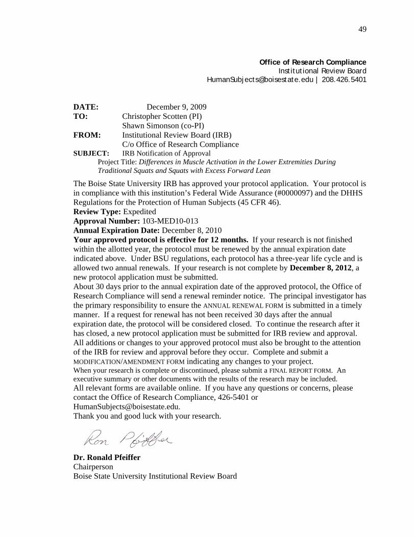
49
Office of Research Compliance
Institutional Review Board [email protected] | 208.426.5401
DATE: December 9, 2009 TO: Christopher Scotten (PI) Shawn Simonson (co-PI) FROM: Institutional Review Board (IRB) C/o Office of Research Compliance SUBJECT: IRB Notification of Approval
Project Title: Differences in Muscle Activation in the Lower Extremities During Traditional Squats and Squats with Excess Forward Lean
The Boise State University IRB has approved your protocol application. Your protocol is in compliance with this institution’s Federal Wide Assurance (#0000097) and the DHHS Regulations for the Protection of Human Subjects (45 CFR 46). Review Type: Expedited Approval Number: 103-MED10-013 Annual Expiration Date: December 8, 2010 Your approved protocol is effective for 12 months. If your research is not finished within the allotted year, the protocol must be renewed by the annual expiration date indicated above. Under BSU regulations, each protocol has a three-year life cycle and is allowed two annual renewals. If your research is not complete by December 8, 2012, a new protocol application must be submitted. About 30 days prior to the annual expiration date of the approved protocol, the Office of Research Compliance will send a renewal reminder notice. The principal investigator has the primary responsibility to ensure the ANNUAL RENEWAL FORM is submitted in a timely manner. If a request for renewal has not been received 30 days after the annual expiration date, the protocol will be considered closed. To continue the research after it has closed, a new protocol application must be submitted for IRB review and approval. All additions or changes to your approved protocol must also be brought to the attention of the IRB for review and approval before they occur. Complete and submit a MODIFICATION/AMENDMENT FORM indicating any changes to your project. When your research is complete or discontinued, please submit a FINAL REPORT FORM. An executive summary or other documents with the results of the research may be included. All relevant forms are available online. If you have any questions or concerns, please contact the Office of Research Compliance, 426-5401 or [email protected]. Thank you and good luck with your research.
Dr. Ronald Pfeiffer Chairperson Boise State University Institutional Review Board

50
APPENDIX B
Informed Consent Form

51
Boise State University - Department of Kinesiology
Research Project Differences in Muscle Activation in the Lower Extremities During Traditional Squats and
Squats with Excess Forward Lean Consent to be a research participant
A. Purpose and Background
Chris Scotten and Shawn Simonson, Ed.D., in the Department of Kinesiology at the Boise
State University are conducting research to determine the differences in lower extremity and trunk
muscle activation while performing squats using two different techniques. The study is aimed to verify
the differences in muscle activity between these techniques. If the claims of this study are supported
by the findings then the traditional technique will be found to activate the leg muscles more, while the
excess forward lean squat will be found to activate the lower back muscles more. This will show that
performing traditional squats will improve activation in targeted muscles, while decreasing lower back
fatigue compared to the excess forward lean squats. From this study, hopefully developing training
will be safer and more efficient.
B. Procedures
If I agree to volunteer and participate in the study, the following will take place:
1. I will complete the study contraindications questionnaire to ascertain my ability to
participate in this study. If I do not meet safe study participation guidelines, I will not be
selected to participate in this study.
2. If I am selected for the study and I agree to participate, I will have my 10 repetition
maximum in the squat exercise determined, participate in isometric testing to determine
my maximum voluntary contraction activity of muscles monitored in the study, and
perform traditional squats and excess forward lean squats to determine the muscle activity
elicited in the monitored muscles by the two different techniques.
3. My 10 repetition maximum will be determined at least three days, but no more than two
weeks, prior to data collection. I will be visually monitored by the primary investigator in
order to validate my 10 repetition maximum weight.
52
4. I will come to the biomechanics lab 3-14 days after determining my 10 repetition
maximum for the squat exercise session.
5. I will be fitted with the silver-silver chloride EMG electrode pads, which will monitor
muscle activity during isometric testing and while performing the two squatting
techniques.
6. I will then have my maximum voluntary contraction values for each monitored muscle
group determined using a series of isometric exercises described by the lab technician.
7. I will then be asked to perform two different squatting techniques using 75% of my 10
repetition maximum for a series of two sets of ten repetitions, with five minutes rest
between sets.
C. Risks/Discomforts
1. Performing several repetitions of squats with added weight may be uncomfortable for some
individuals. Discomfort may be caused by a heavy load being squatted, in which I can use
a padded that can be wrapped around the squat bar. If I feel uncomfortable, the test will be
stopped if I so choose.
2. Soreness the next day may take place due to lifting weights with a full body exercise. I
will be informed by the investigators on how to lessen this soreness.
3. Spotters will be present during all squats and if I need help while performing squats I will
verbally notify the spotter that I need assistance. I may discontinue testing if I feel
uncomfortable after needing help from the spotter.
4. Participation in research may involve loss of privacy; however, my records will be handled
as confidentially as possible. Only Chris Scotten, Shawn Simonson and the lab
technicians will have access to my records. No individual’s identities will be used in any
report or publication that my result from this study.
D. Consent to be a Research Participant
My permission to participate in this study is voluntary. I am free to deny consent or stop the
test at any point, if I so desire. I have read the above and I understand the test procedures that I will

53
perform. For additional questions, I can contact Chris Scotten at 406-570-1369 or Professor
Simonson at 208-426-3973.
If I have any comments or concerns about participation in this study, I should first talk with
the investigators. If for some reason I do not wish to do this, I may contact the Institutional Review
Board, which is concerned with the protection of volunteers in research projects. I may reach the
board office between 8:00 AM and 5:00 PM, Monday through Friday, by calling 208-426-1574 or by
writing:
Institutional Review Board
Office of Research Administration
Boise State University
1910 University Drive
Boise, ID 83725-1135
I understand that the data gathered from the results of this study will be treated as privileged
and confidential and will not be released to any person without my consent. The data, however, will
be used as anonymous data for publication of scientific research with my right to privacy retained.
I give my consent to participate in this study:
____________________________ _________ Signature of participant Date ____________________________ _________ Signature of test supervisor Date
The Boise State University Institutional Review Board has reviewed this project for the protection of
human participants in research.
54
APPENDIX C
Questionnaire

55
Boise State University - Department of Kinesiology
Research Project Differences in Muscle Activation in the Lower Extremities during Traditional
Squats and Squats with Excess Forward Lean Study Contraindications Screening Questionnaire
Par-Q Has your doctor ever said that you have a heart condition and that you should only do physical activity recommended by a doctor?
___YES ___NO
Do you feel pain in your chest when you do physical activity?
___YES ___NO
In the past month, have you had chest pain when you were not doing physical activity?
___YES ___NO
Do you lose your balance because of dizziness or do you ever lose consciousness?
___YES ___NO
Do you have a bone or joint problem (for example, back, knee or hip) that could be made worse by a change in your physical activity?
___YES ___NO
Is your doctor currently prescribing drugs (for example, water pills) for your blood pressure or heart condition?
___YES ___NO
Do you know of any other reason why you should not do physical activity? ___YES ___NO Have you ever had any of the following:
1. Major knee injury or surgery ___Yes ____No 2. Major hip injury or surgery ___Yes ____No 3. Major ankle injury or surgery ___Yes ____No 4. Major back injury or surgery ___Yes ____No 5. Doctor say you have high blood pressure ___Yes ____No
How long have you been participating in an exercise program?
How long have you been training with weights?
How many days a week do you lift weights for exercise?
How long have you been weight training with this frequency?

56
How many days a week do you use a version of the free weight squat exercise in your
exercise routine?
Height _____ Weight_______ Age___ Name:_____________________________ Signature:_______________________ Test Supervisor:_____________________ Signature:_______________________ Date:________

57
APPENDIX D
Recruitment Flyer

58
BOISE STATE UNIVERSITY - DEPARTMENT OF KINESIOLOGY
RESEARCH PROJECT PARTICIPANTS NEEDED
Differences in Muscle Activation in the Lower Extremities During Traditional Squats and Squats with Excess Forward Lean
The Department of Kinesiology at Boise State University will be conducting a research project to
compare muscle activation in the lower extremities according to two different techniques. Men between the ages of 18-30 years are needed for this project. If you are interested in participating please contact Chris Scotten, a graduate student in exercise science at BSU, at the following phone number 406-570-1369. You may also contact Chris via email at [email protected].
Research Description
Electromyography (using surface electrodes to monitor electrical activity in the muscle) will be used to measure muscle activation in several muscles in the lower body while performing two different squatting techniques. Illustrations of the two squatting techniques finishing positions are provided below.
Traditional technique Excess forward lean technique
The traditional technique is performed by keeping the heels in contact the entire motion and keeping the knees from passing the toes by a lot. The excess forward lean technique has the subject raise the heels off the ground and have their knees pass their toes by a lot. The testing procedure will not cost anything and will take place in the biomechanics lab at BSU. Feeling sore the next day and some discomfort while performing the squats may occur. Learning more about proper technique and contributing to the discovery of different muscle activation during different squat techniques are just a few benefits from participating. Interested participants need to have no prior back, knee, or ankle injuries that required surgery. Participants must also be involved in an exercise program that includes squatting. Thank you for your help. The Boise State University Institutional Review Board has reviewed this project for the protection of human participants in research

59
APPENDIX E
Isometric Motions for MVIC Data Collection (Visual Aides)

60
Figure E.1 MVIC gluteus maximus.

61
Figure E.2 MVIC adductor longus position.

62
Figure E.3 MVIC for vastus medialis, vastus lateralis, biceps femoris and semitendinosus.

63
Figure E.4 MVIC gastrocnemius position.

64
Figure E.5 MVIC erector spinae position.
65
APPENDIX F
Noraxon EMG Electrode Placements

66
Figure F.1 EMG electrode placement.
(Electrodes placed at sites 9, 12, 13, 14, 15, 33, 45 and 46)

67
APPENDIX G
Retro Reflective Spherical Marker Placements

68
Figure G.1 Retro reflective spherical marker placement.

69
APPENDIX H
Raw Data

70
Table H.1 Raw data for normalized peak EMG activity.
Table H.2 Raw data for total EMG area.

71
Table H.3 Raw data for percent contribution of normalized peak EMG activity (X 100%).
Table H.4 Raw data for percent contribution of total EMG area (X 100%).

72
Table H.5 Raw data for COP and joint range of motions.

73
APPENDIX I
Statistics

74
Descriptive Statistics for Peak EMG
Mean Std. Deviation N
TradGlut 4.7608 4.48452 13
NonGlut 4.4655 4.46856 13
TradAdd 1.6312 1.38187 13
NonAdd 1.6822 1.58602 13
TradGas .4330 .25504 13
NonGas .6742 .23338 13
TradVM 2.7072 2.92300 13
NonVM 1.8555 1.25187 13
TradVL 1.9829 1.06082 13
NonVL 1.7288 .92796 13
TradBF 1.3670 .82520 13
NonBF 1.6656 1.21692 13
TradSem .4678 .32818 13
NonSem .6388 .37499 13
TradES 1.4634 .71528 13
NonES 1.4949 .67840 13
Repeated Measures ANOVA
Tests of Within-Subjects Effects for Peak EMG
Measure:MEASURE_1
Source Type III Sum
of Squares df
Mean
Square F Sig.
Muscle Sphericity Assumed 297.926 7 42.561 6.291 .000
Error(Muscle) Sphericity Assumed 568.278 84 6.765 Squat Sphericity Assumed .300 1 .300 .967 .345
Error(Squat) Sphericity Assumed 3.723 12 .310 Muscle * Squat Sphericity Assumed 6.572 7 .939 2.092 .053
Error(Muscle*Squat) Sphericity Assumed 37.700 84 .449

75
Descriptive Statistics for Total Area EMG
Mean Std. Deviation N
TradGlut 1048.0000 910.47723 13
NonGlut 893.3846 762.05663 13
TradAdd 310.1131 236.70210 13
NonAdd 284.8023 270.01179 13
TradGas 66.7615 43.74240 13
NonGas 103.1554 33.89250 13
TradVM 506.2631 338.56858 13
NonVM 371.7638 261.49472 13
TradVL 463.6731 265.41971 13
NonVL 353.8208 219.22547 13
TradBF 330.6854 206.57177 13
NonBF 344.3715 235.26143 13
TradSem 80.7823 55.45325 13
NonSem 111.8977 68.06849 13
TradES 349.7477 184.77451 13
NonES 336.8792 182.85065 13
Repeated Measures ANOVA
Tests of Within-Subjects Effects for Total Area EMG
Measure:MEASURE_1
Source Type III Sum
of Squares df
Mean
Square F Sig.
Muscle Sphericity Assumed 1.379E7 7 1969396.499 8.713 .000
Error(Muscle) Sphericity Assumed 1.899E7 84 226037.649 Squat Sphericity Assumed 102944.522 1 102944.522 7.080 .021
Error(Squat) Sphericity Assumed 174474.415 12 14539.535 Muscle * Squat Sphericity Assumed 269828.607 7 38546.944 4.359 .000
Error(Muscle*Squat) Sphericity Assumed 742761.954 84 8842.404

76
Paired Samples T-test for Total Area EMG
Paired Differences
t df
Sig.
(2-
tailed)Mean
Std.
Deviation
Std.
Error
Mean
95% Confidence
Interval of the
Difference
Lower Upper
Pair 1 TradGlut -
NonGlut
154.6153 325.5950
2
90.303
81
-42.13972 351.3704
9
1.712 12 .113
Pair 2 TradAdd -
NonAdd
25.31077 69.10490 19.166
25
-16.44890 67.07044 1.321 12 .211
Pair 3 TradGas -
NonGas
-
36.39385
24.56228 6.8123
5
-51.23669 -
21.55101
-
5.342
12 .000
Pair 4 TradVM -
NonVM
134.4992
3
111.9474
5
31.048
64
66.85006 202.1484
0
4.332 12 .001
Pair 5 TradVL - NonVL 109.8523
1
107.6565
0
29.858
54
44.79614 174.9084
8
3.679 12 .003
Pair 6 TradBF - NonBF -
13.68615
98.41031 27.294
11
-73.15491 45.78260 -.501 12 .625
Pair 7 TradSem -
NonSem
-
31.11538
21.70935 6.0210
9
-44.23422 -
17.99655
-
5.168
12 .000
Pair 8 TradES - NonES 12.86846 84.87507 23.540
11
-38.42103 64.15796 .547 12 .595

77
Descriptive Statistics for Contribution of Peak EMG per Muscle
Mean Std. Deviation N
TradGlut .2915 .18561 13
NonGlut .2757 .17266 13
TradAdd .1032 .05786 13
NonAdd .1092 .07661 13
TradGas .0367 .02630 13
NonGas .0552 .02570 13
TradVM .1745 .10095 13
NonVM .1398 .07485 13
TradVL .1397 .04639 13
NonVL .1242 .04093 13
TradBF .1004 .05314 13
NonBF .1199 .06533 13
TradSem .0335 .02229 13
NonSem .0471 .02799 13
TradES .1203 .07696 13
NonES .1295 .08254 13
Repeated Measures ANOVA
Tests of Within-Subjects Effects for Contribution of Peak EMG per Muscle
Measure:MEASURE_1
Source Type III Sum
of Squares df
Mean
Square F Sig.
Muscle Sphericity Assumed 1.046 7 .149 9.580 .000
Error(Muscle) Sphericity Assumed 1.310 84 .016 Squat Sphericity Assumed 3.077E-7 1 3.077E-7 3.459 .088
Error(Squat) Sphericity Assumed 1.067E-6 12 8.894E-8 Muscle * Squat Sphericity Assumed .018 7 .003 2.785 .012
Error(Muscle*Squat) Sphericity Assumed .076 84 .001

78
Paired Samples Test for Contribution for Peak EMG per Muscle
Paired Differences
t df
Sig.
(2-
tailed) Mean
Std.
Dev.iatio
n
Std.
Error
Mean
95% Confidence
Interval of the
Difference
Lower Upper
Pair 1 TradGlut -
NonGlut
.0158
5
.06122 .01698 -.02115 .05284 .933 12 .369
Pair 2 TradAdd - NonAdd -
.0060
0
.04081 .01132 -.03066 .01866 -.530 12 .606
Pair 3 TradGas -
NonGas
-
.0185
4
.01323 .00367 -.02653 -.01055 -5.054 12 .000
Pair 4 TradVM - NonVM .0347
7
.06017 .01669 -.00159 .07113 2.084 12 .059
Pair 5 TradVL - NonVL .0155
4
.02899 .00804 -.00198 .03306 1.933 12 .077
Pair 6 TradBF - NonBF -
.0195
4
.03371 .00935 -.03991 .00083 -2.090 12 .059
Pair 7 TradSem -
NonSem
-
.0135
4
.01421 .00394 -.02213 -.00495 -3.435 12 .005
Pair 8 TradES - NonES -
.0091
5
.03679 .01020 -.03139 .01308 -.897 12 .387

79
Descriptive Statistics for Contribution for Total Area EMG per Muscle
Mean Std. Deviation N
TradGlut .2942 .17618 13
NonGlut .2874 .17557 13
TradAdd .0955 .05687 13
NonAdd .0924 .06210 13
TradGas .0258 .01848 13
NonGas .0438 .02450 13
TradVM .1652 .06871 13
NonVM .1363 .06836 13
TradVL .1487 .04304 13
NonVL .1262 .04466 13
TradBF .1114 .05632 13
NonBF .1265 .06261 13
TradSem .0266 .01708 13
NonSem .0408 .02372 13
TradES .1324 .08084 13
NonES .1464 .09986 13
Repeated Measures ANOVA
Tests of Within-Subjects Effects for Contribution for Total Area EMG per Muscle
Measure:MEASURE_1
Source Type III
Sum of
Squares df
Mean
Square F Sig.
Muscle Sphericity Assumed 1.196 7 .171 11.505 .000
Error(Muscle) Sphericity Assumed 1.247 84 .015 Squat Sphericity Assumed .000 1 .000 .000 1.000
Error(Squat) Sphericity Assumed 1.500E-6 12 1.250E-7 Muscle * Squat Sphericity Assumed .015 7 .002 4.192 .001
Error(Muscle*Squat) Sphericity Assumed .044 84 .001

80
Paired Samples Test for Contribution for Total Area EMG per Muscle
Paired Differences
t df
Sig.
(2-
tailed) Mean
Std.
Dev.iati
on
Std.
Error
Mean
95% Confidence
Interval of the
Difference
Lower Upper
Pair 1 TradGlut -
NonGlut
.00685 .04366 .01211 -.01954 .03323 .565 12 .582
Pair 2 TradAdd -
NonAdd
.00308 .02437 .00676 -.01165 .01780 .455 12 .657
Pair 3 TradGas -
NonGas
-.01800 .01282 .00356 -.02575 -.01025 -
5.063
12 .000
Pair 4 TradVM - NonVM .02892 .02099 .00582 .01624 .04161 4.968 12 .000
Pair 5 TradVL - NonVL .02246 .02521 .00699 .00723 .03769 3.213 12 .007
Pair 6 TradBF - NonBF -.01515 .03504 .00972 -.03633 .00602 -
1.559
12 .145
Pair 7 TradSem -
NonSem
-.01415 .01237 .00343 -.02163 -.00668 -
4.126
12 .001
Pair 8 TradES - NonES -.01400 .04646 .01289 -.04208 .01408 -
1.086
12 .299
Descriptive Statistics for Kinematics
Mean Std. Deviation N
TradCOP .5223 .09257 13
NonCOP .7008 .04192 13
TradKnee 101.5592 6.68151 13
NonKnee 93.2977 14.51590 13
TradHip 110.9562 25.76456 13
NonHip 101.5031 19.24339 13
TradAnk 26.5023 4.22473 13
NonAnk 20.6192 6.08576 13
TradPelvis 36.5438 22.39360 13
NonPelvis 32.6554 17.38536 13
TradTorso 189.7800 156.62006 13

81
Descriptive Statistics for Kinematics
Mean Std. Deviation N
TradCOP .5223 .09257 13
NonCOP .7008 .04192 13
TradKnee 101.5592 6.68151 13
NonKnee 93.2977 14.51590 13
TradHip 110.9562 25.76456 13
NonHip 101.5031 19.24339 13
TradAnk 26.5023 4.22473 13
NonAnk 20.6192 6.08576 13
TradPelvis 36.5438 22.39360 13
NonPelvis 32.6554 17.38536 13
TradTorso 189.7800 156.62006 13
NonTorso 123.3885 156.20278 13
Repeated Measures ANOVA
Tests of Within-Subjects Effects for Kinematics
Measure:MEASURE_1
Source Type III Sum
of Squares df Mean Square F Sig.
Kinematic Sphericity Assumed 462439.135 5 92487.827 13.504 .000
Error(Kinematic) Sphericity Assumed 410920.406 60 6848.673 Squat Sphericity Assumed 9511.175 1 9511.175 5.387 .039
Error(Squat) Sphericity Assumed 21186.203 12 1765.517 Kinematic * Squat Sphericity Assumed 20487.707 5 4097.541 4.138 .003
Error(Kinematic*Squat) Sphericity Assumed 59407.529 60 990.125
Paired Samples Test for Kinematics
Paired Differences
t df
Sig.
(2-
tailed) Mean
Std.
Deviation
Std.
Error
Mean
95% Confidence
Interval of the
Difference
Lower Upper

82
Pair 1 TradCOP -
NonCOP
-.17846 .09091 .02521 -.23340 -.12353 -
7.078
12 .000
Pair 2 TradKnee -
NonKnee
8.26154 11.80633 3.27449 1.12705 15.39603 2.523 12 .027
Pair 3 TradHip - NonHip 9.45308 23.88050 6.62326 -
4.97777
23.88392 1.427 12 .179
Pair 4 TradAnk - NonAnk 5.88308 5.96775 1.65516 2.27680 9.48935 3.554 12 .004
Pair 5 TradPelvis -
NonPelvis
3.88846 22.60851 6.27047 -
9.77373
17.55065 .620 12 .547
Pair 6 TradTorso -
NonTorso
66.3915
4
110.3442
5
30.6039
9
-.28883 133.0719
0
2.169 12 .051
APPENDIX J

83
Charts (Produced from averaged data but not discussed in results)
Figure J.1 Average hip flexion/extension.

84
Figure J.2 Average hip moment.
Figures J.3 Average knee moment.

85
Figures J.4 Average ankle moment.