
Al-Shifa Journal of Ophthalmology161 Al-Shifa Journal of Ophthalmology Editorial inquiries should be...
60
105 ISSN 1990-3863 Al-Shifa Journal of Ophthalmology Vol. 13, No. 4, October – December 2017 (Index Issue) Logo • Editorial: Recent Advances in Glaucoma Pharmacological Therapy • Astigmatism and Visual Functions in Amblyopic Children • Combined Extraction by a Fellow During Glaucoma Fellowship • Pattern of Ocular Diseases in Children • Single Versus Double Site Peribulbar Anaesthesia Injection • Different Morphological Patterns of Cataract a Teaching Hospital • Vision Related Quality of Life Among Keratoconus Patients Abstracts available at http://www.alshifa-eye.org.pk/Journal.php and http://www.pakmedinet.com/ASJO Indexed in Index Medicus -EMR Recognized by Pakistan Medical & Dental Council – IP/033 HEC approved Journal A S J O QUARTERLY PUBLISHED
Transcript of Al-Shifa Journal of Ophthalmology161 Al-Shifa Journal of Ophthalmology Editorial inquiries should be...

105
ISSN 1990-3863
Al-Shifa
Journal of Ophthalmology
Vol. 13, No. 4, October – December 2017 (Index Issue)
Logo
• Editorial: Recent Advances in Glaucoma Pharmacological Therapy
• Astigmatism and Visual Functions in Amblyopic Children
• Combined Extraction by a Fellow During Glaucoma Fellowship
• Pattern of Ocular Diseases in Children
• Single Versus Double Site Peribulbar Anaesthesia Injection
• Different Morphological Patterns of Cataract a Teaching Hospital
• Vision Related Quality of Life Among Keratoconus Patients
Abstracts available at http://www.alshifa-eye.org.pk/Journal.php and http://www.pakmedinet.com/ASJO
Indexed in Index Medicus -EMR
Recognized by Pakistan Medical & Dental Council – IP/033
HEC approved Journal
A
S
J
O QUARTERLY PUBLISHED
158
Al-Shifa Journal of Ophthalmology
A Journal of
Al-Shifa Trust Eye Hospital, Rawalpindi
Aims and Scope: ASJO, the official journal of Al-Shifa Trust Eye Hospital, Rawalpindi,
publishes original reports of research in Ophthalmology mostly in the form of clinical studies.
Topics may include new diagnostic and surgical techniques, treatment methods, atypical case
reports, major and mini-reviews, preventive ophthalmology including health economics and
applied research findings.
Editor-in-Chief
Prof. Dr. Wajid Ali Khan
Editor
Prof. Dr. Tayyab Afghani
Associate Editor
Dr. Mahmood Ali
Assistant Editors
Dr. Hassan Mansoor Dr. Abdul Hannan
EDITORIAL BOARD
Prof. Dr. Jahangir Akhtar, Anterior Segment
Prof. Dr. Mustafa Kamal Akbar, Anterior Segment
Prof. Dr. Nadeem Qureshi, Surgical Retina
Prof. Dr. Mazhar Ishaq, Medical Retina
Prof. Dr. Nadeem Ishaq, Medical Retina
Prof. Dr. Zafarul Islam, Orbit and Oculoplastics
Prof. Dr. Aamir Yaqub, Orbit and Oculoplastics
Prof. Dr. Farah Akhtar, Glaucoma
Prof. Dr. Saemah Nuzhat Zafar, Pediatric Ophthalmology
Prof. Dr. Abdul Moqeet Khan, Allied Health Sciences
Prof. Dr. Sorath Noorani Siddiqui, Pediatric Ophthalmology
INTERNATIONAL EDITORS
Prof. Dr. Ayesha Khan, Pediatric Ophthalmology, Canada
Prof. Dr. James Standefer, Minnesota, USA
Prof. Dr. Golam Haider, NIO& H Dhaka, Bangladesh
Prof. Dr. Shehzad Naroo, Aston University UK
Dr. Pablo Goldschmidt, Paris, France
Dr. Assad Jalil, Manchester Royal Hospital, UK
Dr. Jodhbir Singh Mehta, SNEC Singapore
Dr. Nadia Waheed, TUSM Boston, USA
Dr. Ashbala Khattak, JHAH Kingdom of Saudi Arabia
Dr. Qazi Khalid Ali, Auckland, New Zealand
Dr. M Shoaib Mustafa, Moorfields Hospital, UAE
Dr. Syed Asad Ali, Moorfields Hospital, UAE
Inquiries or comments regarding the journal may be directed to the editorial board,
anonymously if so desired. Addresses of board members may be obtained from the editorial
office or official website of Al-Shifa Trust; www.alshifaeye.org
159
Information for Authors Authors are required to enclose the following statement, properly signed, with the manuscript
at the time of submission.
“In consideration of the Al-Shifa Journal of Ophthalmology’s taking action in reviewing and
editing my (our) submission, the author(s) undersigned hereby transfer(s), assign(s), or
otherwise convey(s) all copyright ownership to the Al-Shifa Journal of Ophthalmology in the
event that such work is published by the Al-Shifa Journal of Ophthalmology”.
Type DOUBLE-SPACE on 8½ x 11- inch white sheets, leaving ONE INCH margin on ALL
SIDES. Arrange contents as follows:
1. TITLE PAGE should be numbered as page 1, and should have on it only (a) the title, (b)
name(s) of author(s), (c) the institution(s), (d) address for reprints and inquiries, and (e) the
name(s) of sponsoring organization(s) - NOTHING ELSE.
2. ABSTRACT should be the only material on page 2. It should be no more than 250 words.
Give here the author’s OWN exact data, amount, percentages etc., as presented in the paper
and the conclusions drawn there from. Use “active voice” in writing.
3. TEXT of the articles should be divided in sections of: (A) INTRODUCTION, (B)
PARTICIPANTS AND METHODS (or CASE REPORT), (C) RESULTS and (D)
DISCUSSION. Write the whole paper in “active voice” and avoid “passive voice”.
4. ACKNOWLEDGEMENT: Keep these to an absolute minimum, and be specific, e.g.,
“thanks are due to Mr. …for Fig.2”.
5. REFERENCES should be consecutively cited in the body of the paper, and listed at the
end in the same order following Vancouver citation style [For Journal Articles; Author(s)-
Family name and initials. Title of article. Title of journal –abbreviated Publication year,
month, day (month & day only if available); volume(issue): pages]
Each listed reference must give full title of the paper or book and the names of ALL the
authors and don’t use ‘et al’. Adhere to the following style in typing them.
FOR ARTICLES:
1. Afghani T, Qureshi N, Chaudhry KSA. Screening for Diabetic Retinopathy: a
comparative study between hospital and community-based screening and between
paying and non-paying. J Ayub Med Coll Abbottabad. 2007; 19; 16-22.
2. Cochereau I, Goldschmidt P, Goepogui A, Afghani T, Delval L, Pouliquen P, Bourcier
T, Robert PY. Efficacy and safety of short duration azithromycin eye drops versus
azithromycin single oral dose for the treatment of trachoma in children - a randomised,
controlled, double-masked clinical trial. Br J Ophthalmol. 2007;91:667-72.
(Reconfirm the spelling of names, Vol. pages, year, title, etc).
FOR BOOKS
1. Newell FW:Ophthalmology: Principles and Concepts. 6th ed., St. Louis.C.V. Mosby
Company, 1986, p.73.
2. Duke- Elder S, and Leigh AG: Diseases of the Outer Eye. Cornea and Sclera. In Duke-
Elder S (ed): System of Ophthalmology, Vol. 8, Part 2. St. Louis C.V. Mosby Company,
1965, pp.110-114.
(Recheck publisher, City, etc.).
FOR CITING FROM INTERNET SOURCES
Step 1: Name the author, last name first. If no author is listed, then skip this step.
Step 2: Put the title of the work next. This is not the title of the website but the title of the
page within the website that you are accessing. Put this information in quotation marks.
Step 3: Place the title of the overall website next and underline it. Look at the web address
or find the link to the homepage in order to find the title.

160
Step 4: List the publication information. Most articles (or web pages) have a “last updated”
date if you can’t find an actual date for the specific article you are quoting.
Step 5: Include the date of access. This is the date you accessed the Internet source.
Step 6: Place the URL (the website address) at the end of the citation. Copy and paste the
URL so that you ensure you have it down accurately.
Step 7: Check your Internet citation for accuracy. The final Internet source citation should
look like this: Structure: Author or originator. Title of item. Title of website[Online] Date of document or download (day, month, year). URL
<http://address/filename>
Example:
U.S. Census Bureau. "American Fact Finder: Facts About My Community." [Online] 17
Aug 2001. <http://factfinder.census.gov/servlet/BasicFactsServlet>.
6. FIGURES should be numbered in order of appearance in the text. Each figure should have
pasted on its back a label with (1) figure’s number, (2) the last names of authors, and (3)
an arrow indicating the top of the figure. Nothing else should be written or pasted on the
back of the figure or a photograph. Legends of the figure should be typed DOUBLE-
SPACED on a SEPARATE SHEET, and should include description of features shown,
name of author, name of structures, kind of stain, magnification, etc. Example
Figure 1 (Haq, Afghani, and Qadir). Right eye. Histologic section of tumor, spindle-B type
malignant epithelioid cells at the right upper corner, (Hematoxyline and eosin x 400).
7. TABLES: should be typed DOUBLE-SPACED, with NOTHING underlined TRIPLE-
CHECK all numbers and percentages.
Previously published material and figures should include permission to reproduce from
original publication and original author. Photographs with faces should be accompanied by
permission to publish from the subject of the photograph or by a parent in case of minor.
Photographs should be color printed.
THE JOURNAL only accepts manuscripts in ENGLISH
Type EVERY THING double-spaced, and underline nothing. An abbreviated title of four
or less words, the last names of the authors and the page number should be provided in the
upper right-hand corner of all pages. DON’T use abbreviations. DOUBLE-CHECK the
number and percentages in tables. Incomplete manuscripts will not be acknowledged, and
those received without duplicate will be returned to the authors. Papers will be accepted on
the understanding that these are not simultaneously being submitted to any journal or
publication, and that these have not been previously published. All papers will be subject
to reviews by referees and, if necessary to revisions. THE JOURNAL will also consider for
publication, letters, short notes, useful diagnostic and therapeutic tips, announcements, and
interesting photographic documentation. However, it should be preferable to send your
manuscript in a CD (Office 2011 or latest). You may also send a manuscript as attached
file of Microsoft word document via e-mail at one of the following addresses:
[email protected] OR [email protected] with attention to Dr. Tayyab
Afghani. Send CD and hard copies of two or more sets of completed manuscripts and
figures at the following address:
Prof. Dr. Tayyab Afghani, Editor Al-Shifa Journal of Ophthalmology. Al-Shifa Trust
Eye Hospital, Jhelum Road, Rawalpindi, Pakistan.
You may also submit your articles online via ASJO official website;
http://alshifajournal.org/
161
Al-Shifa Journal of Ophthalmology
Editorial inquiries should be addressed to Prof. Dr. Tayyab Afghani, Department of Orbit
and Oculoplastics, Al-Shifa Trust Eye Hospital, Jhelum Road Rawalpindi, Pakistan.
Tel: 0092 51 5487820-25, Fax: 0092 51 5487827: Email:[email protected] ;
Web site: www.alshifaeye.org.pk
Editorial: Recent Advances in Pharmacological Therapy of Glaucoma
Mahmood Ali
163
Exploring Astigmatism and Visual Functions in Amblyopic Children
Sana Azam, Hina Sharif, Shadab Hassan
This cross-sectional study was conducted at Pediatric Department of Al-Shifa
Trust and Eye Hospital Rawalpindi on 150 children to explore different types
of astigmatism and levels of visual functions in amblyopia. The most common
type of astigmatism was Simple Myopic (33.3%) followed by Mixed
Astigmatism (32.7%). Reduced stereopsis was more common in Simple
Myopic astigmatism (14%) than other types of astigmatism. Stereoacuity and
contrast both were found statistically related to type of astigmatism (p-value <
0.05). Colour Vision had no statistically significant relation with type of
astigmatism (p-value > 0.05).
166
Outcomes of Combined Extraction with Intraocular Lens Implant by A
Fellow During Glaucoma Fellowship
Yousaf Jamal Mahsood, Saima Farooq, Mahmood Ali, Farah Akhtar
This retrospective study was conducted to determine the outcomes of combined
extraction with intraocular lens implant performed by a fellow during glaucoma
fellowship. All of the surgeries were performed under supervision of faculty
and all intraoperative and early postoperative complications were recorded.
Total 19 eyes of 19 patients were included in the study, among which 11
(57.9%) were males and 8 (42.1%) were right eyes. Best corrected visual acuity
(BCVA) improved in 17 (89.5%) eyes from the baseline while 1 eye showed
no change and deterioration in 1 eye. There was improvement in IOP from
baseline 20.11 + 7.36 mmHg to 13.56 + 3.28 mmHg (p = 0.01).
174
Pattern of Ocular Diseases in Children Presented at Eye Out Patient
Department, Khyber Teaching Hospital, Peshawar
Hussain Ahmad, Muhammad Naeem, Yousaf Jamal Mahsood, Muhammad
Sajid Khan, Maqsood Ahmad, Ubaidullah
This descriptive cross-sectional study was conducted at department of
Ophthalmology, Khyber Teaching Hospital, Peshawar from 1st September
2014 to 31 August 2015 to report ocular disease pattern in children for early
diagnosis and treatment of such conditions. Total of 4800 children attended eye
OPD during this study duration. Conjunctiva was the most commonly involved
180

162
structure i.e. in 1850(38.54%)children, followed by NLD blockage involving
1040(21.66%) children. Refractive errors were detected in 775(16.14%)
children while disorders of lens accounted for 607(12.64%).
Comparative Study of Single Versus Double Site Peribulbar Anaesthesia
Injection for Cataract Surgery in Terms of Efficacy and Safety
Maqsood Ahmad, Hussain Ahmad, Muhammad Sajid, Ubaidullah
This study was conducted to compare the efficacy & safety of single versus
double-site injections of peribulbar anaesthesia for cataract surgery. A total of
190 patients presenting with age related cataract were randomly allocated in
two groups, patients in group A were subjected to fixed single injection
peribulbar anaesthesia and in group B to classic double injection peribulbar
anaesthesia. The mean akinesia score in group A was 1.7 + 1.3 while in group
B it was 2.1 + 1.4 (p value 0.102). Efficacy was 85.3% in group A and 74.7%
in group B.
186
Different Morphological Patterns of Cataract at Doctors Trust Teaching
Hospital Sargodha
Muhammad Hisham Sultan, Firas Dost Alvi, Danish Gani
This study presents the frequency of various types of cataracts among adult
population, presenting at a teaching hospital in Sargodha region of Punjab.
Total patients were 100 in number. The mean age of presentation was of 57.78
(±9.63) years, with a range of age from 35 years to 80 years. The most frequent
type was nuclear cataract in 67% of patients. Frequency of nuclear + posterior
sub capsular cataract was 23.68% which was comparatively more in subjects
with age <60. The frequency of nuclear cataract and nuclear +posterior sub
capsular cataract was highest in housewives.
196
Vision Related Quality of Life Among Keratoconus Patients and Factors
Affecting the Quality
Asima Rehman, Hina Sharif, Khizar Nabeel
This cross-sectional study was conducted on 312 keratoconus patients out of
which 109 had undergone corneal cross linkage (CXL) surgery. Convenient
non-randomized sampling technique was used for selection of sample. Data
were collected by using reliable scale of NEI VFQ-25. Vision related quality of
life was found to be good in 51% patients while 35% respondents said that they
remained worry about their eye sight most of the time. Visual acuity, education
status, CXL surgery and type of CXL showed significant association with
VQOL (p<0.05).
201
Authors Index
210
Subject Index 216
163
Recent Advances in Pharmacological Therapy of Glaucoma Mahmood Ali
Glaucoma is the leading cause of
irreversible blindness worldwide with an
estimated global prevalence of 60.5 million
people, affected by primary open-angle
glaucoma and primary angle-closure
glaucoma, in 20101. The total number of
people, aged 40-80 years, with glaucoma
was estimated to be 64.3 million, in 2013.
Asia alone accounted for approximately
60% of the world’s total glaucoma cases
followed by Africa, contributing 13% of
cases2.
The recommendations in the management
of glaucoma typically includes usage of
drugs to lower the intraocular pressure
(IOP), and regular follow-ups at clinics to
monitor drug’s efficacy and progression of
disease. Other treatment modalities include
laser and or surgery. Large clinical studies
have shown that, with reduction in IOP,
optic disc damage and progressive visual
loss can be slowed.
There are five groups generally used as
antiglaucoma drugs, which include β-
blockers, α-agonists,
parasympathomimetics (miotics), carbonic
anhydrase inhibitors (CAI’s), and
prostaglandin analogues3. Many patients
require more than one drug to control IOP,
but unfortunately, they don’t work for all
patients. Clinical trials are underway for the
discovery of new drugs, and, if successful,
these drugs will greatly expand the
glaucoma armamentarium that
ophthalmologists require.
One such example of drugs is the Rho
kinase (ROCK) inhibitors, a novel potential
class of glaucoma therapeutics, proposed to
act specifically on the target cells in the
trabecular meshwork resulting in
enhancement of aqueous humor outflow.
These agents work by relaxing the
trabecular meshwork through inhibition of
the actin cytoskeleton contractile tone of
smooth muscle. This results in increased
aqueous outflow directly through the
trabecular meshwork, achieving lower
intraocular pressures in a range similar to
prostaglandins4. Recently, FDA has
approved Rhopressa® (netarsudil
ophthalmic solution 0.02%) for the
lowering of elevated IOP in patients with
open-angle glaucoma or ocular
hypertension.
ROCK signaling regulates a wide spectrum
of fundamental cellular events, including
cell adhesion, motility, proliferation,
differentiation, and apoptosis. Previous
studies, found that ROCK inhibitors reduce
IOP via a direct effect on the conventional
aqueous outflow pathway, by regulation of
contractile properties, fibrotic activity, and
permeability of the trabecular meshwork
and Schlemm's canal tissues, influencing
extracellular matrix production. Moreover,
long-term treatment also exerted an
additional IOP-lowering effect, especially
in ocular hypertension, suggesting that late-
onset remodeling of the extracellular matrix
may elicit mild and delayed changes in IOP.
Additionally, ROCK inhibitors have shown
several beneficial effects, including
increased retinal perfusion, neuroprotection
against various types of stress, and
regulation of wound healing; these benefits
may potentially be helpful in glaucoma
manageement5,6.
FDA has also approved the New Drug
Application (NDA) for Vyzulta
(latanoprostene bunod ophthalmic solution
0.024%), a nitric oxide-donating
EDITORIAL
164
prostaglandin F2-a analogue, indicated for
the reduction of IOP in patients with open-
angle glaucoma or ocular hypertension.
Latanoprostene bunod is a dual mechanism
molecule, which increases aqueous outflow
through both unconventional and
conventional pathway. It consists of
latanoprost acid, which enhances
uveoscleral outflow by up regulating matrix
metalloproteinase expression. The
enhancement of trabecular meshwork /
Schlemm's canal outflow is linked to an
NO-donating moiety, which causes
remodeling of the ciliary muscle's
extracellular matrix, by inducing
cytoskeletal relaxation via the soluble
guanylyl cyclase-cyclic guanosine
monophosphate (sGC-cGMP) signaling
pathway7. In various clinical trials, once-
daily latanoprostene bunod 0.024%
reduced IOP significantly more than
latanoprost with comparable side effects 8,9.
Another drug, Trabodenoson (Inotek
Pharmaceuticals), is currently in phase III
clinical studies. Trabodenoson, an
adenosine mimetic agent, acts by
stimulating the A1 adenosine receptor in
the trabecular meshwork. This stimulation
leads to an upregulation of proteases, which
work by digesting and removing
accumulated proteins that impair the flow
of aqueous humor. In phase II trials the
IOP-reduction efficacy of trabodenoson
was in the range of the prostaglandins and
was sustained over time10.
These recent overtures have offered
promising technological advances and in
parallel, towering incursions have been
made to understand the neurobiological
basis of glaucoma. Recent clinical studies
have also inexorably validated their
immense potential in improving outcomes
and paring down of the side effects
associated with current medicines. The
dearth of new innovative products in the
treatment of glaucoma offer momentous
opportunities and titanic challenges for the
decades to come.
References:
1. Quigley HA, Broman AT. The number
of people with glaucoma worldwide in
2010 and 2020. British journal of
ophthalmology. 2006;90(3):262-7.
2. Tham YC, Li X, Wong TY, Quigley
HA, Aung T, Cheng CY. Global
prevalence of glaucoma and projections
of glaucoma burden through 2040: a
systematic review and meta-analysis.
Ophthalmology. 2014;121(11):2081-
90.
3. Suman RK, Deshmukh Y, Mohanty IR,
Gore VS. Drug utilization studies in
glaucoma patients at MGM Medical
College and Hospital. International
Journal of Scientific Research. 2013;
2(7):433-5.
4. Honjo M, Tanihara H. Impact of the
clinical use of ROCK inhibitor on the
pathogenesis and treatment of
glaucoma. Japanese journal of
ophthalmology. 2018; 14:1-8.
5. Tanihara H, Inatani M, Honjo M,
Tokushige H, Azuma J, Araie M.
Intraocular pressure–lowering effects
and safety of topical administration of a
selective ROCK inhibitor, SNJ-1656, in
healthy volunteers. Archives of
Ophthalmology. 2008 ;126(3):309-15.
6. Wang SK, Chang RT. An emerging
treatment option for glaucoma: Rho
kinase inhibitors. Clinical
Ophthalmology (Auckland, NZ).
2014;8:883.
7. Kaufman PL. Latanoprostene bunod
ophthalmic solution 0.024% for IOP
lowering in glaucoma and ocular
hypertension. Expert opinion on
pharmacotherapy. 2017;18(4):433-44.
8. Weinreb RN, Ong T, Sforzolini BS,
Vittitow JL, Singh K, Kaufman PL. A
randomised, controlled comparison of
latanoprostene bunod and latanoprost
0.005% in the treatment of ocular
hypertension and open angle glaucoma:
the VOYAGER study. British Journal
of Ophthalmology. 2014:bjophthalmol-
2014.
Al-Shifa Journal of Ophthalmology, Vol. 13, No. 4, October –December 2017

165
9. Al-Abbassi MG. Oculohypotensive
Effect of Nitric Oxide Donor in
Glaucomatous Male Rabbits. Journal of
Global Pharma Technology. 2018 Jan
20.
10. Ortiz JM. Recent A dvances in the
Pharmacotherapy for Glaucoma.
Advances in Ophthalmology and
Optometry. 2016;1(1):371-88.
Editorial: Recent Advances in Glaucoma Pharmacological Therapeutics
166
Exploring Astigmatism and Visual Functions in Amblyopic
Children Sana Azam1, Hina Sharif 2, Shadab Hassan1
Abstract:
Objectives: To explore different types of astigmatism and levels of visual functions in
amblyopic children. Also, to determine relationship of Astigmatism with color vision,
stereoacuity and contrast.
Subjects and Methods: Cross-sectional study conducted at Pediatric Department of Al-Shifa
Trust and Eye Hospital Rawalpindi with 150 children who were follow up cases of astigmatic
amblyopia selected by convenient sampling.
Results: The most common type of astigmatism was Simple Myopic (33.3%) followed by
Mixed Astigmatism (32.7%). Reduced stereopsis was more common in Simple Myopic
astigmatism (14%) than other types of astigmatism by using TNO test for stereoacuity. About
88% of participants had stereopsis ranging from 1.80-2.25 with Pelli-Robson contrast chart.
The same levels of contrast were reported in majority of Mixed Astigmatism (31%). Color
vision was found normal in almost 87.3% of participants. Astigmatism showed a statistically
significant relationship with amblyopia with p-value < 0.05. Stereoacuity and contrast both
were found statistically related to type of astigmatism (p-value < 0.05). Colour Vision had no
statistically significant relation with type of astigmatism (p-value > 0.05).
Conclusion: The most common types of astigmatism found in amblyopic children were Simple
Myopic and Mixed Astigmatism. Visual functions were found more affected in Simple Myopic
Astigmatism and least affected in Mixed Astigmatism. So there is a need for earlier and
aggressive correction of astigmatism and to enhance the compliance with given treatment. Al-
Shifa Journal of Ophthalmology 2017; 13(4): 166-73. © Al-Shifa Trust Eye Hospital,
Rawalpindi, Pakistan.
1. Al-Shifa Trust Eye Hospital,
Rawalpindi
2. Al-Shifa School of Public Health,
Rawalpindi
Originally Received: 26 October 2017
Revised: 15 November 2017
Accepted: 23 November 2017
Correspondence to:
Miss. Sana Azam
BSc. Hons Optometry and Orthoptics
Al-Shifa Trust Eye Hospital Rawalpindi
Introduction:
Astigmatism is derived from a Greek letter
‘astigma’ meaning absence of point, so
astigmatism is absence of point focus on
retina inducing blur image due to formation
of line foci. Physiological astigmatism is
present in majority of population but it
rarely causes any symptoms so it remains
undetected. Almost 8-10% of population
has astigmatism >1D which is significant to
cause symptoms. An uncorrected
astigmatic error of even 1D can cause
significant deterioration in vision and if not
corrected may affect quality of life by
impairing visual functions 1. It is found that
earlier correction of astigmatism gives
better visual acuity and at the same time
reduces the risk of refractive amblyopia 2.
ORIGINAL ARTICLE

167
Amblyopia, commonly known as Lazy Eye
which may be a consequence of
astigmatism because in astigmatism vision
suffers in both near and distance 3.
Amblyopia is main cause of visual deficit
in children 4. The term Meridional
Amblyopia is also associated with
astigmatism. This type is most commonly
observed in myopic and mixed astigmatism
and can badly affects visual functions5.
There are also evidences of reduced
contrast in astigmatic amblyopes 6. At the
same time, it affects stereopsis 7.
Astigmatism should be corrected as earlier
as possible in order to avoid all these
serious consequences.
The objective of this study was to find out
different type of astigmatism in amblyopic
children and to determine level of visual
functions in astigmatic amblyopes.
Subjects and Methods:
A cross-sectional study was conducted
from 1st October 2016 to 31st January 2017
in Pediatric Department of Al-Shifa Trust
Eye Hospital Rawalpindi. A sample of 150
patients was taken by considering
amblyopic population as 2230, reported in
one year at hospital setting and prevalence
as 50%. The sampling technique was non-
random convenient sampling. All patients
with ages 5-15years having Best Corrected
Visual Acuity (BCVA) of less than 6/9 in
either eye and having astigmatism of >1.5
DC were included. Cases with squint,
ocular trauma, ocular pathologies and post-
operative eyes were excluded.
A structured questionnaire was developed
and pilot testing was done. Later it was
administrated to all follow up amblyopes to
collect data after recording Best Corrected
Visual Acuity using ETDRS chart.
Moreover, contrast was measured by using
Pelli-Robson chart. TNO and PV-16 were
used for determination of levels of
stereopsis and colour vision respectively.
Data analysis was done by using SPSS
version 17. Descriptive statistics with
number and percentages were used to report
qualitative variables. Association of
astigmatism with amblyopia and reduced
visual functions was found by fisher’s exact
test with p-value <0.05 as significant.
Results:
PARTICIPANTS’ INFORMATION:
A total of 150 patients participated in the
study. Both the genders male (N=76,
50.7%) and female (N=74, 49.3%) were
almost equal in proportion in our sample.
Mean age of participants was 9.51 years
(SD=2.996) ranging from 5-15 years. A
proportion of 26.7% parents had bachelors
or higher education followed by 32 (21.3%)
parents with matriculation. Almost 64%
participants were from rural areas.
Table 1 Demographic Features of respondents
Demographic Variables
Gender N (%)
Male 76 (50.7%)
Female 74 (49.3%)
Residence N(%)
Urban 54 (36%)
Rural 96 (64%)
Parents Educational Status N (%)
Illiterate 11 (7.3%)
Primary-Middle 39 (26%)
Matriculation-Intermediate 50 (40 %)
Bachelors or higher 40 (27%)
Azam et al. Astigmatism and Visual Function in Amblyopia
168
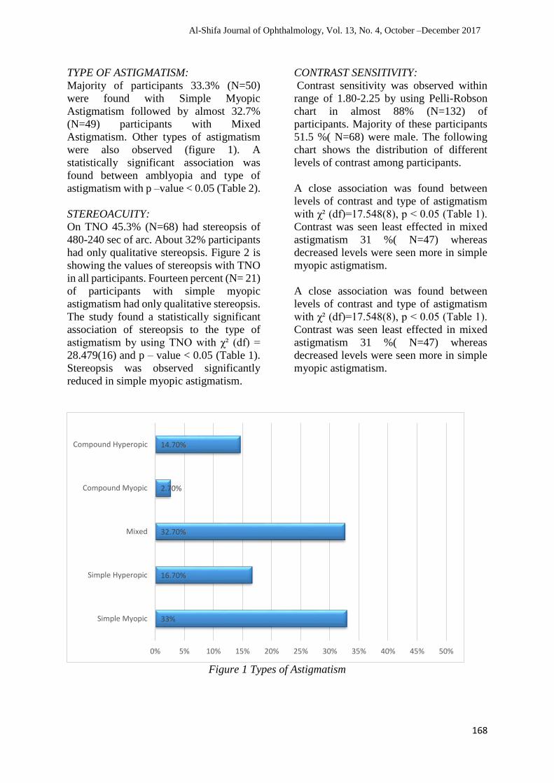
TYPE OF ASTIGMATISM:
Majority of participants 33.3% (N=50)
were found with Simple Myopic
Astigmatism followed by almost 32.7%
(N=49) participants with Mixed
Astigmatism. Other types of astigmatism
were also observed (figure 1). A
statistically significant association was
found between amblyopia and type of
astigmatism with p –value < 0.05 (Table 2).
STEREOACUITY:
On TNO 45.3% (N=68) had stereopsis of
480-240 sec of arc. About 32% participants
had only qualitative stereopsis. Figure 2 is
showing the values of stereopsis with TNO
in all participants. Fourteen percent (N= 21)
of participants with simple myopic
astigmatism had only qualitative stereopsis.
The study found a statistically significant
association of stereopsis to the type of
astigmatism by using TNO with χ² (df) =
28.479(16) and p – value < 0.05 (Table 1).
Stereopsis was observed significantly
reduced in simple myopic astigmatism.
CONTRAST SENSITIVITY:
Contrast sensitivity was observed within
range of 1.80-2.25 by using Pelli-Robson
chart in almost 88% (N=132) of
participants. Majority of these participants
51.5 %( N=68) were male. The following
chart shows the distribution of different
levels of contrast among participants.
A close association was found between
levels of contrast and type of astigmatism
with χ² (df)=17.548(8), p < 0.05 (Table 1).
Contrast was seen least effected in mixed
astigmatism 31 %( N=47) whereas
decreased levels were seen more in simple
myopic astigmatism.
A close association was found between
levels of contrast and type of astigmatism
with χ² (df)=17.548(8), p < 0.05 (Table 1).
Contrast was seen least effected in mixed
astigmatism 31 %( N=47) whereas
decreased levels were seen more in simple
myopic astigmatism.
Figure 1 Types of Astigmatism
33%
16.70%
32.70%
2.70%
14.70%
0% 5% 10% 15% 20% 25% 30% 35% 40% 45% 50%
Simple Myopic
Simple Hyperopic
Mixed
Compound Myopic
Compound Hyperopic
Al-Shifa Journal of Ophthalmology, Vol. 13, No. 4, October –December 2017

169
Fig 2: percentages of participants with different levels of stereopsis
Figure 3: Distribution of levels of contrast
1.3
6.7
45.3
32
14.7
30-15sec of arc
120-60sec of arc
480-240sec of arc
qualitative streopsis
absent
Levels of Stereopsis
percentages of participants
88
11.3
7.7
0 10 20 30 40 50 60 70 80 90 100
1.80-2.25
1.20-1.65
1.05 0r below
percentages of participants
Azam et al. Astigmatism and Visual Function in Amblyopia

170
Table 2: Statistical Relationship between Type of Astigmatism and different variables
Amblyopic
Eye
Type of Astigmatism N (%)
χ²(df)
p-value *
SM SH MIX CM CH
Right
Left
B0th
9(6) 2(1.3) 18(12) 0(0) 7(5)
7(5) 7(5) 12(8) 0(0) 9(6)
34(23) 16(11) 19(13) 4(3) 6(4)
23.245(8)
0.003
Stereopsis
(sec. of arc)
N (%)
30-15
1(0.7)
120-60
4(2.7)
480-240
16(11)
Qualitative
2 (14)
Absent
8(5.3)
1(0.7) 0(0) 1(0.7) 0(0) 0(0)
4(3) 1(0.7) 5(3) 0(0) 0(0)
16(11) 11(7) 32(21) 3(2) 6(4)
2(14) 11(7) 6(4) 1(0.7) 9(6)
8(5.3) 2(1.3) 5(3.3) 0(0) 7(5)
28.47(16)
0.002
Levels of
Contrast
1.05 or
below
1.20-1.65
1.80-2.25
2 (1.3) 0(0) 1(0.7) 0(0) 4(3)
6(4) 1(0.7) 1(0.7) 1(0.7) 2(1.3)
42(28) 24(16) 47(31) 3(2) 16(11)
17.548(8)
0.025
*SM: Simple myopic, SH: Simple hyperopic, CM: Compound myopic, CH: Compound hyperopic
Discussion:
In the present study, most common type of
astigmatism found in amblyopic children
was simple myopic astigmatism (33%)
followed by mixed astigmatism (32.7%).
This study had also found a statistically
significant association between type of
astigmatism and amblyopia (p<0.05).
These two types of astigmatism were also
reported in another study in which simple
myopic and mixed astigmatism were seen
in astigmatic amblyopes due to significant
impact causing reduction in acuity for
horizontal gratings 8. However, compound
hyperopic astigmatism was also reported
with higher prevalence in amblyopic
children than other types of astigmatism 9.
In contrast to previous studies this study
Al-Shifa Journal of Ophthalmology, Vol. 13, No. 4, October –December 2017
171
had ruled out the common types of
astigmatism leading towards Meridian
Amblyopia on the basis of foci 10. In this
study visual functions were also determined
and were seem to be affected by type of
astigmatism (p<0.05). Most of the
participants (88%) had normal levels of
contrast this result is not consistent with
previous studies which supported the
reduction of contrast in amblyopes 11. The
present study normal levels of contrast were
reported in majority of participants (31%)
with mixed astigmatism (p<0.05). Whereas
in case of simple myopic astigmatism
decreased levels of contrast were more as
compare to other types of astigmatism.
These results were different from a
previously conducted study in which
contrast was found reduction in hyperopic
astigmatism 12. The result is same as in
another study conducted earlier 5. Likewise,
another study in Great Britain showed the
normal values of contrast in majority of
astigmatic amblyopes (p>0.05) 13. This
study depicted that the factor which seems
to be related to normal levels of contrast is
use of spectacles by participants. All of the
participants were follow up amblyopes and
were using glasses reported normal
contrast.
In this study, levels of stereopsis were also
measured through different charts. Levels
of stereopsis varied depending on type of
astigmatism and density of amblyopia.
Previously conducted studies showed a
reduction in stereopsis related to astigmatic
amblyopia 5,7.
This reduced stereopsis may be due to
difficulty in fusion of images in
astigmatism which is a more complicated
phenomenon in astigmatism as compared to
pure spherical refractive error 14. However,
in this study gross stereopsis was reported
but with reduced fine stereopsis. This might
be due to large number of participants who
were using glasses and we tested their
stereoacuity with glasses that’s why only
fine stereopsis was found missing. A
statistically significant association was
recorded between types of astigmatism and
stereoacuity (p<0.05). these results were
supported by a previous study in which
reduced stereoacuity was reported in
astigmatic participants 15. However, in
present study along with amblyopia might
be another contributing factor towards
reduced stereopsis.
The results of this study exhibited that
colour vision tests were consistent with
previous studies and colour vision was
recorded normal 16. However, another study
showed reduction in colour vision 17. There
is no certain reason for reduction in colour
vision, probably it might be related to
suppression of fovea and decrease in visual
acuity 18.
The strength of the study was that it
included patients from different areas of
Pakistan as it was a hospital-based study.
Moreover, the use of appropriate
instrumentation was also a strong point.
Every participant included in study was
selected after examination by trained
optometrist and ophthalmologist.
The main limitation of study was small
sample size and convenient sampling as the
time duration was limited. The data were
collected under supervision of qualified
doctors with latest technology. The study
showed a significant impact of astigmatism
on developing amblyopia in children. At
the same time visual functions were found
to be affected in astigmatism related
amblyopia.
Conclusions:
The study showed a significant impact of
astigmatism on developing amblyopia in
children. At the same time visual functions
were found to be affected in astigmatism
related amblyopia. These results had
showed an increase need for screening and
to provide spectacle treatment as earlier as
possible. Moreover, quality of life greatly
depends on visual functions. Reduced
contrast and stereopsis can make a child
Azam et al. Astigmatism and Visual Function in Amblyopia

172
dependent and unable to cope with different
challenges of life as compare to a normal
subject. This can make a child unable to
express all of his/her energies. As children
are future of nation, so efforts should be
made to lessen this burden of astigmatism
related meridional amblyopia.
Recommendations:
These results had showed an increase need
for screening and to provide spectacle
treatment as earlier as possible. Moreover,
it had revealed that there is a need for
proper counseling of parents/guardians of
children reported in hospitals for good
compliance with any given treatment.
References:
1. Wolffsohn JS, Bhogal G, Shah S. Effect
of uncorrected astigmatism on vision. J
Cataract Refract Surg. 2011;37(3):454–
60.
2. Friedburg D, Klöppel KP. [Early
correction of hyperopia and
astigmatism in children leads to better
development of visual acuity]. Klin
Monbl Augenheilkd [Internet]. 1996 Jul
[cited 2017 Feb 8];209(1):21–4.
3. Read SA, Vincent SJ, Collins MJ. The
visual and functional impacts of
astigmatism and its clinical
management. Ophthalmic Physiol Opt
[Internet]. 2014 May [cited 2017 Feb
8];34(3):267–94.
4. Shah M, Khan MT, Khan MD, Habib ur
Rehman H ur. Clinical profile of
amblyopia in Pakistani children age 3 to
14 years. J Coll Physicians Surg Pak
[Internet]. 2005 Jun [cited 2016 Aug
29];15(6):353–7.
5. Harvey EM, Dobson V, Miller JM,
Clifford CE, M. HE. Amblyopia in
astigmatic children: Development and
treatment. J Vis [Internet]. 2005 Dec 1
[cited 2017 Jan 1];5(12):38–38.
6. Freeman RD, Thibos LN. Contrast
sensitivity in humans with abnormal
visual experience. J Physiol [Internet].
1975 Jun [cited 2017 Feb
9];247(3):687–710.
7. Wallace DK, Lazar EL, Melia M, Birch
EE, Holmes JM, Hopkins KB, et al.
Stereoacuity in children with
anisometropic amblyopia. J Am Assoc
Pediatr Ophthalmol Strabismus
[Internet]. 2011 Oct [cited 2017 Feb
9];15(5):455–61.
8. Dobson V, Miller JM, Harvey EM,
Mohan KM. Amblyopia in astigmatic
preschool children. Vision Res.
2003;43(9):1081–90.
9. Xiao X, Liu W-M, Zhao W-X, Wang Y,
Zhang Y-J. [Prevalence of astigmatism
in 2023 children with amblyopia].
Zhongguo Dang Dai Er Ke Za Zhi
[Internet]. 2011 Jun [cited 2017 Feb
8];13(6):462–5
10. Huynh SC, Wang XY, Ip J, Robaei D,
Kifley A, Rose KA, et al. Prevalence
and associations of anisometropia and
aniso-astigmatism in a population based
sample of 6 year old children. Br J
Ophthalmol [Internet]. 2006 May 1
[cited 2017 Oct 15];90(5):597–601
11. Mitchell DE, Freeman RD, Millodot M,
Haegerstrom Meridional amblyopia:
Evidence for modification of the human
visual system by early visual
experience. Vision Res.
1973;13(3):535–I.
12. Mitchell DE, Wilkinson F. The effect of
early astigmatism on the visual
resolution of gratings. J Physiol
[Internet]. 1974 Dec 1 [cited 2017 Mar
3];243(3):739–56.
13. Harvey EM, Dobson V, Miller JM,
Clifford-Donaldson CE. Amblyopia in
astigmatic children: Patterns of deficits.
Vision Res. 2007;47(3):315–26.
14. Sethi Sumita1,* KR. impact of
refractive error on stereopsis in school
going children of Rural Huryana. Indian
J Clin Exp Ophthalmol Year 2015,
Vol 1, Issue 2 First page ( 91) Last
page ( 94)
15. 15.Yang J-W, Huang T-Y, Yang K-J,
Lee J-S, Ku W-C, Yeung L, et al. The
effects of hyperopic and astigmatic
ametropia on stereoacuity by Titmus
Al-Shifa Journal of Ophthalmology, Vol. 13, No. 4, October –December 2017

173
stereo test. Taiwan J Ophthalmol.
2012;2(1):22–4.
16. Rajavi Z, Sabbaghi H, Baghini AS,
Yaseri M, Sheibani K, Norouzi G.
Prevalence of Color Vision Deficiency
and its Correlation with Amblyopia and
Refractive Errors among Primary
School Children. J Ophthalmic Vis Res
[Internet]. 2015 [cited 2017 Oct
22];10(2):130–8.
17. Koçak-Altintas AG, Satana B, Koçak I,
Duman S. Visual acuity and color
vision deficiency in amblyopia. Eur J
Ophthalmol [Internet]. [cited 2017 Mar
4];10(1):77–81.
18. Almog Y, Nemet A. The correlation
between visual acuity and color vision
as an indicator of the cause of visual
loss. Am J Ophthalmol [Internet]. 2010
Jun [cited 2017 Nov 7];149(6):1000–4.
Authors Contribution:
Concept and Design: Sana Azam, Hina Sharif
Data Collection / Assembly: Sana Azam
Drafting: Sana Azam, Shadab Hassan
Statistical expertise: Hina Sharif
Critical revision: Shadab Hassan
Azam et al. Astigmatism and Visual Function in Amblyopia
174
Outcomes of Combined Extraction with Intraocular Lens
Implant by A Fellow During Glaucoma Fellowship Yousaf Jamal Mahsood1, Saima Farooq2, Mahmood Ali3, Farah Akhtar3
Abstract
Objectives: To determine the outcomes of combined extraction with intraocular lens implant
performed by a fellow during glaucoma fellowship.
Study design: A retrospective study.
Methods: All the combined cataract and glaucoma surgeries performed by a glaucoma fellow
during 1st November 2015 to 30th June 2016 were included in the study. All of the surgeries
were performed under supervision of faculty and all intraoperative and early postoperative
complications were recorded. The primary outcomes were the change in mean intraocular
pressure (IOP) and best corrected visual acuity (BCVA) from the baseline. Secondary
outcomes were the complications rate during surgery and in early postoperative phase.
Results: Total 19 eyes of 19 patients were included in the study, among which 11 (57.9%)
were males and 8 (42.1%) were right eyes. Best corrected visual acuity (BCVA) improved in
17 (89.5%) eyes from the baseline while 1 eye showed no change and deterioration in 1 eye.
There was improvement in IOP from baseline 20.11 + 7.36 mmHg to 13.56 + 3.28 mmHg (p
= 0.01).
Conclusion: Combined cataract and glaucoma surgeries performed by glaucoma fellow have
good results in terms of IOP control and BCVA. Al-Shifa Journal of Ophthalmology 2017;
13(4): 174-79. © Al-Shifa Trust Eye Hospital, Rawalpindi, Pakistan.
1. Lady Reading Hospital, Peshawar
2. Hayatabad Medical Complex,
Peshawar
3. Al-Shifa Trust Eye Hospital,
Rawalpindi
Originally Received: 13 August 2017
Revised: 21 October 2017
Accepted: 23 November 2017
Correspondence to:
Dr. Yousaf Jamal Mahsood,
Department of Ophthalmology,
Lady Reading Hospital, Peshawar.
E-mail:[email protected]
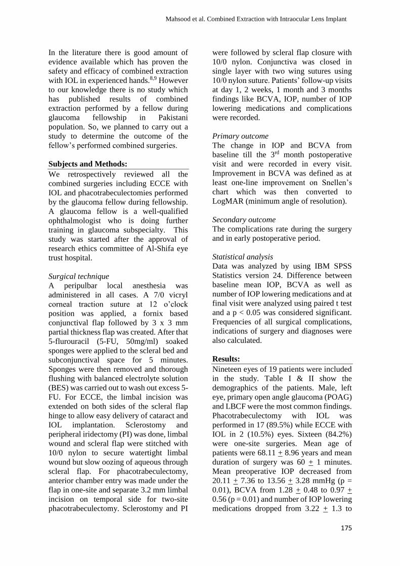
Introduction:
Cataract is the leading cause of blindness
worldwide followed by glaucoma.1
Although both are unrelated to each other
in pathogenesis yet they can coexist at the
same time. This can be explained by the
argument that these entities are age-related
and there is fair amount of chance that as
the life-expectancy increases we will see
more patients with coexistent cataract and
glaucoma. In such situations, combining
cataract and glaucoma surgery in one sitting
is considered as a primary option by many
glaucoma surgeons.2-5 Previously,
conventional extracapsular cataract
extraction (ECCE) with trabeculectomy
was the procedure performed but since the
advent of phacoemulsification, which is
less traumatic to the eye,
phacotrabeculectomy (Phacoemulsification
and trabeculectomy) has gained much
acceptance worldwide.6,7
ORIGINAL ARTICLE
175
In the literature there is good amount of
evidence available which has proven the
safety and efficacy of combined extraction
with IOL in experienced hands.8,9 However
to our knowledge there is no study which
has published results of combined
extraction performed by a fellow during
glaucoma fellowship in Pakistani
population. So, we planned to carry out a
study to determine the outcome of the
fellow’s performed combined surgeries.
Subjects and Methods:
We retrospectively reviewed all the
combined surgeries including ECCE with
IOL and phacotrabeculectomies performed
by the glaucoma fellow during fellowship.
A glaucoma fellow is a well-qualified
ophthalmologist who is doing further
training in glaucoma subspecialty. This
study was started after the approval of
research ethics committee of Al-Shifa eye
trust hospital.
Surgical technique
A peripulbar local anesthesia was
administered in all cases. A 7/0 vicryl
corneal traction suture at 12 o’clock
position was applied, a fornix based
conjunctival flap followed by 3 x 3 mm
partial thickness flap was created. After that
5-flurouracil (5-FU, 50mg/ml) soaked
sponges were applied to the scleral bed and
subconjunctival space for 5 minutes.
Sponges were then removed and thorough
flushing with balanced electrolyte solution
(BES) was carried out to wash out excess 5-
FU. For ECCE, the limbal incision was
extended on both sides of the scleral flap
hinge to allow easy delivery of cataract and
IOL implantation. Sclerostomy and
peripheral iridectomy (PI) was done, limbal
wound and scleral flap were stitched with
10/0 nylon to secure watertight limbal
wound but slow oozing of aqueous through
scleral flap. For phacotrabeculectomy,
anterior chamber entry was made under the
flap in one-site and separate 3.2 mm limbal
incision on temporal side for two-site
phacotrabeculectomy. Sclerostomy and PI
were followed by scleral flap closure with
10/0 nylon. Conjunctiva was closed in
single layer with two wing sutures using
10/0 nylon suture. Patients’ follow-up visits
at day 1, 2 weeks, 1 month and 3 months
findings like BCVA, IOP, number of IOP
lowering medications and complications
were recorded.
Primary outcome
The change in IOP and BCVA from
baseline till the 3rd month postoperative
visit and were recorded in every visit.
Improvement in BCVA was defined as at
least one-line improvement on Snellen’s
chart which was then converted to
LogMAR (minimum angle of resolution).
Secondary outcome
The complications rate during the surgery
and in early postoperative period.
Statistical analysis
Data was analyzed by using IBM SPSS
Statistics version 24. Difference between
baseline mean IOP, BCVA as well as
number of IOP lowering medications and at
final visit were analyzed using paired t test
and a p < 0.05 was considered significant.
Frequencies of all surgical complications,
indications of surgery and diagnoses were
also calculated.
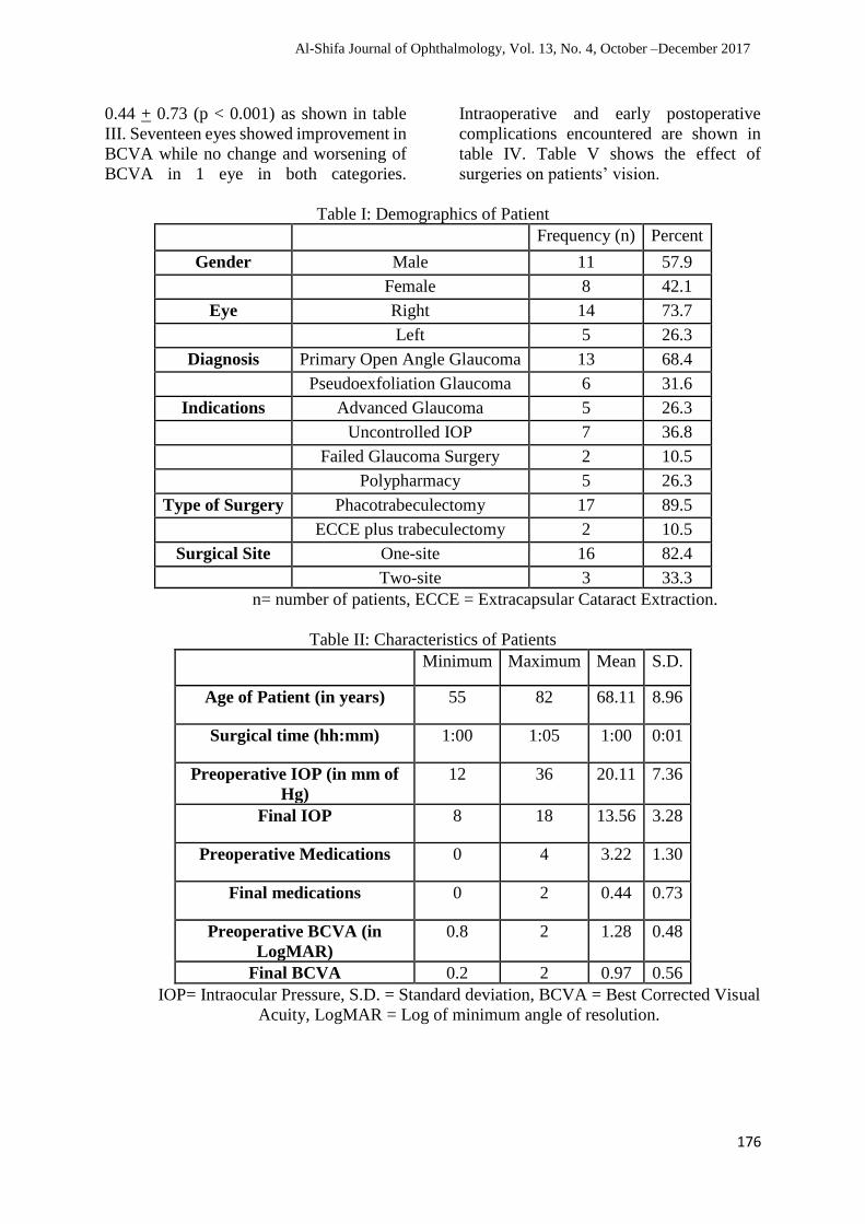
Results:
Nineteen eyes of 19 patients were included
in the study. Table I & II show the
demographics of the patients. Male, left
eye, primary open angle glaucoma (POAG)
and LBCF were the most common findings.
Phacotrabeculectomy with IOL was
performed in 17 (89.5%) while ECCE with
IOL in 2 (10.5%) eyes. Sixteen (84.2%)
were one-site surgeries. Mean age of
patients were 68.11 + 8.96 years and mean
duration of surgery was 60 + 1 minutes.
Mean preoperative IOP decreased from
20.11 + 7.36 to 13.56 + 3.28 mmHg (p =
0.01), BCVA from 1.28 + 0.48 to 0.97 +
0.56 (p = 0.01) and number of IOP lowering
medications dropped from 3.22 + 1.3 to
Mahsood et al. Combined Extraction with Intraocular Lens Implant
176
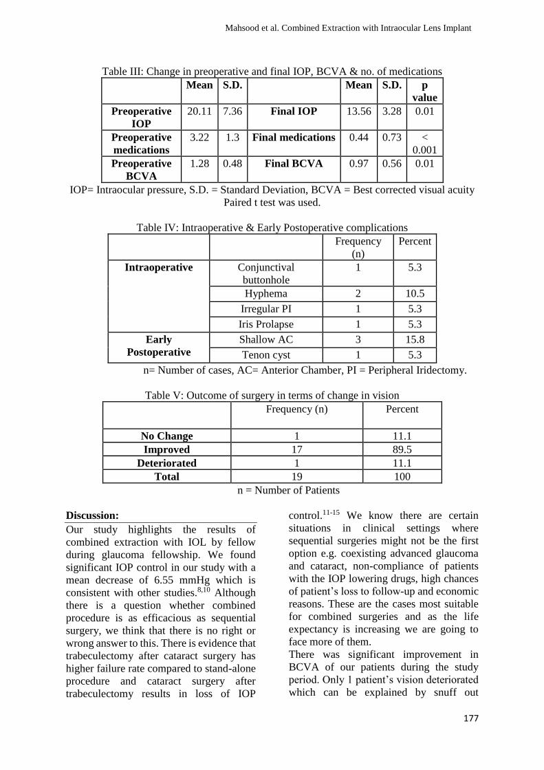
0.44 + 0.73 (p < 0.001) as shown in table
III. Seventeen eyes showed improvement in
BCVA while no change and worsening of
BCVA in 1 eye in both categories.
Intraoperative and early postoperative
complications encountered are shown in
table IV. Table V shows the effect of
surgeries on patients’ vision.
Table I: Demographics of Patient
Frequency (n) Percent
Gender Male 11 57.9
Female 8 42.1
Eye Right 14 73.7
Left 5 26.3
Diagnosis Primary Open Angle Glaucoma 13 68.4
Pseudoexfoliation Glaucoma 6 31.6
Indications Advanced Glaucoma 5 26.3
Uncontrolled IOP 7 36.8
Failed Glaucoma Surgery 2 10.5
Polypharmacy 5 26.3
Type of Surgery Phacotrabeculectomy 17 89.5
ECCE plus trabeculectomy 2 10.5
Surgical Site One-site 16 82.4
Two-site 3 33.3
n= number of patients, ECCE = Extracapsular Cataract Extraction.
Table II: Characteristics of Patients Minimum Maximum Mean S.D.
Age of Patient (in years) 55 82 68.11 8.96
Surgical time (hh:mm) 1:00 1:05 1:00 0:01
Preoperative IOP (in mm of
Hg)
12 36 20.11 7.36
Final IOP 8 18 13.56 3.28
Preoperative Medications 0 4 3.22 1.30
Final medications 0 2 0.44 0.73
Preoperative BCVA (in
LogMAR)
0.8 2 1.28 0.48
Final BCVA 0.2 2 0.97 0.56
IOP= Intraocular Pressure, S.D. = Standard deviation, BCVA = Best Corrected Visual
Acuity, LogMAR = Log of minimum angle of resolution.
Al-Shifa Journal of Ophthalmology, Vol. 13, No. 4, October –December 2017
177
Table III: Change in preoperative and final IOP, BCVA & no. of medications
Mean S.D. Mean S.D. p
value
Preoperative
IOP
20.11 7.36 Final IOP 13.56 3.28 0.01
Preoperative
medications
3.22 1.3 Final medications 0.44 0.73 <
0.001
Preoperative
BCVA
1.28 0.48 Final BCVA 0.97 0.56 0.01
IOP= Intraocular pressure, S.D. = Standard Deviation, BCVA = Best corrected visual acuity
Paired t test was used.
Table IV: Intraoperative & Early Postoperative complications
Frequency
(n)
Percent
Intraoperative Conjunctival
buttonhole
1 5.3
Hyphema 2 10.5
Irregular PI 1 5.3
Iris Prolapse 1 5.3
Early
Postoperative
Shallow AC 3 15.8
Tenon cyst 1 5.3
n= Number of cases, AC= Anterior Chamber, PI = Peripheral Iridectomy.
Table V: Outcome of surgery in terms of change in vision
Frequency (n) Percent
No Change 1 11.1
Improved 17 89.5
Deteriorated 1 11.1
Total 19 100
n = Number of Patients
Discussion:
Our study highlights the results of
combined extraction with IOL by fellow
during glaucoma fellowship. We found
significant IOP control in our study with a
mean decrease of 6.55 mmHg which is
consistent with other studies.8,10 Although
there is a question whether combined
procedure is as efficacious as sequential
surgery, we think that there is no right or
wrong answer to this. There is evidence that
trabeculectomy after cataract surgery has
higher failure rate compared to stand-alone
procedure and cataract surgery after
trabeculectomy results in loss of IOP
control.11-15 We know there are certain
situations in clinical settings where
sequential surgeries might not be the first
option e.g. coexisting advanced glaucoma
and cataract, non-compliance of patients
with the IOP lowering drugs, high chances
of patient’s loss to follow-up and economic
reasons. These are the cases most suitable
for combined surgeries and as the life
expectancy is increasing we are going to
face more of them.
There was significant improvement in
BCVA of our patients during the study
period. Only 1 patient’s vision deteriorated
which can be explained by snuff out
Mahsood et al. Combined Extraction with Intraocular Lens Implant
178
phenomenon that can happen in advanced
glaucoma patients. One downside of
combined procedure is the delayed visual
recovery which may take 4-6 weeks
because of early hypotony, inflammation
and wound healing. With the
phacotrabeculectomy, this recovery time
has been shortened to 3-4 weeks and may
be in near future with new techniques we
may be able to overcome this shortcoming.
During intraoperative stage we had few
complications like conjunctival buttonhole,
hyphema and iris prolapse which were
managed on the table with no
consequences. Shallow anterior chamber
was the most common early postoperative
complication which was managed
conservatively. We did not look in to the
long-term complications and the failure
rates of our surgeries which will give us
more details.
The strength of our study is that it is the first
study in Pakistani population where the
results of a fellow are published. However,
there are few limitations like retrospective
design, smaller sample size and no direct
comparison with the experienced surgeon’s
results. So, we recommend addressing all
these shortcomings and conducting a
multicenter approach where all the fellows
are involved, this will be a very good
project for investigators in future.
Conclusion:
Combined surgeries done by a glaucoma
fellow is safe and effective as compared to
the international data available.
Phacotrabeculectomy offers shorter visual
rehabilitation time as compared to ECCE
with trabeculectomy.
References:
1. Thylefors B, Negrel AD,
Pararajasegaram R, Dadzie KY. Global
data on blindness. Bulletin of the world
health organization. 1995;73(1):115-
21.
2. Gimbel HV, Meyer D. Small incision
trabeculotomy combined with
phacoemulsification and intraocular
lens implantation. Journal of Cataract &
Refractive Surgery. 1993 Jan
1;19(1):92-6.
3. Parihar JK, SM R, Sahoo PK, Misra RP,
Vats DP, SM V, Kamath AP, Rodrigues
FE. Phacotrabeculectomy versus
conventional combined technique in
coexisting glaucoma and cataract.
Medical journal, Armed Forces India.
2005 Feb;61(2):139-42.
4. Carlson DW, Alward WL, Barad JP,
Zimmerman MB, Carney BL. A
randomized study of mitomycin
augmentation in combined
phacoemulsification and
trabeculectomy. Ophthalmology. 1997
Apr 1;104(4):719-24.
5. Wedrich A, Menapace R, Radax U,
Papapanos P. Long-term results of
combined trabeculectomy and small
incision cataract surgery. Journal of
Cataract & Refractive Surgery. 1995
Jan 1;21(1):49-54.
6. Wishart PK, Austin MW. Combined
cataract extraction and trabeculectomy:
phacoemulsification compared with
extracapsular technique. Ophthalmic
surgery. 1993 Dec;24(12):814-21.
7. Percival SP. Glaucoma triple procedure
of extracapsular cataract extraction,
posterior chamber lens implantation,
and trabeculectomy. British journal of
ophthalmology. 1985 Feb 1;69(2):99-
102.
8. Lochhead J, Casson RJ, Salmon JF.
Long term effect on intraocular
pressure of phacotrabeculectomy
compared to trabeculectomy. British
journal of ophthalmology. 2003 Jul
1;87(7):850-2.
9. Chang L, Thiagarajan M, Moseley M,
Woodruff S, Bentley C, Khaw PT,
Bloom P. Intraocular pressure outcome
in primary 5FU phacotrabeculectomies
compared with 5FU trabeculectomies.
Journal of glaucoma. 2006 Dec
1;15(6):475-81.
10. Murthy SK, Damji KF, Pan Y, Hodge
WG. Trabeculectomy and
Al-Shifa Journal of Ophthalmology, Vol. 13, No. 4, October –December 2017
179
phacotrabeculectomy, with mitomycin-
C, show similar two-year target IOP
outcomes. Canadian Journal of
Ophthalmology. 2006 Jan 1;41(1):51-9.
11. Husain R, Liang S, Foster PJ, Gazzard
G, Bunce C, Chew PT et al. Cataract
surgery after trabeculectomy: the effect
on trabeculectomy function. Archives
of ophthalmology. 2012 Feb
1;130(2):165-70.
12. Inal A, Bayraktar S, Inal B, Bayraktar
Z, Yilmaz ÖF. Intraocular pressure
control after clear corneal
phacoemulsification in eyes with
previous trabeculectomy: a controlled
study. Acta Ophthalmologica. 2005 Oct
1;83(5):554-60.
13. Klink J, Schmitz B, Lieb WE, Klink T,
Grein HJ, Sold-Darseff J et al. Filtering
bleb function after clear cornea
phacoemulsification: a prospective
study. British journal of
ophthalmology. 2005 May 1;89(5):597-
601.
14. Rebolleda G, Muñoz-Negrete FJ.
Phacoemulsification in eyes with
functioning filtering blebs: a
prospective study. Ophthalmology.
2002 Dec 31;109(12):2248-55.
15. Ehrnrooth P, Lehto I, Puska P,
Laatikainen L. Long‐term outcome of
trabeculectomy in terms of intraocular
pressure. Acta Ophthalmologica. 2002
Jun 1;80(3):267-71.
Authors Contribution:
Concept and Design: Yousaf Jamal Mahsood, Farah Akhtar
Data Collection / Assembly: Yousaf Jamal Mahsood, Mahmood Ali
Drafting: Yousaf Jamal Mahsood, Saima Farooq
Statistical expertise: Yousaf Jamal Mahsood, Saima Farooq
Critical revision: Mahmood Ali, Farah Akhtar
Mahsood et al. Combined Extraction with Intraocular Lens Implant
180
Pattern of Ocular Diseases in Children Presenting at Eye
Out Patient Department, Khyber Teaching Hospital,
Peshawar Hussain Ahmad1, Muhammad Naeem2, Yousaf Jamal Mahsood3, Muhammad Sajid Khan1,
Maqsood Ahmad1, Ubaidullah1
ABSTRACT
Objective: To report ocular disease pattern in children in order to enable early diagnosis and
treatment of such conditions.
Subjects and Methods: This was a descriptive cross-sectional study conducted in Department
of Ophthalmology, Khyber Teaching Hospital, Peshawar from 1st September 2014 to 31 august
2015. In this study 4800 children in the age group of up to 16 years were included which
presented to eye OPD. A proforma was developed to record personal history, clinical
examination and treatment given.
Results: Total of 4800 children attended eye OPD during this study duration which is 25.34%
of total patients (18940). Out of this 3100(64.58%) were male and 1700 (35.42%) were female
children. Conjunctiva was the most commonly involved structure i.e. in 1850(38.54%)children,
followed by NLD blockage involving 1040(21.66%) children. Refractive errors were detected
in 775(16.14%) children while disorders of lens accounted for 607(12.64%). Regarding
management 2004 (41.75%) children got medical treatment alone while 1966(40.96%)
children got both medical and surgical treatment. In other treatment modalities, 783(16.31%)
children were treated optically and orthoptically and 47(0.98%) children were referred to
specialized units.
Conclusion: Most children affected by eye conditions were male and conjunctival diseases
were the commonest. This is followed by NLD blockage which required probing. Cataracts
were the common disease which required surgery which is preventable blindness. Also
refractive errors were high which need refraction and glasses. Al-Shifa Journal of
Ophthalmology 2017; 13(4): 180-85. © Al-Shifa Trust Eye Hospital, Rawalpindi, Pakistan.
1. Department of Ophthalmology, Khyber
Teaching Hospital, Peshawar
2. Department of Ophthalmology,
Hayatabad Medical Complex,
Peshawar.
3. Department of Ophthalmology, Lady
Reading Hospital, Peshawar.
Originally Received: 07 September 2017
Revised: 08 December 2017
Accepted: 14 December 2017
Correspondence to:
Dr. Hussain Ahmad
Registrar, Department of Ophthalmology,
Khyber Teaching Hospital, Peshawar
Email: [email protected]
Introduction
There is wide variation of presentation of
eye diseases worldwide 1,2. Factors
affecting the type of eye diseases include
age, sex, residence, socioeconomic,
occupation, geographical, racial, dietary,
customs, tradition, and the major
environmental factors prevalent in that
region. Children are mostly affected by eye
diseases that can be treated. Ocular diseases
in children are important because of its
impact on child development. These
disorders are due to causes in prenatal,
neonatal and childhood periods.
ORIGINAL ARTICLE

181
The prevalence of blindness in children
ranges from approximately 0.3/1000
children in developed countries to 1.5/1000
in developing countries 4. Globally there
are estimated to be 1.4 million blind
children, almost three quarters of who live
in the poorest regions of Africa and Asia5.
Almost 50% of all childhood blindness in
poor countries are avoidable. Population-
based data on the prevalence and major
causes of childhood blindness are essential
for every country, in order to set priorities
for control programs and to monitor the
changing patterns over time.
According to a population census in 1998,
Pakistan has an estimated population of 142
million in 2003. It is estimated that 40% of
the population is below 16 years of age. The
prevalence of blindness in children in
Pakistan is estimated to be about 10 per
10,000 children, which means there are
about 60,000 blind children. A further
100,000 to180,000 children are estimated
to have low vision 6. Cataract is the main
preventable cause of blindness. Almost
190,000 children worldwide are blind from
cataract alone7. Therefore, children need
special attention for early diagnosis and
treatment of their ocular diseases so that
they can live happy and productive life.
This will lessen economic burden on their
family and they will be better for their
country.
Subjects and Methods:
This was a hospital based descriptive cross-
sectional study conducted at eye
department, Khyber teaching hospital,
Peshawar from 1st September 2014 to 31st
august 2015. Children up to ages 16 years
were included in this study. A proforma
was developed to record personal history,
clinical examination and treatment given.
During the study, a detailed history of each
child, father’s occupation, immunization,
was asked. Children of 0-3 years were
examined with a magnifying loop. All
children of age group 7-15 were examined
on slit lamp and visual acuity checked with
illiterate Snellen E chart directly and with
pinhole. Refraction was performed under
cycloplegia. Posterior segment
examination was performed after pupil
dilatation with direct and indirect
ophthalmoscope and with 78D.Intraocular
pressure was checked with Perkin’s
tonometer. Squint assessment was done
with prisms and tests of stereopsis.
Treatment was medical, surgical and
optical.
Results:
A total of 4800 children were examined and
enrolled in this study at Eye department
Khalifa Gul Nawaz Teaching Hospital,
Bannu. In this 3700 (64.58%) were male
and 1700 (35.42%) were female. Their age
wise distribution is given in table1.
In 1850(38.54%) children, conjunctiva was
involved (table2). In conjunctival diseases,
vernal keratoconjunctivitis was diagnosed
in 863 (46.64%), bacterial conjunctivitis in
426 (23.04%), follicular conjunctivitis in
395(21.35%), vitamin A deficiency in
111(6%) and subconjunctival hemorrhage
in 55(2.97%) children. Cornea and sclera
were involved in 195(4.06%) children.
Corneal foreign body was present in
107(54.87%), corneal ulcer in 42 (21.55%),
Corneoscleral perforation in 37(18.97%)
and keratoconus was diagnosed in 9
(4.61%) children. Lens disorders were seen
in 607(12.64%) of total children which
were either congenital cataracts or
traumatic cataracts. Disorders of the whole
globe were present in 52 (1.08%) children
in which buphthalmos was in 33 (63.46%)
and phthisis in 19 (36.54%). Disorders of
vitreous and retina were present in
86(1.79%) of total children, including
retinoblastoma in 26(30.23%), retinitis
pigmentosa in 24(27.90%), Stargardt’s
disease in 15(17.44%), congenital vitreous
degenerations in 13(15.11%) and retinal
detachment in 8(9.32%) cases.
Lids and orbit were involved in 75 (1.56%)
of total children. In the diseases of lid and
Ahmad et al. Ocular Diseases in Children

182
orbit, blepharitis was seen in 28(37.35%)
children, chalazion in 21(28%), stye in
13(17.33%), ptosis in 8(10.66%) and
congenital malformation were present in
5(6.66%) cases. Nasolacrimal duct was
involved in 1040(21.66%) of children.
Refractive errors were present in
775(16.14%) of children in which
astigmatism was seen in 315(40.65%),
hypermetropia in 278(35.87%) and myopia
in 182(23.48%) of patients. Ocular motility
was affected in 106(2.24%) children.
Among these 76(71.39%) were convergent
squints,25(23.38%) were divergent squints
and 6(5.33%) cases were of Duane’s
syndrome.
Medical treatment was provided to 2004
(41.75%) patients, while medical and
surgical treatment was given to
1966(40.96%) children. Optical and
orthoptic correction was advised in
783(16.31%) children. In addition,
47(0.98%) children were referred to
specialized units for further management.
Table 1: Age wise distribution of patients
Patient’s age Numbers Percentage
0-5 1752 36.51%
6-10 2192 45.66%
11-16 856 17.83%
Table 2: Disease wise distribution of patients
Disease No of patients
Conjunctiva involved 1850(38.54%)
Cornea/sclera involved 196(4.06%)
Globe involved 52(1.08%)
Lens disorders 607(12.64%)
Uveitis 14(0.29%)
Vitreous and retina 86(1.79%)
Ocular motility 106(2.24%)
Lids and orbit 75(1.56%)
NLD related disorders 1040(21.66%)
Refractive errors 775(16.14%)
Al-Shifa Journal of Ophthalmology, Vol. 13, No. 4, October –December 2017

183
Discussion:
Total 4800 children were examined, of
which 64.58% were males and 35.42%
were female children. These findings are
comparable to a study done at Mayo
Hospital Lahore, with 63.6% male and
36.4% female.
In this study conjunctiva was involved in
38.54% cases. Vernal keratoconjunctivitis
was the most common disorder affecting
46.64% of children and is a recurrent,
bilateral, external, ocular inflammation
primarily affecting boys and young adults
living in warm, dry climates. Bacterial
conjunctivitis which was found in 23.04%
patients. Bacterial conjunctivitis was
diagnosed on the presence of purulent
discharge in eye. This is different from
results obtained by Awan and Usman i.e.
34.18%.8
Conjunctivitis of the newborn is a very
serious problem in many parts of the
developing world. The great damage to
sight is infection, which involves the
cornea. As the condition usually affects
both eyes, the tragedy of an otherwise
healthy child becoming blind is avoidable
[9]. Bacterial conjunctivitis could be due
several factors i.e. due to overcrowding,
poor hygiene, poor nutrition and lack of
health education of mothers, as the children
were coming from very poor community
having large family size, and in most of
families, absence of mothers from home, as
they were also working.
Corneal diseases accounted for 4.06% of all
the childhood ocular diseases. This is less
in comparison to other studies. Afghani et
al in a survey of blind school reported
corneal diseases as a cause of blindness in
12% 10. Rahi et al in their study in a blind
school in India showed 26.4% of corneal
diseases responsible for visual impairment
and blindness 11. Most of corneal problems
were due to trauma and corneal foreign
body. Large families and lack of proper
care put children at risk of sustaining
trauma. This is in comparison with a study
conducted at Hayatabad Medical Complex
Peshawar which reported 49% of childhood
ocular trauma 12. Corneal ulcers in this
study were mostly due to foreign body,
bacterial keratitis, and herpes infections and
lime burn. Vitamin A deficiency may be a
contributing factor to corneal opacity,
which was found in 6% children and which
could not be confirmed due to lack of
laboratory support. According to WHO
report about 3 million children have
clinically xerophthalmia throughout the
world13.Majority of corneal ulcers follow
the occurrence of often trivial corneal
abrasions. In a study by Jatoi S, causes of
corneal ulcers were found to be trauma in
63.5% of cases 14.
In Pakistan, various studies have been
carried out which have so far suggested
Vitamin A deficiency as a problem. No in-
depth survey has been carried out. A survey
in four districts of Punjab covering 21
villages showed prevalence of Vitamin A
deficiency in children as 6.4 % and among
these, frequency in children of 5-6 years
age was 50 %15.
Cataract in this study was 12.64% of cases,
including all types, congenital, traumatic,
and developmental and it found to be the
cause of monocular blindness. Cataract is
leading cause of preventable blindness
globally. Bilateral congenital cataract is the
most common cause of treatable childhood
blindness. Nuclear cataract is usually
present at birth and is non-progressive
whereas lamellar cataract usually develops
later and is progressive16.The major causes
of bilateral cataract in South Asia are
idiopathic (50%), hereditary (25%)and
secondary to Rubella infection (25%)11.
In our study ocular motility disorders were
seen in 2.24% cases. Squints are common
all over the world but do not have any
special association with developing
countries or tropical environments17. A
study from Nepal reported that the
Ahmad et al. Ocular Diseases in Children

184
prevalence of squint was 1.6% 18. Diagnosis
of squint at early age is important because
of its amblyogenic effect. Esotropia is more
likely to be amblyogenic than exotropia 19.
Congenital glaucoma causing buphthalmos
constitute about 33(63.46%) cases out of 52
cases of whole globe involvement. The
incidence of congenital glaucoma varies
among different geographic locations and
ethnic groups with the highest recorded
incidence found in the Gypsy population of
Slovakia (1:1250), followed by general
population of Middle East (1:2500) and the
western nations (1:10,000) 20. In our study
there were 26 cases of retinoblastoma. In a
study done at Agha Khan University
hospital, Karachi, 101 cases of
retinoblastoma were reported between
1998-200221. In order to detect
retinoblastoma as early as possible, health
education for parents and heath care
provider and improved training for
ophthalmologist is essential. Genetic
testing of siblings and children for
retinoblastoma and identification of high
risk children would also be helpful.
Refractive errors in our study were reported
in 16.14% of cases. A population-based
study conducted by Memon in 1987- 90
showed that 11.4% of blindness was due to
refractive errors and amblyopia22.
Refractive errors which account mostly for
low vision are the third largest cause of
treatable blindness in Pakistan23. It
accounts for 8% cases of uniocular
blindness in Khyber Pakhtunkhwa province 24. Astigmatism was most common and
accounted for 40.65% of refractive errors in
our study. This is different from other
studies where hypermetropia was common
error of refraction in children 25.
In our study, congintal NLD blockage was
seen in 21.66% which is very high as
compared to other studies done like study
conducted at Khyber Teaching Hospital,
Peshawar which showed NLD blockage in
5.4%26. Eyelid problems (Chalazion, stye)
in our study were 21 and 13 cases
respectively. In countries with warm
climate, the prevalence of staphylococci is
as high as 95% in culture, and sub clinical
and mild lid infections are quite common 27.
Conclusion:
Most common eye disease in this study was
conjunctivitis either allergic or bacterial
which is mostly due to warm and dry
climate and unhygienic conditions in this
area which is comparable to other areas in
Pakistan. Males were affected more than
females and most common age group
affected was between 7-10years. This study
showed that pediatric eye diseases are quite
common and most of them are treatable so
there is need for proper training of
ophthalmic care providers in paedriatic
ophthalmology.
References:
1. Topalovo AV. Incidence of Eye
Diseases in different parts of the World.
Ophthalmology 1984; 6:374-7.
2. Ajayeoba AI, Scott SCO. Risk factors
associated with eye disea se in Ibadan,
Nigeria. Afr J Biomed Res 2002; 5:1-3.
3. Canavan YM, O’Flaherty MJ, Archer
DB. A Ten -year Survey of Eye-Injuries
in Northern Ireland. Br J Ophthal 1980;
64:618-25.
4. Gilbert C, Foster A. Childhood
blindness in the context of VISION
2020—the right to sight. Bull World
Health Organ 2001; 79:227–32.
5. World Health Organization. Preventing
blindness in children. Report of a
WHO/IAPB scientific meeting.
Geneva, Switzerland: WHO, 2000;
PBL/00.71
6. Current status of etiology, prevalence
and distribution of childhood blindness
in Pakistan. A Report, 2003 Pakistan,
Sight Savers International;18.
7. Muhit MA. Childhood cataract: Home
to Hospital. Community Eye
Health.2004; 17:19-22.
Al-Shifa Journal of Ophthalmology, Vol. 13, No. 4, October –December 2017

185
8. Awan AH, Usman J. Acute Bacterial
conjunctivitis in Southern Punjab. Pak J
Ophthalmol. 2000; 16; 30.
9. Prevention of child hood blindness:
Community eye health No 4:
International center for eye health.
1998; 23.
10. Afghani T. Causes of Childhood
Blindness and severe visual impairment
– Survey of the blind Children from a
rural Population and schools for blind in
an urban area. Pak J Ophthalmol. 2000;
16: 16.
11. Rahi J, Sripathi S, Gilbert C, et al.
Childhood blindness in India causes in
1318 blind School student in nine states
Eye 1995: 545-50.
12. Sethi S, Khan MD.Childhood Ocular
Trauma.J Postgrad Med Inst 2001;
15:51-5.
13. F.D, Sight and life slides on Vitamin A
deficiency disorders (VADD), third
edition 2001, Switzerland; 4.
14. Jatoi S, Qureshi M, Laghari N, et al.
Etiological Diagnosis of infective
ulcerative keratitis. Pak J Ophthalmol.
2002; 18: 40.
15. Shah SRA. Dissertation on Magnitude
of Vitamin A Deficiency in poor
communities of four selected districts
of Punjab (Pakistan). Pico/0006/98:
16. Zetterstrom C, Lundvall A. Cataract in
Children. Cataract Refract J 2005;
31:824-40.
17. Sanford J Eye diseases in hot climate
3rd edition; Butter Worth– Heinemann,
Oxford. 1997; 290.
18. Nepal BP,Koirala S,Adhi Kary
S,Sharma AK. Ocular Morbidity in
School children in Khatmundu.Br Jr
Ophthalmol 2003;87:531-34.
19. Mohney BG, Huffaker RK.common
form of childhood exotropia.
Ophthalmology 2003; 110:2093-6.
20. Bejjani BA, Lewis RA, Tomey
KF.Mutations in CYP1B1, the gene for
cytochrome P4501B1, is the
predominant cause of primary
congenital glaucoma in Saudi Arabia.
Am J Hum Genet 1998; 62:325-33.
21. Bhurgri Y,Muzaffar S,Ahmad
R,Ahmad N,Farid N.Retinobastoma in
Karachi Pakistan. Asian Pac J Cancer
Pre 2004;5:159-63.
22. Memon MS. Prevalence and causes of
Blindness in Pakistan. J Pak Med
Assoc.1992; 42:196-8.
23. Durani J.Blindness statistics for
Pakistan. Pak J Ophthalmol 1999; 15:1-
2.
24. Khan MA,Gullab A, Khan
MD.Prevalence of blindness and low
vision in North West Frontier Province
of Pakistan.Pak J Of Ophthalmol
1994;10:39-42.
25. Gilbert C, Awan H.Blindness in
Children (editorial).BMJ 2003;
327:760-61.
26. Sethi S,Sethi MJ,Iqbal R,Khan T.
Pattern Of common eye diseases in
children attending outpatient eye
department, Khyber Teaching Hospital,
Peshawar. J Med Sci 2008; 16(2):98-
101.
27. Biswas J, Madhavan H. Blephritis and
lid ulcer as initial ocular manifestation
in acquired immunodeficiency
Syndrome patient: Indian J
Ophthalmol. 1997; 45: 234.
Authors Contribution:
Concept and Design: Hussain Ahmad, Muhammad Naeem
Data Collection / Assembly: Muhammad Sajid Khan, Ubaidullah
Drafting: Hussain Ahmad, Yousaf Jamal Mahsood
Statistical expertise: Hussain Ahmad, Maqsood Ahmad, Ubaidullah
Critical revision: Hussain Ahmad, Muhammad Naeem
Ahmad et al. Ocular Diseases in Children
186
Comparative Study of Single Versus Double Site
Peribulbar Anaesthesia Injection for Cataract Surgery in
Terms of Efficacy and Safety Maqsood Ahmad1, Hussain Ahmad1, Muhammad Sajid1, Ubaidullah1
Objective: To compare the efficacy & safety of single versus double-site injections of
peribulbar anaesthesia for cataract surgery
Subjects and Methods: This study was conducted in the department of Ophthalmology,
Khyber Teaching Hospital Peshawar from March 2016-August 2016. Through a randomized
controlled trial Study Design, a total of 190 patients presenting with age related cataract were
randomly allocated in two groups, patients in group A were subjected to fixed single injection
peribulbar anaesthesia and in group B to classic double injection peribulbar anaesthesia. All
patients were followed up 15 minutes to record efficacy in terms of akinesia and safety in terms
of chemosis & subconjunctival haemorrhage.
Results: The mean age of the patients in group A was 50.5 + 6.1 years while in group B it was
51.8 + 4.8 years (p value 0.098). The mean akinesia score in group A was 1.7 + 1.3 while in
group B it was 2.1 + 1.4 (p value 0.102). Efficacy was 85.3% in group A and 74.7% in group
B. Chemosis was seen in 14.7% patients of group A compared to 5.3% of group B.
Subconjunctival hemorrhage was seen in 5.3% of group A and 14.7% of group B. 80% of safety
was observed in both group A and group B with a p value of 1.000.
Conclusion: Although we didn’t find any significant difference in efficacy and safety of single
vs double peribulbar injection anesthesia, the overall study result suggest single injection is
effective than double injection. However, we recommend further research to develop future
guidelines. Al-Shifa Journal of Ophthalmology 2017; 13(4): 186-95. © Al-Shifa Trust Eye
Hospital, Rawalpindi, Pakistan.
1. Department of Ophthalmology, Khyber
Teaching Hospital, Peshawar
Originally Received: 11 September 2017
Revised: 16 November 2017
Accepted: 11 December 2017
Correspondence to:
Dr. Hussain Ahmad
Registrar, Department of Ophthalmology,
Khyber Teaching Hospital, Peshawar
Ph:03348805687
Email: [email protected]
Introduction:
Cataract is the leading cause of blindness
worldwide. Approximately 17.6 million
people, accounting for 39% of all the causes
of blindness, are blind as a result of bilateral
cataract globally.1 In Pakistan cataract
accounts for 51.5% of the avoidable causes
of blindness.2 Approximately 570,000
adults are estimated to be blind and
3,560,000 have visual impairment as a
result of cataract in Pakistan.3
Phacoemulsification, a standard technique
for cataract surgery in developed countries,
is not routinely practiced in developing
countries including Pakistan due to its
complex and expensive equipment and the
requirement for a more advanced surgical
training.4 Manual Small Incision Cataract
Surgery (MSICS) is a good alternative to
ORIGINAL ARTICLE

187
phacoemulsification that is gaining
popularity in developing countries
including Pakistan due to its early wound
stability, minimally induced astigmatism,
low complication rate and cost-
effectiveness.5-6.
Despite various modifications that have
been devised over the decades to reduce the
potential risks of injuring intra orbital
structures, the "blind" insertion of a needle
into the retrobulbar space has never been
completely free of several sight and life-
threatening complications which includes7
haemorrhage, ptosis, conjunctival or eyelid
bruising, globe penetration, optic nerve
damage, central retinal vein and artery
occlusion, and brain stem anaesthesia and
death. The advantages of peribulbar
anaesthesia include its ease of application,
minimal to absent discomfort on
administration, rapid onset of anaesthesia
and, most importantly, elimination of the
potential risks associated with retrobulbar
injections8. In addition to all of these
advantages, the technique is economical,
avoids undesirable cosmetic adverse
effects, and allows instant visual
rehabilitation9.
The single-injection technique for
percutaneous peribulbar anaesthesia with a
short needle as described by Rizzo et al has
been proved to be a simple and easy-to-
perform technique with less pain using a
decreased volume of local anaesthetic. It
requires a single puncture rather than
multiple punctures and provides adequate
analgesia and akinesia10, 11. Low-volume
two-injection technique gives acceptable
conditions for ophthalmic operation.
However, it is suggested that percutaneous
single injection is safer in eye surgery12.
Several studies have demonstrated that
peribulbar anaesthesia provided optimal
conditions for cataract surgery.
Furthermore, the injection of local
anesthetic external to the muscle cone may
decrease the likelihood of optic nerve
damage and globe perforation13. In one
study, both techniques provided similar
successful akinesia after 10 min (87% vs
76%) and 15 min (93% vs 84%) in the
single-injection group and the double-
injection group respectively. Moreover,
chemosis was recorded (16% vs 5%),
subconjunctival hemorrhage (9% vs 2%)
and moderate to severe pain (19% vs 53%)
in single-injection group and the double-
injection group respectively11.
The present study is designed to compare
the efficacy & safety of single versus
double-site injections of peribulbar
anaesthesia for cataract surgery. Local
anaesthesia is the most commonly chosen
method of anaesthesia before cataract
surgery and peribulbar method is most
commonly utilized by our
ophthalmologists. This study will provide
us with local statistics for the efficacy &
safety of single versus double-site
injections of peribulbar anaesthesia before
cataract surgery and in our study single site
injection is found to either equally or more
effective and safer than double site, we
want to share the results of this study and
will suggest local ophthalmologists for
further research and its routine use for
administering anaesthesia before cataract
surgery.
Subjects and Methods:
This was a Randomized Controlled Trial
conducted over a period of 6 months at
Department of Ophthalmology, Khyber
teaching Hospital Peshawar. Sample size
was 95 in each group using 16% vs 5%11
proportion of chemosis in single-injection
group and the double-injection group
respectively (peribulbar anaesthesia), 95%
confidence interval and 80% power of the
test using WHO sample size estimation
formula. Sampling Technique was
consecutive (non-probability) sampling.
Inclusion Criteria:
• All patients with age related
cataract admitted for
phacoemulsification.
Ahmad et al. Single Versus Double Site Peribulbar Anaesthesia

188
• Adults with age range of 40-60
years
• Either gender.
Exclusion Criteria:
• Patients with history of chronic
glaucoma on medical records.
• Patients with trauma to the eye on
history.
• Any history of ocular or refractive
surgery.
• Alcoholic or heavy smoker.
• Patients with history of cardiac or
neurologic disease.
The study was conducted after approval
from hospital ethical and research
committee.
All patients meeting the inclusion criteria
i.e. patients with age related cataract were
included in the study through OPD
department. The purpose and benefits of the
study were explained to all patients and a
written informed consent was obtained.
A detailed history followed by a complete
ophthalmologic examination was done on
all patients that will also include slit lamp
examination and fundoscopy where
possible. The patients were randomly
allocated in two groups by lottery method.
Patients in group A were subjected to single
site injection of peribulbar anaesthesia
while patients in group B were subjected to
double site injection of peribulbar
anaesthesia.
In the single-injection group, the injection
site was percutaneous in the inferior margin
of the orbit and in the same line with the
inferior lacrimal canaliculus. The needle
was advanced in posterior direction for half
of its length and then obliquely in the
direction of the optic foramen. After
negative aspiration, 6 mL of the local
anaesthetic solution was slowly injected. In
the double injection group, the first
injection of 3 mL of the local anaesthetic
was given percutaneously at the junction of
the lateral one- third and medial two-thirds
of the lower orbital margin and the second
injection of 3 mL was given percutaneously
just lateral to the supratrochlear notch.
Mechanical orbital compression was
applied for 10 min in both groups, using a
Honan balloon set at 30 mm Hg.
Once injected, a gap of 15 minutes was left
before the patient is examined for akinesia,
chemosis and subconjunctival haemorrhage
for determining the efficacy and safety of
procedure.
All the above-mentioned information was
recorded in a pre-designed proforma. Strict
exclusion criteria were followed to control
confounders and bias in the study results.
Data collected was analyzed in SPSS
version 10. Mean + SD was calculated for
numerical variable like age. Frequency and
percentages were calculated for categorical
variables like gender, akinesia, chemosis,
subconjunctival haemorrhage, safety and
efficacy. Chi square test was used to
compare the efficacy & safety of both the
groups while keeping ‘p’ value of < 0.05 as
significant. Efficacy & safety in both the
groups were stratified among age & gender
to see the effect modification.
Results:
The study comprised a total of 190 patients
diagnosed with age related cataract
according to operational definitions. The
mean age of the patients of the whole study
population was 51.23 + 5.5 years. The mean
age of patients in group A was 50.5 + 6.1
years while in group B it was 51.8 + 4.8
years. The difference was statistically not
significant while applying Student T test
with a p value of 0.098.
We also distributed the patients with
regards to different age groups. We took
four age group i.e. up to 45 years, 45.01 to
50.00 years, 50.01 to 55.00 years and 55.01
to 60.00 years. We distributed patients as
per these age groups for both study groups.
Al-Shifa Journal of Ophthalmology, Vol. 13, No. 4, October –December 2017
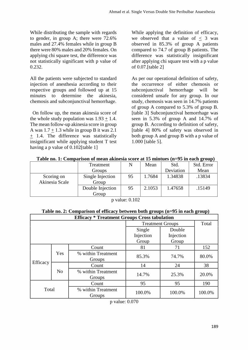
189
While distributing the sample with regards
to gender, in group A; there were 72.6%
males and 27.4% females while in group B
there were 80% males and 20% females. On
applying chi square test, the difference was
not statistically significant with p value of
0.232.
All the patients were subjected to standard
injection of anesthesia according to their
respective groups and followed up at 15
minutes to determine the akinesia,
chemosis and subconjunctival hemorrhage.
On follow up, the mean akinesia score of
the whole study population was 1.93 + 1.4.
The mean follow-up akinesia score in group
A was 1.7 + 1.3 while in group B it was 2.1
+ 1.4. The difference was statistically
insignificant while applying student T test
having a p value of 0.102[table 1]
While applying the definition of efficacy,
we observed that a value of < 3 was
observed in 85.3% of group A patients
compared to 74.7 of group B patients. The
difference was statistically insignificant
after applying chi square test with a p value
of 0.07.[table 2]
As per our operational definition of safety,
the occurrence of either chemosis or
subconjunctival hemorrhage will be
considered unsafe for any group. In our
study, chemosis was seen in 14.7% patients
of group A compared to 5.3% of group B.
[table 3] Subconjunctival hemorrhage was
seen in 5.3% of group A and 14.7% of
group B. According to definition of safety,
[table 4] 80% of safety was observed in
both group A and group B with a p value of
1.000 [table 5].
Table no. 1: Comparison of mean akinesia score at 15 mintues (n=95 in each group)
Treatment
Groups
N Mean Std.
Deviation
Std. Error
Mean
Scoring on
Akinesia Scale
Single Injection
Group
95 1.7684 1.34838 .13834
Double Injection
Group
95 2.1053 1.47658 .15149
p value: 0.102
Table no. 2: Comparison of efficacy between both groups (n=95 in each group)
Efficacy * Treatment Groups Cross tabulation
Treatment Groups Total
Single
Injection
Group
Double
Injection
Group
Efficacy
Yes
Count 81 71 152
% within Treatment
Groups 85.3% 74.7% 80.0%
No
Count 14 24 38
% within Treatment
Groups 14.7% 25.3% 20.0%
Total
Count 95 95 190
% within Treatment
Groups 100.0% 100.0% 100.0%
p value: 0.070
Ahmad et al. Single Versus Double Site Peribulbar Anaesthesia
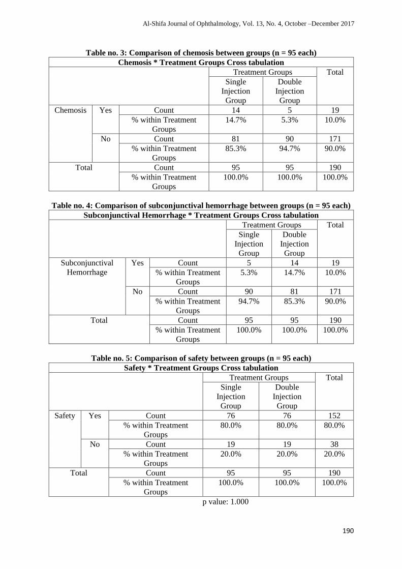
190
Table no. 3: Comparison of chemosis between groups (n = 95 each)
Chemosis * Treatment Groups Cross tabulation
Treatment Groups Total
Single
Injection
Group
Double
Injection
Group
Chemosis Yes Count 14 5 19
% within Treatment
Groups
14.7% 5.3% 10.0%
No Count 81 90 171
% within Treatment
Groups
85.3% 94.7% 90.0%
Total Count 95 95 190
% within Treatment
Groups
100.0% 100.0% 100.0%
Table no. 4: Comparison of subconjunctival hemorrhage between groups (n = 95 each)
Subconjunctival Hemorrhage * Treatment Groups Cross tabulation
Treatment Groups Total
Single
Injection
Group
Double
Injection
Group
Subconjunctival
Hemorrhage
Yes Count 5 14 19
% within Treatment
Groups
5.3% 14.7% 10.0%
No Count 90 81 171
% within Treatment
Groups
94.7% 85.3% 90.0%
Total Count 95 95 190
% within Treatment
Groups
100.0% 100.0% 100.0%
Table no. 5: Comparison of safety between groups (n = 95 each)
Safety * Treatment Groups Cross tabulation
Treatment Groups Total
Single
Injection
Group
Double
Injection
Group
Safety Yes Count 76 76 152
% within Treatment
Groups
80.0% 80.0% 80.0%
No Count 19 19 38
% within Treatment
Groups
20.0% 20.0% 20.0%
Total Count 95 95 190
% within Treatment
Groups
100.0% 100.0% 100.0%
p value: 1.000
Al-Shifa Journal of Ophthalmology, Vol. 13, No. 4, October –December 2017
191
Discussion:
Peribulbar anesthesia remains a popular
choice for patients undergoing cataract
surgery14. Several studies have
demonstrated that peribulbar anesthesia
provided optimal conditions for cataract
surgery15. Furthermore, the injection of
local anesthetic external to the muscle cone
may decrease the likelihood of optic nerve
injury and globe perforation16.
Local anaesthesia is now the main method
employed for cataract surgery17-20. When
compared with general anaesthesia, it is
more popular with surgeons18, more cost-
effective18, 21 and improves patient well
being22,23. Compared with retrobulbar
techniques, peribulbar anaesthesia takes
longer to establish24 and requires larger
volumes of local anaesthetic25 but has fewer
complications17, 19, 26, 27 and provides better
patient comfort and operating conditions17.
Reported complications of peribulbar
anaesthesia include damage to intra-orbital
contents, central nervous and intravascular
penetration24, 27, and unpredictable increase
in intra-ocular pressure28-30. The volume of
potential intra-orbital space available for
injection has been shown to vary
considerably31. This is due to the variation
in orbit and globe sizes seen in the general
population32 and the density of connective
tissue surrounding the eye ball32,33.
Ophthalmologists use a variety of methods
to decide how much local anaesthetic
should be injected. However, no
universally accepted end-point marker of
the local anaesthetic volume sufficient to
provide akinesia or anaesthesia exists.
Usual practice appears to involve injecting
predetermined volumes between 5 and
10 ml34, 35. An excessive increase in intra-
ocular pressure may cause intolerable pain
on injection36, triggering of the
oculocardiac reflex 27, acute ischaemic
optic neuropathy and, rarely,
amaurosis24,27.
Our results demonstrated that both
techniques are similar in terms of efficacy
(both groups had a comparable adequate
akinesia), with a lower total volume of local
anesthetic in the single-injection group.
These findings are in agreement with those
of Mahfouz and Katheri36, Clausel et al8,
and Riad and Nauman37. Previous studies
using B-scan ultrasonography to determine
the exact pattern of distribution of the
injectate in peribulbar anesthesia38,39
reported that a small volume of local
anesthetic (5–6.5 mL) injected in this space
is adequate to surround the eyeball and
produce analgesia. The circumferential
diffusion of the local anesthetic with the
addition of hyaluronidase to sub-Tenon’s
space where the extraocular muscles and
sensory and motor nerves of the eye are
located explains the adequate akinesia
achieved and the higher incidence of
chemosis in the single-injection group40.
Needle length is an important consideration
in needle-related complications. Relatively
short needles (16 or 25 mm) used in single-
or double - injection techniques,
respectively, were associated with a low
incidence of needle-related complications
(hematoma and globe perforation) in both
groups. Moreover, the shorter needles were
associated with a lower incidence of
moderate and severe pain in patients
undergoing the single-injection
technique36. The higher incidence of
subconjunctival hemorrhage in the short-
needle technique is attributable to the
restriction of bleeding to the anterior orbit.
Our results, with high effecacy in achieving
akinesia by 15 min after injection, are
compare with other studies where adequate
akinesia was achieved in 90.6–100% of
cases while using ocular compression35.
Different criteria for a successful block
were used, making accurate comparison
difficult.
In a study, the medial canthus single-
injection peribulbar anesthesia was
Ahmad et al. Single Versus Double Site Peribulbar Anaesthesia
192
significantly less painful and required less
anesthetic agent than the double-injection
peribulbar anesthesia. Akinesia score and
the reinjection rate were similar in the 2
groups, whereas chemosis was significantly
more frequent in the double-injection
group41. The results of 50 consecutive
single-injection peribulbar anaesthetics
performed by the same surgeon are reported
and a description of the procedure is
included. The technique produced adequate
conditions for surgery in all cases. Only 4%
(two cases) were considered to have
inadequate akinesia five minutes after the
injection and required additional injections.
None of the complications described with
retrobulbar and other forms of peribulbar
anaesthesia were observed. The procedure
is relatively easy to master. The study
shows it to be a safe and reliable alternative
to previously described methods of local
anaesthesia42.
The delivery of single percutaneous
peribulbar technique is effective and
reliable in providing both analgesia and
akinesia43. It appears to get closer to the
characteristics of an ideal block than the
classic peribulbar technique. It uses smaller
injectate volume and had speed of onset
comparable to the classic technique. It also
prevents many of the complications
associated with the double injection
technique. Another advantage of the single
percutaneous technique is that it is usually
associated with only minor discomfort,
which may explain the excellent degree of
patient acceptability38. Single percutaneous
peribulbar technique was more comfortable
for the patient at the time of anesthetic
administration and during surgery.
Supplemental block in peribulbar
anesthesia remains the major constraint of
the technique. The reported incidence is
between 5 and 63% in various studies44.45.
According to Ball et al45 an adequate block
can be achieved with a single peribulbar
injection placed either inferotemporally
(classic technique)16 or medially (single
percutaneous technique)40 and there is no
evidence that a second primary injection
decreases the rate of supplemental injection
required. We also therefore propose that a
second primary peribulbar injection is
unnecessary and many carry an increased
risk of globe perforation.
The sensitivity of the eyeball is provided by
the ciliary nerves, which cross the
episcleral space after they emerge from the
globe. The fascial sheath of the eyeball
(tenon’s capsule) extends to rectus muscle
sheath. This explains why the anesthetic is
preferentially guided to this muscle sheath
to produce good akinesia; also, the fascial
sheath of the eyeball guides the injected
solution to the lids, especially to the
orbicularis muscle preventing blinking
during surgery without performing any
facial nerve block. This explains why single
percutaneous technique is more effective
than the classic peribulbar technique.
Anesthesia - related chemosis,
subconjunctival hemorrhage, ecchymosis
occurred mainly in the first group. No
patient had globe injury, perforation or
retrobulbar hemorrhage in both the groups.
In the peribulbar anesthesia by a medial
percutaneous single injection (group 2),
infrequent complications were observed;
this site of injection is relatively avascular,
which may decrease the risk of
hematoma46.
Conclusion:
Based on our results, we can conclude that
the application of the single percutaneous
technique provides a level of comfort
during intraocular surgery. This technique
is a simple and satisfactory alternative
approach for ocular regional anesthesia.
The advantages include decreased pain
with percutaneous and short needle
insertion, decreased volume of anesthetic,
single rather than multiple punctures with
less complications.
Al-Shifa Journal of Ophthalmology, Vol. 13, No. 4, October –December 2017

193
References:
1. Foster A, Gilbert C, and Johnson G.
Changing patterns in global blindness:
1988–2008. Community Eye Health.
2008;21(67):37–39.
2. Dineen B, Bourne RA, Jadoon Z, Shah
SP, Khan MA, Foster A et al. Causes of
blindness and visual impairment in
Pakistan. Br J Ophthalmol
2009;91:1005-10.
3. Jadoon Z, Shah SP, Bourne R, Dineen
B, Khan MA, Gilbert CE et al. Cataract
prevalence, cataract surgical coverage
and barriers to uptake of cataract
surgical services in Pakistan: the
Pakistan National Blindness and Visual
Impairment Survey. Br J Ophthalmol
2009;91:1269-1273.
4. Zaman M, Shah AA, Hussain M, Babar
TF, Khan MT, Dawar S. Outcome of
sutureless manual extra capsular
extraction. J Ayub Med Coll Abbotabad
2009;21(1):39-42.
5. Khan MT, Jan S, Hussain Z, Karim S,
Khalid MK, Mohammad L. Visual
outcome and complications of manual
sutureless small incision cataract
surgery. Pak J Ophthalmol
2010;26(1):32-38.
6. Venkatesh R, Colin SH, Sengupta S,
Ravilla D, Krishnan T, David F.
Phacoemulcification versus manual
small-incision cataract surgery for
white cataract. J Cataract Refract Surg
2010;36:1849-1854.
7. Fazel MRZ, Forghani D, Aghadoost.
Retrobulbar versus topical anesthesia
for phacoemulsification. Pak J Biol.
Sci. 2010;11:2314-9.
8. Clausel H, Touffet L, Havaux M,
Lamard M, Savean J, Cochener B et al.
Peribulbar anesthesia: efficacy of a
single injection with a limited local
anesthetic volume. J Fr Ophtalmol
2011;31:781–5.
9. Budd JM, Brown JP, Thomas J,
Hardwick M, McDonald P, Barber K. A
comparison of sub-Tenon's with
peribulbar anaesthesia in patients
undergoing sequential bilateral cataract
surgery. Anaesthesia. 2009;64:19–22.
10. Ghali AM, Mahfouz A, Hafez A.
Single-injection percutaneous
peribulbar anesthesia with a short
needle versus sub-Tenon’s anesthesia
for cataract extraction. Saudi Journal of
Anaesthesia. 2011;5(2):138-141.
11. Ghali AM, Hafez A. Single-injection
percutaneous peribulbar anesthesia
with a short needle as an alternative to
the double-injection technique for
cataract extraction. Anesth Analg.
2010;110:245–7.
12. El Said TM, Kabeel MM. Comparison
of classic peribulbar anesthesia and new
entry point (single percutaneous
injection technique) in vitroretinal
surgery. Saudi Journal of Anaesthesia.
2010;4(2):80-85.
13. Budd M, Brown JPR, Thomas J,
Hardwick M, McDonald P, Barber K. A
comparison of sub-Tenon’s with
peribulbar anaesthesia in patients
undergoing sequential bilateral cataract
surgery. Anaesthesia, 2009;64:19–22.
14. Bedi A, Carabine U. Peribulbar
anaesthesia: a double-blind comparison
of three local anaesthetic solutions.
Anaesthesia 1999;54:67–71
15. Kumar CM, Dowd TC. Complications
of ophthalmic regional blocks: their
treatment and prevention.
Ophthalmologica 2006;220:73–82
16. Whitsett J, Baleyat H, McClure B.
Comparison of one injectionsite
peribulbar anesthesia and retrobulbar
anesthesia. J Cataract Refract Surg
1990;16:243–5.
17. Patel BCK, Burns TA, Crandall A, et al.
A comparison of topical and retrobulbar
anesthesia for cataract surgery.
Ophthalmology 1996; 103: 1196 203.
18. Hodgkins PR, Luff AJ, Morrell AJ,
Teye Botchway L, Featherston TJ,
Fielder AR. Current practice of cataract
extraction and anaesthesia. British
Journal of Ophthalmology 1992; 76:
323 6.
Ahmad et al. Single Versus Double Site Peribulbar Anaesthesia
194
19. Murdoch IE. Peribulbar versus
retrobulbar anaesthesia. Eye 1990; 4:
445 9.
20. Forrester JV. Local anaesthesia for eye
surgery (Editoral). British Journal of
Ophthalmology 1992; 76: 705.
21. Barker JP, Vafidis GC, Robinson PN,
Hall GM. Plasma catecholamine
response to cataract surgery: a
comparison between general and local
anaesthesia. Anaesthesia 1991; 46: 642
5.
22. Patel BCK, Clinch TE, Burns TA,
Shomaker ST, Jessen R, Crandall AS.
Prospective evaluation of topical versus
retrobulbar anesthesia: a converting
surgeon's experience. Journal of
Cataract and Refractive Surgery 1998;
24: 853 60.
23. Watts MG & Pearce JL. Day-case
cataract surgery (Letter). Lancet 1989;
i: 110 11.
24. Davis II DB & Mandel MR. Efficacy
and complication rate of 16, 224
consecutive peribulbar blocks: a
prospective multicenter study. Journal
of Cataract and Refractive Surgery
1994; 20: 327 37.
25. Bedi A, Carabine U. Peribulbar
anaesthesia: a double-blind comparison
of three local anaesthetic solutions.
Anaesthesia 1999; 54: 67 71.
26. Barker JP, Vafidis GC, Hall GM. Post-
operative morbidity following cataract
surgery. Anaesthesia 1996; 51: 435 7.
27. Rubin AP. Complications of local
anaesthesia for ophthalmic surgery.
British Journal of Anaesthesia 1995;
75: 93 6.
28. Hulbert MFG, Yang YC, Pennefather
PM, Moore JK. Pulsatile ocular blood
flow and intra-ocular pressure during
retrobulbar injection of lignocaine:
Influence of additives. Journal of
Glaucoma 1998; 7: 413 6.
29. O'Donoghue E, Batterbury M, Lavy T.
Effect on intraocular pressure of local
anaesthesia in eyes undergoing
intraocular surgery. British Journal of
Ophthalmology 1994; 78: 605 7.
30. Bowman R, Liu C, Sarkies N.
Intraocular pressure changes after
peribulbar injections with and without
ocular compression. British
31. Johnson RW. Anatomy for ophthalmic
anaesthesia. British Journal of
Anaesthesia 1995; 75: 80 7.
32. Hamilton RC. Techniques of orbital
regional anaesthesia. British Journal of
Anaesthesia 1995; 75: 88 92.
33. Liu C & Redmond R. Choice in local
anaesthesia (Editorial). British Journal
of Ophthalmology 1993; 77: 758.
34. Bjornstrom L, Hansen A, Otland N,
Thim K, Corydon L. Peribulbar
anaesthesia: a clinical evaluation of two
different anaesthetic mixtures. Acta
Ophthalmologica 1994; 72: 712 4.
35. Mahfouz AK, Al Katheri HM.
Randomized trial of superficial
peribulbar compared with conventional
peribulbar anesthesia for cataract
extraction. Clin Ophthalmol
2007;1:55–60
36. Riad W, Nauman A. Single injection
peribulbar anesthesia with a short
needle combined with digital
compression. Anesth Analg
2008;107:1751–3
37. Rizzo L, Marini M, Rosati C, Calamai
I, Nesi M, Salvini R, Mazzini C,
Campana F, Brizzi E. Peribulbar
anesthesia: a percutaneous single
injection technique with a small volume
of anesthetic. Anesth Analg
2005;100:94–6
38. Winder S, Walker SB, Atta HR.
Ultrasonic localization of anesthetic
fluid in sub-Tenon’s, peribulbar, and
retrobulbar techniques. J Cataract
Refract Surg 1999;25:56–9
39. Ripart J, Metge L, Prat-Pradal D, Lopez
FM, Eledjam JJ. Medial canthus single
injection episcleral (sub-tenon
anesthesia): computed tomography
imaging. Anesth Analg 1998;87:42–5
40. Deruddre S, Benhamou D. Medial
canthus single-injection peribulbar
anesthesia: A prospective randomized
comparison with classic double-
Al-Shifa Journal of Ophthalmology, Vol. 13, No. 4, October –December 2017
195
injection peribulbar anesthesia.
Regional anesthesia and pain medicine,
2005;30(3):255-259.
41. Apel, A. and Woodward, R. Single-
injection peribulbar local anaesthesia
—A study of fifty consecutive cases.
Australian and New Zealand Journal of
Ophthalmology, 1991;19:149–153.
42. Bloomberg LB. Anterior periocular
anesthesia: five years experience. J
Cataract Refract Surg. 1991;17:508–11.
[PubMed]
43. Atkinson WS. Retrobulbar injection of
anesthetic within the muscular cone.
Arch Ophthalmol. 1986;16:494–503.
44. Döpfmer UR, Maloney DG, Gaynor
PA, Ratcliffe RM. Döpfmer SPrilocaine
3% is superior to a mixture of
bupivacaine and lignocaine for
peribulbar anaesthesia. Br J Anaesth.
1996;76:77–80.
45. Ball JL, Woon WH, Smith S. Globe
perforation by the second peribulbar
injection. Eye. 2002;16:663–5.
46. Dareau S, Gros T, Bassoul B, Causse L,
Eledjam JJ. Orbital haemorrhage after
medial canthus episclera (sub-Tenon‘s)
anaesthesia. Ann Fr Anesth Reanim.
2003;22:474–6.
Authors Contribution:
Concept and Design: Maqsood Ahmad, Hussain Ahmad
Data Collection / Assembly: Maqsood Ahmad, Muhammad Sajid, Ubaidullah
Drafting: Maqsood Ahmad, Hussain Ahmad
Statistical expertise: Hussain Ahmad, Muhammad Sajid
Critical revision: Maqsood Ahmad, Hussain Ahmad
Ahmad et al. Single Versus Double Site Peribulbar Anaesthesia
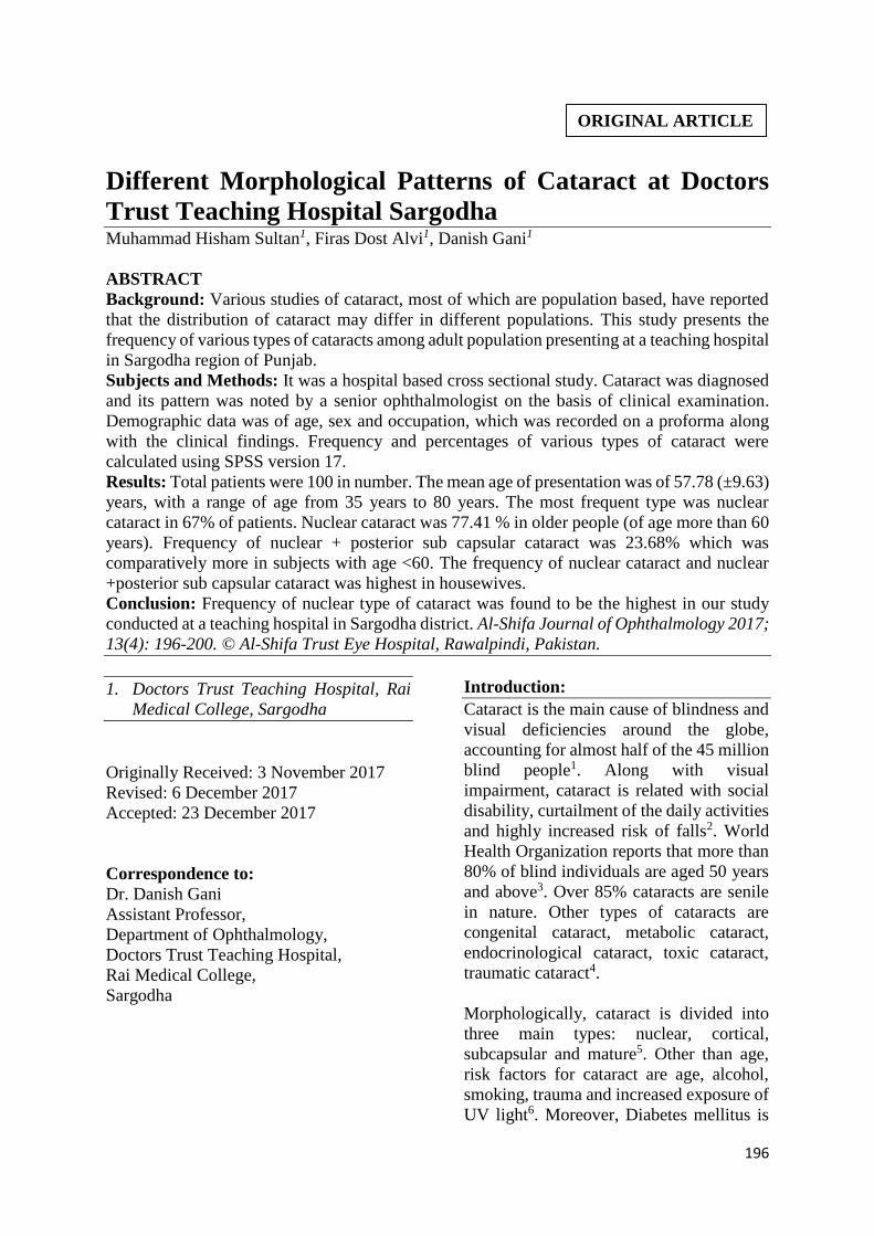
196
Different Morphological Patterns of Cataract at Doctors
Trust Teaching Hospital Sargodha Muhammad Hisham Sultan1, Firas Dost Alvi1, Danish Gani1
ABSTRACT
Background: Various studies of cataract, most of which are population based, have reported
that the distribution of cataract may differ in different populations. This study presents the
frequency of various types of cataracts among adult population presenting at a teaching hospital
in Sargodha region of Punjab.
Subjects and Methods: It was a hospital based cross sectional study. Cataract was diagnosed
and its pattern was noted by a senior ophthalmologist on the basis of clinical examination.
Demographic data was of age, sex and occupation, which was recorded on a proforma along
with the clinical findings. Frequency and percentages of various types of cataract were
calculated using SPSS version 17.
Results: Total patients were 100 in number. The mean age of presentation was of 57.78 (±9.63)
years, with a range of age from 35 years to 80 years. The most frequent type was nuclear
cataract in 67% of patients. Nuclear cataract was 77.41 % in older people (of age more than 60
years). Frequency of nuclear + posterior sub capsular cataract was 23.68% which was
comparatively more in subjects with age <60. The frequency of nuclear cataract and nuclear
+posterior sub capsular cataract was highest in housewives.
Conclusion: Frequency of nuclear type of cataract was found to be the highest in our study
conducted at a teaching hospital in Sargodha district. Al-Shifa Journal of Ophthalmology 2017;
13(4): 196-200. © Al-Shifa Trust Eye Hospital, Rawalpindi, Pakistan.
1. Doctors Trust Teaching Hospital, Rai
Medical College, Sargodha
Originally Received: 3 November 2017
Revised: 6 December 2017
Accepted: 23 December 2017
Correspondence to:
Dr. Danish Gani
Assistant Professor,
Department of Ophthalmology,
Doctors Trust Teaching Hospital,
Rai Medical College,
Sargodha
Introduction:
Cataract is the main cause of blindness and
visual deficiencies around the globe,
accounting for almost half of the 45 million
blind people1. Along with visual
impairment, cataract is related with social
disability, curtailment of the daily activities
and highly increased risk of falls2. World
Health Organization reports that more than
80% of blind individuals are aged 50 years
and above3. Over 85% cataracts are senile
in nature. Other types of cataracts are
congenital cataract, metabolic cataract,
endocrinological cataract, toxic cataract,
traumatic cataract4.
Morphologically, cataract is divided into
three main types: nuclear, cortical,
subcapsular and mature5. Other than age,
risk factors for cataract are age, alcohol,
smoking, trauma and increased exposure of
UV light6. Moreover, Diabetes mellitus is
ORIGINAL ARTICLE
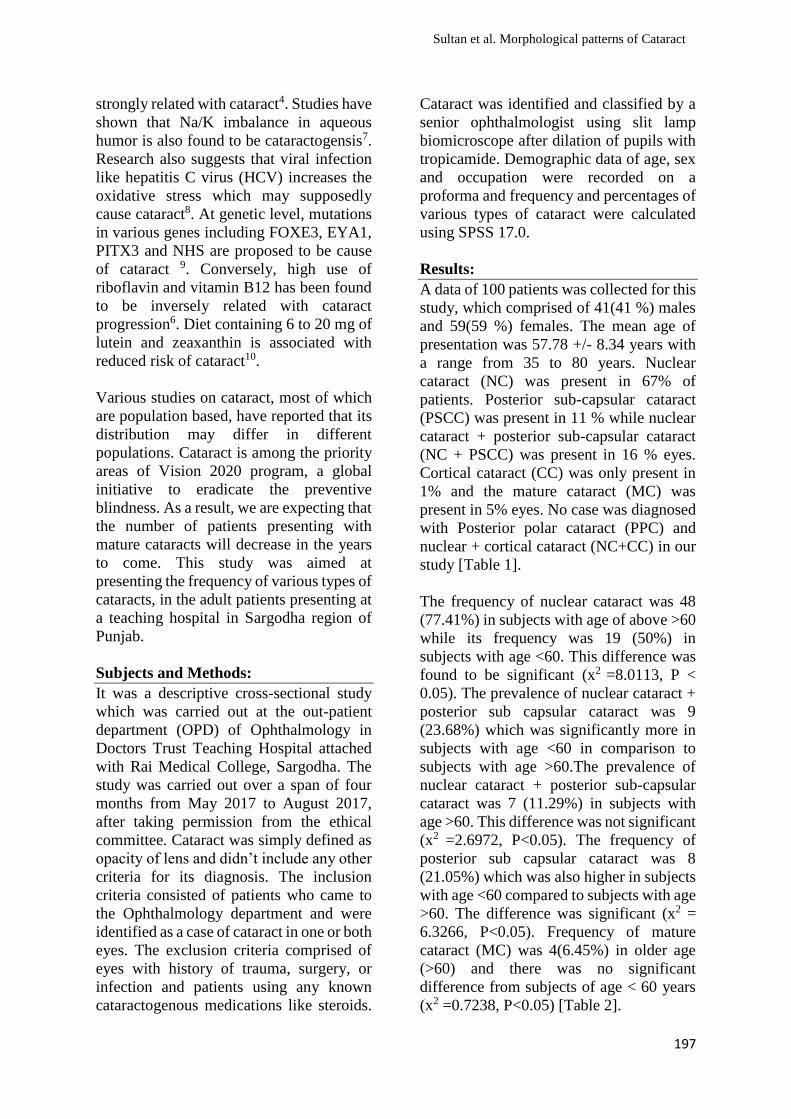
197
strongly related with cataract4. Studies have
shown that Na/K imbalance in aqueous
humor is also found to be cataractogensis7.
Research also suggests that viral infection
like hepatitis C virus (HCV) increases the
oxidative stress which may supposedly
cause cataract8. At genetic level, mutations
in various genes including FOXE3, EYA1,
PITX3 and NHS are proposed to be cause
of cataract 9. Conversely, high use of
riboflavin and vitamin B12 has been found
to be inversely related with cataract
progression6. Diet containing 6 to 20 mg of
lutein and zeaxanthin is associated with
reduced risk of cataract10.
Various studies on cataract, most of which
are population based, have reported that its
distribution may differ in different
populations. Cataract is among the priority
areas of Vision 2020 program, a global
initiative to eradicate the preventive
blindness. As a result, we are expecting that
the number of patients presenting with
mature cataracts will decrease in the years
to come. This study was aimed at
presenting the frequency of various types of
cataracts, in the adult patients presenting at
a teaching hospital in Sargodha region of
Punjab.
Subjects and Methods:
It was a descriptive cross-sectional study
which was carried out at the out-patient
department (OPD) of Ophthalmology in
Doctors Trust Teaching Hospital attached
with Rai Medical College, Sargodha. The
study was carried out over a span of four
months from May 2017 to August 2017,
after taking permission from the ethical
committee. Cataract was simply defined as
opacity of lens and didn’t include any other
criteria for its diagnosis. The inclusion
criteria consisted of patients who came to
the Ophthalmology department and were
identified as a case of cataract in one or both
eyes. The exclusion criteria comprised of
eyes with history of trauma, surgery, or
infection and patients using any known
cataractogenous medications like steroids.
Cataract was identified and classified by a
senior ophthalmologist using slit lamp
biomicroscope after dilation of pupils with
tropicamide. Demographic data of age, sex
and occupation were recorded on a
proforma and frequency and percentages of
various types of cataract were calculated
using SPSS 17.0.
Results:
A data of 100 patients was collected for this
study, which comprised of 41(41 %) males
and 59(59 %) females. The mean age of
presentation was 57.78 +/- 8.34 years with
a range from 35 to 80 years. Nuclear
cataract (NC) was present in 67% of
patients. Posterior sub-capsular cataract
(PSCC) was present in 11 % while nuclear
cataract + posterior sub-capsular cataract
(NC + PSCC) was present in 16 % eyes.
Cortical cataract (CC) was only present in
1% and the mature cataract (MC) was
present in 5% eyes. No case was diagnosed
with Posterior polar cataract (PPC) and
nuclear + cortical cataract (NC+CC) in our
study [Table 1].
The frequency of nuclear cataract was 48
(77.41%) in subjects with age of above >60
while its frequency was 19 (50%) in
subjects with age <60. This difference was
found to be significant (x2 =8.0113, P <
0.05). The prevalence of nuclear cataract +
posterior sub capsular cataract was 9
(23.68%) which was significantly more in
subjects with age <60 in comparison to
subjects with age >60.The prevalence of
nuclear cataract + posterior sub-capsular
cataract was 7 (11.29%) in subjects with
age >60. This difference was not significant
(x2 =2.6972, P<0.05). The frequency of
posterior sub capsular cataract was 8
(21.05%) which was also higher in subjects
with age <60 compared to subjects with age
>60. The difference was significant (x2 =
6.3266, P<0.05). Frequency of mature
cataract (MC) was 4(6.45%) in older age
(>60) and there was no significant
difference from subjects of age < 60 years
(x2 =0.7238, P<0.05) [Table 2].
Sultan et al. Morphological patterns of Cataract
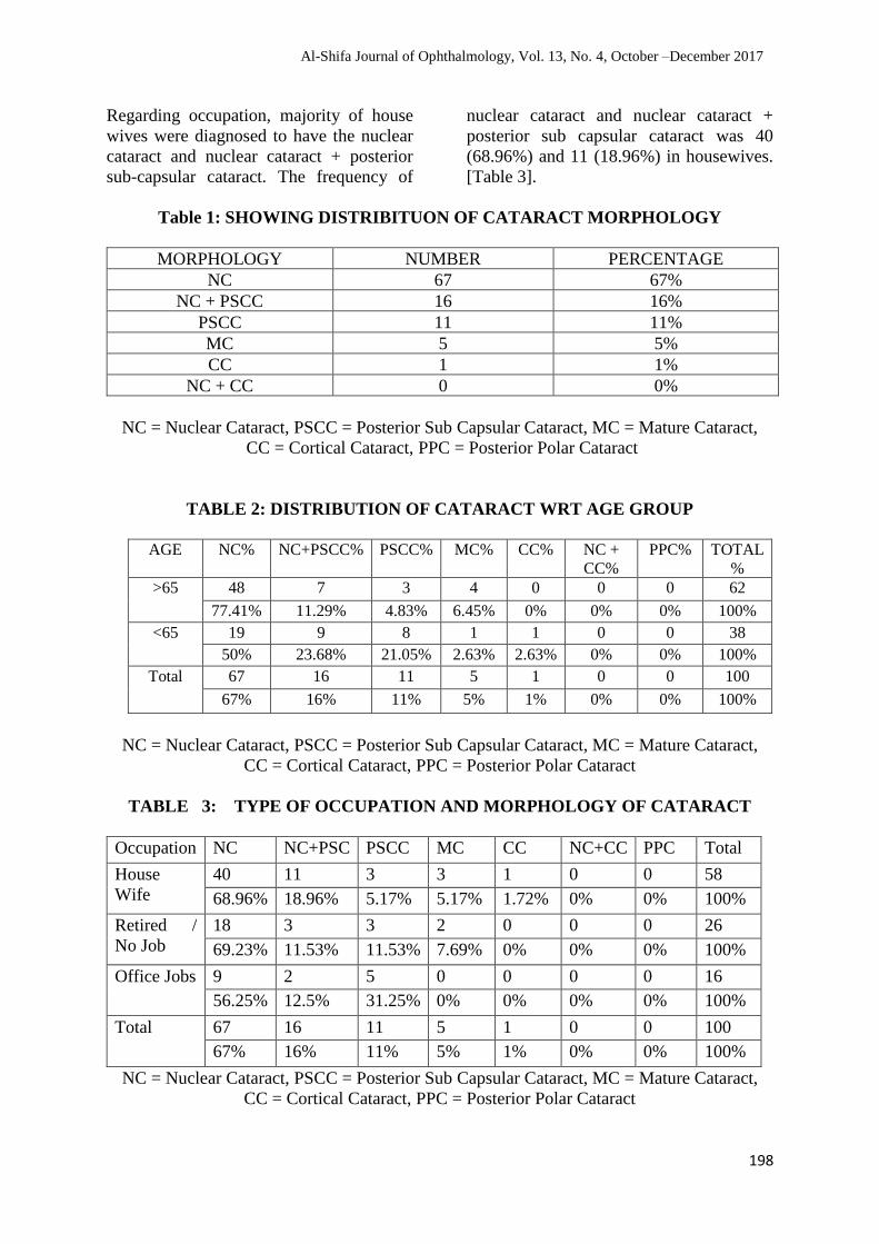
198
Regarding occupation, majority of house
wives were diagnosed to have the nuclear
cataract and nuclear cataract + posterior
sub-capsular cataract. The frequency of
nuclear cataract and nuclear cataract +
posterior sub capsular cataract was 40
(68.96%) and 11 (18.96%) in housewives.
[Table 3].
Table 1: SHOWING DISTRIBITUON OF CATARACT MORPHOLOGY
MORPHOLOGY NUMBER PERCENTAGE
NC 67 67%
NC + PSCC 16 16%
PSCC 11 11%
MC 5 5%
CC 1 1%
NC + CC 0 0%
NC = Nuclear Cataract, PSCC = Posterior Sub Capsular Cataract, MC = Mature Cataract,
CC = Cortical Cataract, PPC = Posterior Polar Cataract
TABLE 2: DISTRIBUTION OF CATARACT WRT AGE GROUP
AGE NC% NC+PSCC% PSCC% MC% CC% NC +
CC%
PPC% TOTAL
%
>65 48 7 3 4 0 0 0 62
77.41% 11.29% 4.83% 6.45% 0% 0% 0% 100%
<65 19 9 8 1 1 0 0 38
50% 23.68% 21.05% 2.63% 2.63% 0% 0% 100%
Total 67 16 11 5 1 0 0 100
67% 16% 11% 5% 1% 0% 0% 100%
NC = Nuclear Cataract, PSCC = Posterior Sub Capsular Cataract, MC = Mature Cataract,
CC = Cortical Cataract, PPC = Posterior Polar Cataract
TABLE 3: TYPE OF OCCUPATION AND MORPHOLOGY OF CATARACT
Occupation NC NC+PSC PSCC MC CC NC+CC PPC Total
House
Wife
40 11 3 3 1 0 0 58
68.96% 18.96% 5.17% 5.17% 1.72% 0% 0% 100%
Retired /
No Job
18 3 3 2 0 0 0 26
69.23% 11.53% 11.53% 7.69% 0% 0% 0% 100%
Office Jobs 9 2 5 0 0 0 0 16
56.25% 12.5% 31.25% 0% 0% 0% 0% 100%
Total 67 16 11 5 1 0 0 100
67% 16% 11% 5% 1% 0% 0% 100%
NC = Nuclear Cataract, PSCC = Posterior Sub Capsular Cataract, MC = Mature Cataract,
CC = Cortical Cataract, PPC = Posterior Polar Cataract
Al-Shifa Journal of Ophthalmology, Vol. 13, No. 4, October –December 2017
199
Discussion:
The average age of presentation in this
study was 57.78 years. This is
comparatively less than the mean age
reported in an Indian study (64.14 years)11.
This could be attributed to difference in
sample size and environmental conditions.
The mean age of cataract in African
population was 66.5 years12.
In our study, we had noticed that nuclear
cataract had the highest incidence in older
age >60 years but nuclear cataract with its
mixed form was found to be higher in
subjects with age <60 years. The frequency
of posterior sub-capsular cataract and
nuclear cataract + posterior sub-capsular
cataract was also high in younger age
subjects. Nuclear cataract typically
develops as the result of age related
sclerotic changes in lens. Because of this
fact, nuclear cataract is sometimes referred
to as an age-related nuclear cataract. Signs
of Nuclear cataract include hardening and
yellowing of the nucleus of the lens, while
in advanced stages the colour may turn
brown or rarely black.
An important finding of this study was that
mature cataract was diagnosed in only 5%
of the total cases. This indicates that there
is a growing level of awareness among our
local population to seek medical
consultation at an earlier level. This might
also be attributed to the services provided
by the surgical eye camps which are
primarily organized to eradicate the
cataract related blindness.
A study shows that subjects who worked
indoor had a high frequency of nuclear
cataract (NC) and cortical cataract13. So, in
our present study nuclear cataract (NC) and
cortical cataract (CC)were found to be
higher in housewives. Surprisingly, in our
study more females presented to the
Ophthalmology department than males.
This is in contrast to majority of studies
conducted in Pakistan. The study was
conducted in a general hospital, so many
subjects presenting to other departments
might have visited the eye department for
their ocular problems and were diagnosed
for cataract. Women in this region are
mostly involved in house hold activities
like cooking. Smoke from firewood might
be a factor causing cataract. A significant
number of patients were not actively
involved in any profession or were living
retirement life.
This study must be appraised as a brief time
study of various morphologies of cataract in
Sargodha. A long-term study must be
carried out in this region to examine the
various trends and morphologies of cataract
and include a higher number of
populations.
Conclusion:
In our study, the most frequent type was
nuclear cataract (NC) alone and also with
its mixed form with a higher frequency in
age >60 years. Further studies must be
carried out to find different types and trends
in this area.
References:
1. Foster PJ, Wong TY, Machin D,
Johnson GJ, Seah SKL. Risk factors for
nuclear, cortical and posterior
subcapsular cataracts in the Chinese
population of Singapore: the
tanjongpagar survey. Br J Opthalmol
2003; 87:1112-1120.
2. Berle D, Steel Z, Essue BM, et al.
Multisite prospective investigation of
psychological outcomes following
cataract surgery in Vietnam. BMJ
Global Health 2017; 2:e000162. Doi
10.1136/bmjgh-2017-00162.
3. Alison GA, Nathan GC, Emily WG.
The New Epidemiology of Cataract.
Opthalmol Clin N Am 2006; 19:415-
425.
4. Chandler RD, Ivan RS. Epidemiology
of cataract- a major cause of
preventable blindness. Bulletin of the
World Health Organization 1981;
59(4): 493-501.
Sultan et al. Morphological patterns of Cataract
200
5. Milan CM, Ann ME, Jeremiah T,
Richard MD. Antioxidant vitamin
supplementation for preventing and
slowing the progression of age-related
cataract. Cochrane Database Syst Rev.;
6: CD004567.
doi:10.1002/14651858.CD004567.pub
2.
6. Tanya SG, Lauren ED, Grace S, et al.
The Association of Dietary
Lutein/Zeaxanthin and B Vitamins with
Cataracts in the Age-Related Eye
Disease Study AREDS Report No.37.
Ophthalmology 2015 July;
122(7):1471-1479.
7. Fatemeh S, Matin G, Parisa NH, Sara
H, Mehdi E, Bahram R. Nutrient
patterns and risk of cataract: a case
control study. Int J Opthalmol 2017;
10(4):586-592.
8. Shih YL, Cheng LL, Shu WJ, I-Kaun
W, Cheng CL, Chih HL, et al.
Increasing risk of cataract in HCV
patients receiving anti-HCV therapy: A
nationwide cohort study 2017.PLoS
ONE 12(3): e0173125.
doi:10.1371/journal.pone.0173125.
9. Shiels A, Hejtmencik JF. Genetics of
human cataract. Clin Genet 2013
August; 84(2): 120-127.
doi:10.1111/cge.12182.
10. Keyvan K, Raluca R, Wenhua L, Lee
RF, Kakarla VC. The Role of Lutein in
Eye-Related Disease. Nutreints 2013;
5: 1823-1839; doi: 10.3390/nu5051823.
11. Seema DB, Yogendra GV, Prajakta PS,
Swati SR. A study of morphology of
cataract in western India. Journal of
clinical ophthalmology and research.
May-August 2015; 3(2):91-93.
12. Natban C, Sbeila KW, Ralf RB,
Antbony k, Beatriz M, Harran M.
Prevelance of the Different Types of
Age-Related Cataract in an African
Population. Invest Opthalmol Vis Sci.
October 2001; 42(11):2478-2482.
13. Seymour Z, Manuel D, Elise T.
Sunlight and human cataracts. Incest
Opthalmol Visual Sci. May
1979;18(5):462-467.
Authors Contribution:
Concept and Design: Muhammad Hisham Sultan, Firas Dost Alvi
Data Collection / Assembly: Danish Gani, Firas Dost Alvi
Drafting: Muhammad Hisham Sultan, Danish Gani
Statistical expertise: Muhammad Hisham Sultan
Critical revision: Hisham Sultan, Danish Gani
Al-Shifa Journal of Ophthalmology, Vol. 13, No. 4, October –December 2017

201
Vision Related Quality of Life Among Keratoconus
Patients and Factors Affecting the Quality Asima Rehman 1, Hina Sharif 2, Khizar Nabeel 2
ABSTRACT:
Objectives: The purpose of this study was to assess vision related quality of life among
keratoconus patient and also to identify associated factors that can affect vision related quality
of life among keratoconus patients.
Subjects and Methods: It was a cross sectional study conducted on 312 keratoconus patients,
out of which 109 had undergone corneal cross linkage (CXL) surgery. Convenient non-
randomized sampling technique was used for selection of sample. Sample was selected from
outpatient department of Al-Shifa Trust eye hospital Rawalpindi. Data were collected by using
reliable scale of NEI VFQ-25 and analyzed by using SPSS version 17. Chi square test of
independence was used to assess any association of vision related quality of life of keratoconus
patients with other variables.
Results: Vision related quality of life was found to be good in 51% patients while 35%
respondents said that they remained worry about their eye sight most of the time. Visual acuity,
education status, CXL surgery and type of CXL showed significant association with VQOL
(p<0.05).
Conclusion: Keratoconus affects vision related quality of life of patients but CXL surgery and
stability in visual acuity can help to improve it. Therefore, our study suggests maintaining
visual acuity of keratoconus patients by timely diagnosis and CXL surgery. Al-Shifa Journal
of Ophthalmology 2017; 13(4): 201-9. © Al-Shifa Trust Eye Hospital, Rawalpindi, Pakistan.
1. Al-Shifa Trust Eye Hospital,
Rawalpindi
2. Al-Shifa School of Public Health,
Rawalpindi
Originally Received: 19 November 2017
Revised: 17 December 2017
Accepted: 26 December 2017
Correspondence to:
Miss Asima Rehman
BSc. Hons Optometry and Orthoptics
Al-Shifa Trust Eye Hospital Rawalpindi
Introduction:
According to WHO health is "a state of
complete physical, mental, and social well-
being of individuals and not merely the
absence of disease". Vision is a sense of
sight through which we perceive shapes,
color, distance and movement of an object.
Visual acuity is the resolving power of an
eye and is a sensitive test for integrity of
visual system. Decrease in vision may
drastically affect the individual’s daily life
activities and may cause social disability.
Quality of life (QOL) is a broad
multidimensional concept that usually
includes subjective evaluations of both
positive and negative aspects of life.
Reduced vision sets limit on daily life
activities, may cause negative effect on
“vision related quality of life”1.
Keratoconus is non-inflammatory,
progressive disease of eye in which normal
ORIGINAL ARTICLE
202
dome shaped cornea becomes thin and
changes into cone shape with the apex of
the cone being forward, this ectasia of the
cornea induces irregular astigmatism,
resulting visual impairment. Incidence of
keratoconus has been estimated to be 5-23
per 10,000 and prevalence of keratoconus
has been estimated to be 5.4 per 10,0002. A
study suggested that the male-to-female
ratio of prevalence of keratoconus was 1:
2.53.
The prevalence of keratoconus varies
according to the geographical location,
ranging from 0.3 per 100,000 in Russia to
the 2300 per 100,000 in central India. The
1st population-based study was done by
Hofstetter using a Placido disc and the
reported incidence of keratoconus in this
study was 600 per 100,000 4.
As keratoconus is usually diagnosed
during young age and it causes painless
progressive deterioration of vision for
near and distance, photophobia and
monocular diplopia, so it has an impact on
vision related quality of life and life
planning of the patients5. ‘Corneal
collagen cross-linking is a medical
procedure to strengthen the corneal tissues
by using riboflavin and UVA irradiation’.
Corneal collagen cross-linking (CXL) is
the first and only treatment option, which
actually stops or slows down the
progression of keratoconus. Corneal
collagen cross-linking is a treatment and
management option which is not only
supposed to prevent disease or improve
prognosis but also has a strong
psychological impact on patients, for their
mental and physical wellbeing, therefore,
it is also believed that there will be change
in quality of life of an individual
comparative to the pre-CXL6. In early age
of life this procedure is effective because
cornea is flexible and vision worsening
chances are more so disease progression
halting is very necessary but in the late 30’s
CXL is not usually required because cornea
stiffens and chances of disease progression
are less7.
The purpose of this study was to assess
vision related quality of life (VQOL)
among keratoconus patient and also to
identify associated factors that can affect
vision related quality of life among
keratoconus patients.
Subjects and Methods:
This cross-sectional study was conducted
on patients having keratoconus of all
stages, not gone through CXL procedure
and also keratoconus patients reporting
after 6 months of CXL procedure at Cornea
clinic of Al Shifa trust eye hospital
Rawalpindi. Study was conducted at
Cornea department of Al-Shifa Trust Eye
hospital from October 2016 to Feb 2017.
Sample size was 312, calculated with the
help of open epi online available software
and sample was collected by non-random
convenient sampling.
Questionnaire included mainly three
sections, demographic, clinical aspects and
modified NEI VFQ-25 which was further
divided into general vision, difficulty with
vision related activities and response
against vision problems. The responses for
NEI VFQ were taken on three-point Likert
scale. The dependent variable (VQOL) was
basically computed by adding all responses
from NEI VFQ and was transformed into
binary variable by taking mean value. Chi-
square test of independence (Fisher’s exact
was used where needed) was used to assess
associations with p-value <0.05 considered
as significant.
Results:
Sixty percent of the respondents were male
and majority of them were from rural areas
(N=200 - 64%). Married participants
constituted 21% (N=65) and 53% reported
education level of primary to matriculation
(Table 1).
Al-Shifa Journal of Ophthalmology, Vol. 13, No. 4, October –December 2017

203
Ninety seven percent (N=303) respondents
had bilateral keratoconus. Majority of the
participants (N=203, 65%) had not
undergone CXL procedure (Table 2).
Among those who went through CXL
procedure, 74% were treated free of cost,
while 48 % had undergone CXL >12
months ago. Unilateral surgery was
reported by 27% respondents and use of
contact lens was not common among
participants (6.4%).
The mean vision related quality of life score
was 99±15, ranging from 42 to 133 (Figure
1). Sixty two percent respondents rated
their health in good state while 35%
respondents said that they remained
worried about their eye sight most of the
time. When they were asked about further
items of NEI VFQ, 49% of them said that
they never felt difficulty in using stairs.
Forty three percent participants highlighted
severe difficulty in recognizing people’s
face and 44% were facing severe difficulty
in watching TV (Table 3).
When respondents were asked about their
response regarding vision problems, 68%
participants said that they go out of their
home for different activities, despite of the
disease. Among those who had good
VQOL, 30% were male and 18% females.
However, statistically significant
association was not found between gender
and VQOL, x2(1) =1.024, p=0.31.
Comparison of VQOL with different age
groups showed 109(35%) participants in
14-22 years age category & there was no
statistical significant association between
age and VQOL in keratoconus patients
(p>0.05) and x2 (df) =1.964(1). Education
was found to be significantly associated
with VQOL, x2(df) =6.48(2) & P<0.05.
There was no significant difference
between impact of unilateral and bilateral
keratoconus on VQOL, with x2(df)
=3.134(1) & P>0.05.
CXL surgery had a significant impact on
VQOL among keratoconus patients, with
x2(df) =5.53(1) & P<0.05, while 58%
participants who had undergone surgery,
showed good VQOL. Seventy five percent
of eyes that had undergone CXL, with
visual acuity of 6/36-6/18 & 6/12-6/6,
reported good VQOL. In comparison, 54%
eyes with CXL having visual acuity CF-
6/60, reported a poor VQOL. The
difference was statistically significant with
x2(df) =7.68(2) & P<0.05.
Patients who had undergone bilateral CXL
surgery (N=25) reported good VQOL in
68% cases as compared to those who
underwent unilateral procedure (N=84),
reporting good VQOL in 55% cases. With
x2(df) =6.88(2) & P<0.05, the difference
was statistically significant. (Table 4)
Rehman et al. Quality of Life in Keratoconus

204
Table 1Demographic Characteristics
Demographic characteristics Frequency (N=312) Percentages
Age(years)
14-22
23-50
201
111
64
36
Gender
Male
Female
186
126
60
40
Education
Illiterate
Primary to metric
Higher
16
166
129
5
53
41
Employment status
Non-Working
Working
233
78
75
25
Monthly income*
Up to 20,000
20-50,000
Above 50,000
67
12
2
83
15
2.5
Living area
Urban
Rural
112
200
36
64
Marital status
Married
Unmarried
Others
65
246
1
21
79
0.3
Province
Punjab
KPK
Kashmir
Others
176
95
32
9
56.4
30.4
10.3
2.9
*N=81(because of missing values)
Al-Shifa Journal of Ophthalmology, Vol. 13, No. 4, October –December 2017
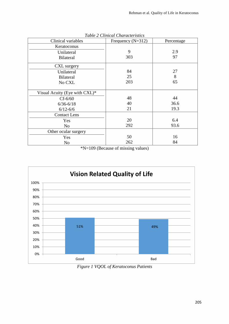
205
Table 2 Clinical Characteristics
Clinical variables Frequency (N=312) Percentage
Keratoconus
Unilateral
Bilateral
9
303
2.9
97
CXL surgery
Unilateral
Bilateral
No CXL
84
25
203
27
8
65
Visual Acuity (Eye with CXL)*
Cf-6/60
6/36-6/18
6/12-6/6
48
40
21
44
36.6
19.3
Contact Lens
Yes
No
20
292
6.4
93.6
Other ocular surgery
Yes
No
50
262
16
84
*N=109 (Because of missing values)
Figure 1 VQOL of Keratoconus Patients
51% 49%
0%
10%
20%
30%
40%
50%
60%
70%
80%
90%
100%
Good Bad
Vision Related Quality of Life
Rehman et al. Quality of Life in Keratoconus
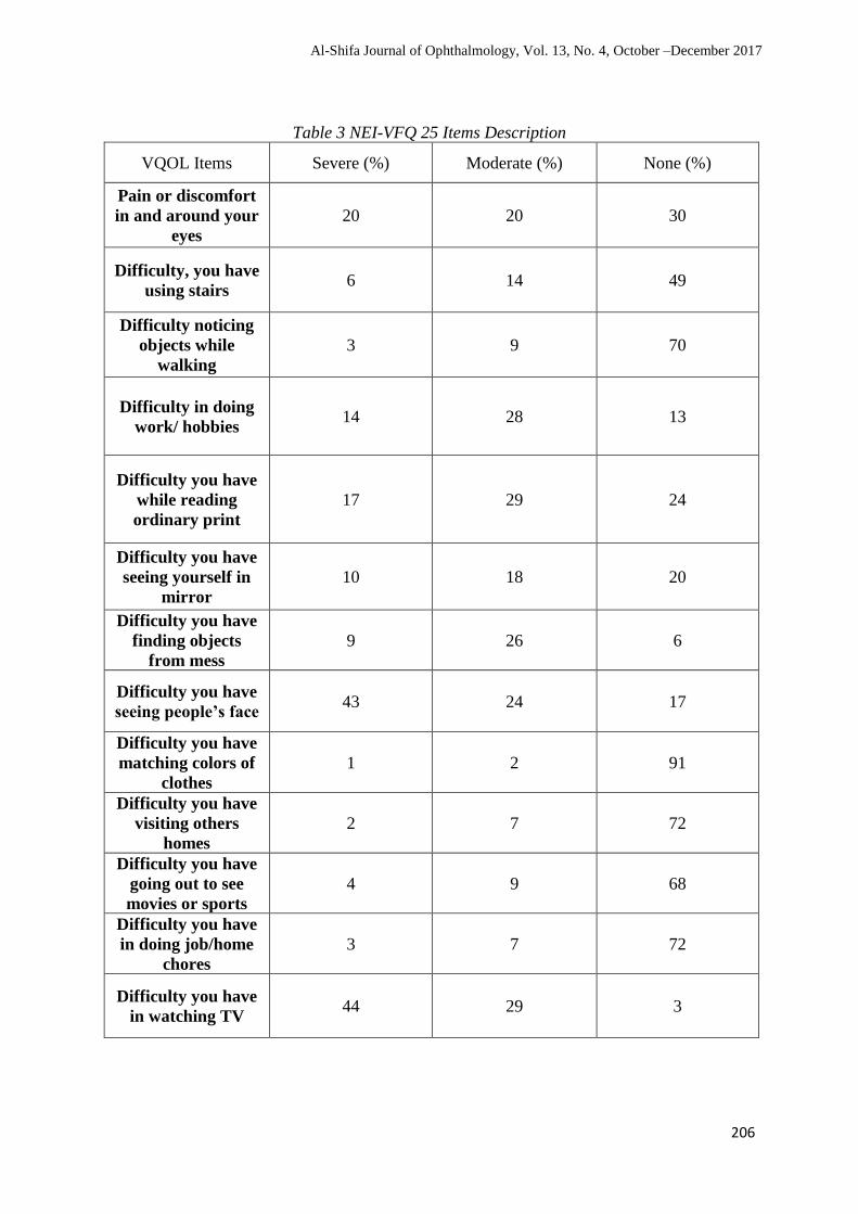
206
Table 3 NEI-VFQ 25 Items Description
VQOL Items Severe (%) Moderate (%) None (%)
Pain or discomfort
in and around your
eyes
20 20 30
Difficulty, you have
using stairs 6 14 49
Difficulty noticing
objects while
walking
3 9 70
Difficulty in doing
work/ hobbies 14 28 13
Difficulty you have
while reading
ordinary print
17 29 24
Difficulty you have
seeing yourself in
mirror
10 18 20
Difficulty you have
finding objects
from mess
9 26 6
Difficulty you have
seeing people’s face 43 24 17
Difficulty you have
matching colors of
clothes
1 2 91
Difficulty you have
visiting others
homes
2 7 72
Difficulty you have
going out to see
movies or sports
4 9 68
Difficulty you have
in doing job/home
chores
3 7 72
Difficulty you have
in watching TV 44 29 3
Al-Shifa Journal of Ophthalmology, Vol. 13, No. 4, October –December 2017
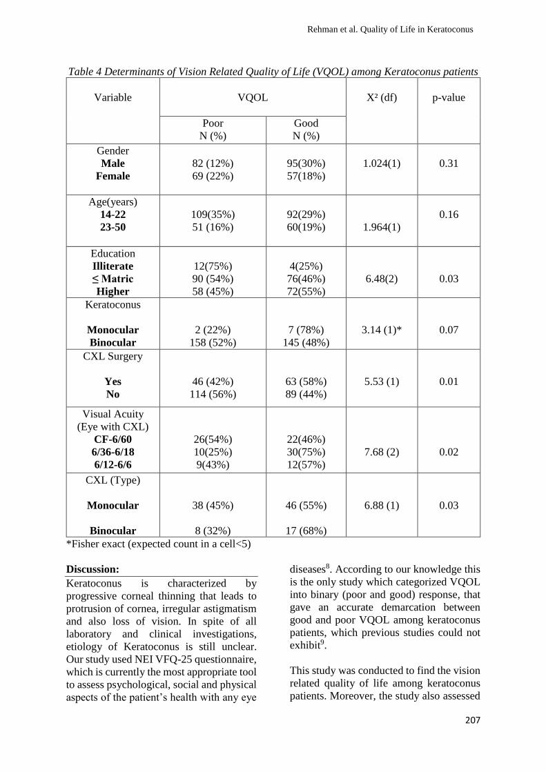
207
Table 4 Determinants of Vision Related Quality of Life (VQOL) among Keratoconus patients
Variable
VQOL
X² (df)
p-value
Poor
N (%)
Good
N (%)
Gender
Male
Female
82 (12%)
69 (22%)
95(30%)
57(18%)
1.024(1)
0.31
Age(years)
14-22
23-50
109(35%)
51 (16%)
92(29%)
60(19%)
1.964(1)
0.16
Education
Illiterate
≤ Matric
Higher
12(75%)
90 (54%)
58 (45%)
4(25%)
76(46%)
72(55%)
6.48(2)
0.03
Keratoconus
Monocular
Binocular
2 (22%)
158 (52%)
7 (78%)
145 (48%)
3.14 (1)*
0.07
CXL Surgery
Yes
No
46 (42%)
114 (56%)
63 (58%)
89 (44%)
5.53 (1)
0.01
Visual Acuity
(Eye with CXL)
CF-6/60
6/36-6/18
6/12-6/6
26(54%)
10(25%)
9(43%)
22(46%)
30(75%)
12(57%)
7.68 (2)
0.02
CXL (Type)
Monocular
Binocular
38 (45%)
8 (32%)
46 (55%)
17 (68%)
6.88 (1)
0.03
*Fisher exact (expected count in a cell<5)
Discussion:
Keratoconus is characterized by
progressive corneal thinning that leads to
protrusion of cornea, irregular astigmatism
and also loss of vision. In spite of all
laboratory and clinical investigations,
etiology of Keratoconus is still unclear.
Our study used NEI VFQ-25 questionnaire,
which is currently the most appropriate tool
to assess psychological, social and physical
aspects of the patient’s health with any eye
diseases8. According to our knowledge this
is the only study which categorized VQOL
into binary (poor and good) response, that
gave an accurate demarcation between
good and poor VQOL among keratoconus
patients, which previous studies could not
exhibit9.
This study was conducted to find the vision
related quality of life among keratoconus
patients. Moreover, the study also assessed
Rehman et al. Quality of Life in Keratoconus
208
difference of outcome for the factors that
can affect VQOL in keratoconus patients.
Our study showed mean level of 99±15
(51% respondents with good VQOL). In
comparison, a case control study reported a
mean composite score of 75.2 ±17.2 for
keratoconus, which is different from our
findings9.
Visual acuity (eye with CXL) was found to
be statistically associated with VQOL and
it is similar to the findings of another study,
while gender of keratoconus patients was
not found to be associated with VQOL,
which is different from previous studies10.
The present study has also found a
statistically significant association between
education & VQOL (p<0.05), this is
supported by another study which showed
education to be significantly associated
with VQOL11. Sixty five percent
respondents did not have CXL surgery and
among these 56% showed poor VQOL. On
the other hand, those who had undergone
CXL showed (58%) good VQOL which
depicted positive effect of this surgery on
VQOL and this association was statistically
significant (P<0.05). Moreover, this finding
was also reported by many other
researches. In this study association
between CXL surgery (monocular or
binocular) and VQOL was also
statistically significant (P<0.05). The
findings revealed a better VQOL among
those who had surgery of either type.
However, binocular surgery exhibited
68% good VQOL as compared to 55%
among monocular, these results were
supported by previous researches as
well10, 11, 12.
The possible limitation of our study can be
the inclusion of all those patients who
reported at a hospital, which are
considered to be more concerned about
their health which may show bias for NEI-
VFQ items. Moreover, we used
convenient sampling. However, our study
filled a conspicuous gap in literature
especially for Pakistan. We further
suggest cohort-based study to analyze
VRQOL with varying stages of
Keratoconus.
References:
1. Rabinowitz YS. Keratoconus. Survey
of ophthalmology. 1998 Jan
1;42(4):297-319.
2. Krachmer JH, Feder RS, Belin MW.
Keratoconus and related
noninflammatory corneal thinning
disorders. Survey of ophthalmology.
1984 Jan 1;28(4):293-322.
3. Yildiz EH, Cohen EJ, Virdi AS,
Hammersmith KM, Laibson PR,
Rapuano CJ. Quality of life in
keratoconus patients after penetrating
keratoplasty. American journal of
ophthalmology. 2010 Mar
1;149(3):416-22.
4. Kymes SM, Walline JJ, Zadnik K,
Sterling J, Gordon MO. Changes in the
Quality-of-Life of People with
Keratoconus. Am J Ophthalmol.
2008;145(4):611–617.
5. Offor N, Greenstein SA, Fry KL, Hersh
PS. Patient Satisfaction and Subjective
Visual Function after Corneal Collagen
Crosslinking. Invest Ophthalmol Vis
Sci. 2011;52(14):5206–5206.
6. Mangione CM, Lee PP, Gutierrez PR,
Spritzer K, Berry S, Hays RD.
Development of the 25-list-item
national eye institute visual function
questionnaire. Archives of
ophthalmology. 2001 Jul
1;119(7):1050-8.
7. Kymes SM, Walline JJ, Zadnik K,
Gordon MO. Quality of life in
keratoconus. American journal of
ophthalmology. 2004 Oct
1;138(4):527-35.
8. Fink BA, Wagner H, Steger-May K,
Rosenstiel C, Roediger T, McMahon
TT, Gordon MO, Zadnik K. Differences
in keratoconus as a function of gender.
American journal of Ophthalmology.
2005 Sep 1;140(3):459-e1.
Al-Shifa Journal of Ophthalmology, Vol. 13, No. 4, October –December 2017
209
9. Cesneková T, Skorkovská K, Petrová S,
Cermáková S. Visual functions and
quality of life in patients with
keratoconus. Ceska a slovenska
oftalmologie: casopis Ceske
oftalmologicke spolecnosti a Slovenske
oftalmologicke spolecnosti. 2011
Apr;67(2):51-4.
10. Aydin Kurna, S., Altun, A., Gencaga,
T., Akkaya, S., & Sengor, T. (2014).
Vision Related Quality of Life in
Patients with Keratoconus. Journal of
Ophthalmology, 2014, 694542.
11. Sahebjada S, Fenwick EK, Xie J,
Snibson GR, Daniell MD, Baird PN.
Impact of keratoconus in the better eye
and the worse eye on vision-related
quality of life. Investigative
ophthalmology & visual science. 2014
Jan 1;55(1):412-6.
12. Erdurmus M, Yildiz EH, Abdalla YF,
Hammersmith KM, Rapuano CJ,
Cohen EJ. Contact lens related quality
of life in patients with keratoconus. Eye
& contact lens. 2009 May 1;35(3):123-
7.
Authors Contribution:
Concept and Design: Asima Rehman
Data Collection / Assembly: Asima Rehman
Drafting: Asima Rehman, Hina Sharif
Statistical expertise: Hina Sharif, Khizar Nabeel
Critical revision: Hina Sharif, Khizar Nabeel
Rehman et al. Quality of Life in Keratoconus
210
Authors Index
Afghani, Tayyab
Situation Analysis of Diabetic Retinopathy services in Muzaffarabad division of Azad Jammu
and Kashmir, Pakistan. 13(1):40-9.
Refractive Errors in Intellectually Disabled People at a Special School. 13(2):93-9.
Barriers for spectacle users in choosing contact lens as an alternative vision correction method.
13(3): 133-39.
Aftab, Muhammad
Anatomical and Physiological Outcome of Scleral Buckling with Subretinal Fluid Drainage.
13(2):100-5.
Ahmad, Hussain
Comparative evaluation of conventional extracapsular cataract extraction versus manual small
incision cataract surgery in terms of visual outcome. 13(2):65-71.
Manual Small Incision Cataract Surgery, Evaluation of Visual Outcome and Complications
Following Surgery in Patients with Pseudoexfoliation. 13(2):72-78.
Clinical Audit of Patients Presenting at Department of Glaucoma as Part of Glaucoma
Fellowship Program. 13(3): 114-20.
Effectiveness Of 0.05% Cyclosporine-A Eye Drops for Allergic Conjunctivitis in Terms of
Improvement in Ocular Itching Score. 13(3): 140-44.
Pattern of Ocular Diseases in Children Presented at Eye Out Patient Department, Khyber
Teaching Hospital, Peshawar. 13(4):180-5.
Comparative Study of Single Versus Double Site Peribulbar Anaesthesia Injection for Cataract
Surgery in Terms of Efficacy and Safety. 13(4):186-95.
Ahmad, Imran
Ocular complications of severe vernal keratoconjunctivitis (VKC) in patients presenting to a
private clinic. 13(1):35-9.
Ahmad, Mushtaq
Comparative evaluation of conventional extracapsular cataract extraction versus manual small
incision cataract surgery in terms of visual outcome. 13(2):65-71.
Manual Small Incision Cataract Surgery, Evaluation of Visual Outcome and Complications
Following Surgery in Patients with Pseudoexfoliation. 13(2):72-78.
Ahmad, Maqsood
Effectiveness Of 0.05% Cyclosporine-A Eye Drops for Allergic Conjunctivitis in Terms of
Improvement in Ocular Itching Score. 13(3): 140-44.
Pattern of Ocular Diseases in Children Presented at Eye Out Patient Department, Khyber
Teaching Hospital, Peshawar. 13(4):180-5.
Comparative Study of Single Versus Double Site Peribulbar Anaesthesia Injection for Cataract
Surgery in Terms of Efficacy and Safety. 13(4):186-95.
Ahmad, Naseer
Comparative evaluation of conventional extracapsular cataract extraction versus manual small
incision cataract surgery in terms of visual outcome. 13(2):65-71.
Ahmad, Saqib Qayyum
Pattern of Placement of Doctors After Post-graduation in Ophthalmology. 13(1):25-9.
Akhtar, Farah
Clinical Audit of Patients Presenting at Department of Glaucoma as Part of Glaucoma
Fellowship Program. 13(3): 114-20.
Outcomes of Combined Extraction with Intraocular Lens Implant by A Fellow During
Glaucoma Fellowship. 13(4):174-9.
Al-Shifa Journal of Ophthalmology, Vol. 13, No. 4, October –December 2017

211
Akbar, Maheen
Congenital cystic eye ball with an intracranial anomaly: A Case Report. 13(3): 153-57.
Ali, Mahmood
Editorial: Recent Advances in Pharmacological Therapy of Glaucoma. 13(4):163-5.
Outcomes of Combined Extraction with Intraocular Lens Implant by A Fellow During
Glaucoma Fellowship. 13(4):174-9.
Alvi, Firas Dost
Different Morphological Patterns of Cataract at Doctors Trust Teaching Hospital Sargodha. 13(4):196-200.
Asif, Muhammad
Frequency of Different Types of Manifest Strabismus in Children. 13(2):86-92.
Azam, Sana
Exploring Astigmatism and Visual Functions in Amblyopic Children13(4):166-73.
Buscaran Covadonga
Situation Analysis of Diabetic Retinopathy services in Muzaffarabad division of Azad Jammu
and Kashmir, Pakistan. 13(1):40-9.
Chaudhry, Adeel
Anatomical and Physiological Outcome of Scleral Buckling with Subretinal Fluid Drainage.
13(2):100-5.
Durrani, Waqar-ud-Din
Situation Analysis of Diabetic Retinopathy services in Muzaffarabad division of Azad Jammu
and Kashmir, Pakistan. 13(1):40-9.
Elbeih, Islam
Situation Analysis of Diabetic Retinopathy services in Muzaffarabad division of Azad Jammu
and Kashmir, Pakistan. 13(1):40-9.
Farooq, Omer
Situation Analysis of Diabetic Retinopathy services in Muzaffarabad division of Azad Jammu
and Kashmir, Pakistan. 13(1):40-9.
Farooq, Saima
Clinical Audit of Patients Presenting at Department of Glaucoma as Part of Glaucoma
Fellowship Program. 13(3): 114-20.
Outcomes of Combined Extraction with Intraocular Lens Implant by A Fellow During
Glaucoma Fellowship. 13(4):174-9.
Gani, Danish
Different Morphological Patterns of Cataract at Doctors Trust Teaching Hospital Sargodha. 13(4):196-200.
Gul, Nazli
Manual Small Incision Cataract Surgery, Evaluation of Visual Outcome and Complications
Following Surgery in Patients with Pseudoexfoliation. 13(2):72-78.
Habiba, Ume
Frequency of Different Types of Manifest Strabismus in Children. 13(2):86-92.
Hassan, Shadab
Causes of Childhood Blindness in a Tertiary Care Eye Hospital. 13(1):9-17.
Exploring Astigmatism and Visual Functions in Amblyopic Children13(4):166-73.
Hussain, Mehfooz
Efficacy of Dorzolamide plus Timolol Maleate in the Treatment of Primary Open Angle
Glaucoma Presented to a Tertiary Care Centre of Khyber Pukhtunkhwa. 13(2):79-85.
Idris, Mohammad
Efficacy of Dorzolamide plus Timolol Maleate in the Treatment of Primary Open Angle
Glaucoma Presented to a Tertiary Care Centre of Khyber Pukhtunkhwa. 13(2):79-85.
Authors Index. Al-Shifa Journal of Ophthalmology. Vol 13. 2017
212
Iqbal, Sara
Frequency of Different Types of Manifest Strabismus in Children. 13(2):86-92.
Kaleemullah
Efficacy of Dorzolamide plus Timolol Maleate in the Treatment of Primary Open Angle
Glaucoma Presented to a Tertiary Care Centre of Khyber Pukhtunkhwa. 13(2):79-85.
Kanwal, Maria
Refractive Errors in Intellectually Disabled People at a Special School. 13(2):93-9.
Karamat, Muhammad Irfan
Anatomical and Physiological Outcome of Scleral Buckling with Subretinal Fluid Drainage.
13(2):100-5.
Khan, Bilal
Efficacy of Dorzolamide plus Timolol Maleate in the Treatment of Primary Open Angle
Glaucoma Presented to a Tertiary Care Centre of Khyber Pukhtunkhwa. 13(2):79-85.
Khan, Hina Mehwish
Comparative evaluation of conventional extracapsular cataract extraction versus manual small
incision cataract surgery in terms of visual outcome. 13(2):65-71.
Manual Small Incision Cataract Surgery, Evaluation of Visual Outcome and Complications
Following Surgery in Patients with Pseudoexfoliation. 13(2):72-78.
Khan, Muhammad Sajid
Pattern of Ocular Diseases in Children Presented at Eye Out Patient Department, Khyber
Teaching Hospital, Peshawar. 13(4):180-5.
Khan, Muhammad Tariq
Situation Analysis of Diabetic Retinopathy services in Muzaffarabad division of Azad Jammu
and Kashmir, Pakistan. 13(1):40-9.
Khan, Waseem Ahmed
Frequency and Types of Refractive Errors in School-going Children in Mirpur, Azad Jammu
& Kashmir (AJK), in Age Group of 5-15 Years. 13(1):30-4.
Khattak, Irfan Aslam
Situation Analysis of Diabetic Retinopathy services in Muzaffarabad division of Azad Jammu
and Kashmir, Pakistan. 13(1):40-9.
Kiani, Ovais Manzoom
Frequency and Types of Refractive Errors in School-going Children in Mirpur, Azad Jammu
& Kashmir (AJK), in Age Group of 5-15 Years. 13(1):30-4.
Mahsood, Yousaf Jamal
Comparative evaluation of conventional extracapsular cataract extraction versus manual small
incision cataract surgery in terms of visual outcome. 13(2):65-71.
Manual Small Incision Cataract Surgery, Evaluation of Visual Outcome and Complications
Following Surgery in Patients with Pseudoexfoliation. 13(2):72-78.
Clinical Audit of Patients Presenting at Department of Glaucoma as Part of Glaucoma
Fellowship Program. 13(3): 114-20.
Outcomes of Combined Extraction with Intraocular Lens Implant by A Fellow During
Glaucoma Fellowship. 13(4):174-9.
Pattern of Ocular Diseases in Children Presented at Eye Out Patient Department, Khyber
Teaching Hospital, Peshawar. 13(4):180-5.
Masood, Ch.Tariq
Frequency and Types of Refractive Errors in School-going Children in Mirpur, Azad Jammu
& Kashmir (AJK), in Age Group of 5-15 Years. 13(1):30-4.
Mansoor, Hassan
Editorial: Genetic Testing in Retinal Dystrophies; A Way of Future. 13(1):6-8.
Al-Shifa Journal of Ophthalmology, Vol. 13, No. 4, October –December 2017

213
Manzoor, Amna
Congenital cystic eye ball with an intracranial anomaly: A Case Report. 13(3): 153-57.
Nabeel Khizar
Vision Related Quality of Life Among Keratoconus Patients and Factors Affecting the Quality. 13(4):201-10. Naeem, Badruddin Athar
Role of B-Scan in Evaluating Posterior Segment Pathologies in Opaque Ocular Media. 13(3):
121-26.
Naeem, Muhammad
Comparative evaluation of conventional extracapsular cataract extraction versus manual small
incision cataract surgery in terms of visual outcome. 13(2):65-71.
Manual Small Incision Cataract Surgery, Evaluation of Visual Outcome and Complications
Following Surgery in Patients with Pseudoexfoliation. 13(2):72-78.
Clinical Audit of Patients Presenting at Department of Glaucoma as Part of Glaucoma
Fellowship Program. 13(3): 114-20.
Pattern of Ocular Diseases in Children Presented at Eye Out Patient Department, Khyber
Teaching Hospital, Peshawar. 13(4):180-5.
Najeeb, Sara
Comparison of Endothelial Cell Loss in Diabetics After Phacoemulsification and Extra-
Capsular Cataract Extraction. 13(1):18-24. Comparison of Peripapillary Retinal Nerve Fibre Layer (RNFL) Thickness in Type II Diabetics and
Normal Population. 13(2):59-64.
Nazir, Tehmina
Role of B-Scan in Evaluating Posterior Segment Pathologies in Opaque Ocular Media. 13(3):
121-26.
Post-Operative Complications After Reconstruction of Contracted Socket Using Amniotic
Membrane. 13(3): 145-52.
Okonkwo, Sunday
Editorial: Evolution of Cataract Surgery: Past, Present and the Future. 13(2): 56-8.
Congenital cystic eye ball with an intracranial anomaly: A Case Report. 13(3): 153-57.
Rabia, Miss
Frequency of Different Types of Manifest Strabismus in Children. 13(2):86-92.
Rasul, Javed
Ocular complications of severe vernal keratoconjunctivitis (VKC) in patients presenting to a
private clinic. 13(1):35-9.
Rehman, Asima
Vision Related Quality of Life Among Keratoconus Patients and Factors Affecting the Quality. 13(4):201-10. Riaz, Nadeemud Din
Anatomical and Physiological Outcome of Scleral Buckling with Subretinal Fluid Drainage.
13(2):100-5.
Sadiq, Muhammad
Editorial: Immunosuppressant Drugs and Ocular Inflammatory Conditions. 13 (3): 111-3.
Sadiq, Muhammad Irfan
Comparison of Endothelial Cell Loss in Diabetics After Phacoemulsification and Extra-
Capsular Cataract Extraction. 13(1):18-24.
Frequency and Types of Refractive Errors in School-going Children in Mirpur, Azad Jammu
& Kashmir (AJK), in Age Group of 5-15 Years. 13(1):30-4.
Authors Index. Al-Shifa Journal of Ophthalmology. Vol 13. 2017

214
Comparison of Peripapillary Retinal Nerve Fibre Layer (RNFL) Thickness in Type II Diabetics
and Normal Population. 13(2):59-64.
Sadiq, Muhammad Usman
Comparison of Endothelial Cell Loss in Diabetics After Phacoemulsification and Extra-
Capsular Cataract Extraction. 13(1):18-24.
Comparison of Peripapillary Retinal Nerve Fibre Layer (RNFL) Thickness in Type II Diabetics
and Normal Population. 13(2):59-64.
Sajid, Muhammad
Effectiveness Of 0.05% Cyclosporine-A Eye Drops for Allergic Conjunctivitis in Terms of
Improvement in Ocular Itching Score 13(3): 140-44.
Comparative Study of Single Versus Double Site Peribulbar Anaesthesia Injection for Cataract
Surgery in Terms of Efficacy and Safety. 13(4):186-95.
Saleem, Zubair
Anatomical and Physiological Outcome of Scleral Buckling with Subretinal Fluid Drainage.
13(2):100-5.
Sarwar, Shamsa
Barriers for spectacle users in choosing contact lens as an alternative vision correction method.
13(3): 133-39.
Shah, Mir Ali
Efficacy of Dorzolamide plus Timolol Maleate in the Treatment of Primary Open Angle
Glaucoma Presented to a Tertiary Care Centre of Khyber Pukhtunkhwa. 13(2):79-85.
Frequency of Different Types of Manifest Strabismus in Children. 13(2):86-92.
Sharif, Hina
Exploring Astigmatism and Visual Functions in Amblyopic Children13(4):166-73.
Vision Related Quality of Life Among Keratoconus Patients and Factors Affecting the Quality. 13(4):201-10.
Siddiqui, Sorath Noorani
Causes of Childhood Blindness in a Tertiary Care Eye Hospital. 13(1):9-17.
Frequency of Different Types of Manifest Strabismus in Children. 13(2):86-92.
Suleri, Ishtiaq
Compliance of Spectacles Wear in School Going Children in District Rawalpindi. 13(3): 127-
32.
Sultan, Muhammad Hisham
Different Morphological Patterns of Cataract at Doctors Trust Teaching Hospital Sargodha. 13(4):196-200.
Taimur, Fariha
Role of B-Scan in Evaluating Posterior Segment Pathologies in Opaque Ocular Media. 13(3):
121-26.
Post-Operative Complications After Reconstruction of Contracted Socket Using Amniotic
Membrane. 13(3): 145-52.
Ubaidullah
Effectiveness Of 0.05% Cyclosporine-A Eye Drops for Allergic Conjunctivitis in Terms of
Improvement in Ocular Itching Score. 13(3): 140-44.
Pattern of Ocular Diseases in Children Presented at Eye Out Patient Department, Khyber
Teaching Hospital, Peshawar. 13(4):180-5.
Comparative Study of Single Versus Double Site Peribulbar Anaesthesia Injection for Cataract
Surgery in Terms of Efficacy and Safety. 13(4):186-95.
Ullah, Irfan
Clinical Audit of Patients Presenting at Department of Glaucoma as Part of Glaucoma
Fellowship Program. 13(3): 114-20.
Al-Shifa Journal of Ophthalmology, Vol. 13, No. 4, October –December 2017
215
Waqar, Saman
Compliance of Spectacles Wear in School Going Children in District Rawalpindi. 13(3): 127-
32.
Barriers for spectacle users in choosing contact lens as an alternative vision correction method.
13(3): 133-39.
Zafar, Saemah Nuzhat
Pattern of Placement of Doctors After Post-graduation in Ophthalmology. 13(1):25-9.
Zafar, Nadeem
Pattern of Placement of Doctors After Post-graduation in Ophthalmology. 13(1):25-9.
Zaheer, Naima
Causes of Childhood Blindness in a Tertiary Care Eye Hospital. 13(1):9-17.
Zondervan, Marcia
Situation Analysis of Diabetic Retinopathy services in Muzaffarabad division of Azad Jammu
and Kashmir, Pakistan. 13(1):40-9.
Authors Index. Al-Shifa Journal of Ophthalmology. Vol 13. 2017
216
Subject Index
Anaesthesia. 13(4):186-95.
Amniotic Membrane Transplant. 13(3): 145-52.
Amblyopic.13(4):166-73.
Astigmatism.13(4):166-73.
B-Scan Ultrasonography. 13(3): 121-26.
Cataract. 13(4):196-200.
Cataract Extraction. 13(1):18-24, 13(2): 56-8, 13(2):65-71, 13(2):72-78.
Childhood Blindness. 13(1):9-17.
Combined Extraction with Intraocular Lens Implant. 13(4):174-9.
Contact Lenses. 13(3): 133-39.
Congenital Eye Ball Anomaly. 13(3): 153-57.
Diabetes. 13(2):59-64.
Diabetic Retinopathy. 13(1):40-9.
Endothelial Cell. 13(1):18-24.
Genetics. 13(1):6-8.
Glaucoma Fellowship Program. 13(3): 114-20, 13(4):174-9.
Immunosuppressant Drugs 13 (3): 111-3, 13(3): 140-44, 13(4):163-5.
Keratoconus. 13(4):201-10.
Medical Education. 13(1):25-9.
Ocular Diseases in Children. 13(4):180-5.
Ocular Inflammatory Conditions. 13 (3): 111-3.
Pharmacology. 13(4):163-5.
Primary Open Angle Glaucoma. 13(2):79-85.
Refractive Errors. 13(1):30-4, 13(2):93-9, 13(3): 127-32.
Retinal Dystrophies. 13(1):6-8.
Retinal Nerve Fibre Layer (RNFL). 13(2):59-64.
Scleral Buckling. 13(2):100-5.
Socket Construction. 13(3): 145-52.
Strabismus. 13(2):86-92.
Vernal Keratoconjunctivitis. 13(1):35-9, 13(3): 140-44.
Vision Related Quality of Life. 13(4):201-10.
Al-Shifa Journal of Ophthalmology, Vol. 13, No. 4, October –December 2017