ACUTE PORPHYRIA WITH -...
10
SODIUM AND CHLORIDE DEPLETION IN ACUTE PORPHYRIA WITH REFERENCE TO THE STATUS OF ADRENAL CORTICAL FUNCTION By F. T. G. PRUNTY (From. the Department of Chemical Pathology, St. Thom:as's Hospital Medical School, London, England) (Received for publication December 27, 1948) INTRODUCTION The fact that depletion of sodium and of chlo- ride may occur in acute porphyria, in the absence of alkalosis due to vomiting, has recently been noted independently by Abrahams, Gavey and Maclagan (1) and by Linder (2). These authors suggested that a possible basis for this deficiency was that of adrenal cortical failure, but the re- sponse of their two patients to substitution ther- apy with cortical hormones does not appear to have given unequivocal evidence for this hy- pothesis. The present paper presents an attempt to ex- plore this question in further detail. The plan of investigation falls under two headings: firstly to study the function of the adrenal cortex during the height of an attack of acute porphyria and secondly to observe the response to desoxycorti- costerone administration in greater detail. Prunty, Forsham and Thorn (3) have recently indicated that the elucidation of adrenal cortical function with respect to its relation to electrolyte metabo- lism is a complex matter, whereas the functional production of the 1 1-oxy-steroid group of hor- mones is more easily assessable by urine analysis and the use of the response to purified adreno- corticotrophic hormone. The severity of the ill- ness of the patient under observation was con- sidered a contraindication precluding the use of the deprivation test of Cutler, Power and Wilder (4). METHODS The patient (Case 1) was confined to bed during the first 80 days of observation. During the period of elec- trolyte study she was maintained on a light diet, kept as constant as possible, with the addition of 7 gm. of sodium chloride daily, the major portion of which was given orally as half normal saline. Urine was collected daily under toluene and analysed the day of completing the collection. The methods of analysis used were as fol- lows. Sodium by the method of Butler and Tuthill (5); chloride by the iodometric method of Van Slyke and Hiller (6); potassium by King's method (7); carbon dioxide combining power by the technique of Peters and Van Slyke (8). Total base in serum was estimated by electrodialysis (Malm [9]). "Neutral reducing steroids" in the urine were determined by the author's modification of the method of Talbot et al. (10),' and 17-ketosteroids by that of Callow, Callow and Emmens (11), using a correction equation for interfering chromogens. The ad- renocorticotrophic hormone test for adrenal cortical re- serve has been fully described by Forsham et al. (12). Total urine porphyrin was estimated by adsorption on a calcium phosphate precipitate followed by solution in hy- drochloric acid as described by Sveinsson, Rimington and Barnes (13), the final assay of porphyrin being carried out in the fluorimeter. OBSERVATIONS Plasma electrolyte changes The trend of the alterations found in the plasma electrolytes is indicated in Table I, and Figures 1 and 2. At the commencement of the period of observation the patient was receiving potassium citrate, 2.7 gm. per diem, on account of mild urinary symptoms. At this time (15th day) there was a moderate deficiency of sodium and of chloride, the values being 125 and 86 MEq. per 1. respectively. The carbon dioxide combining power was normal (30.4 MEq. per 1.). It will also be noted that there was a moderate elevation of blood pressure, a characteristic of this disease. On the 19th day potassium citrate was withdrawn and on the 20th day a six-day period of desoxycorti- costerone acetate administration commenced. This was given in two doses per day of 5 mgm. each intramuscularly. On the fifth day of desoxycorti- costerone administration the serum potassium had 1 The determination of "neutral reducing steroids" in the urine is thought to bear a relation to the endogenous 11-oxygenated corticosteroid production. The reducing substances in the urine have hitherto sometimes been loosely termed "11-oxysteroids." 690
Transcript of ACUTE PORPHYRIA WITH -...
SODIUMAND CHLORIDE DEPLETION IN ACUTE PORPHYRIAWITH REFERENCETO THE STATUS OF ADRENAL
CORTICAL FUNCTION
By F. T. G. PRUNTY
(From. the Department of Chemical Pathology, St. Thom:as's Hospital Medical School,London, England)
(Received for publication December 27, 1948)
INTRODUCTION
The fact that depletion of sodium and of chlo-ride may occur in acute porphyria, in the absenceof alkalosis due to vomiting, has recently beennoted independently by Abrahams, Gavey andMaclagan (1) and by Linder (2). These authorssuggested that a possible basis for this deficiencywas that of adrenal cortical failure, but the re-sponse of their two patients to substitution ther-apy with cortical hormones does not appear tohave given unequivocal evidence for this hy-pothesis.
The present paper presents an attempt to ex-plore this question in further detail. The plan ofinvestigation falls under two headings: firstly tostudy the function of the adrenal cortex duringthe height of an attack of acute porphyria andsecondly to observe the response to desoxycorti-costerone administration in greater detail. Prunty,Forsham and Thorn (3) have recently indicatedthat the elucidation of adrenal cortical functionwith respect to its relation to electrolyte metabo-lism is a complex matter, whereas the functionalproduction of the 1 1-oxy-steroid group of hor-mones is more easily assessable by urine analysisand the use of the response to purified adreno-corticotrophic hormone. The severity of the ill-ness of the patient under observation was con-sidered a contraindication precluding the use ofthe deprivation test of Cutler, Power and Wilder(4).
METHODS
The patient (Case 1) was confined to bed during thefirst 80 days of observation. During the period of elec-trolyte study she was maintained on a light diet, kept asconstant as possible, with the addition of 7 gm. of sodiumchloride daily, the major portion of which was givenorally as half normal saline. Urine was collected dailyunder toluene and analysed the day of completing thecollection. The methods of analysis used were as fol-lows. Sodium by the method of Butler and Tuthill (5);
chloride by the iodometric method of Van Slyke andHiller (6); potassium by King's method (7); carbondioxide combining power by the technique of Peters andVan Slyke (8). Total base in serum was estimated byelectrodialysis (Malm [9]). "Neutral reducing steroids"in the urine were determined by the author's modificationof the method of Talbot et al. (10),' and 17-ketosteroidsby that of Callow, Callow and Emmens (11), using acorrection equation for interfering chromogens. The ad-renocorticotrophic hormone test for adrenal cortical re-serve has been fully described by Forsham et al. (12).Total urine porphyrin was estimated by adsorption on acalcium phosphate precipitate followed by solution in hy-drochloric acid as described by Sveinsson, Rimington andBarnes (13), the final assay of porphyrin being carriedout in the fluorimeter.
OBSERVATIONS
Plasma electrolyte changes
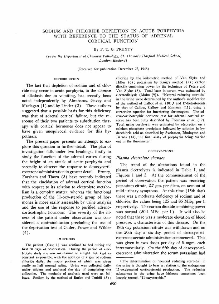
The trend of the alterations found in theplasma electrolytes is indicated in Table I, andFigures 1 and 2. At the commencement of theperiod of observation the patient was receivingpotassium citrate, 2.7 gm. per diem, on account ofmild urinary symptoms. At this time (15th day)there was a moderate deficiency of sodium and ofchloride, the values being 125 and 86 MEq. per 1.respectively. The carbon dioxide combining powerwas normal (30.4 MEq. per 1.). It will also benoted that there was a moderate elevation of bloodpressure, a characteristic of this disease. On the19th day potassium citrate was withdrawn and onthe 20th day a six-day period of desoxycorti-costerone acetate administration commenced. Thiswas given in two doses per day of 5 mgm. eachintramuscularly. On the fifth day of desoxycorti-costerone administration the serum potassium had
1 The determination of "neutral reducing steroids" inthe urine is thought to bear a relation to the endogenous11-oxygenated corticosteroid production. The reducingsubstances in the urine have hitherto sometimes beenloosely termed "11-oxysteroids."
690
SODIUM AND CHLORIDE DEPLETION IN ACUTE PORPHYRIA
TABLE I
Electrolytes MEqper 1. serum (Case 1)
Day Na K CToalC02 Cl Prot. Ht Ureabase
gin. mgm.per cent per cent
15 125 3.5 135 30.4 86 5.4 38 5119 8720 10 mgm. DOCAo.d.21 10 mgm. DOCAo.d. 2.7 138 9022 10 mgm. DOCAo.d.23 10 mgm. DOCAo.d. 32.5 8624 10 mgm. DOCAo.d. 1.9 33.0 87 3225 10 mgm. DOCAo.d.26 129 2.1 138 32.1 89 5.2 34 3529 134 2.7 147 32.5 91 5.9 37 3133 133 2.7 145 30.0 93 6.4 38 3040 4.1 9644 3.5 145 10191 140 5.0 148 28.6 108 6.997 36 28
fallen as low as 1.9 MEq. per 1. There was nodefinite electrocardiographic evidence of potassiumdeficiency, but there was an increase of musclepain suggesting the onset of the potassium de-ficiency syndrome, and it was decided that thedesoxycorticosterone should be stopped after afurther 24 hours. On cessation of desoxycorti-costerone administration the serum potassium wasstill as low as 2.1 MEq. per 1., whereas there had
160
140
120
1oo0MEG/L
80
60
40
201
0
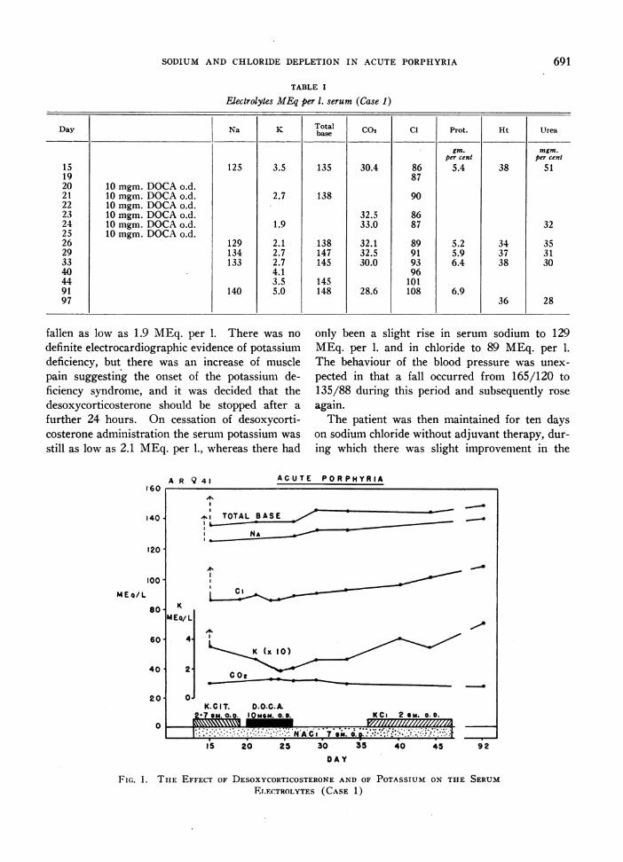
only been a slight rise in serum sodium to 129MEq. per 1. and in chloride to 89 MEq. per 1.The behaviour of the blood pressure was unex-pected in that a fall occurred from 165/120 to135/88 during this period and subsequently roseagain.
The patient was then maintained for ten dayson sodium chloride without adjuvant therapy, dur-ing which there was slight improvement in the
ACUTE PORPHYRIAA R Q 41
KM4 Ea/ L
4.
2-
0.
^, TOTAL BASEm
NNAAl
C
K (x to)
CO2-I-
K.CIT. D.O.C.A.2*7 am. O.. I 0MSM. OD. KCi 2 am. 0.0.
NACi 7 su.. o. ..............
15 20 25 30 35 40 45 92
DAY
FIG. 1. THE EFFECT OF DESOXYCORTICOSTERONEAND OF POTASSIUM ON THE SERUMELECTROLYTES (CASE 1)
0% rlt '* It I - - - - . - - - - --
691
p
F. T. G. PRUNTY
EFFECT OF D. O.C.A. IN ACUTE PORPHYRIA.
He140 X
Ito
100
H T.40
PROTEI N6%7 90- -~~~~~~~~~~~~4UR2
so .~~~~~~~~~~~~~~~~30FIG.2.DES COTIC SERUM C RIDE N B
4 RE40oU RE
t ~~~~~~~~~~~~~~~~~~~20l ~~~~~~~~~~~~~~~~~~~~~~~~~~10
POT. CIT. D 0 C- A 10 me.oo
D A Y
FIG. 2. THE EFFECT OF DESOXYCORTICOSTERONEON SERUMCHLORIDE AND, BLOOD
OD--A
PRESSURE(CASE 1)
sodium and chloride with a persistently low potas-sium. Potassium chloride was given in 2 gm.doses daily from the 36th day and thereafter therewas a marked improvement with respect to potas-sium, chloride and sodium. This corresponded toa period of clinical improvement. The changes inthe carbon dioxide combining power were mini-mal throughout these periods.
Excretion of urine chloride
It will be seen from Table II that in spite ofmoderate hypochloraemia at the beginning of ob-servation the urine chloride excretion amounted
TABLE II
Urine excretion of chloride (Case 1)
Day DOCA Average Cl Urine volume
10 mgm. per day MEqper day ml.18-20 _ 168 259321-23 + 248 352724-26 + 80 174227-29 _ 166 280530-32 _ 166 2687
to as much as 168 MEq. per day. It remainedconsiderable throughout the period of study.
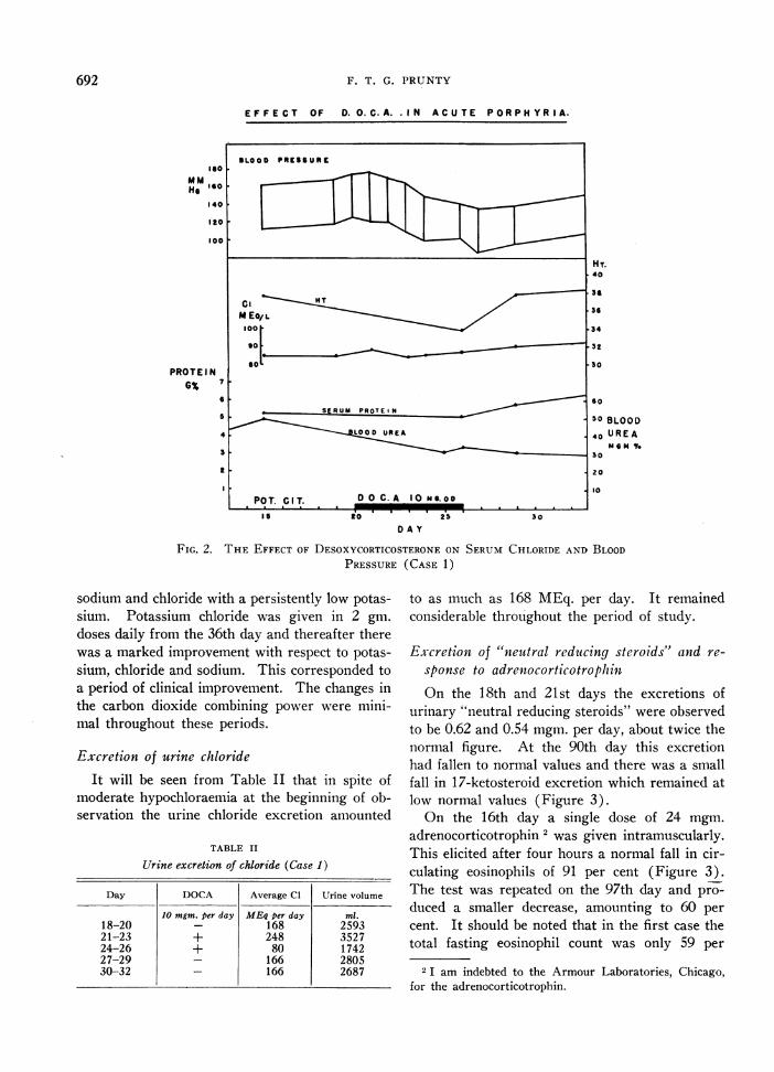
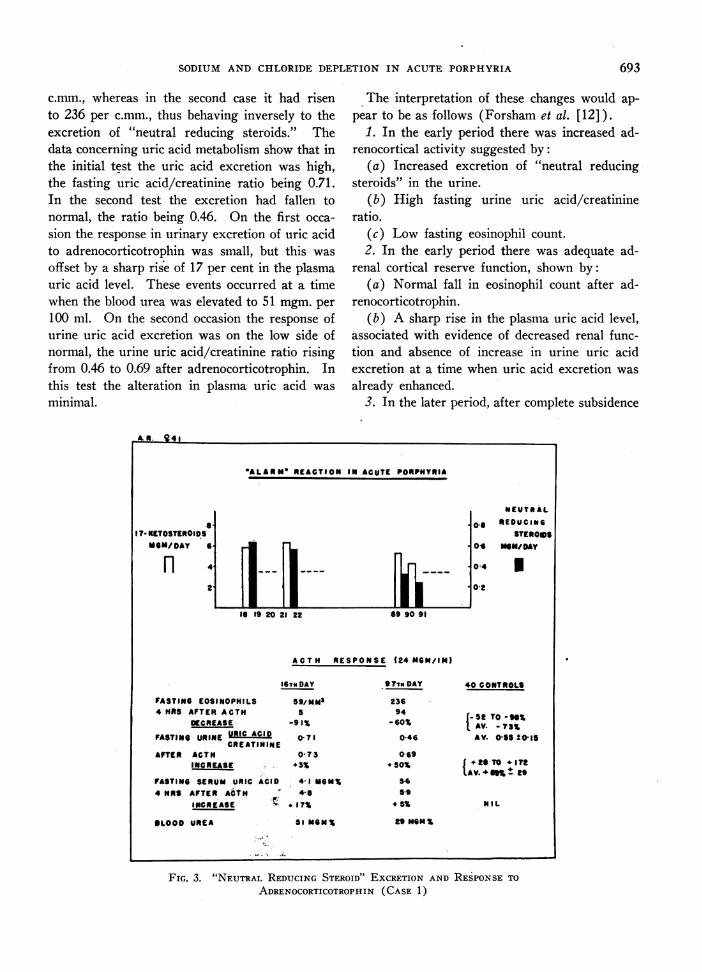
Excretion of "neutral reducing steroids" and re-sponse to adrenocorticotropinOn the 18th and 21st days the excretions of
urinary "neutral reducing steroids" were observedto be 0.62 and 0.54 mgm. per day, about twice thenormal figure. At the 90th day this excretionhad fallen to normal values and there was a smallfall in 17-ketosteroid excretion which remained atlow normal values (Figure 3).
On the 16th day a single dose of 24 mgm.adrenocorticotrophin 2 was given intramuscularly.This elicited after four hours a normal fall in cir-culating eosinophils of 91 per cent (Figure 3).The test was repeated on the 97th day and pro-duced a smaller decrease, amounting to 60 percent. It should be noted that in the first case thetotal fasting eosinophil count was only 59 per
2 I am indebted to the Armour Laboratories, Chicago,for the adrenocorticotrophin.
692
SODIUM AND CHLORIDE DEPLETION IN ACUTE PORPHYRIA
c.mm., whereas in the second case it had risento 236 per c.mm., thus behaving inversely to theexcretion of "neutral reducing steroids." Thedata concerning uric acid metabolism show that inthe initial test the uric acid excretion was high,the fasting uric acid/creatinine ratio being 0.71.In the second test the excretion had fallen tonormal, the ratio being 0.46. On the first occa-sion the response in urinary excretion of uric acidto adrenocorticotrophin was small, but this wasoffset by a sharp rise of 17 per cent in the plasmauric acid level. These events occurred at a timewhen the blood urea was elevated to 51 mgm. per100 ml. On the second occasion the response ofurine uric acid excretion was on the low side ofnormal, the urine uric acid/creatinine ratio risingfrom 0.46 to 0.69 after adrenocorticotrophin. Inthis test the alteration in plasma uric acid wasminimal.
The interpretation of these changes would ap-pear to be as follows (Forsham et al. [12]).
1. In the early period there was increased ad-renocortical activity suggested by:
(a) Increased excretion of "neutral reducingsteroids" in the urine.
(b) High fasting urine uric acid/creatinineratio.
(c) Low fasting eosinophil count.2. In the early period there was adequate ad-
renal cortical reserve function, shown by:(a) Normal fall in eosinophil count after ad-
renocorticotrophin.(b) A sharp rise in the plasma uric acid level,
associated with evidence of decreased renal func-tion and absence of increase in urine uric acidexcretion at a time when uric acid excretion wasalready enhanced.
3. In the later period, after complete subsidence
M.g 41
OALARNOREACTION IN ACUTE PORPHYRIA
a1? KETOSTEROIDS
MOM/DAY
0-*
046
04
0.2
1 19 20 21 22 89 90 91
ACTH RESPONSE124 HCN/IN)
FASTING EOSINOPHILS4 HRS AFTER ACTH
DECREASE
FASTING URINE URIC ACIDCREATININE
AFTER ACTHINCREASE
FASTING SERUM URIC ACID4 HRS AFTER ACtIN
INCREASE
BLOOD UREA
IG DAY
59/NMN5
-91
0 71
0 73+3%
. 4-1 MGM%4-6
+ 17%51 MGM%
97irm DAY
23694
-60%0-46
069* 50%
5*659
* 5%
29 mNm%
FIG. 3. "NEUTRAL REDUCINGSTEROID" EXCRETION AND RESPONSETO
ADRENOCORTICOTROPHIN(CASE 1)
693
NEUTRALREDUCING
STEROIDSNON/DAY
I
40 CONTROLS
t 52 TO -v%AAV. - 73%AV. 0-555015
5+.2TO 172IAV.+S% ±29
NIL
F. T. G. PRUNTY
.... -. -P.....4..
FI(;. 4. ADRENALCORTEX (CASE 3) X 180
of the attack of porphyria, the adrenal corticalactivity had returned to normal, indicated by:
(a) The normal excretion of "neutral reducingsteroids."
(b) The fall to normal of the fasting urine uricacid/creatinine ratio.
(c) The increase in the fasting eosinophilcount.
4. In the later period there was still an ade-quate reserve in adrenal cortical function, shownby:
(a) Normal fall in eosinophil count after ad-renocorticotrophin.
(b) Low normal response in the urine uricacid/creatinine ratio to adrenocorticotrophin.
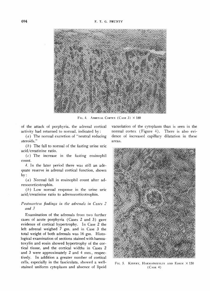
Postnmortcem findings in the adrenials in Cases 2and 3Examination of the adrenals from two further
cases of acute porphyria (Cases 2 and 3) gaveevidence of cortical hypertrophy. In Case 2 theleft adrenal weighed 7 gm. and in Case 3 thetotal weight of both adrenals was 16 gin. Histo-logical examination of sections stained with haema-toxylin and eosin showed hypertrophy of the cor-tical tissue, and the cortical widths in Cases 2and 3 were approximately 2 and 4 mm., respec-tively. In addition a greater number of corticalcells, especially in the fasciculata, showed a well-stained uniform cytoplasm and absence of lipoid
vacuolation of the cytoplasm than is seen in thenormal cortex (Figure 4). There is also evi-dence of increased capillary dilatation in theseareas.
v .w. . w.. : !t All FE
FIG. KIDNEY, HAEMATOXYLINAND EosiN X 150(CASE 4)
6()4
SODIUM AND CHLORIDE DEPLETION IN ACUTE PORPHYRIA6
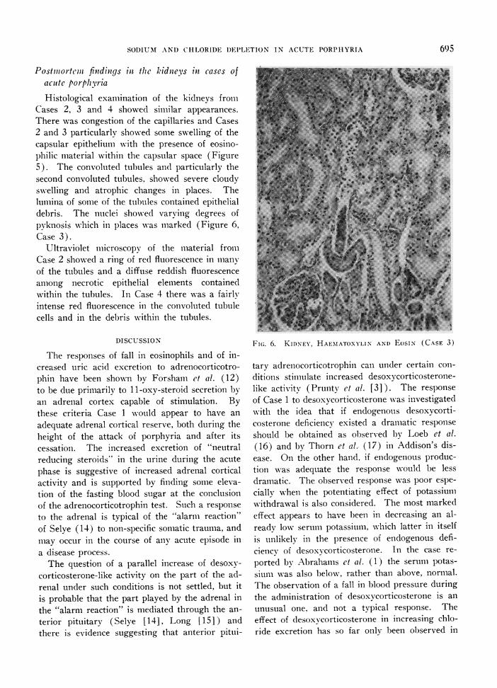
Postinortein findings in thc kidneys in cases ofacute porphlyriaHistological examination of the kidneys from
Cases 2, 3 and 4 showed similar appearances.There was congestion of the capillaries and Cases2 and 3 particularly showed some swelling of thecapsular epithelium with the presence of eosino-philic material within the capsular space (Figure5). The convoluted tubules and particularly thesecond convoluted tubules. showed severe cloudyswelling and atrophic changes in places. Thelumina of some of the tubules contained epithelialdebris. The nuclei showed varying degrees ofpyknosis which in places was marked (Figure 6,Case 3).
Ultraviolet microscopy of the material fromCase 2 showed a ring of red fluorescence in manyof the tubules and a diffuse reddish fluorescenceamong necrotic epithelial elements containedwithin the tubules. In Case 4 there was a fairlyintense red fluorescence in the convoluted tubulecells and in the debris within the tubules.
DISCUSSION
The responses of fall in eosinophils and of in-creased uric acid excretion to adrenocorticotro-phin have been shown by Forsham et al. (12)to be due primarily to 1 1-oxy-steroid secretion byan adrenal cortex capable of stimulation. Bythese criteria Case 1 would appear to have anadequate adrenal cortical reserve, both during theheight of the attack of porphyria and after itscessation. The increased excretion of "neutralreducing steroids" in the urine during the acutephase is suggestive of increased adrenal corticalactivity and is supported by finding some eleva-tion of the fasting blood sugar at the conclusionof the adrenocorticotrophin test. Such a responseto the adrenal is typical of the "alarm reaction"of Selye (14) to non-specific somatic trauma, andmay occur in the course of any acute episode ina disease process.
The question of a parallel increase of desoxy-corticosterone-like activity on the part of the ad-renal under such conditions is not settled, but itis probable that the part played by the adrenal inthe "alarm reaction" is mediated through the an-terior pituitary (Selye [14], Long [15]) andthere is evidence suggesting that anterior pitui-
Fi(;. 6. KIDNEY, HAEMATOXYLIN AND EoSIN (CASE 3)
tary adrenocorticotrophic can under certain con-ditions stimulate increased desoxycorticosterone-like activity (Prunty et al. [3]). The responseof Case 1 to desoxycorticosterone was investigatedwith the idea that if endogenous desoxycorti-costerone deficiency existed a dramatic responseshould be obtained as observed by Loeb et al.(16) and by Thorn et al. (17) in Addison's dis-ease. On the other hand, if endogenous produc-tion was adequate the response would be lessdramatic. The observed response was poor espe-cially when the potentiating effect of potassiumwithdrawal is also considered. The most markedeffect appears to have 1een in decreasing an al-ready low serumn potassium, which latter in itselfis unlikely in the presence of endogenous defi-ciency of desoxycorticosterone. In the case re-
ported by Abrahams et al. (1) the serum potas-siunm was also below, rather than above, normal.The observation of a fall in blood pressure duringthe administration of desoxycorticosterone is an
unusual one, and not a typical response. Theeffect of desoxvcorticosterone in increasing chlo-ride excretion has so far only been observed in
6()5
F. T. G. PRUNTY
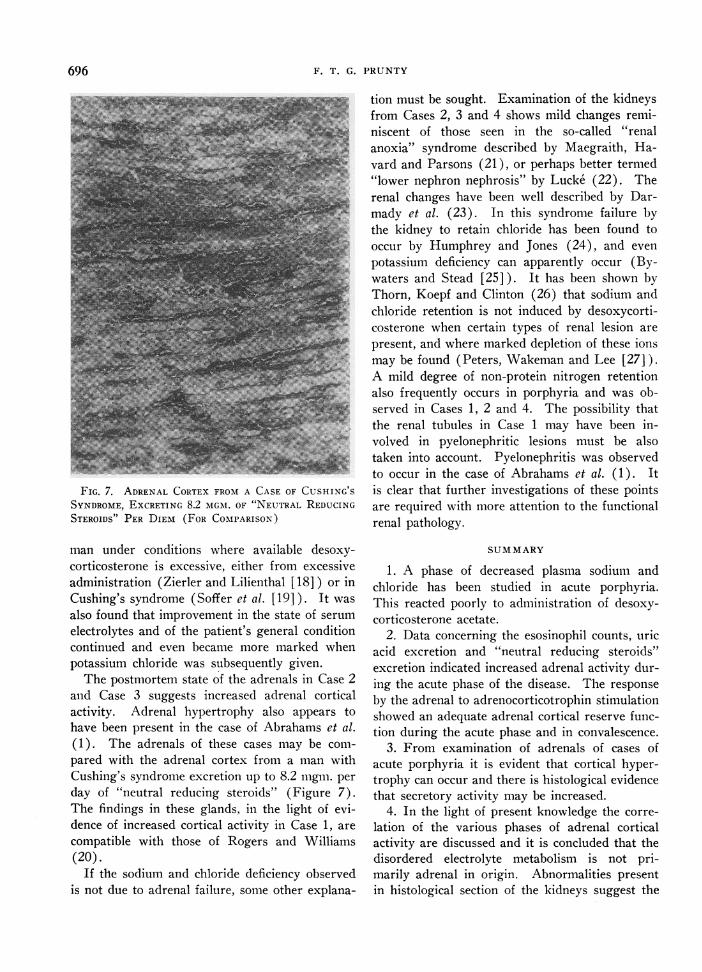
FIG. 7. ADRENALCORTEXFROMA CASE OF CUSHINGS
SYNDROME,EXCRETING 8.2 MGM. OF "NEUTRAL REDUCINGSTEROIDS" PER DIEM (FOR COMPARISON)
man under conditions where available desoxy-corticosterone is excessive, either from excessiveadministration (Zierler and Lilienthal [18]) or inCushing's syndrome (Soffer et al. [19] ). It was
also found that improvement in the state of serum
electrolytes and of the patient's general conditioncontinued and even became more marked whenpotassium chloride was subsequently given.
The postmortem state of the adrenals in Case 2and Case 3 suggests increased adrenal corticalactivity. Adrenal hypertrophy also appears tohave been present in the case of Abrahams et al.(1). The adrenals of these cases may be com-
pared with the adrenal cortex from a man withCushing's syndrome excretion up to 8.2 mgm. per
day of "neutral reducing steroids" (Figure 7).The findings in these glands, in the light of evi-dence of increased cortical activity in Case 1, are
compatible with those of Rogers and Williams(20).
If the sodium and chloride deficiency observedis not due to adrenal failure, some other explana-
tion must be sought. Examination of the kidneysfrom Cases 2, 3 and 4 shows mild changes remi-niscent of those seen in the so-called "renalanoxia" syndrome described by Maegraith, Ha-vard and Parsons (21), or perhaps better termed"lower nephron nephrosis" by Lucke (22). Therenal changes have been well described by Dar-mady et al. (23). In this syndrome failure bythe kidney to retain chloride has been found tooccur by Humphrey and Jones (24), and evenpotassium deficiency can apparently occur (By-waters and Stead [25] ). It has been shown byThorn, Koepf and Clinton (26) that sodium andchloride retention is not induced by desoxycorti-costerone when certain types of renal lesion arepresent, and where marked depletion of these ionsmay be found (Peters, Wakemanand Lee [27]).A mild degree of non-protein nitrogen retentionalso frequently occurs in porphyria and was ob-served in Cases 1, 2 and 4. The possibility thatthe renal tubules in Case 1 may have been in-volved in pyelonephritic lesions must be alsotaken into account. Pyelonephritis was observedto occur in the case of Abrahams et al. (1). Itis clear that further investigations of these pointsare required with more attention to the functionalrenal pathology.
SUMMARY
1. A phase of decreased plasma sodium andchloride has been studied in acute porphyria.This reacted poorly to administration of desoxy-corticosterone acetate.
2. Data concerning the esosinophil counts, uricacid excretion and "neutral reducing steroids"excretion indicated increased adrenal activity dur-ing the acute phase of the disease. The responseby the adrenal to adrenocorticotrophin stimulationshowed an adequate adrenal cortical reserve func-tion during the acute phase and in convalescence.
3. From examination of adrenals of cases ofacute porphyria it is evident that cortical hyper-trophy can occur and there is histological evidencethat secretary activity may be increased.
4. In the light of present knowledge the corre-lation of the various phases of adrenal corticalactivity are discussed and it is concluded that thedisordered electrolyte metabolism is not pri-marily adrenal in origin. Abnormalities presentin histological section of the kidneys suggest the
696
SODIUM AND CHLORIDE DEPLETION IN ACUTE PORPHYRIA
necessity for further investigation of renal functionin this disease.
APPENDIX
Case notes
Case 1. A 42-year-old woman complained that fornine days she had suffered from anorexia and nausea.She was an orphan, and had two healthy children.Since an operation for prolapse eight years previouslyshe had had attacks of cystitis. For a week there hadbeen marked asthenia and several attacks of vomitingwith small quantities of vomitus, and she complained ofan acheing pain in the lower abdomen. The attack coin-cided with one of her menstrual periods which werenormal. She had noticed nocturia. The patient was adark-haired individual with some generalised increasedpigmentation and mild facial hirsutes. There was markedtenderness in both inguinal fossae, but no guarding, andconstipation was severe. Blood pressure was 150/90,pulse rate 100 and temperature ranging up to 99° F.The urine contained a trace of albumin and a few puscells of which 80 per cent were polymorphs. The bloodurea was 44 mgm. per 100 ml. She was given potassiumcitrate on account of her urinary symptoms.
After seven days penicillin therapy was commenced butwithout effect. After 13 days of increased pain and de-veloping rigidity in the lower abdomen exploratory lapa-ratomy was carried out without significant findings. Thepatient had become mentally poorly co-operative and wasslightly hallucinated.
Her urine then was shown to contain porphobilinogenand uroporphyrin and the diagnosis of acute porphyriawas established, the uroporphyrin excretion ranging from12 to 35 mgm. per day. (A study of the porphyrinexcretion is being fully reported elsewhere [McSwiney,Nicholas and Prunty, 1949] [28].)
On the 15th day her blood pressure had risen to 160/115and the pulse rate varied between 120 and 130 for thefollowing three weeks. The plasma protein was 5.4 gn.per cent, albumin 3.3 gm. per cent, globulin plus fibri-nogen 2.1 gm. per cent. Serum cholesterol was 222 rngm.per cent with 68 per cent ester; alkaline phosphatase 2.4units, thymol turbidity 4 units; blood urea 51 mgm. percent and icteric index 4 units. On the 16th day theurobilinogen excretion was 2.9 mgm. per day, and 10.4mgm. per day on the 28th day. It fell to less than 1mgm. per day from the 22nd day onwards. At the con-clusion of the adrenocorticotrophin test on the 16th dayand after 18 hours' fast, the blood sugar was 131 mgm.per cent, but glycosuria was never found. Values forserum electrolytes are recorded in Table I.
Potassium citrate was withdrawn on the 18th day anddesoxycorticosterone acetate 5 mgm. b.d. given intra-muscularly from the 20th to 25th days inclusive, and shewas receiving 7 gm. of added sodium chloride. At theend of this period of six days she complained of increas-ing widespread muscle pain and a constrictive feeling inthe chest. There was a generalised fine somatic tremor.The electrocardiogram was interpreted as being within
normal limits on two occasions, but reading was difficulton account of the tremor. Since the patient's conditionhad apparently deteriorated and the potassium level wasso low (Table I) D.O.C.A. was withdrawn.
On the 29th day the plasma protein was 5.9 gm. percent, albumin 3.0 gm. per cent; serum 'cholesterol 311mgm. per cent, 64 per cent being ester; alkaline phospha-tase 2 units; icteric index 5 units. There was a B. colibacilluria which failed to improve with penicillin therapy.
On the 36th day she was given potassium chloride 2gm. daily in addition to the sodium chloride. From thistime she started to improve clinically and the pulse rategradually returned to normal in the following five weeks.At the end of this period she had greatly improved, theblood pressure had fallen to 130/60, and urea clearancewas 75 per cent of normal. The B. coli bacilluria wascleared up with mandelate therapy. After a further fourweeks she was discharged well.
Case 2. A girl aged 16 was admitted to hospital withconvulsions after a week's acute abdominal pain. Pulserate was 110, temperature 100.50 and blood pressure120/80. Blood urea was 43 mgm. per cent, plasma pro-tein 7.5 gm. per cent, albumin 3.4 gm. per cent. Urineurobilinogen two plus. The urine contained porphobili-nogen, and uroporphyrin amounting to 15 mgm. per day.The attack subsided in three weeks.
Six months later the patient returned with abdominalpain, soon after the onset of which menstruation com-menced. After two weeks ascending paralysis appearedand the patient ultimately died of bronchopneumonia andintercostal paralysis. Investigations during the attackshowed 26,000 white cells; blood urea 62 mgm. per cent,plasma protein 6.5 gm. per cent, albumin 3.2 gm. per cent.There was a slight increase of urine urobilinogen and anoccasional granular cast The urine uroporphyrin ex-cretion ranged up to 68 mgm. per day.
The chief postmortem findings were areas of focalnecrosis in the liver which was shown to contain uro-porphyrin by ultraviolet microscopy and analysis. It alsocontained porphobilinogen. The renal and adrenal find-ings are described above. Detailed investigations of thecase have already been reported. (Prunty [29].)
Case 3. A man aged 20 complained of marked astheniafor several weeks. Six months previously he had anattack of paralysis accompanied by a dark urine, andthree months previously an attack of abdominal pain withsimilar urine.
During the present attack the pain was generalizedwith tenderness in the iliac fossae. There was slightconjunctival icterus. After the onset of mild paralyticsymptoms he died suddenly in ten days. The serumsodium was 126 MEq. per l. and blood urea 34 mgm.per cent nine days before he died. . Porphobilinogen andporphobilin were present in the specimen of urine exam-ined and a moderate amount of uroporphyrin as the zincmetal complex.
At autopsy the myocardium was found to be thin, butotherwise normal. The liver showed areas suggestive ofearly focal necrosis. The hepatic cells contained numer-ous brownish pigment granules, particularly in the cen-
697
F. T. G. PRUNTY
tral portion of the lobules. The adrenals and kidneyshave been described above.
Case 4. A man aged 26 for two months noticed adarkening in the colour of his urine. At birth a menin-gocele was present. He was admitted to hospital witha diagnosis of intestinal obstruction for two days. Con-stipation was complete. He was in shock with a verylow blood pressure, pulse 140, and temperature 1010 F.The blood contained 18,500 white cells; N.P.N. 66 mgm.per cent, carbon dioxide combining power 41 MEq. per 1.,chloride 88 MEq. per 1. The urine had a specific gravityof 1020, no albumin, but hyaline casts and unidentifiedbrown crystals were present. There was a great deal ofporphobilin present in the specimen of urine examined,with much porphyrin metal complex which could not befurther identified with the amount of material available.There was a two plus reaction for urobilinogen.
The patient died at the end of a week and autopsyshowed congenital absence of the left kidney with doublerenal pelvis and ureter on the right. There was extremegut distention and much fluid present. The liver ap-peared normal. Histologically the liver showed the pres-ence of numerous non-iron staining brownish granules inthe liver cells. Ultraviolet microscopy revealed intensered fluorescence in these areas. The pigment appearedto be concentrated particularly in the cells in the centralportions of the lobules, particularly about the central vein.The parenchyma in the centres of some of the lobulesshowed early degenerative changes. The kidney is com-mented upon in the text above.
I am indebted to Drs. Adams and Harewood Littlefor material from Cases 3 and 4.
BIBLIOGRAPHY
1. Abrahams, A., Gavey, C. J., and Maclagan, N. F.,A fatal case of acute porphyria with unusual fea-tures. Brit. M. J., 1947, 2, 327.
2. Linder, G. C., Salt metabolism in acute porphyria.Lancet, 1947, 2, 649.
3. Prunty, F. T. G., Forsham, P. H., and Thorn, G. W.,Desoxycorticosterone-like activity induced by ad-renocorticotrophin in man. Clin. Sci., 1948, 7, 109.
4. Cutler, H. H., Power, M. H., and Wilder, R. M.,Concentrations of chloride, sodium and potassiumin urine and blood; their diagnostic significance inadrenal insufficiency. J. A. M. A., 1938, 111, 117.
5. Butler, A. M., and Tuthill, E., An application of theuranyl zinc acetate method for determination ofsodium in biological material. 3. Biol. Chem.,1931, 93, 171.
6. Van Slyke, D. D., and Hiller, A., Application ofSendroy's iodometric chloride titration to protein-containing fluids. J. Biol. Chem., 1947, 167, 107.
7. King, E. J., Haslewood, G. A. D., Delory, G. E., andBeall, D., Microchemical methods of blood analysis,revised and extended. Lancet, 1942, 2, 207.
8. Peters, J. P., and Van Slyke, D.D., QuantitativeClinical Chemistry. Williams & Wilkins Co.,Baltimore, 1932, Vol. II, p. 245.
9. Malm, O. J., Personal communication, 1947.10. Talbot, N. B., Saltzman, A. H., Wixom, R. L., and
Wolfe, J. K., Colorimetric assay of urinary corti-costeroid-like substances. J. Biol. Chem., 1945,160, 535.
11. Callow, N. H., Callow, R. K., and Emmens, C. W.,Colorimetric determination of substances containingthe grouping -CH2CO- in urine extracts as indi-cation of androgen content. Biochem. J., 1938, 32,1312.
12. Forsham, P. H., Thorn, G. W., Prunty, F. T. G.,and Hills, A. G., Clinical studies with pituitaryadrenocorticotrophin. J. Clin. Endocrinol., 1948,8, 15.
13. Sveinsson, S. L., Rimington, C., and Barnes, H. D.,Complete porphyrin analysis of pathological urines.Scand. J. Clin. Biochem. & Physiol., 1949, in press.
14. Selye, H., General adaptation syndrome and diseasesof adaptation. J. Clin. Endocrinol., 1946, 6, 117.
15. Long, C. N. H., The conditions associated with thesecretion of the adrenal cortex. Federation Proc.,1947, 6, 461.
16. Loeb, R. F., Atchley, D. W., Ferrebee, J. W., andRagan, C., Observations on effect of desoxycorti-costerone esters and progesterone in patients withAddison's disease. Tr. A. Am. Physicians, 1939,54, 285.
17. Thorn, G. W., Howard, R. P., and Emerson, K., Jr.,Treatment of Addison's disease with desoxycorti-costerone acetate, synthetic adrenal cortical hor-mone (preliminary report). J. Clin. Invest., 1939,18, 449.
18. Zierler, K. L., and Lilienthal, J. L., Jr., Sodium lossin man induced by desoxycorticosterone acetate:Study in a subject with myotonic dystrophy.Am. J. Med., 1948, 4, 186.
19. Soffer, L. J., Lesnick, G., Sorkin, S. Z., Sobotka,H. H., and Jacobs, M., Utilization of intravenouslyinjected salt in normals and in patients with Cush-ing's syndrome before and after administration ofdesoxycorticosterone acetate. J. Clin. Invest., 1944,23, 51.
20. Rogers, W. F., Jr., and Williams, R. H., Correla-tions of biochemical and histologic changes in theadrenal cortex. Arch. Path., 1947, 44, 126.
21. Maegraith, B. G., Havard, R. E., and Parsons, D. S.,Renal syndrome of wide distribution induced pos-sibly by renal anoxia. Lancet, 1945, 2, 293.
22. Luck6, B., Lower nephron nephrosis (renal lesionsof crush syndrome, of burns, transfusions and otherconditions affecting lower segments of nephrons).Mil. Surgeon, 1946, 99, 371.
23. Darmady, E. M., Siddons, A. H. M., Corson, T. C.,Langton, C. D., Vitek, Z., Badenoch, A. W., andScott, J. C., Traumatic uraemia. Reports on 8cases. Lancet, 1944, 2, 809.
24. Humphrey, J. H., and Jones, F. A., Oliguria afterabortion. Clin. Sc., 1947, 6, 173.
698
SODIUM AND CHLORIDE DEPLETION IN ACUTE PORPHYRIA
25. Bywaters, E. G. L., and Stead, J. K., Thrombosis ofthe femoral artery with myohaemoglobinuria andlow serum potassium concentration. Clin. Sc.,
1945, S, 195.26. Thorn, G. W., Koepf, G. F., and Clinton, M., Jr.,
Renal failure simulating adrenocortical insuffi-ciency. New England J. Med., 1944, 231, 76.
27. Peters, J. P., Wakeman, A. M., and Lee, C., Total
acid-base equilibrium of plasma in health and dis-ease; hypochloremia and total salt deficiency innephritis. J. Clin. Invest., 1929, 6, 551.
28. McSwiney, R. R., Nicholas, R. E. H., and Prunty,F. T. G., 1949, in preparation.
29. Prunty, F. T. G., Acute porphyria; investigations onthe pathology of the porphyrins and identificationof the excretion of uroporphyrin I. Arch. Int.Med., 1946, 77, 623.
699