ACUTE INTERMITTENT PORPHYRIA
66
Department of Clinical Science and Education Karolinska Institutet, Stockholm, Sweden ACUTE INTERMITTENT PORPHYRIA STUDIES ON CHRONIC DISEASE, RECOMBINANT ENZYME THERAPY AND LATE COMPLICATIONS Eliane Sardh Stockholm 2008
Transcript of ACUTE INTERMITTENT PORPHYRIA
Department of Clinical Science and Education Karolinska Institutet, Stockholm, Sweden
ACUTE INTERMITTENT PORPHYRIA
STUDIES ON CHRONIC DISEASE, RECOMBINANT ENZYME THERAPY AND LATE COMPLICATIONS
Eliane Sardh
Stockholm 2008

All previously published papers were reproduced with permission from the publisher. Published by Karolinska Institutet. © Eliane Sardh, 2008 ISBN 978-91-7409-162-5
2008
Gårdsvägen 4, 169 70 Solna
Printed by

“They who dream by day are cognizant of many things which escape those who dream only by night. - Edgar Allan Poe
To Jan and my beautiful sons, Fabian and Liam


ABSTRACT This thesis comprise five studies on patients with acute intermittent porphyria (AIP) observed under different clinical conditions associated with the disorder. They have generally been monitored at the porphyria out-patient clinic in Stockholm South Hospital, and were biochemically monitored at Porphyria Centre Sweden. Acute intermittent porphyria is an autosomal dominant metabolic disorder with comparatively high prevalence in Sweden; 1:10 000. The disease is caused by a partial deficiency of porphobilinogen deaminase (PBGD), the third enzyme in heme biosynthesis. The clinical penetrance of the underlying gene mutation is relatively limited and characterized by attacks of painful and potentially life threatening symptoms, mainly from the nervous system, and accompanied by accumulation and excretion of the porphyrin precursors porphobilinogen (PBG) and the possibly neurotoxic 5-aminolevulinic acid (ALA). Estimation of the current extent of porphyrin precursor excretion is presently the only tool for assessment of an acute attack of porphyria in the patient with AIP. In this process, precise knowledge of the physiological and pathophysiological fluctuations of these markers is essential for the accurate evaluation of the clinical situation at hand. Beside the few recently reported liver transplantations, there is presently no available way to bring about permanent cure for the metabolic error in AIP. In wait for gene therapy, the obvious measure would be to compensate for the underlying enzyme deficiency by supplementation via administration of the deficient enzyme. In success, would not only acute attacks be inhibited, the lethal late manifestations from liver and kidneys would also be blocked. Our studies thus approach questions concerning the kinetics of PBG and ALA in AIP monitored also under conditions secondary to renal engagement and hepatic tumour. In order to be able to measure the low plasma concentrations of these metabolites, we developed a novel HPLC-MS procedure. One study defines the pharmacokinetic and pharmacodynamic variables of recombinant human PBGD administered in a clinical trial aimed at evaluation of the clinical efficacy of enzyme therapy. In the studies of late complications more than fifty patient case-books were surveyed with regard to beforehand selected variables connected with the development of renal engagement and primary liver cancer. Only patients with biochemical signs of active disease are studied, twenty four currently asymptomatic individuals with permanently increased urinary PBG and ALA being included in either study I or II. Additional three patients with four current attacks participate in study III, and in study IV further three patients burdened by recurrent attacks, chronic hypertension and end-stage renal disease take part. In study V twenty AIP patients with primary liver cancer are included. In study I we established the concentration patterns of PBG and ALA in plasma and urine, monitored during eight hours in ten currently asymptomatic AIP gene carriers. Within each individual, the pattern shows to be constant and there is strong correlation
between these two metabolites in plasma and urine. Their renal clearances were in both cases about 70 mL/min. This study formed the basis for the studies II, III and IV. In studies I, II and III are observed significantly higher morning, than evening plasma concentrations of PBG, pointing to a circadian variation not previously reported, and not found for ALA. In Study II the safety, pharmacokinetics and pharmacodynamics of human recombinant PBGD used in substitution therapy of AIP, was studied. This work, done in collaboration with the company Zymenex A/S, aimed at repairing the PBGD deficient step in the heme biosynthetic pathway. The drug proved to be safe, and the pharmacokinetics and pharmacodynamics of the enzyme were elucidated. The drug effectively removed plasma PBG but showed no effect on the presumably neurotoxic ALA. In Study III we give evidence that plasma PBG is a more sensitive biomarker for the acute attack of porphyria, than plasma ALA, or urinary PBG and ALA. In Study IV is demonstrated that before hemodialysis PBG accumulates strongly in plasma, but is readily filtered by the hemodialysis membrane. The accumulation of PBG is less evident in the patient on peritoneal dialysis. Accumulated plasma porphyrins are not cleared by either of the dialysis procedures, and both patients studied developed skin lesions. The patient in predialysis showed only a mildly increased accumulation of porphyrins. Study V is a compilation of the clinical and biochemical presentations, histopathological characteristics and therapeutic outcomes of twenty patients with AIP that had developed primary liver cancer. The tumour was earlier recognized in an annual surveillance program, than via other measures, and all patients were detected after the age of 50 years and had increased porphyrin precursor excretion. In AIP patients with, as well as without symptoms in studies I, II and III, the concentrations of PBG in plasma and urine are about twice of those of ALA. This was not observed in study IV, where the patients had developed end stage renal failure. To conclude, through the thorough clinical and biochemical revisions of the patient materials of the study a greatly deepened insight is gained in the natural history of the disease caused by mutations in the gene for porphobilinogen deaminase, i.e. acute intermittent porphyria.

LIST OF PUBLICATIONS The thesis is based on the following original articles, referred to in the text by their Roman numerals.
I. Variations in porphobilinogen and 5-aminolevulinic acid concentrations in plasma and urine from asymptomatic carriers of the acute intermittent porphyria gene with increased porphyrin precursor excretion. Ylva Floderus, Eliane Sardh, Christer Möller, Claes Andersson, Lillan Rejkjaer, Dan EH Andersson, Pauline Harper. Clin Chem 2006; 52: 701-707.
II. Safety, pharmacokinetics and pharmacodynamics of recombinant human porphobilinogen deaminase in healthy subjects and asymptomatic carriers of the acute intermittent porphyria gene who have increased porphyrin precursor excretion. Eliane Sardh, Lillan Rejkjaer, Dan EH Andersson, Pauline Harper. Clin Pharmacokinet 2007; 46: 335-349.
III. Plasma porphobilinogen as a sensitive biomarker to monitor the clinical and therapeutic course of acute intermittent porphyria attacks. Eliane Sardh, Pauline Harper, Dan EH Andersson, Ylva Floderus. Eur J Intern Med 2008; available online 8 Aug 2008.
IV. Porphyrin precursors and porphyrins in three patients with overt acute intermittent porphyria and end-stage renal disease under different therapy regimes. Eliane Sardh, Dan EH Andersson, Ann Henrichson, Pauline Harper. Submitted manuscript.
V. Primary liver cancer in acute porphyria: a single centre experience of 20 cases. Eliane Sardh, Staffan Wahlin, Mikael Björnstedt, Pauline Harper#, Dan EH Andersson#. Submitted manuscript. # Shared senior authorship

CONTENTS INTRODUCTION.......................................................................................................11
Background ............................................................................................................11 Studies on chronic disease, enzyme replacement therapy and late complications 12
REVIEW OF LITERATURE....................................................................................13
Heme ......................................................................................................................13 The heme biosynthetic pathway.............................................................................13 Regulation of the heme biosynthetic pathway .......................................................15 Acute intermittent porphyria ..................................................................................15
Clinical manifestations of AIP .....................................................................16 Initiation of the attack...................................................................................17 Pathogenesis .................................................................................................17 Diagnosis ......................................................................................................19 Current treatment..........................................................................................20
New therapeutic options.........................................................................................21 Enzyme substitution .....................................................................................21 Gene therapy.................................................................................................21 Liver transplantation.....................................................................................21
Late complications .................................................................................................22 The life perspective of AIP gene carriers...............................................................22
APPENDIX ..................................................................................................................23 AIMS OF THE STUDY..............................................................................................25
The specific aims....................................................................................................25
MATERIALS AND METHODS ...............................................................................27
Recruitment of participants ....................................................................................27 Studies I and II .......................................................................................................27
Study participants .........................................................................................27 Study design .................................................................................................28
Study III .................................................................................................................30 Study participants .........................................................................................30 Study design .................................................................................................30
Study IV .................................................................................................................31 Study participants .........................................................................................31 Study design .................................................................................................31
Study V ..................................................................................................................31 Study participants .........................................................................................31

Study design................................................................................................. 31 Materials ................................................................................................................ 31 Methods ................................................................................................................. 32
Statistical methods (studies I, II and III)...................................................... 34
RESULTS.................................................................................................................... 35
Studies on chronic active disease and enzyme replacement therapy..................... 35
Study I ......................................................................................................... 35 Study II ......................................................................................................... 36 Study III........................................................................................................ 37
Studies on late complications ................................................................................ 38 Study IV........................................................................................................ 38 Study V ......................................................................................................... 41
DISCUSSION.............................................................................................................. 43
Studies on asymptomatic but biochemically active AIP patients.......................... 44
Renal clearances of PBG and ALA ............................................................. 45 Ratios of PBG to ALA in plasma and urine ................................................ 45 Circadian variation of PBG.......................................................................... 46
Enzyme replacement therapy ................................................................................ 46 Late complications................................................................................................. 47
Renal impairment......................................................................................... 47 Primary liver cancer..................................................................................... 49
CONCLUSIONS......................................................................................................... 51
Physiology and pathophysiology of ALA and PBG ............................................. 51 Enzyme therapy ..................................................................................................... 51 Late complications................................................................................................. 52
Renal impairment......................................................................................... 52 Primary liver cancer..................................................................................... 52
Challenges..................................................................................................................... 53 Health care aspects ................................................................................................ 53 Future research ...................................................................................................... 53
ACKNOWLEDGEMENTS....................................................................................... 55 REFERENCES ........................................................................................................... 57
LIST OF ABBREVIATIONS AIP acute intermittent porphyria ALA 5-aminolevulinic acid ALAD 5-aminolevulinic acid dehydratase ALAS 5-aminolevulinic acid synthase AUC area under the concentration-time curve C0 concentration at time zero CAR constitutive androstane receptor CC cholangiocarcinoma cHCC-CC combined hepatocellular carcinoma and cholangiocarcinoma Cmax maximum concentration eGFR estimated glomerular filtration rate ELISA enzyme linked immunosorbent assay GABA gamma aminobutyric acid HCC hepatocellular cancer HMB hydroxymethylbilane HMBS hydroxymethylbilane synthase HPLC high performance liquid-chromatography MS masspectrometry PBG porphobilinogen PBGD porphobilinogen deaminase PLC primary liver cancer PXR pregnane xenobiotic receptor rhPBGD recombinant human porphobilinogen deaminase SIADH syndrome of inappropriate antidiuretic hormone secretion t1/2 half-life for the elimination phase
11
INTRODUCTION Background The porphyrias are a group of inherited metabolic diseases connected with enzyme deficiencies in the heme biosynthetic pathway. Heme, the prosthetic group of many important proteins, is synthesized in all living cells but is most active in the erythroid bone marrow for the formation of hemoglobin, and in the liver, particularly for the formation of members of the cytochrome P450 enzyme system. There are eight enzymes involved in heme biosynthesis. With the exception of the initial enzyme, a genetic defect leading to enzymatic deficiency at any subsequent step in the heme biosynthetic chain may result in accumulation of toxic or presumably toxic heme precursors. Based upon the principal site of expression of the enzymatic deficiency, the porphyrias may be classified as hepatic or erythropoietic, but on grounds of the main clinical presentation they are more often referred to as acute or cutaneous. The acute porphyrias mainly give rise to symptoms from the nervous system, while the cutaneous porphyrias are characterized by dermal photosensitivity. The most common of the acute hepatic porphyrias is acute intermittent porphyria (AIP), caused by a partial deficiency of porphobilinogen deaminase (PBGD), the third enzyme in the heme biosynthetic pathway. It has a worldwide prevalence of about 1:20 000 [1]. The clinical penetrance of PBGD deficiency is high compared with the other autosomal dominant acute porphyrias, and women are more affected than men [2, 3]. The disease is characterized by acute attacks with predominantly neuropsychiatric symptoms. The attack of acute porphyria is precipitated by any factor that gives rise to significantly increased mitochondrial activity of the rate-limiting enzyme in hepatic heme biosynthesis, i.e. ubiquitous 5-aminolevulinate synthase (ALAS1) [4]. The consequentially increased flux of metabolites through the PBGD-deficient pathway results in accumulation of the porphyrin precursors porphobilinogen (PBG) and 5-aminolevulinic acid (ALA), which appear in increased concentration in plasma and urine [1, 5]. Treatment of the acute attack includes removal of factors that trigger the condition and appropriate supportive measures for alleviating the symptoms, as well as measures for repression of ALAS1 activity via administration of carbohydrates and heme. After recovery from the acute porphyric crisis the patient may continue to present with increased excretion of PBG and ALA also during the asymptomatic phase, a state which may persist for a long period of time [1, 6]. A few patients develop recurrent acute attacks requiring repeated hospitalizations and specific treatment. In-between attacks these patients present with permanently high excretion of PBG and ALA that increases further during subsequent porphyric crisis [1, 6]. This chronic active condition has been associated with the late complications related to acute porphyria, e.g. hypertension, renal impairment, chronic neuropathies and primary liver cancer [7].

12
Studies on chronic disease, enzyme replacement therapy and late complications In this thesis, are presented the findings in five studies on AIP gene carriers under different clinical conditions associated with the disorder. In Study I we investigated the concentration patterns of plasma and urinary porphyrin precursors as monitored during eight hours in ten asymptomatic AIP-gene carriers with permanently increased urinary excretion. The development of a combined high performance liquid-chromatographic masspectrometric (HPLC-MS) method for measuring porphyrin precursors in the low concentration range made it possible for us to study the variation of these metabolites in plasma, and to determine their renal clearance. This work formed the basis for the studies II, III and IV. In Study II we investigated the safety, pharmacokinetics and pharmacodynamics of the recombinant human PBGD enzyme (rhPBGD) used in substitution therapy. This work, was done in collaboration with the company Zymenex A/S, Denmark, and attempted to repair the PBGD-deficiency in AIP gene carriers. In Study III we studied the changing patterns of porphyrin precursors in plasma and urine during four acute attacks in three AIP-patients, and characterized the metabolic effect of the given therapy. In Study IV we investigated the patterns of porphyrin precursors and porphyrins in plasma and urine, as well as the clinical outcome in three patients with chronic porphyric disease accompanied by hypertension, end-stage renal disease and chronic peripheral neuropathy. Study V is a compilation of the clinical and biochemical presentation, histopathological characteristics and therapeutic outcome in twenty patients with AIP that had developed primary liver cancer.

13
REVIEW OF LITERATURE Heme Heme is a complex of ferrous iron and protoporphyrin IX, which associated with different proteins is essential for life in all aerobic cells [1]. Heme serves as a prosthetic group for hemoproteins involved in an array of crucial biological functions. It is synthesized in the bone marrow for the formation of hemoglobin, the transporter of oxygen, and by the liver for the formation of microsomal cytochromes P450 responsible for detoxification of drugs and environmental contaminants, and for the metabolism and syntheses of steroid hormones. Heme is also engaged in the transfer of electrons in the cytochromes of the respiratory chain and serves as the prosthetic group of numerous hemoproteins that synthesize important regulatory or signaling molecules including cyclic guanosine monophosphate (cGMP) (guanylate cyclase) and nitric oxide (NO synthase). The heme biosynthetic pathway Heme biosynthesis involves eight enzymes that sequentially convert glycine and succinyl-CoA to heme, figure 1. The first and the final three enzymes are situated in the mitochondria, and the others in the cytosol. The heme biosynthetic pathway is composed of four basic processes, formation of the pyrrole, assembly of the tetrapyrrole, modification of the tetrapyrrole side chains followed by oxidation of protoporphyrinogen IX to protoporphyrin IX, and the insertion of a single ferrous iron [1, 8]. The unique property of heme, an iron ion coordinately bound to a tetrapyrrole, permits binding of heme to a number of proteins for oxygen transport or electron transfers in oxidation-reduction reactions. The first enzyme of the pathway, 5-aminolevulinic acid synthase (ALAS), catalyses the formation of 5-aminolevulinic acid (ALA), which is exclusively committed to the synthesis of heme. Aminolevulinic acid dehydratase (ALAD), the next enzyme in the pathway, condensates two molecules of ALA to the monopyrrole porphobilinogen (PBG). The third enzyme, porphobilinogen deaminase (PBGD), catalyzes the stepwise condensation of four molecules of PBG to form the linear tetrapyrrole hydroxymethylbilane (HMB), also called preuroporphyrinogen. Nonenzymatically, HMB forms uroporphyrinogen I, which does not participate in the subsequent steps of heme biosynthesis. The intra-molecular rearrangements and ring closure of HMB to form uroporphyrinogen III, a cyclic tetrapyrrole with eight carboxyl side chains, is catalyzed by uroporphyrinogen III synthase (UROS). Uroporphyrinogen III is then stepwise decarboxylated by uroporphyrinogen decarboxylase (UROD) and coproproporphyrinogen oxidase (CPO) as depicted in figure 1, forming protoporphyrinogen IX, with two carboxyl side chains. This molecule is oxidized to protoporphyrin IX by the seventh enzyme of the pathway, protoporphyrinogen oxidase (PPOX). The final step in the heme biosynthetic chain is the insertion of iron by the enzyme ferrochelatase (FC).

14
Figure 1. The heme biosynthetic pathway. The subcellular distribution of enzymes and intermediates in the cytoplasm and in mitochondria is shown. = COOH, ALAS = 5-aminolevulinic acid synthase, ALAD = 5-aminolevulinic acid dehydratase, PBGD = porphobilinogen deaminase, UROS = uroporphyrinogen III synthase, UROD = uroporphyrinogen decarboxylase, CPO = coproporphyrinogen oxidase, PPOX = protoporphyrinogen oxidase, FC = ferrochelatase.
ALA and PBG are also nominated porphyrin precursors
Cytoplasm Mitochondria
Coproporphyrinogen III Protoporphyrinogen IX
6. CPO
Uroporphyrinogen III Protoporphyrin IX
5. UROD
HemeHydroxymethylbilane
7. PPOX
Porphobilinogen 5-aminolevulinic acid
Glycin + Succinyl-CoA
4. UROS 8. FC
3. PBGD
2. ALAD 1. ALAS
Cytoplasm Mitochondria
Coproporphyrinogen III Protoporphyrinogen IX
6. CPO
Uroporphyrinogen III Protoporphyrin IX
5. UROD
HemeHydroxymethylbilane
7. PPOX
Porphobilinogen 5-aminolevulinic acid
Glycin + Succinyl-CoA
4. UROS 8. FC
3. PBGD
2. ALAD 1. ALAS
15
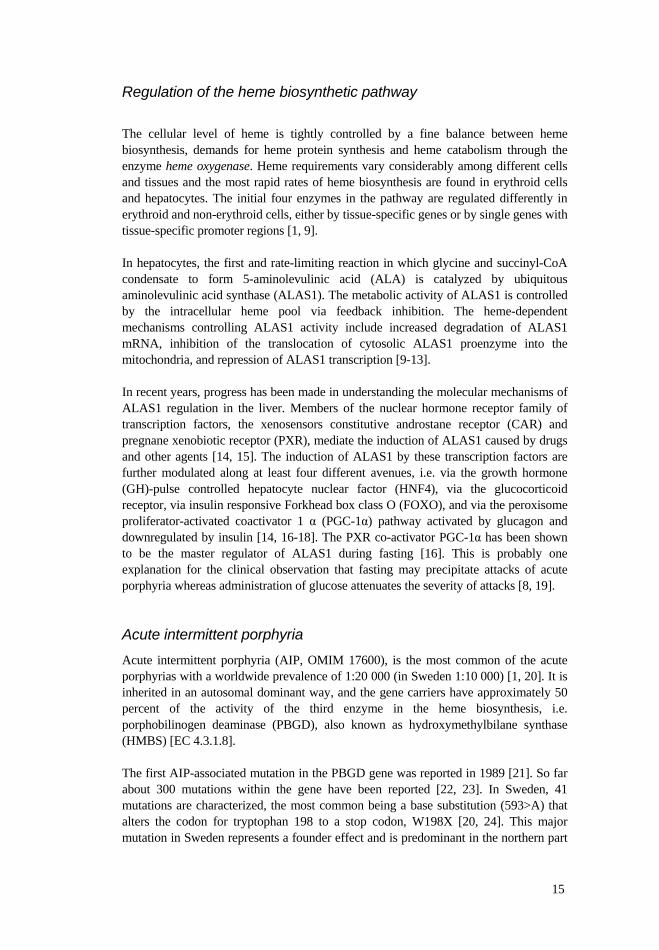
Regulation of the heme biosynthetic pathway The cellular level of heme is tightly controlled by a fine balance between heme biosynthesis, demands for heme protein synthesis and heme catabolism through the enzyme heme oxygenase. Heme requirements vary considerably among different cells and tissues and the most rapid rates of heme biosynthesis are found in erythroid cells and hepatocytes. The initial four enzymes in the pathway are regulated differently in erythroid and non-erythroid cells, either by tissue-specific genes or by single genes with tissue-specific promoter regions [1, 9]. In hepatocytes, the first and rate-limiting reaction in which glycine and succinyl-CoA condensate to form 5-aminolevulinic acid (ALA) is catalyzed by ubiquitous aminolevulinic acid synthase (ALAS1). The metabolic activity of ALAS1 is controlled by the intracellular heme pool via feedback inhibition. The heme-dependent mechanisms controlling ALAS1 activity include increased degradation of ALAS1 mRNA, inhibition of the translocation of cytosolic ALAS1 proenzyme into the mitochondria, and repression of ALAS1 transcription [9-13]. In recent years, progress has been made in understanding the molecular mechanisms of ALAS1 regulation in the liver. Members of the nuclear hormone receptor family of transcription factors, the xenosensors constitutive androstane receptor (CAR) and pregnane xenobiotic receptor (PXR), mediate the induction of ALAS1 caused by drugs and other agents [14, 15]. The induction of ALAS1 by these transcription factors are further modulated along at least four different avenues, i.e. via the growth hormone (GH)-pulse controlled hepatocyte nuclear factor (HNF4), via the glucocorticoid receptor, via insulin responsive Forkhead box class O (FOXO), and via the peroxisome proliferator-activated coactivator 1 (PGC-1 ) pathway activated by glucagon and downregulated by insulin [14, 16-18]. The PXR co-activator PGC-1 has been shown to be the master regulator of ALAS1 during fasting [16]. This is probably one explanation for the clinical observation that fasting may precipitate attacks of acute porphyria whereas administration of glucose attenuates the severity of attacks [8, 19]. Acute intermittent porphyria Acute intermittent porphyria (AIP, OMIM 17600), is the most common of the acute porphyrias with a worldwide prevalence of 1:20 000 (in Sweden 1:10 000) [1, 20]. It is inherited in an autosomal dominant way, and the gene carriers have approximately 50 percent of the activity of the third enzyme in the heme biosynthesis, i.e. porphobilinogen deaminase (PBGD), also known as hydroxymethylbilane synthase (HMBS) [EC 4.3.1.8]. The first AIP-associated mutation in the PBGD gene was reported in 1989 [21]. So far about 300 mutations within the gene have been reported [22, 23]. In Sweden, 41 mutations are characterized, the most common being a base substitution (593>A) that alters the codon for tryptophan 198 to a stop codon, W198X [20, 24]. This major mutation in Sweden represents a founder effect and is predominant in the northern part
16
of the country with a local prevalence of 1:1000. The penetrance of the disease is relatively low, only 20-50 % of the carriers experience clinical symptoms [25]. Women are more often affected than men [2, 3, 26], and symptoms are often related to the menstrual cycle. Overt disease, with a peak occurrence in the third decade, is exceptionally rare before puberty [27] and less likely to occur after menopause [2]. Clinical manifestations of AIP
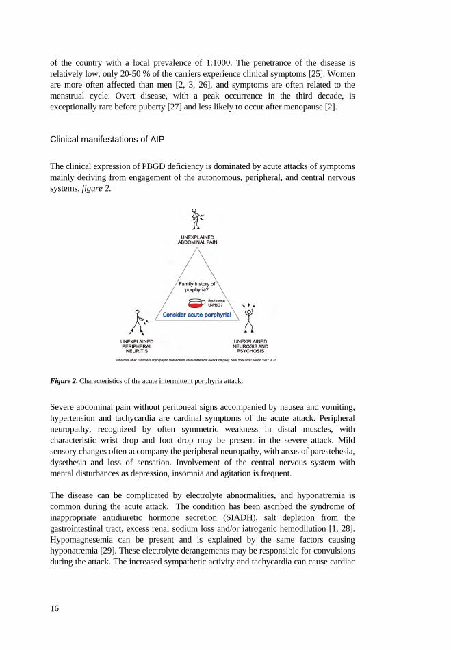
The clinical expression of PBGD deficiency is dominated by acute attacks of symptoms mainly deriving from engagement of the autonomous, peripheral, and central nervous systems, figure 2.
Figure 2. Characteristics of the acute intermittent porphyria attack.
Severe abdominal pain without peritoneal signs accompanied by nausea and vomiting, hypertension and tachycardia are cardinal symptoms of the acute attack. Peripheral neuropathy, recognized by often symmetric weakness in distal muscles, with characteristic wrist drop and foot drop may be present in the severe attack. Mild sensory changes often accompany the peripheral neuropathy, with areas of parestehesia, dysethesia and loss of sensation. Involvement of the central nervous system with mental disturbances as depression, insomnia and agitation is frequent. The disease can be complicated by electrolyte abnormalities, and hyponatremia is common during the acute attack. The condition has been ascribed the syndrome of inappropriate antidiuretic hormone secretion (SIADH), salt depletion from the gastrointestinal tract, excess renal sodium loss and/or iatrogenic hemodilution [1, 28]. Hypomagnesemia can be present and is explained by the same factors causing hyponatremia [29]. These electrolyte derangements may be responsible for convulsions during the attack. The increased sympathetic activity and tachycardia can cause cardiac

17
arrhythmias and sudden death, especially in the presence of hypomagnesemia. Other serious potentially lethal complications are bulbar paralysis causing respiratory failure. Initiation of the attack
Xenobiotics, including several prescription drugs, and certain reproductive hormones, catabolic conditions and several other factors, in practice not always possible to identify, may trigger the attack of acute porphyria, figure 3. Most probably they act through activation of the nuclear receptors, or co-activators of these, which initiate transcription of the ALAS1 gene.
Figure 3. The pathophysiology of the acute porphyria attack. Effect of ALAS1 induction subsequent to decreased cellular heme pool in acute porphyria.
Simultaneous depletion of the hepatic intracellular regulatory heme pool enhances the inductive response of ALAS1 by permitting mitochondrial entry of the enzyme transcribed. The consequentially accelerated flux of metabolites through the heme biosynthetic pathway will risk to overload the deficient PBGD step. If heavy enough, ALAS1-induction will thus give rise to accumulation of the porphyrin precursors porphobilinogen (PBG) and 5-aminolevulinic acid (ALA) [1, 30] which accumulate and are found in increased concentrations in plasma and urine [30]. A few patients, predominantly women, may be affected by recurrent acute attacks, often related to the menstrual cycle. After recovery from the acute attack, a majority of the patients may continue to present with increased excretion of PBG and ALA also during the asymptomatic phase, a condition which may persist for a long period of time [1, 6]. Pathogenesis
There is currently a fairly complete understanding of the molecular genetics as well as the biochemistry of the acute porphyrias. However, the pathogenesis behind the neurovisceral symptoms and the mechanisms by which a partial defect in the heme biosynthesis causes transient dysfunction of the nervous system are not understood [31].

18
Hypotheses have been put forward that the cause of the symptoms may be found in neurotoxic effects from (1) porphyrin precursors ALA and PBG or porphyrins that accumulate, or from products derived from ALA and PBG, e.g. neurotoxic free radicals; (2) a relative heme deficiency in the nervous system secondary to impaired biosynthesis of heme leading to impaired hemoprotein function in neural tissue; (3) depletion of substrates or cofactors essential for nerve function as a result of disturbed heme biosynthesis [31]. Porphyrin precursors, neurotoxicity
The current major hypothesis is that porphyrin precursors, and especially ALA, cause the porphyric neuropathy. This hypothesis is supported by the fact that ALAD porphyria, tyrosinemia type I and lead poisoning, all are associated with increased urinary excretion of ALA and accompanied by neurological symptoms resembling those of acute porphyria. Direct administrations of ALA to neuromuscular, muscular and spinal cord preparations of cultured cells from various species have demonstrated neurotoxic effects, however not in human spinal cord neurons [31]. In vivo experiments of ALA administration to rodents, involving parenteral [32] and oral administration during several weeks [33], have, however failed to give evidence of any significant toxic effect. Also, in a heroic experiment, oral administration of ALA to a male volunteer during 3.8 days reaching concentrations corresponding to those prevailing during an attack of acute porphyria, did not cause any subjective symptoms or pathophysiological changes in concordance with an attack of acute porphyria [34]. There are several different mechanisms proposed for ALA neurotoxicity. The compound ALA has close structural similarity with the inhibitory neurotransmitter gamma aminobutyric acid (GABA) and the excitatory amino acid L-glutamic acid. Animal experiments in vitro using ALA concentrations corresponding to those recorded in the central nervous system during an acute attack [35], have shown that ALA can act as a partial GABA agonist [36], reducing the presynaptic release of this inhibitory neurotransmitter. This could explain the CNS dysfunction, particularly seizures and delirium, and possibly also some effects in the gut [31]. In vivo studies in man confirming this hypothesis are, however, still lacking. Another hypothesis put forward is that the neuropathological damage could derive from reactive oxygen species generated from the enolic form of ALA [37, 38]. Being carcinogenic, it has been suggested that reactive oxygen species of this origin also may be implicated in the development of primary liver cancer in patients with acute porphyria [37, 39, 40]. Neurotoxic effects of PBG and porphyrins have been questioned since patients with ALAD porphyria and lead poisoning have similar symptoms as noted in individuals with AIP, although only ALA is increased. There are only a few reports in the literature, where neurotoxic effects of PBG are investigated. In one in vivo study it was noted that PBG as well as porphyrins failed to exhibit any pharmacological action in

19
rabbits [41]. In a contradicting in vitro experiment in rat an inhibitory effect of PBG on neuromuscular excitability was demonstrated [42]. An argument put forward against the assumption that porphyrin precursors cause the porphyric neurotoxicity, is based on the clinical observation that a high urinary excretion of ALA (and PBG) often persists during the asymptomatic period after recovery from an attack of acute porphyria. A Finnish survey of 145 AIP patients noted increased urinary ALA excretion in 61 % of the patients during clinical remission [6]. Our own experience confirms this observation, as the AIP patients with high urinary excretion of ALA (and PBG) included in studies I and II were asymptomatic. On the other hand, acute neurological symptoms have never been reported in gene carriers of acute porphyria with normal urinary excretion of ALA and PBG. Evidently, accumulation of the precursors is a necessary, even if not sufficient, prerequisite for the emergence of neurotoxicity during the attack of acute porphyria [43]. Heme deficiency manifestations
The second major hypothesis is that a critical deficiency of heme could lead to decreased levels of key hemoproteins such as cytochromes P450 involved in the hepatic oxidative metabolism, to decreased synthesis of regulatory or signal molecules such as guanylate cyclase and nitric oxide, or to decreased activity of tryptophan dioxygenase altering tryptophan metabolism in a way that give rise to augmented serotonergic activity [31, 44-46]. However, hemoproteins involved in mitochondrial electron transport, such as cytochromes critical for aerobic metabolism and energy supply, seem to be conserved [47-49]. Diagnosis
No single clinical sign is exclusively characteristic for the attack of acute porphyria, and 5-10 % of the patients may not exhibit the most common features, e.g. abdominal pain and tachycardia [50]. The diagnosis of an acute attack in a patient not previously recognized as an AIP- gene carrier relies on the clinician’s awareness of the possibility of acute porphyria as a cause for symptoms such as, e.g., abdominal pain and autonomic dysfunction, and on his or her knowledge of the differential diagnostic value of a demonstration of increased concentrations of porphyrin precursors in urine. The urine in the bladder spontaneously forms oxidation products of PBG and of porphyrins, which give the urine a red or red-brownish tint, varying from port to diluted strawberry sap [30]. The oxidation and polymerization is enhanced by exposure to air and light, and ocular inspection of the urine, by the physician or the patient himself, can convey suspicion of the diagnosis. In a patient known to be a carrier of a gene for acute porphyria, the acute attack is usually easy to diagnose when the differential diagnoses explaining the symptoms have been ruled out. Increased urinary excretion of PBG confirms the attack and the increase is usually more marked than that of ALA and porphyrins [30, 51-53].
20
The diagnosis of AIP gene carriership is not problematic in cases where the person belongs to a family with an identified mutation in the PBGD gene, which makes diagnosis by gene analysis possible. In contrast, the diagnosis in an asymptomatic patient belonging to a family not previously known to harbour AIP, requires a combination of biochemical methods, but due to their poor sensitivity the non-carrier condition can never be excluded with full certainty. Faulty specificity can also give rise to overdiagnosis [30]. In most of the AIP carriers, the activity of erythrocyte PBGD, is decreased to approximately 50 % [1, 54]. However, erythrocyte PBGD activity has a wide normal range that overlaps the range of patients with PBGD deficiency [55]. Also, dependent on alternative splicing of the erythroid and housekeeping isoforms, erythrocyte PBGD activity is normal in AIP gene carriers who have a mutation in the first exon or intron of the PBGD gene [6, 56]. Further, conditions affecting the erythropoiesis in a way to increase the proportion of young red cell elements in the blood, e.g. malignancies and most forms of anemia, will increase the erythrocyte PBGD activity, while e.g. chronic iron deficiency with an aged peripheral red cell population will give rise to a decrease. Identification of the underlying gene defect by molecular biological techniques, whenever possible is the most reliable form of diagnosis. The identification of AIP-gene carriership is of extreme importance and should preferably be established before puberty. It makes it possible to provide counseling aimed at informing the individual of how to avoid factors that may trigger the outbreak of acute disease, and thus to avoid the severe late manifestations of the disease. A warning card, stating the assessed AIP-condition of the individual, and the importance of this fact in the treatment of the patient, can also be supplied to show in contacts with health care personnel. Current treatment
After recognition of the acute attack, any drug or other potentially porphyrogenic agent should as far as possible be removed. Hospitalization is generally required and appropriate symptomatic treatment should be started using medicaments known to be safe in acute porphyria [57, 58]. Specific treatment is based on administration of carbohydrate-based calories by oral or intravenous administration of glucose, at a minimum of 300-500 g of carbohydrates per day. Blood electrolytes should be closely monitored and if necessary corrected. In other attacks than mild, and in prolonged attacks, heme arginate, (Normosang®) 3-4 mg/kg body weight is infused into a large vein for 4 days [50, 59-64]. Thrombophlebitis is avoided by dissolving heme arginate in human serum albumin [65]. The specific measures aim at repressing the rate of heme biosynthesis by inhibiting inappropriate hepatic ALAS1-induction, and thus the overproduction of ALA and PBG. However, this form of treatment has only a temporary effect and is unable to offer long-time protection against further acute attacks or against AIP related long-term complications. It is therefore motivated to explore new therapeutic alternatives.
21
New therapeutic options Since decades it has been assumed that the liver is the most important organ in initiating the AIP disease, since the acute attacks often are precipitated by factors that induce hepatic ALAS1 and/or consume hepatic heme [1, 4, 9]. Medical and technological advancements have provided the prerequisites for new therapeutics options aimed at restoring decreased hepatic PBGD activity in AIP. Enzyme substitution
The development of recombinant DNA technology has made it possible to produce enzymes that have proved to be therapeutic in a few metabolic diseases such as e.g. Gaucher type I, Pompe and mucopolysaccharidosis type I, II, VI [66]. Recombinant human PBGD was manufactured by Zymenex A/S, Hillerød, Denmark, as a new therapeutic alternative in AIP aimed at repairing the deficient PBGD step. The enzyme proved to be effective in reducing plasma and urinary PBG in a PBGD-deficient mouse model exhibiting typical biochemical features of human AIP crises, with massively increased urinary excretion of PBG and ALA after induction of ALAS by phenobarbital [67, 68]. Human trials with recombinant human PBGD were thereafter initiated and the combined phase I-II study is presented in study II. The administration of recombinant enzyme was found to be safe and effective for removal of accumulated PBG from plasma and urine. The pharmacokinetic profile showed dose proportionality and the elimination half-life of the recombinant enzyme was established. The clinical grounds were thus established to investigate the efficacy of the enzyme during periods of overt disease in a phase III study, from which the results not yet have been published. Gene therapy
The hepatic gene defect causing PBGD deficiency in AIP has been corrected in animal models using adenovirus vectors [69], and long-term expression has been achieved using adeno-associated virus vectors that are less harmful to the liver [70, 71]. This therapy would, if possible to apply on human carriers of AIP, correct the deficient enzymatic step for longer periods than e.g. enzyme, or cell therapy. Liver transplantation
Liver exchange in acute porphyria was for the first time attempted in 1992 in a young boy with ALAD-deficiency porphyria [72]. The neuromuscular state, two and a half years after the transplantation, was about the same as before the liver exchange and he was confined to wheel chair probably due to irreversible secondary nerve damage. A few more crisis occurred with respiratory distress and the boy died two years and 9 months after the transplantation [73].
22
In 2004 a young AIP carrier with a recalcitrant picture of recurrent attacks was successfully treated by liver transplantation [74]. Liver exchange due to advanced cirrhosis also cured cutaneous manifestations in a man affected by variegate porphyria [75]. Until today a few other liver transplantations have been performed, although the outcomes are not yet published. These experiences point to a new therapeutic option for seriously affected patients and might also be considered for patients that have developed primary liver cancer, or for patients with chronic active disease and terminal renal failure, in combination with kidney transplantation. However, the decision to transplant is not easy an easy one in acute porphyria since the disease has an unpredictable clinical course and instances of spontaneous remission occur. Late complications Late complications in AIP, i.e. hypertension [76-79], renal impairment [80, 81], chronic peripheral neuropathy [31, 82] and hepatocellular cancer (HCC) [83-86], are more often seen in AIP patients with chronic active disease. In a study from northern Sweden 27 percent of deceased AIP gene carriers had developed HCC [84], and other reports have confirmed a high incidence of HCC in acute hepatic porphyrias [83, 85]. The complication seldom occurs before the age of 50 years and is more frequent in women than men. Furthermore, it is not associated with the globally common risk factors of HCC like cirrhosis or hepatitis. The life perspective of AIP gene carriers In earlier reports, the prognosis of patients suffering from acute attacks was poor, and the mortality ranged from 18-58 % [7]. During the last decades, the emphasis on early recognition of AIP gene carriership and subsequent counseling has had an important positive impact in reducing the morbidity and mortality in the disease. In the event of an acute attack, the AIP patient can promptly be given adequate treatment, including heme therapy which significantly has been shown to improve the prognosis and outcome of the attack [7, 87]. Still, the mortality due to symptomatic AIP was reported to be three-fold compared to the general population in the United States during the years 1940-1988 [87] and the late complications in acute porphyria impair the life perspective and prognosis of the disease [7].
23
APPENDIX Characteristics and definitions of the clinical and biochemical presentations of AIP, as applied in this thesis The attack of acute porphyria is a clinical condition presenting secondary to inborn PBGD-deficiency and characterized by an episode of clinical symptom requiring hospitalization and specific treatment. The most common symptoms are nausea, vomiting and abdominal pain without peritoneal signs. Hypertension and tachycardia are generally present. Motor neuropathy is common and engagement of respiratory functions may lead to respiratory arrest. Hyponatremia may take place. Seizures can be a consequence of hyponatremia or hypomagnesemia or be secondary to central nervous system involvement. Psychiatric manifestations such as depression, insomnia, agitation, confusion and hallucinosis may be present. The attack of acute porphyria is by definition accompanied by increased concentrations of porphyrin precursors in plasma and urine. The occasional attack of acute porphyria: Occasional attacks of acute porphyria (as described above) are arbitrarily defined as less than 4 attacks per year. Recurrent attack of acute porphyria: Attacks of acute porphyria requiring hospitalization and specific treatment, arbitrarily defined as on more than 4 occasions per year. Recurrent attacks may be related to the menstruation cycle, but in many cases the triggering factor is not identified. The patient has usually permanently increased concentrations of porphyrin precursors in plasma and urine and the concentrations may further increase during relapse. Subacute/subchronic symptoms: Prolonged symptoms of varying, although lesser intensity, may even if not requiring medical care, be troublesome for the patient. The clinical manifestations generally include abdominal pain or discomfort, constipation, back or thigh pain, paresthesia, fatigue, insomnia, or psychic distress without any other conceivable cause than AIP. This presentation is referred to as subacute/subchronic symptoms. The patient has increased concentrations of porphyrin precursors in plasma and urine, and has often been the subject of previous attacks of acute porphyria. Chronic active disease: Deficiency of PBGD presenting with clinical manifestations on and off, during several years, is referred to as chronic active disease. Asymptomatic but biochemically active AIP: This term refers to a condition characterized by increased concentrations of porphyrin precursors in plasma and urine in the absence of clinical symptoms. The condition may persist for several years, but the patients may have experienced symptoms of acute porphyria earlier. Latent AIP: Quiescent stage of the disorder characterized by the absence of clinical or biochemical signs of AIP. Most AIP gene carriers belong to this group. The gene
24
carriers with the latent disorder in general have never experienced AIP symptoms, but some have been affected by active disease that has become latent. Late complications: Neuronal motor involvement during acute attacks may become permanent and leave sequelae in the form of permanent pareses or spastic contractures. Chronic hypertension generally affects individuals with long term active disease and may lead to renal impairment. A few patients develop end-stage renal disease, especially individuals with chronic acute attacks. Primary liver cancer is a late complication and generally met with after 50 years of age. Cutaneous engagement in acute porphyrias: Skin fragility and blisters are usually seen in variegate porphyria and hereditary coproporphyria, with or without neurological symptoms. Accumulation of uroporphyrin-I may occur in AIP in connection to end-stage renal disease and dialysis, and skin lesions otherwise not present may become the main complaint.
25
AIMS OF THE STUDY The prime aim of the study was to investigate the physiology and pathophysiology of the porphyrin precursors porphobilinogen (PBG) and 5-aminolevulinic acid (ALA) in asymptomatic and symptomatic patients with acute intermittent porphyria (AIP) under different clinical conditions associated with the disorder. The specific aims
1) To develop a HPLC-MS method for quantification of the concentrations of PBG and ALA in plasma.
2) To select a large number of genetically well characterized asymptomatic AIP
patients with high urinary excretion of PBG and ALA.
3) To determine in these patients the concentrations of PBG and ALA in plasma and urine during 8 hours. To investigate the relationship between the plasma and urinary metabolites and determine their respective clearance.
4) To investigate in these patients the safety, pharmacokinetics and
pharmacodynamics of administered human recombinant porphobilinogen deaminase (PBGD, the deficient enzyme in AIP) as a candidate therapy in acute intermittent porphyria.
5) To study in symptomatic patients, the pattern of the porphyrin precursors during
the course of an acute porphyric attack and the response to given therapy. 6) To evaluate the patterns of porphyrin precursors and porphyrins in plasma and
urine in patients with recurrent acute attacks and renal failure in dialysis or in predialysis.
7) To retrospectively evaluate 20 years of an annual surveillance program applied
to AIP gene carriers after 50 years of age, for early detection of primary liver cancer.

27
MATERIALS AND METHODS The specific mutation in the PBGD gene was known in all AIP patients [20], and the urinary excretion pattern of PBG, ALA and porphyrins had been regularly analysed at the laboratory of Porphyria Centre Sweden during several years. The majority of patients had been monitored clinically at the porphyria out-patient clinic and specialist ward at Stockholm South Hospital. Recruitment of participants All subjects included in studies I, II and III were voluntary recruited and written informed consent was obtained from all participants. Studies I, II, III and V were approved by the local Ethics Committee of Karolinska Institutet.
Studies I and II Study participants
Asymptomatic but biochemically active acute intermittent porphyria patients
The 24 AIP patients included in studies I and II are presented in table 1. They were all asymptomatic but biochemically active, i.e. exhibited biochemical markers for PBGD-overload in the form of increased urinary excretion of urinary PBG and ALA.
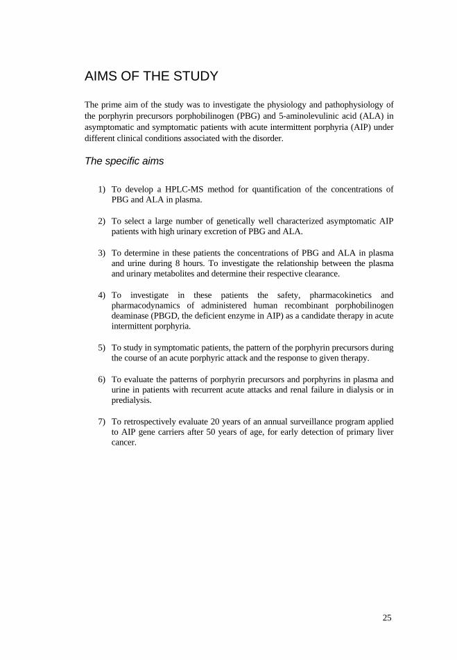
A pre-study, including two clinically asymptomatic but biochemically active AIP patients (patients 3 and 7, table 1), had been undertaken to investigate the urinary excretion pattern of PBG and ALA over a period of three weeks. The porphyrin precursors were measured in random morning samples during two-four days each week. The excretion of PBG and ALA was found to be relatively constant over time, (figure 4), as was the urinary ratio between PBG and ALA which was a prerequisite for studies I and II.
0
5
10
15
20
25
30
0
5
10
15
20
25
30 U-ALAU-PBG
[mm
ol/m
ol c
reat
inin
e][m
mol
/mol
cre
atin
ine]
Week 1 Week 2 Week 3
AIP patient 7, table 1
AIP patient 3, table 1
0
5
10
15
20
25
30
0
5
10
15
20
25
30 U-ALAU-PBG
[mm
ol/m
ol c
reat
inin
e][m
mol
/mol
cre
atin
ine]
Week 1 Week 2 Week 3
AIP patient 7, table 1
AIP patient 3, table 1
Figure 4: Urinary concentrations of PBG and ALA expressed as mmol/mol creatinine, measured in random morning samples of urine from two asymptomatic but biochemically active AIP patients during 3 weeks. Each bar represents a single determination of the concentration in the respective sample. In AIP patient 3 the mean (SD) urinary PBG concentration was 21.09 (2.97) and that of ALA 8.43 (0.90) mmol/mol creatinine. In AIP patient 7 the corresponding values were 19.57 (1.57) and 11.49 (2.14) mmol/mol creatinine. The mean (SD) urinary PBG/ALA ratios in the two patients were 2.51 (0.32) and 1.71 (0.27), respectively.

28
Table 1: Characteristics of AIP patients included in studies I and II. Subject (No)
Gender (Male/Female)
Age(years)
Mutation (in HMBS gene)c
U-PBG (<1.2 mmol/ mol crea)d
U-ALA (<3.1 mmol/ mol crea) d
Erc-PBGD (>70 pkat/ g Hb) d
PBG/ALA ratiof
1 a M 67 499-1G>A 19.8 8.8 47.1 2.3 2 M 65 593G>A 27.6 11.5 59.1 2.4 3 a M 61 730_731delCT 26.5 9.9 64.3 2.7 4 M 54 593G>A 28.4 11.4 47.0 2.5 5 M 48 593G>A 10.1 4.8 56.0 2.1 6 M 46 593G>A 20.4 9.1 76.6 2.2 7b M 45 593G>A 23.4 8.8 32.9 2.7 8b M 43 593G>A 20.0 10.5 57.5 1.9 9 M 40 593G>A 29.4 10.4 68.2 2.8 10a M 30 33G>T 18.5 9.4 79.3e 2.0 11 F 61 499-1G>A 3.3 2.8 62.4 1.2 12 F 59 593G>A 5.5 2.7 69.9 2.0 13 F 57 593G>A 11.0 3.6 63.9 3.1 14 F 55 593G>A 8.8 2.8 76.1 3.1 15 F 55 593G>A 9.3 3.3 65.5 2.8 16 F 54 499-1G>A 6.8 3.4 64.0 2.0 17b F 53 345-2A>G 23.1 6.6 70.5 3.5 18 F 47 593G>A 19.2 11.8 ------ 1.6 19b F 44 593G>A 34.1 7.3 57.1 4.7 20 F 43 593G>A 7.0 3.4 51.0 2.1 21b F 35 76C>T 27.2 15.9 57.1 1.7 22 F 33 499-1G>A 5.6 4.6 62.3 1.2 23 b F 32 499-1G>A 24.6 6.1 48.7 4.0 24 a F 27 499-1G>A 27.7 15.0 59.2 1.8
a Participants in study I ; b Participants in study I and study II; c The HMBS gene (HUGO Nomenclature committee) codes for the PBGD enzyme; d Reference value; e Mutation in exon 1 of the HMBS gene, f In urine. Healthy subjects as normal controls
Five healthy subjects, three men and two women, were included in study I. The mean age (SD) was 24 (4) years. In study II twenty healthy males, with a mean age of 31.6 (11.5) years were included. The healthy subjects had no known heredity for acute porphyria, or any clinical history or biochemical markers of acute porphyria, and medical examination and routine laboratory screening showed no abnormalities.
Study design
Study I
The individual characteristics of the ten AIP patients included in study I are shown in table 1. The inclusion criteria were (a) urinary PBG concentrations 4 times the upper reference limit, i.e. 4.8 mmol/mol creatinine (upper reference limit 1.2), arbitrarily assigned in order to ensure high plasma concentrations of PBG, (b) at least 30 days without AIP symptoms (e.g. abdominal or muscle pain), heme therapy or other AIP-

29
specific treatment, (c) negative pregnancy test, (d) no clinical history or laboratory signs of liver disease or alcohol abuse. The study participants were admitted to hospital early in the morning day 1, bringing a portion of the first morning urine (random morning urine sample), and stayed for 8 hours. During this period heparinized venous samples and urine samples were obtained each hour from 08:00 until 12:00, and afterwards at 14:00 and 16:00. Thus a total of 7 blood samples and 6 urine samples were obtained during 8 hours. The blood samples were immediately centrifuged at 1600g for 10 minutes and plasma was stored at -80 ºC until analyzed. The volumes of the urine portions were measured, and aliquots were stored at -80 ºC until analyzed. The participants left the hospital at 16:00 and continued to collect urine until 08:00 on day 2 and delivered it to the hospital, thus a 24-h urine collection was carried out. All samples were kept protected from light.
Study II
The individual characteristics of the twenty AIP patients included in study II are given in table 1. The inclusion criteria were (a) urinary PBG concentration 4 times the upper reference limit, i.e. 4.8 mmol/mol creatinine (upper reference limit 1.2), (b) at least 6 months without symptoms of acute porphyria (c) age between 18 and 65 years (d) no concurrent disease, drug or alcohol abuse, (e) women were required to have contraception for the previous 3 months and (f) no history of anaphylactic reaction or drug hypersensitivity. The 20 healthy subjects were all men. The study was performed in accordance with Good Clinical Practice (GCP) [88].
After admission to hospital four different doses of rhPBGD were administered intravenously as a single bolus dose (part A, open-labeled), or as repeated doses (part B, randomized, double-blinded, placebo controlled). The rhPBGD-doses chosen were based on earlier protocols applied in studies in the PBGD-deficient mouse model [89]. A summary of the protocol is given in figure 5.
Figure 5. Study protocol. Part A: 12 AIP patients, open single dose design. Doses: 0.5, 1.0, 2.0 and 4.0-mg/kg body weight. Part B: 20 AIP patients and 20 healthy men, double-blinded, placebo controlled. Doses: 0.25, 0.5, 1.0, 2.0 mg/kg body weight, given each 12 h for four consecutive days.
Stud
ypo
pula
tion
Part BRandomized, double-blinded, placebo controlled
Part AOpen label single dose design
Dose level 1 Dose level 1
Dose level 2 Dose level 2
Dose level 3
Dose level 4
Dose level 3
Dose level 4
Stud
ypo
pula
tion
Part BRandomized, double-blinded, placebo controlled
Part AOpen label single dose design
Dose level 1Dose level 1 Dose level 1Dose level 1Dose level 1
Dose level 2 Dose level 2
Dose level 3
Dose level 4
Dose level 3
Dose level 4
Dose level 2 Dose level 2
Dose level 3
Dose level 4
Dose level 3
Dose level 4
Dose level 2Dose level 2 Dose level 2Dose level 2
Dose level 3
Dose level 4
Dose level 3
Dose level 4

30
In part A twelve AIP patients participated. The enzyme was administered intravenously as a single bolus dose at four different dose levels; 0.5, 1.0, 2.0 and 4.0-mg/kg body weight, respectively, three subjects being included at each dose level. Part B included 40 subjects, i.e. 20 AIP patients and 20 healthy men. The daily amount of enzyme administered was the same as in part A, but divided into two equal doses that were given in 12-hour intervals for four consecutive days, i.e. 0.25, 0.5, 1.0 and 2.0-mg/kg body weight on each occasion. Ten subjects were included at each dose level: 5 AIP patients and 5 healthy subjects. Eight individuals received the active drug and 2 received placebo. The subjects were randomized in a double-blinded placebo controlled way with one AIP patient and one healthy subject receiving placebo at each dose level. The AIP patients that had participated in part A were included in part B after a washout period of 2 weeks. The study participants were admitted to hospital in the morning and heparinized venous blood samples were obtained prior to injection of the respective dose, and intermittently during the trial period. Blood and urine samples were collected at the appropriate time intervals, starting at 07:55 (base-line). The sampling intervals were chosen according to those used in previous animal studies [89]. In part A samples were collected from 08:00 until 20:00. In part B samples were collected from 08:00 until 20:00 on days 1 and 4. In-between, i.e. on days 2 and 3, only one sample was collected prior to dose administration (base line). The blood samples were protected from light and promptly centrifuged at 3000 g, at 4oC. The plasma was stored at –20 oC until analyzed. From each sampling occasion three tubes of plasma were separately stored for later measurements of rhPBGD, PBG, ALA and porphyrin concentrations. The urine samples were protected from light and stored at –20 oC for later analysis of PBG, ALA, and porphyrin concentrations. Study III Study participants
Acute intermittent porphyria patients during acute attacks
Three AIP patients participated during four attacks of acute porphyria.
Study design
The patients were admitted to the hospital due to an episode of clinical and biochemical signs consistent with an attack of acute porphyria. Immediately after admission the patients received symptomatic treatment and carbohydrate loading, i.e. 200 grams carbohydrates over 24 hours according to the Swedish recommendations [58]. The patients were also prescribed and encouraged to consume carbohydrate rich meals and had free access to nutrition beverages. The analgesic doses, mainly morphine, were individually adjusted after need and the daily intake recorded. The clinical outcome was monitored, and in case of unsatisfactory therapeutic response to carbohydrate administration, heme arginate was given. The selected acute attacks were of such clinical character that it was possible, under medical surveillance, to delay the start of heme therapy, for two to four days. The study period ranged from four to nine days.
31
Study IV Study participants
Acute intermittent porphyria patients with recurrent acute attacks and chronic renal failure
This study included three AIP patients with similar clinical features, i.e. recurrent acute attacks, chronic hypertension and chronic renal impairment. One patient was still in a predialysis state, one was treated with hemodialysis and one was on peritoneal dialysis. Study design
Data were collected and compiled from medical journals covering several years. The clinical outcome of the different therapy regimens and also the biochemical patterns of the porphyrin precursors and porphyrins measured in urine and plasma during the development of end-stage renal failure, are reported in the study. Study V Study participants
Acute porphyria patients diagnosed with primary liver cancer.
This retrospective study included twenty patients diagnosed with acute porphyria who developed primary liver cancer during the years 1987-2007. All patients had been monitored at the porphyria out-patient clinic at Stockholm South Hospital.
Study design
Data were collected and compiled from medical journals covering the study period of twenty years. Tumour and liver tissue were examined for histopathological diagnosis whenever there was available tissue. The beforehand selected variables regarding patient data (including history of alcohol consumption), diagnostic procedures, histopathological findings, clinical and biochemical findings and therapeutic measures and outcomes, were examined and compared with findings in previous reports on primary liver cancer in acute porphyria. MATERIALS Recombinant human PBGD (study II)
Recombinant human PBGD (rhPBGD, Porphozym™) was manufactured as the erythroid isoform (mol weight 42 kD, 344 amino acids) by Zymenex A/S (former HemeBiotech A/S), by genetic engineering using E. coli as host organism, and formulated in isotonic phosphate buffer solution, pH 8.0. The composition of the drug product was: rhPBGD 5 mg/mL, Na2HPO4 3.16 mM, NaH2PO4 0.51 mM, glycine 27 mM, Mannitol 222 mM, and sterile water for injection. The placebo had the same composition with the exception of rhPBGD.

32
Methods Analyses of PBG and ALA in urine (studies I, II, III, IV and V)
PBG and ALA in urine were analyzed with ion-exchange chromatography [90, 91] using the Bio-Rad PBG/ALA-Test. The concentrations of urinary PBG and ALA were expressed as micromoles excreted per unit time or as micromoles per mol creatinine, i.e. normalized to the creatinine concentration of the specimen [92]. The inter-assay variations (as CVs) for the ion-exchange chromatography procedures were 2.3 % for PBG and 3.3 % for ALA. The variations for PBG and ALA, expressed per mol creatinine, were 3.5 % and 4.3 % respectively. Detection limits were 1.9 µmol/L for PBG and 1.8 µmol/L for ALA.
Analyses of PBG and ALA in plasma (studies I, II, III and IV)
Quantitative determinations of PBG and ALA concentrations in plasma were made by HPLC–MS developed by us (study I) [93] and further modified (HPLC-tandem mass spectrometry, MS/MS) (study II) in collaboration with the commercial laboratory, BEKanalys AB, Sweden. The lower limit of quantification of PBG as well as ALA in plasma was 0.12 µmol/L, established by using ten quality control samples. The intra-assay precision for ALA was 3.3 % to 12 % and accuracy was 89 % to 103 %, based on results from six-double determinations of three concentrations performed on three different days. The respective intra-assay precision for PBG was 1.5 % to 12 % and accuracy 87 % to 108 %. The inter-assay precision for ALA was in the range 6.5 to 9.0 % and accuracy 95 to 99 %. The corresponding values for PBG were 9.0 to 11 % and 93 to 100 %, respectively. Relative recovery was 103 % for ALA and 85 % for PBG.
Determination of PBG and ALA clearance (study I) Renal clearances of PBG and ALA were determined in the 10 AIP patients and of ALA in the 5 healthy individuals using the formula:
CPBG (or ALA) = UPBG (or ALA) * V / PPBG (or ALA) (mL/min) Where U and P are the urine and plasma concentrations respectively and V the urine flow rate. The clearances were calculated from the pooled 8 h urine concentration values (µmol/L) and the mean plasma concentration values (µmol/L) of the 7 blood samples from each participant. In the healthy subjects, plasma PBG concentrations were under the limit of detection of the method and thus the clearance for this metabolite was not possible to determine. Calculation of PBG/ALA ratios (studies I, II, III and IV)
The PBG/ALA ratios in plasma and urine were calculated in AIP patients. The urinary ratio in healthy subjects and in AIP gene carriers in latent phase is about 0.3 (Porphyria Centre Sweden).

33
Normalized plasma and urinary PBG and ALA concentrations (sudies I, II and III)
In order to evaluate concentration changes over time and after treatment, in studies I, II and III the concentration of PBG and ALA at admission were put to 100 %. Calculation of estimated glomerular filtration rate (eGFR) (study IV)
The estimated glomerular filtration rate (eGFR) was calculated according to the formula from Cockcroft & Gault:
Ccr (women) = 52 * ((140-age)* bodyweight (kg)) / (S-creatinine (µmol/L) * 50) The stage of kidney disease was described according to the NFK DOQI™ classification stages of kidney disease [94] . Analyses of porphyrins in urine and plasma (studies II and IV)
Urinary total porphyrin concentration was quantified by anion-exchange chromatography with the Bio-Rad Porphyrin-Test. The inter-assay variation (as CV) for the ion-exchange chromatography method was 5.4 %. The limit of detection was 0.04 µmol/L. Total plasma porphyrin concentration was assayed by fluorescence spectrophotometry [95]. The inter-assay variation (as CV) was 7.4 % and the limit of detection was 0.5 nmol/L. Plasma and urine porphyrins were fractionated by a reversed-phase high-performance liquid chromatography (HPLC) gradient system [96].
Analyses of rhPBGD concentration in plasma (study II)
The concentration of rhPBGD in plasma was analysed by Scantox A/S, Denmark, using an enzyme-linked immunosorbent assay (ELISA), as specified in study II, page 341. Validation of the method showed that the accuracy was 93.0 % at 5.3 µg/mL and 100.1 % at 265 ng/mL and the precision was 10.3 % at 5.3 µg/mL and 17.4 % at 265 ng/mL. The lower limit of detection of the method was 50 ng/mL.
Analyses of rhPBGD antibodies (study II)
The concentration of rhPBGD antibodies was determined by Scantox A/S, Denmark, using an ELISA method on plates coated with rhPBGD. Protein G conjugated with horseradish peroxidase (HRP) was used to quantify the amount of rhPBGD antibodies. Values for antibody titers above 8000 dilution factor (df), i.e. ratio 1:1 were considered positive.
Pharmacokinetic calculations (study II)
Pharmacokinetic calculations were made on the basis of data for plasma rhPBGD concentration from healthy men and from AIP patients after bolus intravenous injection of rhPBGD. Calculations were performed using the computational rules for WinNonLin® noncompartmental analysis engine, version 4.1 (kindly provided by Pharsight Corporation Mountain View, Ca, USA).
34
The area under the plasma rhPBGD concentration-time curves (AUC) was estimated by the linear trapezoidal rule. The maximum concentration (Cmax) was equivalent to the measured peak plasma rhPBGD concentration. The concentration at time zero (C0) was obtained by back-extrapolating from the two first concentration values of rhPBGD in plasma, i.e. at 5 and 15 minutes after drug administration. The correlations between the administered doses and the respective AUC from time zero to infinity (AUC ), and C0, were investigated for all dose levels in both part A and part B. The t1/2 for rhPBGD was estimated using log linear regression including all the measured rhPBGD concentrations in the terminal elimination phase, according to the computational rules for WinNonLin®’s noncompartmental analysis. Pharmacodynamic calculations (study II)
The study of the effect of rhPBGD administration on plasma PBG concentration included only AIP patients, and was based on the change in plasma concentration of PBG over time after the administration of a single or of repeated doses of rhPBGD. Healthy subjects were not included as their plasma PBG concentration was not measurable by the method. In order to compare the effect of the different doses of rhPBGD on plasma PBG concentration, each individual’s plasma PBG concentration at time zero (initial concentration, PBG0) was put to be equivalent to 100 %. The plasma PBG concentration at time t after injection of rhPBGD, was expressed as per cent of PBG0 calculated as: (PBGt / PBG0)*100 ( %) for all timepoints. The effect of the enzyme on ALA and porphyrin concentrations in plasma and urine were also studied.
Liver histopathological diagnosis (study V)
Tumour and liver tissue were examined for histopathological diagnosis whenever there were specimens available, i.e. after biopsy or tumour resection. The specimens were sirius stained to grade fibrosis, and routinely stained with hematoxylin and eosin for histological diagnosis. Cholangiocarcinoma was classified after extensive immunohistochemistry including cytokeratin 7.
Statistical methods (studies I, II and III) Statistical analyses and graphics were performed with Microsoft Excel® software with the Analyse-It™ (Analyse-It Software Ltd) statistical software add-in (study I) and XLSTAT statistical software add-in (studies II and III). The specific methods are specified in each study. P values 0.05 was considered statistically significant.
35
RESULTS Studies on chronic active disease and enzyme replacement therapy Study I
The mean (SD) plasma PBG and ALA concentrations for the 10 AIP patients were 3.1 (1.0) µmol/L [range 1.7 - 5.1] and 1.7 (0.7) µmol/L [range 0.9 - 3.6], respectively. The corresponding values for the excreted amounts of urinary PBG and ALA were 102 (25) µmol/8-hour [range 68 – 146] and 56 (18) µmol/8-hour [range 32 – 91]. In the 5 healthy individuals, plasma PBG concentrations were below the detection limit of the method (<0.12 µmol/L), while the mean value for plasma ALA was determined to be 0.38 (0.03) µmol/L [range 0.36 - 0.41]. The mean values for urinary PBG and ALA in the healthy individuals were 2.9 (0.7) µmol/8-hour [range 2.3 - 4.1] and 9.3 (1.2) µmol/8-hour [range 7.8 - 10.5], respectively. Thus in healthy subjects the concentrations of ALA in plasma and urine are considerable higher than those for PBG. The total (SD) urine excretion of PBG in 24 hours was 244 (51.4) µmol in AIP patients, and 6.5 (1.1) µmol in healthy individuals. For ALA the corresponding values were 136 (39.4) and 25.8 (3.7) µmol, respectively. In AIP patients as well as in healthy individuals the amount of PBG and ALA excreted during 8 hours was about 40 % of the total amount collected in 24 hours. AIP patients showed no significant difference in urinary PBG concentration in the random morning sample compared with the pooled urine collected during 8 hours or 24 hours (normalized to creatinine) (p = 0.257 and p = 0.426 respectively). The same was also true for urinary ALA concentration (p = 0.203 and p = 0.229). The diagnostic accuracy of the HPLC-MS technology was confirmed by the following findings. In the AIP patients, there was a high correlation between plasma concentrations of PBG and ALA quantified by the HPLC-MS method and the urine concentrations quantified by certified laboratory methods according to the Swedish Board for Accreditation and Conformity (SWEDAC). This was true both where the mean values were compared and where the individual values over time were studied (r = 0.678, p < 0.05 and r = 0.856, p < 0.01, respectively). The concentration of PBG in the AIP patients was twice that of ALA, in plasma as well as in urine. The mean ratio for the PBG/ALA ratio in plasma was 2.0 (0.8) [range 1.2 - 3.3] and in urine 2.0 (0.5) [range 1.3 - 3.1]. In healthy individuals the urine PBG/ALA ratio was 0.32 (0.07) [range 0.23 - 0.41], i.e. the same range as found in AIP patients in latent phase. In healthy subjects, the plasma PBG/ALA ratio was not calculated as the PBG concentrations were below the detection limit of the method.

36
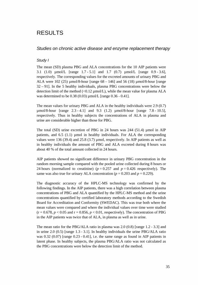
The mean values for renal clearences are shown in figure 6. There was no significant difference between the values for renal clearance of PBG and of ALA in the AIP patients (p = 0.730; paired t-test). No correlation could be demonstrated between creatinine clearance and PBG or ALA clearance (r = 0.630, p = 0.13 and r = 0.179, p = 0.70, respectively)
In AIP patients, the general concentration patterns for plasma and urinary PBG and ALA during the 8 hours observation time were relatively constant in each individual. The concentrations of plasma and urinary PBG were significantly higher in the morning sample than in the sample drawn at 16:00, (p < 0.001 and p < 0.002, respectively). We observed no differences when comparing the morning and afternoon samples with regard to the concentrations of ALA in plasma or urine. Study II
No serious adverse events following the administration of rhPBGD were observed, but thirteen AIP patients (65 %) reported a total of 31 non-severe reactions. Abdominal pain was the most frequent complaint and occurred in 31 % of the cases. Twelve healthy men (60 %) reported 23 diverse adverse events, mostly with infection-related symptoms. Increased anti-rhPBGD IgG titers were found in 4 healthy subjects and in 3 AIP gene patients. None of the 7 cases had any clinical signs of allergic reaction. The adverse events and immunogenicity noted were not related to the dose of rhPBGD administered. The pharmacokinetic variables of rhPBGD are shown in Table II, study II, page 346. In all subjects receiving the enzyme, the highest concentration of plasma rhPBGD was found in the first collected sample, i.e. at 5 minutes after administration. The median value for t1/2 in the AIP patients at dose levels 2.0 and 4.0 mg/kg was 121 minutes (2.0 h). The corresponding median t1/2 in the healthy subjects was 140 minutes (2.3 h) at the 2.0-mg/kg dose. No statistical differences were found in the half-lives (t1/2) obtained at the 2 and 4 mg/kg doses in either subject group (p = 0.723). There was no difference between the AIP patients and the healthy subjects regarding the AUC s on day 1 (p = 0.625) but a slight difference on day 4 (p < 0.077) may be noted. The corresponding
0
20
40
60
80
100
120
140
160
180
':´
ALA clearencePBG clearence
Creatinine clearence
AIP patients Healthy controls
Ren
alcle
aren
ce [
mL/
min
]
0
20
40
60
80
100
120
140
160
180
':´
ALA clearencePBG clearence
Creatinine clearence
AIP patients Healthy controls
Ren
alcle
aren
ce [
mL/
min
]
Figure 6. In AIP patients the mean (SD) value for renal clearance of PBG was 71.0 (14.6) mL/min and 69.5 (13.4) mL/min for ALA. In healthy individuals the corresponding clearance of ALA was 51.1 (9.3) mL/min. The mean value for creatinine clearance in the AIP patients was 90.4 (12.0) mL/min during 8 h. The corresponding value for the healthy participants was 149 (25.8) mL/min. The reference interval is 90 - 150 mL/min for individuals up to 50 years of age.

37
comparison of C0 showed no differences between the subject groups (p = 0.607 and p= 0.454 on day 1 and day 4, respectively). The enzyme was shown to be effective at all doses, in single dose mode as well as when repeated doses were given. The plasma PBG concentration remained undetectable during approximately 2 hours, after which it slowly started to rise. At the highest doses of rhPBGD there was a significant prolonged reducing effect on plasma PBG concentration (p < 0.05). Twelve hours after the administration of the enzyme, i.e. at a time when the plasma rhPBGD concentration was undetectable at all dose levels, the plasma PBG concentration reached values between 40 - 114 % (mean 70 %) of the initial concentration. There was no significant difference between day 1 and day 4 in the initial plasma PBG concentration (p = 0.227). Thus, there was no additative reducing effect of rhPBGD on plasma PBG concentration when the enzyme was administered at 12 h intervals for four consecutive days. The individual and arithmetic means of AUCs , as well as the individual and arithmetic means of C0 were compared between day 1 and day 4 for PBGD-deficient subjects and healthy subjects. There were no differences observed between the compared variables (p = 0.890 and p = 0.978, respectively), confirming the absence of accumulative effect of rhPBGD when repeatedly administered at 12 h intervals for four consecutive days. There was a strong correlation between the AUCs and the respective enzyme doses in both part A and part B (r = 0.977, p < 0.023 and r = 0.886, p < 0.014, respectively). In the AIP patients receiving placebo there was a 20-30 % increment and/or decline in plasma PBG concentration during the first hours of observation. After that the plasma PBG level stabilized at about 70 % of the initial concentration. No change in plasma ALA concentration was observed. In the groups of AIP patients receiving rhPBGD, there was a substantial increment of porphyrin concentration in plasma (Figure 3, study II, page 345) and urine during the first hours after drug administration. The major porphyrin fraction found in plasma and urine was uroporphyrin I. The probable explanation for this increased porphyrin fraction is a non-enzymatical condensation of four molecules of PBG to form hydroxyl-methylbilane, which spontaneously is cyclized into the tetrapyrrole uroporphyrin I. Study III
Three AIP patients (A, B and C) were studied after admission to hospital during four acute porphyria attacks. Patient B presented with considerably increased concentrations of urinary PBG and ALA compared to remission 6 weeks earlier. The other two had on admission increased urinary PBG and ALA concentrations but in the same range as in symptom-free periods analyzed 6 months earlier in patient A and 18 months earlier in patient C.
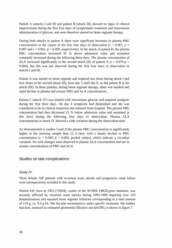
38
Patient A (attacks I and II) and patient B (attack III) showed no signs of clinical improvement during the first four days of symptomatic treatment and intravenous administration of glucose, and were therefore started on heme arginate therapy. During both attacks in patient A there were significant increases in plasma PBG concentration in the course of the first four days of observation (r = 0.967, p = 0.007 and r = 0.961, p = 0.009, respectively). In the attack of patient B, the plasma PBG concentration increased 35 % above admission value and remained constantly increased during the following three days. The plasma concentration of ALA increased significantly in the second attack (II) of patient A (r = 0,979 p = 0.004), but this was not observed during the first four days of observation in attacks I and III. Patient A was started on heme arginate and required two doses during attack I and four doses in her second attack (II), from day 5 until day 8, as did patient B in his attack (III). In these patients, during heme arginate therapy, there was marked and rapid decline in plasma and urinary PBG and ALA concentrations. Patient C (attack IV) was treated with intravenous glucose and required analgesic during the first three days. On day 4 symptoms had diminished and she was considered to be in clinical remission and released from hospital. The plasma PBG concentration had then decreased 25 % below admission value and remained at this level during the following four days of observation. Plasma ALA concentrations in attack IV showed a wide variation during the observation time. As demonstrated in studies I and II the plasma PBG concentration is significantly higher in the morning sample than 12 h later, with a steady decline in PBG concentration (r = 0.940, p = 0.003, pooled values), which indicate a circadian variation. No such changes were observed in plasma ALA concentration and nor in urinary concentrations of PBG and ALA. Studies on late complications
Study IV
Three female AIP patients with recurrent acute attacks and progressive renal failure were retrospectively included in this study. Patient ED, born in 1953 (†2004), carrier of the W198X PBGD-gene mutation, was severely afflicted by recurrent acute attacks during 1993-1999 requiring over 220 hospitalizations and repeated heme arginate infusions corresponding to a total amount of 110 g, i.e. 9.4 g Fe. She became normotensive under specific treatment. Her kidney function, assessed as estimated glomerular filtration rate [eGFR], is shown in figure 7.
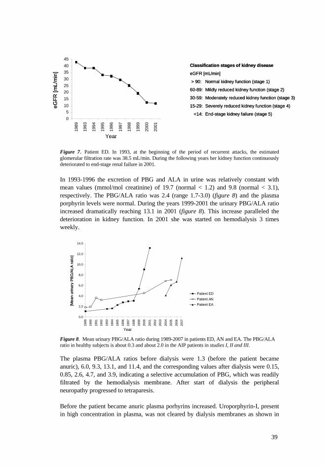
39
Figure 7. Patient ED. In 1993, at the beginning of the period of recurrent attacks, the estimated glomerular filtration rate was 38.5 mL/min. During the following years her kidney function continuously deteriorated to end-stage renal failure in 2001. In 1993-1996 the excretion of PBG and ALA in urine was relatively constant with mean values (mmol/mol creatinine) of 19.7 (normal < 1.2) and 9.8 (normal < 3.1), respectively. The PBG/ALA ratio was 2.4 (range 1.7-3.0) (figure 8) and the plasma porphyrin levels were normal. During the years 1999-2001 the urinary PBG/ALA ratio increased dramatically reaching 13.1 in 2001 (figure 8). This increase paralleled the deterioration in kidney function. In 2001 she was started on hemodialysis 3 times weekly. Figure 8. Mean urinary PBG/ALA ratio during 1989-2007 in patients ED, AN and EA. The PBG/ALA ratio in healthy subjects is about 0.3 and about 2.0 in the AIP patients in studies I, II and III. The plasma PBG/ALA ratios before dialysis were 1.3 (before the patient became anuric), 6.0, 9.3, 13.1, and 11.4, and the corresponding values after dialysis were 0.15, 0.85, 2.6, 4.7, and 3.9, indicating a selective accumulation of PBG, which was readily filtrated by the hemodialysis membrane. After start of dialysis the peripheral neuropathy progressed to tetraparesis. Before the patient became anuric plasma porhyrins increased. Uroporphyrin-I, present in high concentration in plasma, was not cleared by dialysis membranes as shown in
Classification stages of kidney disease
eGFR [mL/min]
> 90: Normal kidney function (stage 1)
60-89: Mildly reduced kidney function (stage 2)
30-59: Moderately reduced kidney function (stage 3)
15-29: Severely reduced kidney function (stage 4)
<14: End-stage kidney failure (stage 5)
eGFR
[mL/
min
]
05
1015202530354045
1989
1993
1994
1995
1996
1997
1998
1999
2000
2001
Year
0,0
2,0
4,0
6,0
8,0
10,0
12,0
14,0
1989
1990
1991
1992
1993
1994
1995
1996
1997
1998
1999
2000
2001
2002
2003
2004
2005
2006
2007
Patient EDPatient ANPatient EA[M
ean
urin
ary
PBG
/ALA
ratio
]
Year
0,0
2,0
4,0
6,0
8,0
10,0
12,0
14,0
1989
1990
1991
1992
1993
1994
1995
1996
1997
1998
1999
2000
2001
2002
2003
2004
2005
2006
2007
Patient EDPatient ANPatient EA[M
ean
urin
ary
PBG
/ALA
ratio
]
Year
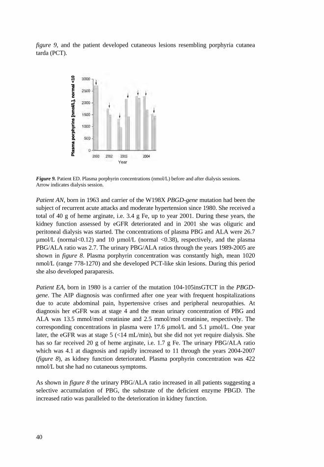
40
figure 9, and the patient developed cutaneous lesions resembling porphyria cutanea tarda (PCT). Figure 9. Patient ED. Plasma porphyrin concentrations (nmol/L) before and after dialysis sessions. Arrow indicates dialysis session. Patient AN, born in 1963 and carrier of the W198X PBGD-gene mutation had been the subject of recurrent acute attacks and moderate hypertension since 1980. She received a total of 40 g of heme arginate, i.e. 3.4 g Fe, up to year 2001. During these years, the kidney function assessed by eGFR deteriorated and in 2001 she was oliguric and peritoneal dialysis was started. The concentrations of plasma PBG and ALA were 26.7 µmol/L (normal<0.12) and 10 µmol/L (normal <0.38), respectively, and the plasma PBG/ALA ratio was 2.7. The urinary PBG/ALA ratios through the years 1989-2005 are shown in figure 8. Plasma porphyrin concentration was constantly high, mean 1020 nmol/L (range 778-1270) and she developed PCT-like skin lesions. During this period she also developed paraparesis. Patient EA, born in 1980 is a carrier of the mutation 104-105insGTCT in the PBGD-gene. The AIP diagnosis was confirmed after one year with frequent hospitalizations due to acute abdominal pain, hypertensive crises and peripheral neuropathies. At diagnosis her eGFR was at stage 4 and the mean urinary concentration of PBG and ALA was 13.5 mmol/mol creatinine and 2.5 mmol/mol creatinine, respectively. The corresponding concentrations in plasma were 17.6 µmol/L and 5.1 µmol/L. One year later, the eGFR was at stage 5 (<14 mL/min), but she did not yet require dialysis. She has so far received 20 g of heme arginate, i.e. 1.7 g Fe. The urinary PBG/ALA ratio which was 4.1 at diagnosis and rapidly increased to 11 through the years 2004-2007 (figure 8), as kidney function deteriorated. Plasma porphyrin concentration was 422 nmol/L but she had no cutaneous symptoms. As shown in figure 8 the urinary PBG/ALA ratio increased in all patients suggesting a selective accumulation of PBG, the substrate of the deficient enzyme PBGD. The increased ratio was paralleled to the deterioration in kidney function.
Plas
ma
porp
hyrin
s [n
mol
/L],
norm
al <
10
Year
Plas
ma
porp
hyrin
s [n
mol
/L],
norm
al <
10
Year
41
Study V
Twenty patients diagnosed with acute hepatic porphyria (18 AIP, 1 variegate porphyria and 1 hereditary coproporphyria) were included in this retrospective study of patients who had developed primary liver cancer (PLC). The patients were all monitored at the porphyria out-patient clinic, Stockholm South Hospital. There were 16 women and 4 men and the mean age at PLC diagnosis in women was 68.6 years (range 56-82) and 64.0 years (range 53-75) in men. The overall mean age at diagnosis was 67.7 years. Primary liver cancer was detected in ten patients via an annual surveillance program instigated for the purpose. Five patients were diagnosed at the porphyria out-patient clinic due to suspected relapse of symptoms of acute porphyria, and five patients were diagnosed at their local clinic based on symptoms from the gastrointestinal tract not associated with porphyria. In the ten cases where PLC was found in the annual surveillance program, the mean tumour diameter was 4.3 cm (2-7.5), while the tumours found outside surveillance had a mean diameter of 10.4 cm (2-19). Seven patients presented with high concentration of urinary porphyrin precursors at diagnosis and this group mainly included patients with the highest tumour burden. Among these, three patients presented with concomitant symptoms of acute porphyria. The remaining 13 patients had virtually unchanged urinary excretion of PBG and ALA compared to previous controls and had no symptoms related to porphyria. Tumour and liver tissue were re-examined by the same experienced liver pathologist whenever there was available tissue. In 11 of the 17 patients that underwent biopsy there was adjacent liver tissue available to examine the grade of fibrosis, and four showed to have cirrhosis. In all sixteen patients diagnosed with hepatocellular carcinoma (HCC) six patients had well differentiated HCC, and five moderately differentiated. Two patients had poorly differentiated HCC with pleomorphic growth pattern. Three patients were diagnosed by standard radiological criteria [97] without biopsy. Three patients had cholangiocarcinoma and one patient combined hepatocellular carcinoma and cholangiocarcinoma with sarcomatous transformation. Eight patients were subjected to surgical resection of the tumour. Other treatments were radio therapy (6 cases), systemic chemotherapy (4 cases), radiofrequency ablation (2 cases), percutaneous ethanol injection (1 case) and transarterial chemoemobolization (1 case). Among the ten deceased patients, the median survival time was 1.9 years after diagnosis (mean 3.9, range 0.3-13.3). Among the ten patients that are presently alive the median survival time after diagnosis is 2.4 years (mean 3.6, range 1.1-10.8). In the two patients with the longest survival time (10.8 and 13.3 years, respectively), the tumour was surgically resected.


43
DISCUSSION
Monitoring PBG and ALA in patients with acute porphyria in periods of active disease or remission has over decades been done by measurements of the metabolites in urine. These methods, which are mostly based on ion exchange chromatography in combination with spectrophotometry, have a high sensitivity and are readily performed in the clinical chemistry laboratory. Only a few reports have described methods for measurement of plasma PBG and/or ALA concentrations for monitoring AIP. Some workers have used ion exchange chromatography alone [5], or in combination with thin layer chromatography [35]. Other methods applied are fluorimetric enzyme assay [98], high pressure liquid chromatograpy [99], and gas liquid chromatography with electron-capture detection [100], but these methods have not been used routinely in the clinical chemistry laboratory. The manufacturing of human recombinant porphobilinogen deaminase (rhPBGD) (Zymenex A/S) and subsequent evaluation of its therapeutic efficacy in AIP, made it necessary to develop a reliable method to measure PBG and ALA in blood samples. A novel method, based on the combination of reverse phase HPLC and masspectometry, was therefore developed by us for the quantification of PBG and ALA in plasma. The method was first applied in pre-clinical studies in the AIP mouse model [89], in which minute plasma samples were collected for analysis of the metabolites before and after phenobarbital induction of ALAS1, and later during the enzyme administration trials. The diagnostic accuracy of the HPLC-MS method was further evaluated in asymptomatic AIP patients presenting with high excretion of the precursors in urine (study I). We found a strong correlation between the plasma concentrations of PBG and ALA measured by our HPLC-MS method, and the concentrations in urine measured by the Bio Rad® ion exchange chromatography method. Also, the ratio PBG to ALA in plasma, in these subjects with high excretion of the precursors, was found to be the same as in urine, i.e. approximately 2. The development of a validated HPLC-MS method permitted us to investigate during 8 hours the porphyrin precursor patterns in plasma and urine in ten asymptomatic AIP patients with permanently high excretion, and thus to calculate the renal clearance for these metabolites, which was a prerequisite for the investigation of the pharmacokinetics and pharmacodynamics of rhPBGD in a larger group of asymptomatic AIP patients, also with permanently high excretion. It also made possible a study of the variation of PBG and ALA in plasma during the attack of acute porphyria, and to evaluate the effect of different therapeutic regimens on the accumulation of the precursors as reflected in their plasma concentrations over time. Further, it gave us the tool to monitor the concentrations of PBG and ALA in plasma of patients with recurrent acute attacks, as well as in patients with chronic renal failure with and without dialysis.

44
Studies on asymptomatic but biochemically active AIP patients In study I we could corroborate, that the concentrations of PBG and ALA in plasma and urine were relatively constant within each individual AIP patient during 8 hours of observation time but differed between the individuals. The plasma concentrations of the metabolites showed a high correlation with those in urine. Our results from analyses of plasma are in accordance with those reported by Miyagi et al. [5] in five asymptomatic but biochemically active AIP patients, where the concentrations of serum PBG were higher than the concentrations of ALA. This was also true regarding the admission concentrations of plasma PBG and ALA in the asymptomatic but biochemically active AIP patients included in study II (table I, page 28). One of the patients in study I, patient 7, had a peak-like rise of plasma ALA concentration, which showed to be reflected one hour later in the ALA concentration in urine. No change in plasma PBG was observed, but there was a slight and sustained increase in urine PBG in this individual. These observations are in line with the higher correlation observed between the concentrations of ALA in plasma and in urine, compared to the correlation between plasma and urine PBG (Figure 1, study I, page 705). This patient also participated in study III (patient C, attack IV), and on day 2 of the attack the plasma ALA peak observed (Figure 2a, study III, page 4) was not associated with clinical impairment, nor with any corresponding increase in plasma PBG. No concentration peak of ALA was observed in the urine portion next sampled, which may be explained by blunting by the ongoing intravenous infusion, or by imprecision in the sampling procedure. During the acute attacks (study III), the changes observed in plasma PBG and ALA concentrations before heme therapy were not reflected in the urinary excretion patterns. This discrepancy may point to the fact that fluctuations in PBG and ALA levels are better detected in spot plasma samples than in urine. The values of pooled urine presented in study III, represent the integrated mean concentration of the metabolite excreted during a time period of 4 hours (daytime) and 12 hours (night-time). Moreover, these metabolites are strongly concentrated during renal filtration, which may have as a result that urine determinations are less sensitive to small concentration changes. Similar observations were made by Gorchein and Webber [35], who noted that fluctuations in plasma ALA were not reflected in the urinary excretion pattern, a circumstance tentatively interpreted as a result of rapid oscillations taking place in hepatic ALA production or release, not detected in collected urine. The concentration patterns of PBG and ALA during the acute attacks may have been partially blunted by the glucose administration that was started in all the patients immediately after admission. The mechanisms by which glucose down regulates and represses ALAS1 have been described by Handschin and Thunell [14, 16]. The beneficial therapeutic effect of glucose, recognized since several decades [101], seems to be effective in mild attacks, as in patient C (attack IV), and is also reflected in plasma PBG concentrations, which constantly remained below the concentration presented at admission. In contrast, the patients A and B (attacks I, II and III) did not experience clinical improvement in their attacks during the first four days, and plasma PBG concentrations increased constantly (Figure 1a, study III, page 3). This showed
45
that the symptomatic treatment and administration of glucose had not been sufficient, and that heme treatment was indicated. The clinical improvement during the consequently administration of heme arginate was reflected in a rapid decline in plasma and urinary levels of PBG and ALA. The effect of glucose is totally overridden by that of heme arginate, a therapy that causes a prompt clinical and biochemical remission, (Figure 1-4b, study III, page 3-5). This observation is entirely in line with the current recommendations that in other than mild attacks should heme therapy be started early [3, 50, 65] The lack of difference between the values of urinary PBG and ALA in remission and the values at admission to hospital in patients A and C is difficult to interpret. In an acute attack the excretion of PBG usually is at least doubled compared to the asymptomatic phase [102, 103], but the inter-individual variation is wide [6] and thus the concentration to which urinary PBG must change to confirm or exclude an acute attack is not fully known. The importance of careful evaluation of clinical manifestations must be stressed [6]. It should also be considered that patient A and C had previously been afflicted by acute attacks, and had good knowledge about how to cope with symptoms. Under these circumstances the patients may already have taken measures at home like increased sugar intake and/or analgesics before seeking medical care and that the urinary excretion peak of porphyrin precursors may have been ameliorated prior to admission to hospital. In patient B, with less knowledge of the disease and less awareness of the value of self-treatment, the quantitative PBG and ALA values at admission reflect the characteristic concentration peak, pathognomonic of an attack of acute porphyria. Renal clearances of PBG and ALA
The renal clearances of PBG and ALA was 71.0 mL/min and 69.5 mL/min, respectively, and there is thus no significant difference between the values (figure 6, page 36). Previously, the clearance of PBG has been determined in two patients by Miyagi et. al. [5] and found to be in the same range as found in our study I. In the AIP patients the clearance values for PBG and ALA were both about 77 % of that of creatinine. The renal clearance of ALA, determined by Gorchein and Webber [35], was 12 % lower than that of creatinine, and thus in the same range as our. In the healthy participants in study I, the clearance of ALA was about 34 % of that for creatinine (figure 6, page 36). This is not unexpected since they were younger and had a mean creatinine clearance of 149 mL/min and a mean plasma ALA concentration of 0.38 µmol/L, compared to the AIP patients’ 90.4 mL/min and 1.7 µmol/L, respectively. Ratios of PBG to ALA in plasma and urine The PBG/ALA ratio in urine in the healthy participants in study I was approximately 0.3, which is the same as in latent AIP patients (Porphyria Centre Sweden). In contrast, in AIP patients with increased excretion of porphyrin precursors, the PBG/ALA ratio was approximately 2.0 both in plasma and urine (studies I, II and III). This finding

46
cannot be explained by differences in renal handling, as the renal clearances of the two metabolites were about the same (figure 6, page 36). The two-fold concentration of PBG in comparison with ALA may reflect the selective accumulation of the substrate for the deficient PBGD enzyme. The reasons for the relative increase in ALA concentration are not obvious but may reflect a state of up-regulated ALAS1 and/or a possible inhibitory effect of PBG on ALA-dehydratase [9]. In at least two other studies, where the patterns of PBG and ALA have been observed in AIP patients in symptomatic periods, the ratio for PBG/ALA was also found to be increased during attacks, and normalized after treatment with heme arginate [104] or liver transplantation [74]. During the acute attacks followed in study III, the plasma and urine PBG/ALA ratios were about the same as noted in studies I and II, i.e. approximately 2, at admission as well as under the symptomatic treatment and administration of glucose. The ratio changed rapidly on heme arginate therapy and eventually a specific pattern was no longer possible to discern. Circadian variation of PBG
When studying the general variation patterns of PBG and ALA in studies I, II and III, we found a significant decline in plasma PBG concentration from morning to afternoon, reflecting a circadian variation of this variable that has not been described before. One explanation for the phenomenon can be found in the fact that ALAS1 has been identified as circadianly expressed, and that heme biosynthesis and the circadian clock are reciprocally regulated [12, 13]. On the other hand PXR/CAR transactivation of ALAS1 transcription is modulated by the adrenal corticosterone secretion and hepatic glucose production, which in turn are under circadian control [105]. Like Gorchein and Webber [35], we did not find any circadian variation in plasma ALA concentration. Enzyme replacement therapy The development of recombinant DNA technology has made it possible to produce enzymes that have proved to be therapeutic in a few metabolic diseases [66]. Recombinant human PBGD was manufactured by Zymenex A/S, Hillerød, Denmark, as a possible new therapeutic alternative in AIP aimed at repairing the deficient PBGD step in heme biosynthesis. The enzyme proved effective as far as plasma PBG concentrations were reduced in a phenobarbital treated AIP animal model [67, 68]. In study II the recombinant human porphobilinogen deaminase (rhPBGD, Porphozym™) was administered to healthy subjects as well as to asymptomatic AIP patients in order to study its tolerability, pharmacokinetics and metabolic effects. No serious adverse events were reported. Adverse reactions noted were of mild severity, and none required any treatment. Seven subjects developed plasma antibodies against rhPBGD, but no allergic reactions took place. As in the animal model, the recombinant enzyme was effective in reducing the plasma PBG concentration, which almost instantaneously after administration reached

47
concentrations below the limit of detection of the method. The effect persisted for at least two hours, after which the plasma PBG concentrations increased again to reach about 70 % of the baseline values in 8-12 hours. The half-life of the enzyme in plasma was about 2 hours and the AUCs were proportional to the respective dose. After establishing the safety, and defining the pharmacokinetic and pharmacodynamic variables for the drug, the clinical trials in overt disease were started. Plasma and urinary porphyrin concentrations increased rapidly after administration of the enzyme and returned to initial concentrations within 12 - 24 hours after drug administration. The increased porphyrin concentrations are a result of spontaneously formed uroporphyrin I [1, 106]. The concentration patterns of PBG, ALA and porphyrins in urine were the same as in plasma, indicating unimpaired renal clearance under the enzyme therapy. There were no changes in plasma or urinary porphyrin concentrations in AIP patients that received placebo, or in healthy subjects receiving either the enzyme or placebo. Late complications Renal impairment
In spite of being under continuous specialized medical attention the three patients in study IV were afflicted by recurrent acute attacks and developed hypertension, peripheral neuropathy and progressive renal failure with accumulation of porphyrins in blood. The progression of the complications may have been secondary to accumulation of toxic metabolites. The possible contribution of PBG and ALA in this process is unclear. There is thus no experimental evidence of PBG neurotoxicity and ALA, the metabolite suspected to cause nerve damage in acute porphyrias, was present in blood only in low concentration after development of end-stage renal disease (ESRD). The accumulation of uroporphyrins will sensitize the skin to the sun, but porphyrins have not been associated with nerve cell damage [1]. Cutaneous lesions were not seen in the patients until advanced renal failure arose. The causes for ESRD were not elucidated by renal biopsy in any of the patients. Renal failure in AIP have been reported to be secondary to factors such as hypertension [76-79], interstitial tubular damage [107, 108] and noxious effects of porphyrin precursors on renal parenchyma [38, 109]. The repeated heme administrations may also have contributed [110, 111], as may also secondary kidney hemosiderosis (the 10 mL ampoule of Normosang® contain 250 mg human hemin, i.e. about 20 mg iron), however none of our patients had any biochemical signs of iron overload. In studies I, II and III the plasma and urinary PBG/ALA ratios were both approximately 2, and all patients had conserved renal function for their age. In the three patients in study IV where the urinary excretion pattern had been followed during several years, it was observed that the increase in urinary PBG/ALA ratio paralleled the impairment in glomerular filtration (figure 7, page 39 and figure 9, page 40). The pathophysiological mechanism underlying the observation that it is mostly PBG that accumulates is unclear.

48
One hypothesis could be a further inhibition of hepatic PBGD by its substrate PBG. It has been reported that the conversion of PBGD from apo-enzyme to holo-enzyme needs the two molecules of PBG linked together, i.e. a dipyrromethane component [106]. In vitro, excess of PBG strongly inhibits hepatic PBGD [5, 112], which can explain why an additional reduction of the residual activity of the enzyme is seen in AIP patients during acute attacks. This mechanism may also be the explanation for the selective accumulation of PBG found in our patients. The reason for the relatively low ALA concentrations observed in the three patients is unclear. The concentration of ALA was decreased in both plasma and urine, thus neither this observation can be explained by changes in the renal filtration of the metabolites. The theory that accumulated PBG would give rise to product inhibition of ALA-dehydratase [9] is not validated by these observations. Porphobilinogen is readily filtrated by the dialysis membrane used, as indicated by the fact that the plasma PBG/ALA ratio decreased after each dialysis session to about 70 % of the value before dialysis (Figure 1, study IV). Hemodialysis is an intermittent procedure and caused a huge accumulation of PBG between the dialysis sessions. The high plasma concentrations of uroporphyrin I are not cleared by dialysis membranes (figure 8, page 39), and not affected by high flux hemodialysis, an alternative recommended in patients with PCT and ESRD [113]. In overt PCT and ESRD the combined therapeutic efforts of dialysis, erythropoietin administration and small venesectios reduce the porphyrin levels in blood and remove hepatic iron giving relief from symptoms. In AIP patients with chronic active disease and ESRD the pathophysiology is different, as the source of elevated porphyrin levels, the overproduction of PBG, persists. Peritoneal dialysis regimens seem to be more beneficial, as plasma PBG/ALA ratios were almost equal to those found in high excreters with normal kidney function (figure 9, page 40, patient AN), but it does not either clear the high levels of uroporphyrins that give rise to cutaneous photosensitivity. In 2004 the first case of curative liver transplantation was performed in a young AIP patient afflicted with recurrent acute attacks [74]. Kidney transplantation has been made in AIP patients without complications [114, 115]. Combined liver and kidney transplantations have been undertaken with acceptable outcome in several other metabolic disorders [116]. In 1999, when patient ED was considered for combined transplantation, there was no experience of this procedure in AIP and liver exchange was abstained from because of the poor general condition of the patient and the seriously deteriorated peripheral veins resulting from repeated heme arginate infusions. Presently, considering the good result obtained in at least one reported case of liver transplantation in acute porphyria [74], the possibility of a combined liver and kidney transplantation should be considered in seriously affected AIP patients with chronic active disease and progressively impaired renal function, at a clinical stage when this therapeutic alternative still is an option. This is not an easy decision for the patient to make, neither for the involved physicians.

49
Primary liver cancer
Since the first report of an association between hepatocellular cancer (HCC) and acute porphyria, several studies have been published. Most of them are from northern Sweden [84, 86, 117-120] with cases that mainly derive from the same AIP cohort of patients. A Finnish report [85] is a retrospective study including 7 cases of HCC among 82 deceased patients with acute porphyria (AIP and VP). No cases of HCC were found among the still living. Previously published reports include only HCC, and no cases of cholangiocarcinoma. The patients described in study V had in 80 % of the cases HCC and in 20 % cholangiocarcinoma or combined hepatocellular cancer and cholangiocarcinoma. Whether this reflects similar pathogenetic mechanisms truly related to acute porphyria, or if it rather is a random finding, is unclear. In the reports from Finland and Sweden the mean age at HCC diagnosis was over 65 years [84, 85], which is consistent with the 67.7 years found in study V. In the French report, the patients were considerably younger and the youngest only 37 years old [83]. Male dominance characterizes the global incidence of HCC, rates typically being 2-4 times higher in men than in women [121]. The previous Nordic and French reports show a possible slight dominance of the female sex in AIP patients presenting with HCC, with an average ratio of women to men of 1.2:1. The ratio observed in our patient material is 16:4. We can see no immediate reason for this female dominance. There is however strong evidence that fertile women are more prone to symptomatic acute porphyria than men [2, 3], and symptoms usually ease after menopause [2]. The mean age at primary liver cancer (PLC) diagnosis in the women included in Table 1, study V was 68.6 years, i.e. long after menopause. In Sweden and Europe alcohol is a major risk factor for HCC. In our patient group the alcohol consumption was low. Globally, HCC is associated with underlying cirrhosis in 80-90 % of the cases [122], and with chronic hepatitis B and C infection [121]. In our patients, cirrhosis was found in 36 % of the assessable HCC cases, and none had positive hepatitis serology. Elevated levels of serum alpha-fetoprotein are generally considered a marker of HCC but around 30 % of the cases have normal levels at the time of diagnosis [123]. Among our HCC patients only 2 of 17 (12 %) had elevated serum alpha-fetoprotein levels (Table 1, study V), in both cases below 200 ng/mL. The histopathological examination of our cases revealed that 16 of the patients had HCC, where 6 had had well differentiated HCC and 5 moderately differentiated HCC. Two patients had poorly differentiated HCC with a pleomorphic growth pattern, indicating a more complex DNA damage, and one had combined hepatocellular cancer and cholangiocarcinoma, also demonstrating sarcomatous transformation. This, as well as previously published studies, suggest that acute porphyria in itself is a considerable risk factor for PLC [83-85]. As the common risk factors for HCC hardly are relevant to the development of liver cancer in patients with acute porphyria, the primary pathogenetic event leading to PLC conceivable is to be found in the porphyric condition. Factors such as reduced heme synthesis leading to mitochondrial cytochrome deficiency, and the high intrahepatic concentrations of ALA, may be implicated. Both factors promote the production of reactive oxygen species [37, 38], driving oxidative
50
stress which provides a procarcinogenic microenvironment that mediates cellular and DNA damage. Porphobilinogen, the substrate of the deficient enzyme in AIP, has not been shown to be involved. This and other studies have found no clear correlation to a porphyria related genotype [25]. The W198X mutation is roughly as dominant among our PLC cases as it is in the national Swedish cohort [24]. All patients presented in study V, had had periods of symptoms of porphyria and/or increased urinary excretion of PBG and ALA. At the time for PLC diagnosis, seven patients presented with very high urinary excretion of PBG and ALA, and three of these had concomitant acute symptoms of porphyria. In six patients the development of PLC coincided with a relapse of the porphyric condition. The remaining patients had no current symptoms of acute porphyria and exhibited unchanged urinary excretion of PBG and ALA. The relevance of high concentrations of porphyrin precursors in the development of PLC is not clear and is a subject for further studies. Among 160 patients with acute porphyria registered in our porphyria out-patient clinic, 15 have developed PLC during the period 1987-2007, corresponding to a cumulative incidence of 9.4 %, i.e a relative risk of 65.9 compared to the age matched population in the Stockholm area. The approximated annual risk is thus nearly 1 %, which is a risk level clearly motivating close ultrasound surveillance [97] above the age of 50 years. Whether a 6 or 12 month surveillance interval should be recommended is unclear, since no data are available on tumour growth rate in this, or in the previous studies. But the intervals should definitively be shorter in those cases where radiology comes up with abnormal but unspecific findings. Also, seemingly unprovoked clinical and biochemical symptoms of acute porphyria after a long period of latency at middle age or later, should arise suspicion of PLC and prompt radiological examination of the liver. Efforts have been made to include all patients with acute porphyria in Stockholm to the PLC surveillance programme during these last two decades. In the present study, ten patients were diagnosed with PLC in an annual surveillance program. The tumour burden in these patients was smaller compared to that of the other patients.

51
CONCLUSIONS The major achievements of the work in this thesis are: Physiology and pathophysiology of ALA and PBG
The novel HPLC-MS procedure developed by us has the unique capacity to
quantify the low plasma concentrations of PBG and ALA in AIP patients with high urinary concentration of these metabolites, allowing monitoring of their fluctuations over time in studies I, II, III and IV. There is no significant difference between the PBG and ALA concentrations
in the random morning sample compared to those in pooled urine collected during 8 hours or 24 hours, thus confirming that random urine sample is a reliable method for evaluation of AIP patients in clinical chemistry laboratory (study I).
There is a circadian variation in plasma PBG with higher morning values,
which is not the case for plasma ALA (studies I, II and III). In asymptomatic AIP patients the renal clearances are the same for PBG and
ALA, i.e. about 70 mL/min, and they do not differ from that of creatinine (study 1).
Within the individual asymptomatic AIP patient the plasma concentration
pattern of PBG and ALA is constant and there is a strong correlation between plasma concentrations and urinary concentrations of PBG and of ALA, respectively (study 1).
In asymptomatic as well as in symptomatic AIP patients the concentrations
of PBG in plasma and urine are about twice of those of ALA (study I, II and III).
Plasma PBG is a sensitive biomarker to monitor the acute porphyria attack
(study III). Enzyme therapy
The recombinant enzyme was well tolerated and safe to administer. In asymptomatic AIP patients and healthy controls the elimination half-lives
of intravenously administered rhPBGD is about 2 hours. There is no significant difference between asymptomatic AIP patients and
healthy controls with regard to AUC for rhPBGD on day 1, but a slight difference is present on day 4. With regard to C0 there is no difference between the two groups.
52
Enzyme replacement therapy in asymptomatic AIP patients by way of
intravenous administration of rhPBGD, counteracts the accumulation of PBG in plasma characteristic for the disorder, but does not affect the concentrations of ALA, the presumptive neurotoxic metabolite.
Late complications Renal impairment
In end-stage renal disease, PBG accumulates more than ALA and the increase in PBG/ALA ratio parallels the deterioration in kidney function.
The peripheral neuropathy progressed during the development of end-stage
renal disease. PBG and ALA are readily filtered by the dialysis membrane.
Uroporphyrin I, present in high concentrations in these patients is not cleared
by dialysis membranes, and gives rise to cutaneous photosensitivity. Primary liver cancer
In acute porphyria gene carriers, primary liver cancer makes its clinical debut after 50 years of age.
Primary liver cancer in acute porphyria is earlier detected by application of
an annual patient surveillance program than via other measures.
All the patients that had developed primary liver cancer had increased concentrations of urinary PBG and ALA, although in different magnitude.

53
CHALLENGES Health care aspects
Intensify efforts a) to identify gene carriers e.g by family screening, b)
to assure continuity in the systematic work to identify and propagate knowledge of agents known to precipitate symptoms c) to maintain patient and family counseling programs d) to expand liver surveillance program in acute porphyria patients above 50 years e) to educate health care personnel d) to standardize routines for treatment of acute porphyria patients during acute attacks and late complications. These national ambitions should be coordinated with the efforts within the international European community.
Establish consensus for the criterias for organ transplantation in severely
afflicted patients with acute porphyria. Future research
Identify candidate genes that underlie the susceptibility and
vulnerability to triggering agents in the acute porphyrias. Design studies to elucidate the carcinogenic pathomechanisms behind
the development of primary liver cancer in the acute porphyrias.


55
ACKNOWLEDGEMENTS This thesis is not the work of one individual. There are many among family, friends and colleagues who have contributed along the way, each in their own way, with love and support and I express my sincere gratitude to all of you. However, there are a few I would like to thank in particular: My supervisor, Pauline Harper, for your never ending encouragement and support. Without your valuable ideas and profound knowledge of the acute porphyrias, this thesis had never been finished. You are a brilliant scientist and an excellent tutor and have served as a model in my scientific development. Also, your humour, unselfishness and warmth that create an enjoyable atmosphere even in long nights in front of the computer, talking about everything from science to the everyday complexity of life as a woman. My co-supervisor Dan Andersson, for introducing me to the acute porphyrias and teaching me the clinical aspects of the disease. For your generosity in giving me time away from the clinic and letting me spend the best part of the “Porphyria account”. But mostly, for your humour and truly humanitarian view of life, for always having your door open to discuss professional or private matters, no matter big or small, giving support and encouragement. Stig Thunell, for your generous welcoming of me to the “Porphria family,” and for sharing your extensive knowledge from the many years in the front line of porphyria research. Also, for taking the time to read my work and provide important input of how to augment the readability of the texts. Ulrika von Döbeln, for encouragement and providing the facilities and laboratory resources to carry out this work. Christina Häll and Lotta Larsson, for excellent technical assistance and enthusiasm during the early mornings and late evenings of the clinical trials. The warm environment you created may well have added to the placebo effect of the drug. Anne Berger, Anna Karin Strand Nilsson, and all the porphyria specialist nurses at ward 27, for taking such good care of the patients and always provide room for extra blood sampling and added routines, helping me with my research. Zymenex A/S, for developing the recombinant enzyme and for giving me the opportunity to be part of the clinical trials, my co-authors Lillan Rejkjær who I worked so intensively with during the trials and in the following compiling of data and during the writing process, Claes Andersson for developing the HPLC-MS method, the base of the work presented in this thesis, and Christer Möller, for encouragement and support. My co-authors Staffan Wahlin, for your knowledge of primary liver cancers of which you so generously share with an endocrinologist, for friendship and stimulating scientific discussions, and Mikael Björnstedt for your histopathological skill and your enthusiasm, I look forward to our continued collaboration in future research and in the care of porphyria patients.

56
The entire staff at the Porphyria Centre Sweden and CMMS for helpful assistance among computers and copying machines and letting me share the candy in the lunchroom, especially; Ann Henrichson, my co-author and roommate, who so generously provided space for me and all my papers, always taking time for a friendly chat. Also, for the patience you showed when teaching me how to handle the computer, especially the making of illustrations. Ylva Floderus, my co-author, for your organized and competent way of handling data. Henrik Åhlman, for all the help and assistance with the computer. Nils Adner, head of the Endocrinology section, for support and time to finish this thesis. Thomas Nyström, and all other colleagues and friends at the Endocrinology section, for making our section such a good place to work at. Urban Ström, Mårten Söderberg, Hans Ohrling and Anders Ahlgren, collegues, friends and schedule organizers, for providing me with time for research, although there is so much clinical work to be done. Sara Mansten, Cristina Volpe and Carina Ursing, my very best friends and colleagues, for private and professional friendship and support in every way. I miss our everyday talks of life and dinner parties, and hopefully we can see more of each other now. All AIP patients, for your generous and willing participation in the studies. Ella Hellström, in memoriam. You introduced me to my first porphyria patient when you told me the story of your sister. Through the years, you always took an interest in my research and I know you will attend my dissertation, listening from the place you are now. All my dear friends, for your patience and support, your ability to listen and to understand, even though I tire you with the everyday details of science and work. Hopefully, I can now take a break from always being the over-ambitious girl in the group. My in-law family of mother, brother and sisters, especially Siv, for being the pillar of the family and Per, for genuine support and interesting discussions of medicine, philosophy and literature. Marianne, for being such a wonderful grand-mother to my children. My mother Svea, for everything, there are no words that can express my gratitude for the support and endless love you have given me and my children over the years. Jan, my love, for meeting you again, for always believing in me, for making dinner every night these last months and always providing a warm place to rest. My beautiful sons Fabian and Liam, for being the meaning of it all. Always remember your stars……

57
REFERENCES 1. Anderson KE, Sassa S, Bishop DF, Desnick RJ. Disorders of Heme Biosynthesis: X-Linked Sideroblastic Anemia and the Porphyrias. In: Scriver CR, Beaudet AL, Sly WS, Valle D, eds, The Metabolic and Molecular Bases of Inherited Disease. New York: McGraw-Hill. 2001; 2991-3062. 2. Andersson C, Innala E, Bäckström T. Acute intermittent porphyria in women: clinical expression, use and experience of exogenous sex hormones. A population-based study in northern Sweden. Journal of Internal Medicine 2003; 254: 176-83. 3. Hift RJ, Meissner PN. An analysis of 112 acute porphyric attacks in Cape Town, South Africa: Evidence that acute intermittent porphyria and variegate porphyria differ in susceptibility and severity. Medicine 2005; 84: 48-60. 4. Thunell S, Pomp E, Brun A. Guide to drug porphyrogenicity prediction and drug prescription in the acute porphyrias. Br J Clin Pharmacol 2007; 64: 668- 79. Epub 2007 Jun 19. 5. Miyagi K, Cardinal R, Bossenmaier I, Watson CJ. The serum porphobilinogen and hepatic porphobilinogen deaminase in normal and porphyric individuals. Journal of Laboratory and Clinical Medicine 1971; 78: 683-95. 6. Kauppinen R, von und zu Fraunberg M. Molecular and biochemical studies of acute intermittent porphyria in 196 patients and their families. Clinical Chemistry 2002; 48: 1891-900. 7. Kauppinen R, Mustajoki P. Prognosis of acute porphyria: Occurrence of acute attacks, precipitating factors, and associated diseases. Medicine 1992; 71: 1-13. 8. Ajioka RS, Phillips JD, Kushner JP. Biosynthesis of heme in mammals. Biochim Biophys Acta 2006; 1763: 723-36. Epub 2006 Jun 3. 9. Thunell S. Porphyrins, porphyrin metabolism and porphyrias. I. Update. Scandinavian Journal of Clinical and Laboratory Investigation 2000; 60: 509- 40. 10. Cable EE, Miller TG, Isom HC. Regulation of heme metabolism in rat hepatocytes and hepatocyte cell lines: delta-aminolevulinic acid synthase and heme oxygenase are regulated by different heme-dependent mechanisms. Archives of Biochemistry and Biophysics 2000; 384: 280-95. 11. Furuyama K, Kaneko K, Vargas PDt. Heme as a magnificent molecule with multiple missions: heme determines its own fate and governs cellular homeostasis. Tohoku J Exp Med 2007; 213: 1-16. 12. Kaasik K, Lee C. Reciprocal regulation of haem biosynthesis and the circadian clock in mammals. Nature 2004; 430: 467-71. 13. Yin L, Wu N, Curtin J, et al. Rev-erbalpha, a heme sensor that coordinates metabolic and circadian pathways. Science 2007; 318: 1786-89. 14. Thunell S. (Far) Outside the box: Genomic approach to acute porphyria. Physiol Res 2006; 55: S43-66.

58
15. Peyer AK, Jung D, Beer M, et al. Regulation of human liver delta- aminolevulinic acid synthase by bile acids. Hepatology 2007; 46: 1960-70. 16. Handschin C, Lin J, Rhee J, et al. Nutritional regulation of hepatic heme biosynthesis and porphyria through PGC-1alpha. Cell 2005; 122: 505-15. 17. Puigserver P RJ, Donovan J, Walkey CJ, Yoon JC, Oriente F, Kitamura Y, Altomonte J, Dong H, Accili D, Spiegelman BM. Insulin-regulated hepatic gluconeogenesis through FOXO1-PGC-1alpha interaction. Nature 2003; 423: 550-55. 18. Yoon J, Puigserver P, Chen G, et al. Control of hepatic gluconeogenesis through the transcriptional coactivator. Nature 2001; 413: 131. 19. Phillips JD, Kushner JP. Fast track to the porphyrias. Nat Med 2005; 11: 1049- 50. 20. Floderus Y, Shoolingin-Jordan P, Harper P. Acute intermittent porphyria in Sweden. Molecular, functional and clinical consequences of some new mutations found in the porphobilinogen deaminase gene. Clin Genet 2002; 62: 288-97. 21. Grandchamp B, Picat C, Mignotte V, et al. Tissue-specific splicing mutation in acute intermittent porphyria. Proceedings of the National Academy of Sciences 1989; 86: 661-4. 22. The Human Gene Mutation Database at the Institute of Medical Genetics in Cardiff. 2008, http://www.hgmd.cf.ac.uk. Accessed September 2008. 23. Hrdinka M, Puy H, Martasek P. May 2006 update in porphobilinogen deaminase gene polymorphisms and mutations causing acute intermittent porphyria. Comparison with the situation in slavic population. Physiol Res 2007; 55: S119-36. 24. Thunell S, Floderus Y, Henrichson A, Harper P. Porphyria in Sweden. Physiological Research 2006; 55: S109-18. 25. Andersson C, Floderus Y, Wikberg A, Lithner F. The W198X and R173W mutations in the porphobilinogen deaminase gene in acute intermittent porphyria have higher clinical penetrance than R167W. A population-based study. Scand J Clin Lab Invest 2000; 60: 643-8. 26. Anderson KE, Spitz IM, Bardin CW, Kappas A. A gonadotropin releasing hormone analogue prevents cyclical attacks of porphyria. Archives of Internal Medicine 1990; 150: 1469-74. 27. Elder GH. Hepatic porphyrias in children. Journal of Inherited Metabolic Disease 1997; 20: 237-46. 28. Stein JA, Curl FD, Valsamis M, Tschudy DP. Abnormal iron and water metabolism in acute intermittent porphyria with new morphologic findings. American Journal of Medicine 1972; 53: 784-9. 29. Liamis G, Mitrogianni Z, Liberopoulos E, Tsimihodimos V, Elisaf M. Electrolyte disturbances in patients with hyponatremia. Internal medicine 2007; 46: 685-90.

59
30. Thunell S, Harper P, Brock A, Petersen NE. Porphyrins, porphyrin metabolism and porphyrias. II. Diagnosis and monitoring in the acute porphyrias. Scandinavian Journal of Clinical and Laboratory Investigation 2000; 60: 541- 59. 31. Meyer UA, Schuurmans MM, Lindberg RL. Acute porphyrias: pathogenesis of neurological manifestations. Seminars in Liver Disease 1998; 18: 43-52. 32. Edwards SR, Shanley BC, Reynoldson JA. Neuropharmacology of delta- aminolaevulinic acid-I. Effect of acute administration in rodents. Neuropharmacology 1984; 23: 477-81. 33. Edwards S, Jackson D, Reynoldson J, Shanley B. Neuropharmacology of delta- aminolaevulinic acid. II. Effect of chronic administration in mice. Neurosci Lett 1984; 50: 169. 34. Mustajoki P, Timonen K, Gorchein A, Seppalainen AM, Matikainen E, Tenhunen R. Sustained high plasma 5-aminolaevulinic acid concentration in a volunteer: no porphyric symptoms. European Journal of Clinical Investigation 1992; 22: 407-11. 35. Gorchein A, Webber R. Delta-aminolaevulinic acid in plasma, cerebrospinal fluid, saliva and erythrocytes: studies in normal, uraemic and porphyric subjects. Clin Sci 1987; 72: 103-12. 36. Brennan M, Cantrill R. Delta-aminolaevulinic acid is a potent agonist for GABA autoreceptors. Nature 1979 Aug 9; 280: 514-5. 37. Batlle AMdC. Porphyrins, porphyrias, cancer and photodynamic therapy - a model for carcinogenesis. Journal of Photochemistry and Photobiology, B: Biology 1993; 20: 5-22. 38. Thunell S, Andersson C, Carlmark B, et al. Markers for vulnerability in acute porphyria. A hypothesis paper. European Journal of Clinical Chemistry and Clinical Biochemistry 1995; 33: 179-94. 39. Hunter G, Rivera E, Ferreira G. Supraphysiological concentrations of 5- aminolevulinic acid dimerize in solution to produce superoxide radical anions via a protonated dihydropyrazine intermediate. Archives of biochemistry and biophysics 2005; 437: 128-37. 40. Seth AK, Badminton MN, Mirza D, Russell S, Elias E. Liver transplantation for porphyria: Who, when, and how? Liver Transpl 2007; 13: 1219-27. 41. Goldberg A, Paton WDM, Thompson JW. Pharmacology of the porphyrins and porphobilinogen. British Journal of Pharmacology 1954; 9: 91-4. 42. Feldman DS, Levere RD, Lieberman JS, Cardinal RA, Watson CJ. Presynaptic neuromuscular inhibition by porphobilinogen and porphobilin. Proceedings of the National Academy of Sciences 1971; 68: 383-6. 43. Sassa S. Modern diagnosis and management of the porphyrias. Br J Haematol 2006; 135: 281-92. Epub 2006 Sep 4. 44. Litman DA, Correia MA. Elevated brain tryptophan and enhanced 5- Hydroxytryptamine turnover in acute hepatic heme deficiency: Clinical i mplications. Journal of Pharmacology and Experimental Therapeutics 1985; 232: 337-45.

60
45. Puy H, Deybach JC, Baudry P, Callebert J, Touitou Y, Nordmann Y. Decreased nocturnal plasma melatonin levels in patients with recurrent acute intermittent porphyria attacks. Life Sciences 1993; 53: 621-7. 46. Puy H, Deybach JC, Bogdan A, et al. Increased delta aminolevulinic acid and decreased pineal melatonin production. A common event in acute porphyria studies in the rat. Journal of Clinical Investigation 1996; 97: 104-10. 47. Brenner DA, Bloomer JR. Heme content of normal and porphyric cultured skin fibroblasts. Biochemical Genetics 1977; 15: 1061-70. 48. Herrick AL, McColl KEL, Wallace AM, Moore MR, Goldberg A. Elevation of hormone-binding globulins in acute intermittent porphyria. Clinica Chimica Acta 1990; 187: 141-8. 49. Solis C, Martinez-Bermejo A, Naidich TP, Kaufmann WE, Astrin KH, Bishop DF, Desnick RJ. Acute intermittent porphyria: studies of the severe homozygous dominant disease provides insights into the neurologic attacks in acute porphyrias.[see comment]. Archives of Neurology 2004; 61: 1764-70. 50. Anderson KE, Bloomer JR, Bonkovsky HL, Kushner JP, Pierach CA, Pimstone NR, Desnick RJ. Recommendations for the diagnosis and treatment of the acute porphyrias. Ann Intern Med 2005; 142: 439-50. 51. Deacon AC, Elder GH. ACP Best Practice No 165: front line tests for the investigation of suspected porphyria. J Clin Pathol 2001; 54: 500-7. 52. Elder GH, Hift RJ, Meissner PN. The acute porphyrias [see comments]. Lancet 1997; 349: 1613-7. 53. Lamon J, With TK, Redeker AG. The Hoesch test: Bedside screening for urinary porphobilinogen in patients with suspected porphyria. Clin Chem 1974; 20: 1438-40. 54. Andersson C, Thunell S, Floderus Y, et al. Diagnosis of acute intermittent porphyria in northern Sweden: an evaluation of mutation analysis and biochemical methods. Journal of Internal Medicine 1995; 237: 301-8. 55. Lilius L, Lannfelt L, Wetterberg L, Floderus Y, Henrichson A, Thunell S. Porphobilinogen deaminase in acute intermittent porphyria: activity and concentration in erythrocytes and lymphocytes. Clinica Chimica Acta 1991; 197: 77-84. 56. Nordmann Y, Puy H. Human hereditary hepatic porphyrias. Clinica Chimica Acta 2002; 325: 17-37. 57. The Drug Database for Acute Porphyria website. http://www.drugs- porphyria.org/. Accessed, September 2008. 58. Thunell S, Bjernevik K, Thunell G, Harper P. Patient´s and Doctors´s guide to medication in acute porphyria 2005. Swedish Porphyria Association, Karolinska University Hospital. 2005. 59. Elder GH, Hift RJ. Treatment of acute porphyria. Hospital Medicine (London) 2001; 62: 422-5. 60. Harper P, Wahlin S. Treatment options in acute porphyria, porphyria cutanea tarda, and erythropoietic protoporphyria. Curr Treat Options Gastroenterol 2007; 10: 444-55.

61
61. Hift RJ, Meissner PN, Kirsch RE. The clinical diagnosis, prevention and management of the hepatic porphyrias. Trop Gastroenterol 1997; 18: 41-4. 62. Fraunberg MV, Pischik E, Udd L, Kauppinen R. Clinical and Biochemical Characteristics and Genotype-Phenotype Correlation in 143 Finnish and Russian Patients With Acute Intermittent Porphyria. Medicine (Baltimore) 2005; 84: 35-47. 63. Kauppinen R, Timonen K, Mustajoki P. Treatment of the porphyrias. Annals of Medicine 1994; 26: 31-8. 64. Mustajoki P, Tenhunen R, Pierach C, Volin L. Heme in the treatment of porphyrias and hematological disorders. Semin Hematol 1989; 26: 1-9. 65. The European Porphyria Initiative website. http://www.porphyria-europe.com/. Accessed September 2008. 66. Desnick R, Schuchman E. Enzyme replacement and enhancement therapies: lessons from lysosomal disorders. Nat Rev Genet 2002; 3: 954-66. 67. Johansson A. New Approaches for Treatment of Acute Intermittent Porphyria by Enzyme Substitution and Gene Therapy - evaluation in vitro and in vivo -. Centre for Inherited Metabolic Diseases (CMMS), Division of Clinical Chemistry, Department of Laboratory Medicine. Stockholm: Karolinska Institute. 2004. 68. Lindberg RLP, Porcher C, Grandchamp B, et al. Porphobilinogen deaminase deficiency in mice causes a neuropathy resembling that of human hepatic porphyria. Nature Genetics 1996; 12: 195-9. 69. Johansson A, Nowak G, Möller C, Blomberg P, Harper P. Adenoviral-mediated expression of porphobilinogen deaminase in liver restores the metabolic defect in a mouse model of acute intermittent porphyria. Molecular Therapy 2004; 10: 337-43. 70. Fontanellas A, Sampedro A, Mauleon I, et al. AAV-mediated liver-specific expression of porphobilinogen deaminase protects against acute attack induced by phenobarbital injection in a mouse model of acute intermittent porphyria. 2007; S05-5, page 40. 71. Rodríguez-Pena MS, Pañeda Rodriguez MA, González-Aseguinolaza G, Fontanellas A, Hermes W, Preusting H, Twisk J. Vector development for the treatment of acute intermittent porphyria (AIP). 2007; S05-4, page 39. 72. Thunell S, Henrichson A, Floderus Y, et al. Liver transplantation in a boy with acute porphyria due to aminolaevulinate dehydratase deficiency. European Journal of Clinical Chemistry and Clinical Biochemistry 1992; 30: 599-606. 73. Maruno M, Furuyama K, Akagi R, et al. Highly heterogeneous nature of delta- aminolevulinate dehydratase (ALAD) deficiencies in ALAD porphyria. Blood 2001; 97: 2972-8. 74. Soonawalla ZF, Orug T, Badminton MN, Elder GH, Rhodes JM, Bramhall SR, Elias E. Liver transplantation as a cure for acute intermittent porphyria. Lancet 2004; 363: 705-6. 75. Stojeba N, Meyer C, Jeanpierre C, Perrot F, Hirth C, Pottecher T, Deybach JC. Recovery from a variegate porphyria by a liver transplantation. Liver Transplantation 2004; 10: 935-8.
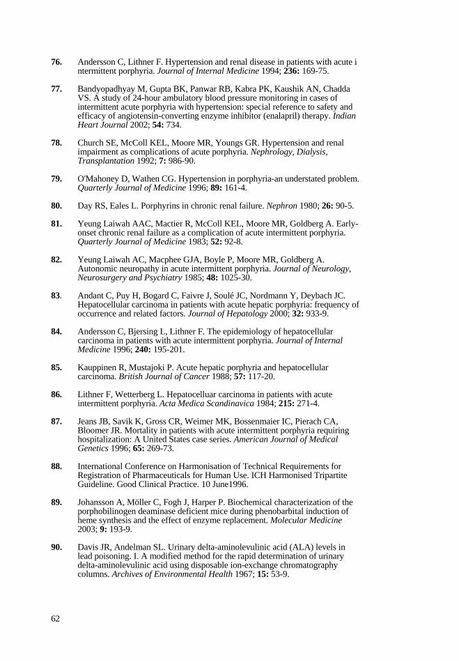
62
76. Andersson C, Lithner F. Hypertension and renal disease in patients with acute i ntermittent porphyria. Journal of Internal Medicine 1994; 236: 169-75. 77. Bandyopadhyay M, Gupta BK, Panwar RB, Kabra PK, Kaushik AN, Chadda VS. A study of 24-hour ambulatory blood pressure monitoring in cases of intermittent acute porphyria with hypertension: special reference to safety and efficacy of angiotensin-converting enzyme inhibitor (enalapril) therapy. Indian Heart Journal 2002; 54: 734. 78. Church SE, McColl KEL, Moore MR, Youngs GR. Hypertension and renal impairment as complications of acute porphyria. Nephrology, Dialysis, Transplantation 1992; 7: 986-90. 79. O'Mahoney D, Wathen CG. Hypertension in porphyria-an understated problem. Quarterly Journal of Medicine 1996; 89: 161-4. 80. Day RS, Eales L. Porphyrins in chronic renal failure. Nephron 1980; 26: 90-5. 81. Yeung Laiwah AAC, Mactier R, McColl KEL, Moore MR, Goldberg A. Early- onset chronic renal failure as a complication of acute intermittent porphyria. Quarterly Journal of Medicine 1983; 52: 92-8. 82. Yeung Laiwah AC, Macphee GJA, Boyle P, Moore MR, Goldberg A. Autonomic neuropathy in acute intermittent porphyria. Journal of Neurology, Neurosurgery and Psychiatry 1985; 48: 1025-30. 83. Andant C, Puy H, Bogard C, Faivre J, Soulé JC, Nordmann Y, Deybach JC. Hepatocellular carcinoma in patients with acute hepatic porphyria: frequency of occurrence and related factors. Journal of Hepatology 2000; 32: 933-9. 84. Andersson C, Bjersing L, Lithner F. The epidemiology of hepatocellular carcinoma in patients with acute intermittent porphyria. Journal of Internal Medicine 1996; 240: 195-201. 85. Kauppinen R, Mustajoki P. Acute hepatic porphyria and hepatocellular carcinoma. British Journal of Cancer 1988; 57: 117-20. 86. Lithner F, Wetterberg L. Hepatocelluar carcinoma in patients with acute intermittent porphyria. Acta Medica Scandinavica 1984; 215: 271-4. 87. Jeans JB, Savik K, Gross CR, Weimer MK, Bossenmaier IC, Pierach CA, Bloomer JR. Mortality in patients with acute intermittent porphyria requiring hospitalization: A United States case series. American Journal of Medical Genetics 1996; 65: 269-73. 88. International Conference on Harmonisation of Technical Requirements for Registration of Pharmaceuticals for Human Use. ICH Harmonised Tripartite Guideline. Good Clinical Practice. 10 June1996. 89. Johansson A, Möller C, Fogh J, Harper P. Biochemical characterization of the porphobilinogen deaminase deficient mice during phenobarbital induction of heme synthesis and the effect of enzyme replacement. Molecular Medicine 2003; 9: 193-9. 90. Davis JR, Andelman SL. Urinary delta-aminolevulinic acid (ALA) levels in lead poisoning. I. A modified method for the rapid determination of urinary delta-aminolevulinic acid using disposable ion-exchange chromatography columns. Archives of Environmental Health 1967; 15: 53-9.
63
91. Mauzerall D, Granick S. The occurrence and determination of delta- aminolevulinic acid and porphobilinogen in urine. J Biol Chem 1956; 219: 435- 46. 92. Bloom KE, Zaider EF, Morledge LJ, Poh-Fitzpatrick MB. Urinary porphyrin . excretion in normal children and adults. Am J Kidney Dis 1991; 18: 483-9. 93. Floderus Y, Sardh E, Moller C, Andersson C, Rejkjaer L, Andersson DE, Harper P. Variations in porphobilinogen and 5-aminolevulinic acid concentrations in plasma and urine from asymptomatic carriers of the acute intermittent porphyria gene with increased porphyrin precursor excretion. Clin Chem 2006; 52: 701-7. 94. National Kidney Foundation KDOQI ™. http://www.kidney.org. Accessed September 2008. 95. Siersema PD, de Rooij FWM, Edixhoven-Bosdijk A, Weimar W, Wilson JHP. Heme synthesis in chronic renal failure. The effects of hemodialysis, peritoneal dialysis and erythropoietin treatment. Nephron 1995; 71: 297-302. 96. Lim CK, Peters TJ. Urine and faecal porphyrin profiles by reversed-phase high- performance liquid chromatography in the porphyrias. Clinica Chimica Acta 1984; 139: 55-63. 97. Bruix J, Sherman M. Management of hepatocellular carcinoma. Hepatology 2005; 42: 1208-36. 98. de Rooij FWM, Edixhoven A, Wilson JHP. Porphyria: A Diagnostic Approach. Elsevier Science (USA). 2003. 99. Miyajima K, Hirata M, Yoshida T, Kosaka H, Okayama A. Study on measurement of delta-aminolevulinic acid in plasma by high-performance liquid chromatography. Journal of Chromatography B: Biomedical Applications 1994; 654: 165-9. 100. Gorchein A. Determination of delta-aminolaevulinic acid in biological fluids by gas-liquid chromatography with electron-capture detection. Biochemical Journal 1984; 219: 883-9. 101. Tschudy DP, Welland FH, Collins A, Hunter G, Jr. The effect of carbohydrate feeding on the induction of delta-aminolevulinic acid synthetase. Metabolism 1964; 13: 396-406. 102. Aarsand AK, Petersen PH, Sandberg S. Estimation and Application of Biological Variation of Urinary {delta}-Aminolevulinic Acid and Porphobilinogen in Healthy Individuals and in Patients with Acute Intermittent Porphyria. Clin Chem 2006; 52: 650-6. 103. Schoenfeld N, Mamet R. Individualized workup - a new approach to the biochemical diagnosis of acute attacks of neuroporphyria. Physiol Res 2007; 55: S103- 8. 104. Wenger S, Meisinger V, Brucke T, Deecke L. Acute porphyric neuropathy during pregnancy--effect of haematin therapy. European Neurology 1998; 39: 187-8. 105. Buijs R, Kalsbeek A. Hypothalamic integration of central and peripheral clocks. Nat Rev Neurosci 2001; 2: 521-26.
64
106. Shoolingin-Jordan PM, Al-Dbass A, McNeill LA, Sarwar M, Butler D. Human porphobilinogen deaminase mutations in the investigation of the mechanism of dipyrromethane cofactor assembly and tetrapyrrole formation. Biochemical Society Transactions 2003; 31: 731-5. 107. Onozato ML, Tojo A, Kamijo A, Taniguchi S, Kimura K, Goto A, Fujita T. Tubulointerstitial nephritis associated with acute intermittent porphyria. Clinical Nephrology 2001; 55: 171-4. 108. Marsden JT, Chowdhury P, Wang J, Deacon A, Dutt N, Peters TJ, Macdougall IC. Acute intermittent porphyria and chronic renal failure. Clin Nephrol 2008; ume 69: 339-46. 109. Shah S. Role of reactive oxygen metabolites in experimental glomerular disease. Kidney Int 1989; 35: 1093-106. 110. Dhar GJ, Bossenmaier I, Cardinal R, Petryka ZJ, Watson CJ. Transitory renal failure following rapid administration of a relatively large amount of hematin in a patient with acute intermittent porphyria in clinical remission. Acta Medica Scandinavica 1978; 203: 437-43. 111. Tracz MJ, Alam J, Nath KA. Physiology and pathophysiology of heme: implications for kidney disease. J Am Soc Nephrol 2007; 18: 414-20. Epub 2007 Jan 17. 112. Shoolingin-Jordan PM, McNeill LA. Molecular changes in porphobilinogen deaminase in AIP. Physiological Research 2003; 52: 24S. 113. Carson RW, Dunnigan EJ, DuBose TD, Goeger DE, Anderson KE. Removal of plasma porphyrins with high-flux hemodialysis in porphyria cutanea tarda associated with end-stage renal disease. Journal of the American Society of Nephrology 1992; 2: 1445-50. 114. Nunez DJ, Williams PF, Herrick AL, Evans DB, McColl KEL. Renal transplantation for chronic renal failure in acute porphyria. Nephrology, Dialysis, Transplantation 1987; 2: 271-4. 115. Warholm C, Wilczek H. Renal transplantation in a case of acute intermittent porphyria. Journal of Clinical Pharmacology 2003; 43: 1158-60. 116. Demirci G, Becker T, Nyibata M, et al. Results of combined and sequential liver-kidney transplantation. Liver Transpl 2003; 9: 1067-78. 117. Bengtsson NO, Hardell L. Porphyrias, porphyrins and hepatocellular cancer. British Journal of Cancer 1986; 54: 115-7. 118. Bjersing L, Andersson C, Lithner F. Hepatocellular carcinoma in patients from Northern Sweden with acute intermittent porphyria: Morphology and mutations. Cancer Epidemiology, Biomarkers and Prevention 1996; 5: 393-7. 119. Hardell L. Aspects of primary liver cancer and its relation to porphyria cutanea tarda and porphyria acuta intermittens. IARC Scientific Publications 1986; 77: 591-2. 120. Hardell L, Bengtsson NO, Jonsson U, Eriksson S, Larsson LG. Aetiological aspects on primary liver cancer with special regard to alcohol, organic solvents and acute intermittent porphyria - an epidemiological investigation. British Journal of Cancer 1984; 50: 389-97.

65
121. Bosch FX, Ribes J, Díaz M, Cléries R. Primary Liver Cancer: Worldwide Incidence and Trends. Journal of Gastroenterology 2004; 127: S5-S16. 122. Leong TY, Leong AS. Epidemiology and carcinogenesis of hepatocellular carcinoma. HPB (Oxford) 2005; 7: 5-15. 123. Bialecki ES, Di Bisceglie AM. Diagnosis of hepatocellular carcinoma. HPB (Oxford) 2005; 7: 26-34.