ACLS PROVIDER MANUAL STUDENT CD FAQ - Online … Study guide.pdf · ACLS PROVIDER MANUAL STUDENT CD...
13
ACLS PROVIDER MANUAL STUDENT CD FAQ 1. I cannot access the ACLS Precourse Self-Assessment Test. - Internet Explorer must be open before the CD is inserted. Remove the CD from the tray; close all other applications, then insert the CD - If you have a pop-up blocker, remove the CD from the tray, re-insert the CD while holding down the “Ctrl” key so Macromedia Flash can run. OR you can go to My Computer > Right Click On the CD-ROM drive > Explore> Double Click on PC_Start or MAC_Start - Make sure you are using Internet Explorer 6.0 or higher (Not AOL, FireFox, Mozilla or Netscape) - Check to make sure Active X Controls are enabled by going to Internet Explorer> Tools> Internet Options> Security Tab> Custom Level> Active X Controls and Plug-ins> Enable - Check to make sure “Allow Active Content CDs to run on my Computer” is checked by going to Tools>Internet Options> Advanced Tab> Security - Download “Adobe Flash Player” and “Adobe Reader” from www.adobe.com if you do not have it already installed on your computer. Restart the computer after you have installed the Adobe Flash Player 2. I cannot play the CD more than “two, three, four times” - Delete “Temp Files” Internet Explorer > Tools > Internet Options > General > Delete Files. Click on OK - Close other programs running in the background - Restart the Computer 3. I cannot open “ACLS Core Drugs” or any other PDF files on the CD - Make sure you have Adobe installed on your computer, otherwise download Adobe Acrobat Reader from www.adobe.com . - 4. I can't hear any sound. What do I do? - Make sure the speakers are turned on and the volume is turned up - Check the Volume and Mute settings on your computer. Make sure Mute is not checked, and adjust Volume as needed. There are multiple ways to check these settings: ■ Click on the speaker icon in your system tray. Adjust Volume if needed and make sure Mute is not checked. ■ Go to Start > Settings > Control Panel>Sounds and Audio Devices>Volume. Make sure Mute is not checked. Then go to Advanced. Adjust Volume if needed and make sure Mute is not checked. ■ Go to Start > Programs > Accessories > Entertainment > Volume Control. - Make sure the volume on the video clip is turned up. The Volume Control button is located at the bottom of the screen on the left.
-
Upload
duongkhuong -
Category
Documents
-
view
223 -
download
2
Transcript of ACLS PROVIDER MANUAL STUDENT CD FAQ - Online … Study guide.pdf · ACLS PROVIDER MANUAL STUDENT CD...
ACLS PROVIDER MANUAL STUDENT CD FAQ
1. I cannot access the ACLS Precourse Self-Assessment Test.
- Internet Explorer must be open before the CD is inserted. Remove the CD from the tray; close all other applications, then insert the CD
- If you have a pop-up blocker, remove the CD from the tray, re-insert the CD while holding down the “Ctrl” key so Macromedia Flash can run. OR you can go to My Computer > Right Click On the CD-ROM drive > Explore> Double Click on PC_Start or MAC_Start
- Make sure you are using Internet Explorer 6.0 or higher (Not AOL, FireFox, Mozilla or Netscape)
- Check to make sure Active X Controls are enabled by going to Internet Explorer> Tools> Internet Options> Security Tab> Custom Level> Active X Controls and Plug-ins> Enable
- Check to make sure “Allow Active Content CDs to run on my Computer” is checked by going to Tools>Internet Options> Advanced Tab> Security
- Download “Adobe Flash Player” and “Adobe Reader” from www.adobe.com if you do not have it already installed on your computer. Restart the computer after you have installed the Adobe Flash Player
2. I cannot play the CD more than “two, three, four times”
- Delete “Temp Files” Internet Explorer > Tools > Internet Options > General > Delete Files. Click on OK
- Close other programs running in the background - Restart the Computer
3. I cannot open “ACLS Core Drugs” or any other PDF files on the CD
- Make sure you have Adobe installed on your computer, otherwise download Adobe Acrobat Reader from www.adobe.com.
- 4. I can't hear any sound. What do I do?
- Make sure the speakers are turned on and the volume is turned up - Check the Volume and Mute settings on your computer. Make sure Mute is not checked, and
adjust Volume as needed. There are multiple ways to check these settings:
■ Click on the speaker icon in your system tray. Adjust Volume if needed and make sure Mute is not checked.
■ Go to Start > Settings > Control Panel>Sounds and Audio Devices>Volume. Make sure Mute is not checked. Then go to Advanced. Adjust Volume if needed and make sure Mute is not checked.
■ Go to Start > Programs > Accessories > Entertainment > Volume Control. - Make sure the volume on the video clip is turned up. The Volume Control button is located at
the bottom of the screen on the left.

Patient Assessment In ACLS, the specific treatment of a given dysrhythmia or condition depends on the patient’s hemodynamic status. In general, patients can be divided into four categories to determine treatment priorities: Asymptomatic,Symptomatic – Stable, Symptomatic – Unstable, or Pulseless.
Asymptomatic patients do not receive treatment, but should be monitored for changes in condition. Any patient with symptoms (even apparently mild symptoms such as palpitations) should be assessed to determine if they are Stable or Unstable. Determination of a patient’s level of hemodynamic compromise can include several factors, including General Appearance, Level of Consciousness, and Vital Signs (especially systolic Blood Pressure).
General Appearance: The first indication of hemodynamic status comes from a patient’s general appearance, including skin signs, level of activity, and work of breathing. If a patient shows signs of compensation (such as pale, cool, or diaphoretic skin) or acute distress, they are unstable. Level of Consciousness: Interaction with the patient allows the provider to evaluate the patient’s level of consciousness, based on the patient’s activity, awareness of their surroundings, and ability to provide information. If a patient shows any level of mental deficit, family or friends should be consulted to determine if this state differs from the patient’s baseline. If the mental deficit is acute, the patient should be considered unstable. Vital Signs: Vital signs provide a diagnostic evaluation of the patient. Blood Pressure is the primary indicator. A systolic blood pressure above 90 mm usually indicates that the patient is stable (although the provider should be alert for changes in blood pressure that might indicate an unstable patient even if blood pressure is normal). Other vital signs may be useful, but should not be relied upon exclusively. Pulse Oximetry can be useful, especially if it rises or falls, but providers should remember that various conditions (such as CO2poisoning) can mask changes in blood oxygen levels, and that a high O2 saturation may be present in unstable patients (such as those in shock). Additionally, heart rate is of no use in determining if a patient is stable or unstable – a patient with a heart rate of 80 can be severely unstable, while a patient with a heart rate of 210 can be stable if they are still perfusing well.
If a patient’s General Appearance, Level of Consciousness, and Vital Signs are all normal, the patient is stable. If possible, treatment should be rendered starting with the least invasive that is appropriate for that patient’s hemodynamic status. In ACLS, the preferential treatment for symptomatic but stable patients is generally Medications, while the preferential treatment for unstable patients is generally Electrical Therapy.
Once treatment is rendered, the provider must reassess the patient. If the patient remains symptomatic, the appropriate treatment (medications or electricity) should be given again depending on the patient’s heart rhythm and current hemodynamic status. (Thus, if a patient was stable before, but becomes unstable after administration of a drug, the patient should receive electrical therapy to continue treating the dysrhythmia rather than additional doses of a medication.)
If a patient’s General Appearance indicates that they may be unconscious, you should check for responsiveness. If the patient is Unresponsive, get help (send someone to call 911 and bring back an AED, call a code, etc.). The BLS Algorithm should then be followed – open the Airway, check for Breathing, and assess Circulation. If the patient is apneic, rescue breathing should be started; if the patient is pulseless, rescuers should begin CPR.
Once you determine that a patient is Pulseless, an AED or EKG monitor should be attached as soon as possible. CPR should be continued with minimal interruptions. After each rhythm check, the patient should be Defibrillated if appropriate (for V-Fib and Pulseless V-Tach). Regardless of the heart rhythm, medications should be given as soon as possible after CPR is resumed (the specific medication determined by the patient’s exact status and heart rhythm).
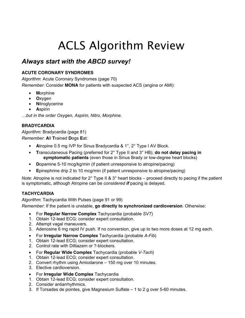
ACLS Algorithm Review Always start with the ABCD survey!
ACUTE CORONARY SYNDROMES Algorithm: Acute Coronary Syndromes (page 70)
Remember: Consider MONA for patients with suspected ACS (angina or AMI):
• Morphine• Oxygen• Nitroglycerine• Aspirin
…but in the order Oxygen, Aspirin, Nitro, Morphine.
BRADYCARDIA Algorithm: Bradycardia (page 81)
Remember: All Trained Dogs Eat:
• Atropine 0.5 mg IVP for Sinus Bradycardia & 1°, 2° Type I AV Block.
• Transcutaneous Pacing (preferred for 2° Type II and 3° HB); do not delay pacing in symptomatic patients (even those in Sinus Brady or low-degree heart blocks)
• Dopamine 5-10 mcg/kg/min (if patient unresponsive to atropine/pacing)
• Epinephrine drip 2 to 10 mcg/min (if patient unresponsive to atropine/pacing)
Note: Atropine is not indicated for 2° Type II & 3° heart blocks – proceed directly to pacing if the patient is symptomatic, although Atropine can be considered if pacing is delayed.
TACHYCARDIA Algorithm: Tachycardia With Pulses (page 91 or 99)
Remember: If the patient is unstable, go directly to synchronized cardioversion. Otherwise:
• For Regular Narrow Complex Tachycardia (probable SVT)1. Obtain 12-lead ECG; consider expert consultation. 2. Attempt vagal maneuvers. 3. Adenosine 6 mg rapid IV push. If no conversion, give up to two more doses at 12 mg each.
• For Irregular Narrow Complex Tachycardia (probable A-Fib)1. Obtain 12-lead ECG; consider expert consultation. 2. Control rate with Diltiazem or ?-blockers.
• For Regular Wide Complex Tachycardia (probable V-Tach)1. Obtain 12-lead ECG; consider expert consultation. 2. Convert rhythm using Amiodarone – 150 mg over 10 minutes. 3. Elective cardioversion.
• For Irregular Wide Complex Tachycardia 1. Obtain 12-lead ECG; consider expert consultation. 2. Consider antiarrhythmics. 3. If Torsades de pointes, give Magnesium Sulfate – 1 to 2 g over 5-60 minutes.

VENTRICULAR FIBRILLATION / PULSELESS VENTRICULAR TACHYCARDIA Algorithm: Pulseless Arrest – Shockable (page 42) Remember: Good ACLS starts with good BLS:
• CPR – start immediately. Push hard and push fast. • Shock – analyze rhythm, and shock if in VF/pulseless VT. • CPR – resume CPR immediately after shock delivery. Continue for 5 cycles / 2 minutes. • Vasopressor – Epi 1 mg q 3-5 min (can replace 1st or 2nd dose of Epi with 40 units Vasopressin).
Give as soon as possible after resuming CPR, circulate with chest compressions. • Shock – analyze rhythm, and shock if in VF/pulseless VT. • CPR – resume CPR immediately after shock delivery. Continue for 5 cycles / 2 minutes. • Antiarrhythmic – Amiodarone 300 mg IV/IO or Lidocaine 1-1.5 mg/kg up to 3 mg/kg.
Give as soon as possible after resuming CPR, circulate with chest compressions. • Shock – analyze rhythm, and shock if in VF/pulseless VT. • CPR – resume CPR immediately after shock delivery. Continue for 5 cycles / 2 minutes.
Note: Minimize interruptions to chest compressions – do not check a pulse or evaluate the heart rhythm after a shock. After each shock, resume CPR immediately and continue for 5 cycles prior to rhythm analysis and possible pulse check. After a second dose of Epinephrine, a second antiarrhythmic dose (Amiodarone 150 mg or Lidocaine 0.5 – 0.75 mg/kg) may given after the next rhythm check. PULSELESS ELECTRICAL ACTIVITY Algorithm: Pulseless Arrest – Not Shockable (page 54) Remember: PEA:
• Possible causes (consider the 6 H’s and 5 T’s). • Epinephrine 1 mg q 3-5 min (can replace 1st or 2nd dose of Epi with 40 units Vasopressin).
Give as soon as possible after resuming CPR, circulate with chest compressions. • Atropine, 1mg IV/IO q 3-5 min to max 3mg (only if electrical rate is < 60)
Give as soon as possible after resuming CPR, circulate with chest compressions. Note: In PEA, the electrical system of the heart is functioning, but there is a problem with the pump, pipes, or volume – a mechanical part of the system is not working. You can use the 6 H’s and 5 T’s to remember the most common reversible causes of PEA: Hypovolemia Hypoxia Tamponade, cardiac Toxins Hypo-/Hyperkalemia Hypoglycemia Tension Pneumothorax Trauma Hydrogen Ion (acidosis) Hypothermia Thrombosis (coronary or pulmonary) ASYSTOLE Algorithm: Pulseless Arrest – Not Shockable (page 54) Remember: DEAD:
• Determine whether to initiate resuscitation. • Epinephrine 1 mg q 3-5 min (can replace 1st or 2nd dose of Epi with 40 units Vasopressin).
Give as soon as possible after resuming CPR, circulate with chest compressions. • Atropine, 1mg IV/IO q 3-5 min to max 3mg
Give as soon as possible after resuming CPR, circulate with chest compressions. • Differential Diagnosis or Discontinue resuscitation – Are they still dead? Consider the 6 H’s and
5 T’s (see above) – check blood glucose; check core temperature; consider Naloxone; etc.

EKG and Electrical Therapy Review The EKG tracing represents electrical activity through the heart. The P wave represents depolarization of the atria; the QRS complex represents depolarization of the ventricles; and the T wave represents the latter stage of repolarization of the ventricles. The interval from the first deflection of the P wave to the be-ginning of the QRS complex is the P-R Interval (PRI), and should be between 0.12 and 0.20 seconds. A normal QRS complex has a duration of 0.12 seconds or less; a longer duration (wide QRS) indicates delayed conduction through the ventricles, often as the result of a ventricular pacemaker focus. The horizontal axis of the EKG strip measures time. Each large box represents 0.20 seconds; each small box represents 0.04 seconds. To obtain a 3-lead EKG tracing, place the white (RA) electrode on the right chest just below the clavicle; the black electrode (LA) on the left chest just below the clavicle; and the Red electrode (LL) laterally on the lower left abdomen. Pacer pads go in the anterior/posterior positions. Defibrillation pads go on the upper right chest and lower left abdomen, although on children and other small patients the pads may need to be placed in the middle of the anterior and posterior chests.
Rhythm Disturbances Treat the patient, not the dysrhythmia. Always assess your patient for pulses, perfusion, and level of consciousness – is the patient Stable, Unstable, or Pulseless? Next, assess the rhythm: Is it fast or slow? Is it life-threatening? As you treat the patient, try to discover the cause of the dysrhythmia – for many patients, their only chance of survival is if you can identify and treat a reversible cause. There are many possible causes of rhythm disturbances or PEA. Common causes include sympathetic stimulation, stress, hypoxia, ischemia, drugs/toxins, and electrolyte disturbances. Although lab draws can be useful, a history of the patient and the current event obtained from a family member or caregiver is often more useful. Defibrillation (Unsynchronized Shock) Fibrillation is a disorganized rhythm that, if present in the ventricles, is life-threatening. Immediate CPR combined with early defibrillation is critical to survival from sudden cardiac arrest. Defibrillation terminates all electrical activity in the pulseless heart in the hopes that it will resume beating in a coordinated fashion. A shock should be delivered about once every 2 minutes if the patient remains in Ventricular Fibrillation. With a monophasic monitor, the recommendation is to deliver a single shock at 360 Joules. If a biphasic monitor is used, the recommended dosage is machine-dependent, and should appear on the front of the monitor. If optimal shock dosage is not known, the consensus is to defibrillate at 200 J. Synchronized Cardioversion Synchronized cardioversion is the preferred treatment for unstable patients with a tachycardia such as Atrial Fibrillation, V-Tach with a pulse, or Supraventricular Tachycardia (SVT). The shock is timed by the monitor to be delivered in coordination with the QRS complex of the heart. If the patient is conscious, consider sedation prior to cardioversion; however, synchronized cardioversion should not be delayed while waiting for sedation in severely symptomatic patients.
With a monophasic monitor, the initial shock is delivered at 100 J; if the rhythm does not terminate, deliver additional shocks in stepwise fashion (200J, 300J, and 360J for subsequent shocks). With a biphasic monitor, dosage and steps are device-dependent; if optimal doses are unknown, begin at 100 J and step up from there. Transcutaneous Pacing (TCP) External cardiac pacing is the recommended treatment for sympto-matic bradycardias. If the patient is conscious, consider sedation; however, pacing should not be delayed while waiting for sedation. Begin pacing at zero milliamps, slowly increasing until capture is achieved. Then, set the rate at 20 beats per minute above the monitored heart rate, with a minimum rate of 50 bpm.


ACLS Medications Review This information on medications meets the standard set by the 2005 American Heart Association for Advanced Cardiac Life Support. It does not supersede local protocols or medical control; consult with your medical director for the most up-to-date guidelines on medication administration where you work.
IV/IO medications should be administered in a peripheral line during CPR, as soon as possible after a rhythm check. It is recommended that you flush with 20 ml of fluid after each drug administration and elevate the extremity. Always use large bore catheters if possible.
A Note on Endotracheal Administration of Medications: This route of medication administration is being de-emphasized by the AHA – the IV or IO routes are preferred. However, the ET route can still be used if providers are unable to gain access by IV/IO. Use the mnemonic “NAVEL” to remember which drugs can be administered via this route: Narcan – Atropine – Vasopressin – Epinephrine – Lidocaine. If using the ET route, the drug dosage must be increased, typically 2-2.5 times the IV/IO bolus dosage (although there is no consensus on Epinephrine or Vasopressin dosing via this route), followed by a 10 ml normal saline flush.
ADENOSINE Class: Indicated for: IV Bolus Dosage (no IO):Endogenous nucleoside PSVT / Regular Narrow- 6 mg rapid IV push – 1st dose; complex Tachycardia 12 mg rapid IV push – 2nd dose 12 mg rapid IV push – 3rd dose
Notes: Doses are followed by a saline flush. Two subsequent doses of 12 mg each may be administered at 1 – 2 minute intervals. Use the port closest to cannulation. The AHA recommends that the dose be cut by half if administering through a central line, or in the presence of Dipyridamole or Carbamazepine. Larger doses may be required in the presence of caffeine or Theophylline.
AMIODARONEClass: Indicated for: IV/IO Bolus Dosage: Antiarrhythmic V-Fib / Pulseless V-Tach 300 mg – 1st dose; 150 mg – 2nd dose Arrhythmias 360 mg over 6 hours (slow) 150 mg over 10 minutes (rapid) Infusion dose 540 mg IV/IO over 18 hours (.5 mg/min)
Notes: Cumulative doses >2.2 g/24 hours are associated with significant hypotension. Do not administer with other drugs that prolong QT interval (i.e., Procainimide). Terminal elimination is extremely long – half life lasts up to 40 days. During arrest, IV bolus should be delivered slowly, over 1 – 3 minutes.

ASPIRIN Class: Indicated for: PO Dosage (no IV/IO): NSAID (Non-Steroidal Chest pain / ACS 160 mg – 325 mg Anti-Inflammatory Drug) Suppository Dose: 300 mg Notes: In suspected ACS, Aspirin can block platelet aggregation and arterial constriction. Also helps with pain control. May cause or exacerbate GI bleeding. ATROPINE Class: Indicated for: IV/IO Bolus Dosage: Parasympathetic Blocker Bradycardia 0.5 mg every 3-5 minutes as needed Asystole, slow PEA 1 mg every 3-5 minutes (up to 3 mg) Notes: Used only in symptomatic bradycardia or in PEA with heart rate < 60. (Not indicated in Second Degree Type II or Third Degree heart block.) Doses < 0.5 mg may result in paradoxical slowing of the heart. ET route discouraged, but can be used if IV/IO access not available. DEXTROSE/GLUCOSE Class: Indicated for: IV/IO Bolus Dosage: Carbohydrate Hypoglycemia 25 g (50 ml) of D50W Notes: Used to reverse documented hypoglycemia in patients with symptomatic bradycardia or during cardiac arrest. Should not be used routinely during cardiac arrest. DILTIAZEM Class: Indicated for: IV Dosage: Calcium Channel Blocker A-Fib / A-Flutter 15-20 mg over 2 minutes Notes: May cause hypotension. Do not use in wide-QRS tachycardias of uncertain origin. DOPAMINE Class: Indicated for: IV Infusion: Catecholamine Symptomatic Bradycardia 2-10 µg/kg/min – cardiac dose Hypotension 10-20 µg/kg/min – vasopressor dose Notes: Titrate to patient response. Correct hypovolemia with volume replacement before initiating Dopamine. May cause tachyarrhythmias. Do not mix with Sodium Bicarbonate. EPINEPHRINE Class: Indicated for: IV/IO Bolus Dosage: Catecholamine Pulseless Arrest 1 mg (1:10,000) every 3-5 minutes Symptomatic Bradycardia Infusion: 1 mg in 500ml of D5W or NaCl
at 1 µg/min titrated to effect. Notes: First line drug in all pulseless rhythms. Increases myocardial oxygen demand, and may cause myocardial ischemia or angina. ET route is discouraged, but if used give 2-2.5 mg of a 1:1000 solution diluted in 10 ml normal saline.

FLUID ADMINISTRATION (e.g., Normal Saline / NaCl) Class: Indicated for: IV/IO Bolus Dosage: Fluid Volume Hypovolemia 250 – 500 cc bolus (repeat as needed) Notes: Use to treat specific reversible causes, such as hypovolemia. Routine administration of fluids during a resuscitation is not indicated, as it can reduce coronary perfusion pressure. HEPARIN (Unfractionated) Class: Indicated for: IV/IO Bolus Dosage: Anticoagulant STEMI (AMI) Initial Dose: 60 IU/kg (max. 4000 IU) Infusion: 12 IU/kg/hr (max. 1000 IU/hr) Notes: Do not use in patients with active bleeding or bleeding disorders; severe hypertension; or recent surgery. Monitor aPTT and platelet count while administering. LIDOCAINE Class: Indicated for: IV/IO Bolus Dosage: Antiarrhythmic V-Fib/Pulseless V-Tach 1-1.5 mg/kg (1st dose) Stable V-Tach Infusion: 1-4 mg/min (30-50 µg/kg/min) Notes: May repeat at 0.5-0.75 mg/kg every 5-10 minutes to a max. dose of 3 mg/kg. Use with caution in presence of impaired liver; discontinue if signs of toxicity develop. Prophylactic use in AMI is contraindicated. ET route discouraged, but can be used if IV/IO access not available. MAGNESIUM SULFATE Class: Indicated for: IV/IO Bolus Dosage: Electrolyte Torsades de pointes or 1-2 g in 10 ml D5W over 5-20 minutes Hypomagnesemia Notes: A fall in blood pressure may be noted with rapid administration. Dose is given over 5-20 minutes during cardiac arrest, 5-60 minutes in living patients. Use with caution in renal failure. MORPHINE SULFATE Class: Indicated for: IV/IO Bolus Dosage: Opiate / Analgesic Chest pain 2-4 mg every 5-30 minutes Pulmonary edema Notes: Administer slowly and titrate to effect; may cause hypotension. May cause respiratory depression – be prepared to support ventilations. Naloxone is the reversal agent. NALOXONE HYDROCHLORIDE (NARCAN) Class: Indicated for: IV/IO Bolus Dosage: Opiate Antagonist Narcotic overdose 0.4-2.0 mg (up to 10 mg in 10 min.) Notes: Monitor for recurrence of respiratory depression. May cause opiate withdrawal. ET route discouraged, but can be used if IV/IO access not available.

NITROGLYCERIN Class: Indicated for: IV Bolus Dosage: Vasodilator Chest pain/ACS 12.5-25 µg in D5W or NaCl Sublingual Dose: 0.3 – 0.4 mg Notes: Most commonly given sublingually as tablet or spray – repeat up to 3 doses at 5 minute intervals. Hypotension may occur. Do not use with Viagra or other phosphodiasterase inhibitors; with severe bradycardia or tachycardia; or in presence of RV infarction or inferior MI. Do not mix with other drugs. OXYGEN Class: Indicated for: Flow Rate: Atmospheric Gas Any cardiopulmonary Stable Patient: 2-6 lpm via NC emergency Unstable Patient: 10-15 lpm via NRB Suspected stroke Notes: Pulse oximetry provides a useful method of titrating oxygen administration; however, it may be inaccurate in low cardiac output states or in patients with specific toxicities (such as Carbon Monoxide exposure). SODIUM BICARBONATE Class: Indicated for: IV Bolus Dosage: Buffer Acidosis, hyperkalemia 1 mEq/kg Notes: Not recommended for routine use in cardiac arrest patients. If available, use arterial blood gas analysis to guide bicarbonate therapy. VASOPRESSIN Class: Indicated for: IV/IO Bolus Dosage: Hormone Pulseless arrest 40 U IV/IO Notes: Only given one time to replace the first or second dose of Epinephrine; Epinephrine dosing can continue 3 to 5 minutes after Vasopressin is administered. Vasopressin should not replace antiarrhythmics (such as Amiodarone). May cause cardiac ischemia and angina. Not recommended for responsive patients with coronary artery disease. ET route discouraged, but can be used if IV/IO access not available. VERAPAMIL Class: Indicated for: IV Bolus Dosage: Calcium Channel Blocker A-Fib/A-Flutter, PSVT 2.5-5 mg over 2-5 minutes Notes: Alternative drug after Adenosine to terminate PSVT with adequate blood pressure and preserved LV function. Can cause peripheral vasodilation and hypotension. Use with extreme caution in patients receiving oral β-blockers.

© 2006 American Heart Association 11/20/05
American Heart Links There are several resources available to you on the American Heart Association website at www.americanheart.org. Here are some helpful links:
• You can find statistics on cardiovascular diseases and risk factors at http://www.americanheart.org/presenter.jhtml?identifier=2007
• You can find out your risk for heart disease at
http://www.americanheart.org/presenter.jhtml?identifier=3003500
• You can access information on the warning signs of heart attack and stroke at http://www.americanheart.org/presenter.jhtml?identifier=3053
• You can find out how to lead a healthy lifestyle at
http://www.americanheart.org/presenter.jhtml?identifier=1200009
• You can also go to the Emergency Cardiovascular Care (ECC) website at http://www.americanheart.org/presenter.jhtml?identifier=3011764, where you can find out about other American Heart Association CPR or First Aid courses and even find a course in your area.
• To find any other topic, use the Heart and Stroke Encyclopedia at this link:
http://www.americanheart.org/presenter.jhtml?identifier=10000056