A practical approach to multicolor flow cytometry … › ... › fcf › assets › pdf ›...
21
Journal of Immunological Methods 243 (2000) 77–97 www.elsevier.nl / locate / jim A practical approach to multicolor flow cytometry for immunophenotyping a ,1 b, * Nicole Baumgarth , Mario Roederer a Department of Genetics, Stanford University Medical School, Beckman Center B-007, Stanford, CA 94305-5318, USA b Department of Stomatology, UCSF, San Francisco, CA, USA Abstract Through a series of novel developments in flow cytometry hardware, software, and dye-chemistry it is now possible to simultaneously measure up to 11 distinct fluorescences and two scattered light parameters on each cell. Such advanced multicolor systems have a number of advantages over current two- and three-color flow cytometric measurements. They provide a large amount of novel information for each sample studied, an exquisitely accurate quantitation of even rare cell populations, and allow identification and characterization of novel cell subsets. In particular, this technology is proving crucial to identifying functionally homogeneous subsets of cells within the enormously complex immune system; such identification and enumeration is important for understanding disease pathogenesis. However, multicolor flow cytometry comes with a new and sometimes difficult set of technical problems that must be overcome by users to derive meaningful results. In this manuscript, we describe the basic aspects of multicolor flow cytometry, including the technical hurdles and artefacts that may occur, and provide some suggestions for how to best overcome these hurdles. While inspired by the 11-color technology that we currently use, these principles apply to all flow cytometric experiments in which more than one fluorescent dye is used. 2000 Elsevier Science B.V. All rights reserved. Keywords: Flow cytometry; Immunofluorescence; Immunophenotyping; Multiparameter flow cytometry 1. Introduction Since the earliest application of flow cytometry to Abbreviations: APC, allophycocyanin; A595, Alexa 595; CasB: Cascade Blue; CasY, Cascade Yellow; Cy5.5APC, Cy7APC, the study of cells, there has been a drive to increase Cy5.5, Cy7 conjugates of allophycocyanin; Cy5PE, Cy5.5PE, the number of distinct measurements for each cell. Cy7PE, Cy5, Cy5.5, and Cy7 conjugates of phycoerythrin; This developmental effort blossomed in the late Cy5.5PerCP: Cy5.5 tandem conjugate of PerCP; FITC, fluores- 1990s, when the number of independently measur- cein; PBP: phycobiliprotein (i.e., PE or APC); PE, phycoerythrin; able ‘colors’ (each color corresponds to a distinct PerCP: peridinin chlorophyll protein; PFC, polychromatic flow cytometry; PMT, photomultiplier tube; TR, Texas Red; TRPE, fluorescence-based measurement of a cellular protein Texas Red-conjugated phycoerythrin; SA, streptavidin or function) increased from five to 11 (Roederer et *Corresponding author. Tel.: 11-415-514-0395; fax: 11-415- al., 1997; Bigos et al., 1999). 476-4204. The success of this developmental effort was due E-mail address: [email protected] (M. Roederer). 1 to the coordinated development of new hardware, Present address: Center for Comparative Medicine, University new fluorochromes, and new software analysis tools of California, Davis, CA 95616, USA. 0022-1759 / 00 / $ – see front matter 2000 Elsevier Science B.V. All rights reserved. PII: S0022-1759(00)00229-5
Transcript of A practical approach to multicolor flow cytometry … › ... › fcf › assets › pdf ›...
Journal of Immunological Methods 243 (2000) 77–97www.elsevier.nl / locate / jim
A practical approach to multicolor flow cytometry forimmunophenotyping
a ,1 b ,*Nicole Baumgarth , Mario RoedereraDepartment of Genetics, Stanford University Medical School, Beckman Center B-007, Stanford, CA 94305-5318, USA
bDepartment of Stomatology, UCSF, San Francisco, CA, USA
Abstract
Through a series of novel developments in flow cytometry hardware, software, and dye-chemistry it is now possible tosimultaneously measure up to 11 distinct fluorescences and two scattered light parameters on each cell. Such advancedmulticolor systems have a number of advantages over current two- and three-color flow cytometric measurements. Theyprovide a large amount of novel information for each sample studied, an exquisitely accurate quantitation of even rare cellpopulations, and allow identification and characterization of novel cell subsets. In particular, this technology is provingcrucial to identifying functionally homogeneous subsets of cells within the enormously complex immune system; suchidentification and enumeration is important for understanding disease pathogenesis. However, multicolor flow cytometrycomes with a new and sometimes difficult set of technical problems that must be overcome by users to derive meaningfulresults. In this manuscript, we describe the basic aspects of multicolor flow cytometry, including the technical hurdles andartefacts that may occur, and provide some suggestions for how to best overcome these hurdles. While inspired by the11-color technology that we currently use, these principles apply to all flow cytometric experiments in which more than onefluorescent dye is used. 2000 Elsevier Science B.V. All rights reserved.
Keywords: Flow cytometry; Immunofluorescence; Immunophenotyping; Multiparameter flow cytometry
1. Introduction
Since the earliest application of flow cytometry toAbbreviations: APC, allophycocyanin; A595, Alexa 595; CasB:Cascade Blue; CasY, Cascade Yellow; Cy5.5APC, Cy7APC, the study of cells, there has been a drive to increaseCy5.5, Cy7 conjugates of allophycocyanin; Cy5PE, Cy5.5PE, the number of distinct measurements for each cell.Cy7PE, Cy5, Cy5.5, and Cy7 conjugates of phycoerythrin; This developmental effort blossomed in the lateCy5.5PerCP: Cy5.5 tandem conjugate of PerCP; FITC, fluores-
1990s, when the number of independently measur-cein; PBP: phycobiliprotein (i.e., PE or APC); PE, phycoerythrin;able ‘colors’ (each color corresponds to a distinctPerCP: peridinin chlorophyll protein; PFC, polychromatic flow
cytometry; PMT, photomultiplier tube; TR, Texas Red; TRPE, fluorescence-based measurement of a cellular proteinTexas Red-conjugated phycoerythrin; SA, streptavidin or function) increased from five to 11 (Roederer et
*Corresponding author. Tel.: 11-415-514-0395; fax: 11-415- al., 1997; Bigos et al., 1999).476-4204.
The success of this developmental effort was dueE-mail address: [email protected] (M. Roederer).1 to the coordinated development of new hardware,Present address: Center for Comparative Medicine, University
new fluorochromes, and new software analysis toolsof California, Davis, CA 95616, USA.
0022-1759/00/$ – see front matter 2000 Elsevier Science B.V. All rights reserved.PI I : S0022-1759( 00 )00229-5
78 N. Baumgarth, M. Roederer / Journal of Immunological Methods 243 (2000) 77 –97
that significantly increase the quality and quantity of can be simultaneously measured. The 11-color PFCmeasurements. This increase comes with a price, that is currently in routine use at Stanford uses dyeshowever, as this new technology has its own set of excited by three different laser lines. The excitationtechnical problems and difficulties that users must be and emission spectra of these dyes and the filters thataware of and must overcome in order to derive were chosen to collect the emitted light from thesemeaningful results. Nonetheless, once these hurdles dyes are shown in Fig. 1.are overcome, this new technology is well worth theeffort, as the information obtained from the measure- 2.1. Characteristics of useful fluorochromesments is not only novel but could not be obtainedotherwise using standard flow cytometric techniques. When designing experiments for the flow cytome-
As we outline below, setting up multicolor flow ter that include the use of new dyes, careful consid-cytometry is not simply achieved by adding new eration must be given to the choice of fluorochromes.reagents to existing reagent combinations, but re- Desirable fluorochromes for cytometric technologiesquires a more involved process of quality control, have several properties: they (i) are biologicallyoptimization, and ‘debugging’. Therefore, to dis- inert; (ii) have high cell-associated fluorescencetinguish multicolor flow cytometry with its unique intensities (‘bright’); (iii) exhibit little spectral over-benefits and technical problems from current stan- lap amongst each other; and (iv) for immuno-dard flow cytometric technologies (i.e., using four or phenotyping, are easily conjugated to monoclonalfewer fluorescent dyes), we refer to it as ‘poly- antibodies.chromatic flow cytometry’ (PFC) and will use theterm throughout this manuscript. 2.1.1. Biological inertness
Flow cytometers capable of collecting data for Most of the fluorochromes that are currently in usemore than three or four colors are now becoming are biologically inert: i.e., they do not affect themore prevalent, as manufacturers have recognized cells, nor do they bind directly to cellular elements.the significant demand for the types of analysis There are, however, exceptions to this: the mostafforded by this technology. Given the bewildering common example is that of the ‘background’ bindingarray of fluorochromes, lasers, hardware, and soft- of Cy5PE (and other Cyanine–PBP tandem dyes) toware that might be used in PFC, we outline here the monocytes and B cells. This background binding isfundamental requirements, interactions, and prob- variable between species and can be extremely highlems associated with setting up this technology, so in some instances. For example, Cy5PE binds strong-that users can make educated decisions about instru- ly to B cells in mice with autoimmune disordersment requirements and the design of their experi- (e.g., non-obese diabetic mice). While there arements. Finally, we provide some examples in which methods available to reduce this background, it is ofthis technology has been of particular benefit. We concern when the particular cell types being studiedhope to provide with this brief review some practical are those that interact ‘nonspecifically’ with thetips and encouragement for those thinking of expand- fluorochrome.ing their current flow cytometric measurements. Thebenefits of true multicolor flow cytometry make this 2.1.2. Cell-associated fluorescence intensitytechnique a particularly useful and probably soon an With regard to the high fluorescence intensities orirreplaceable tool for the study of cell biology and the ‘brightness’ of a fluorochrome, it should be notedimmunology. that the characterization of a fluorescence signal as
‘bright’, i.e., the difference between the unstainedand the stained cells, is still empirical. ‘Bright’
2. Fluorochromes signals result from fluorochromes with the followingcharacteristics: (1) a high extinction coefficient; (2) a
The ability to measure multiple parameters of each high quantum yield; (3) an emission spectrum over-cell is limited by the number of fluorochromes that lapping as little as possible with cellular auto-

N. Baumgarth, M. Roederer / Journal of Immunological Methods 243 (2000) 77 –97 79
Fig. 1. Shown are excitation and emission spectra of the 11 dyes that we currently use for polychromatic flow cytometry (PFC). Indicatedare the wavelengths of the different laser lines used for their excitation (407, 488 and 595 nm; other laser lines that can be used for some ofthe dyes are shown for reference), and the filters chosen for optimal light collection and minimal spillover. The fluorochromes, laser linesand filters are currently in routine use on a modified three-laser hybrid Becton-Dickinson/Cytomation flow cytometer, described previously(Roederer et al., 1997; Bigos et al., 1999).
80 N. Baumgarth, M. Roederer / Journal of Immunological Methods 243 (2000) 77 –97
fluorescence; (4) measurable with high sensitivity others, all of these fluorochromes are useful fordetectors; and (5) the ability to conjugate multiple clearly distinguishing the positively stained cellsfluorochromes to each detecting unit (e.g., a high from the unstained cells.ratio of fluorochrome to antibody). It is important to point out that the brightness of a
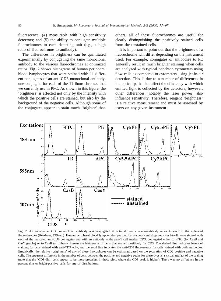
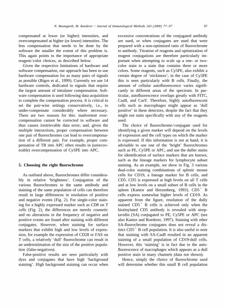
The differences in brightness can be quantitated fluorochrome will differ depending on the instrumentexperimentally by conjugating the same monoclonal used. For example, conjugates of antibodies to PEantibody to the various fluorochromes at optimized generally result in much brighter staining when cellsratios. Fig. 2 shows histograms of human peripheral are analyzed with typical benchtop cytometers usingblood lymphocytes that were stained with 11 differ- flow cells as compared to cytometers using jet-in-airent conjugates of an anti-CD8 monoclonal antibody, detection. This is due to a number of differences inone conjugate for each of the 11 fluorochromes that the optical paths that affect the efficiency with whichwe currently use in PFC. As shown in this figure, the emitted light is collected by the detectors; however,‘brightness’ is affected not only by the intensity with other differences (notably the laser power) alsowhich the positive cells are stained, but also by the influence sensitivity. Therefore, reagent ‘brightness’background of the negative cells. Although some of is a relative measurement and must be assessed bythe conjugates appear to stain much ‘brighter’ than users on any given instrument.
Fig. 2. An anti-human CD8 monoclonal antibody was conjugated at optimal fluorochrome–antibody ratios to each of the indicatedfluorochromes (Roederer, 1997a,b). Human peripheral blood lymphocytes, purified by gradient centrifugation over Ficoll, were stained witheach of the indicated anti-CD8 conjugates and with an antibody to the pan-T cell marker CD3, conjugated either to FITC (for CasB andCasY graphs) or to CasB (all others). Shown are histograms of cells that stained positively for CD3. The dashed line indicates levels ofstaining for cells stained with anti-CD3 only, and the solid line indicates the anti-CD8 fluorescence for cells stained with both antibodies.Empirically, the relative ‘brightness’ of any of these fluorophores can be estimated based on the separation of CD8 positive and negativecells. The apparent difference in the number of cells between the positive and negative peaks for these dyes is a visual artefact of the scaling(note that the ‘CD8-dim’ cells appear to be more prevalent in those plots where the CD8 peak is higher). There was no difference in thepercent dim or bright-positive cells for any of distributions.
N. Baumgarth, M. Roederer / Journal of Immunological Methods 243 (2000) 77 –97 81
2.1.3. Spectral overlaps gle protein molecules (PE, APC) require a slightlyThe spectral overlaps that exist between dyes more complex procedure, but reactive fluorochromes
currently represent the biggest hurdle in PFC analy- can be easily prepared, are stable for long periods ofsis. Due to the similarities and overlaps in the time, can be used in a simple conjugation procedure.
2emission spectra of different dyes (see Fig. 1), it is (3) Tandem dyes (Cy5PE, Cy5.5PE, Cy7PE,not possible to choose emission filters that uniquely Cy5.5APC, Cy7APC, Cy5.5PerCP), require the usemeasure only one of the dyes in a multicolor of more complex procedures which involve first theexperiment. The appropriate choice of filter, how- generation of the reactive tandems that can then, in aever, can greatly reduce collection of light from second step, be used for conjugation to antibodies.other fluorochromes (i.e., reduce ‘spillover’). Note Generating the reactive tandems requires carefulfrom Fig. 1 that a set of up to four dyes excited by testing of different conditions for each batch ofthree different lasers can be chosen that exhibit tandem dye to optimize the ratio of the two dyesessentially no spectral overlap; thus, four- or fewer- used. However, once the chemistry has been opti-color experiments can be carefully designed to avoid mized, a large batch of reactive dye can be preparedthe problems caused by spillover. However, the for use in many conjugations.necessity of having three lasers for a four-colorexperiment renders this an impractical solution. 2.2. Other fluorochromes.
Because of the spectral overlap, each fluoro-chrome will contribute a signal to several detectors, New fluorochromes for immunophenotyping appli-therefore the contribution in detectors not assigned to cations are constantly being developed. At the timethat fluorochrome must be subtracted from the total of writing, several dyes exist that are useful forsignal in those detectors. This process, termed ‘com- immunophenotyping applications and which can bepensation’, is discussed in detail below. used in addition to or instead of some dyes listed in
Fig. 1.2.1.4. Conjugation to antibodies
For many applications of flow cytometry, a further2.2.1. PerCPrequirement for a fluorochrome is that it can be
This is a chlorophyll-like protein that can bereadily conjugated to antibodies. The 11 dyes wedirectly conjugated to antibodies. It has an emissioncommonly use (Figs. 1 and 2) meet this criterion.spectrum similar to Cy5PE, but with no excitation inThere are, however, varying degrees of difficultythe red, and hence very little spillover into the APCinvolved in carrying out the conjugation reactions. Itdetector compared to Cy5PE. On the other hand, itis likely that any laboratory carrying out flowhas several disadvantages: it is easily ‘bleached’ bycytometric analyses with five or more dyes will needlasers, limiting its utility to low-power instrumentsto conjugate antibodies, as commercial conjugatessuch as benchtop instruments; it is less bright thanare not yet available for a number of these dyes.Cy5PE; and it is only available in conjugates from aEven if all of the dyes become available as antibodysingle vendor (Becton-Dickinson, San Jose, CA).conjugates, it is unlikely that every combination of
antibody and fluorochrome necessary for each userwill be available. 2.2.2. Cy5.5PerCP
Detailed procedures for conjugating the dyes can A relatively new tandem conjugate of PerCP thatbe found on the web (Roederer, 1997a) or from has excitation and emission spectra very similar tocommercial vendors who supply conjugation kits. Cy5.5PE. Unlike PerCP, it can be used in high powerBriefly, the procedures for conjugating these dyes laser instruments as it does not ‘bleach’ easily, but isfall into three classes. (1) Small organic molecules also only available in conjugated form through(FITC, TR, A595, A430, CasB, and CasY) are Becton-Dickinson.conjugated to antibodies in relatively simple, short
2reactions that require only one or two column Cy5PE is the same as ‘Tricolor’, Cy7APC is the same asseparations (or other desalting procedures). (2) Sin- ‘Allo-7’ and ‘PharRed’, and Cy5.5PerCP is the same as ‘TruRed’.
82 N. Baumgarth, M. Roederer / Journal of Immunological Methods 243 (2000) 77 –97
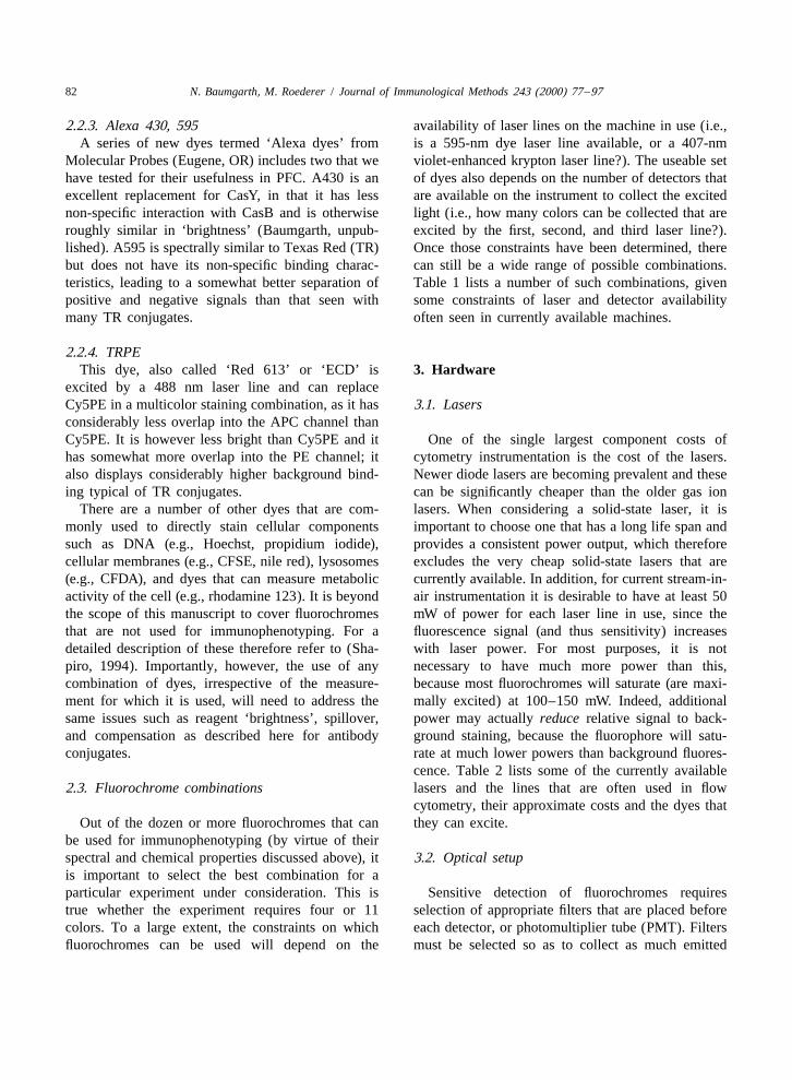
2.2.3. Alexa 430, 595 availability of laser lines on the machine in use (i.e.,A series of new dyes termed ‘Alexa dyes’ from is a 595-nm dye laser line available, or a 407-nm
Molecular Probes (Eugene, OR) includes two that we violet-enhanced krypton laser line?). The useable sethave tested for their usefulness in PFC. A430 is an of dyes also depends on the number of detectors thatexcellent replacement for CasY, in that it has less are available on the instrument to collect the excitednon-specific interaction with CasB and is otherwise light (i.e., how many colors can be collected that areroughly similar in ‘brightness’ (Baumgarth, unpub- excited by the first, second, and third laser line?).lished). A595 is spectrally similar to Texas Red (TR) Once those constraints have been determined, therebut does not have its non-specific binding charac- can still be a wide range of possible combinations.teristics, leading to a somewhat better separation of Table 1 lists a number of such combinations, givenpositive and negative signals than that seen with some constraints of laser and detector availabilitymany TR conjugates. often seen in currently available machines.
2.2.4. TRPEThis dye, also called ‘Red 613’ or ‘ECD’ is 3. Hardware
excited by a 488 nm laser line and can replaceCy5PE in a multicolor staining combination, as it has 3.1. Lasersconsiderably less overlap into the APC channel thanCy5PE. It is however less bright than Cy5PE and it One of the single largest component costs ofhas somewhat more overlap into the PE channel; it cytometry instrumentation is the cost of the lasers.also displays considerably higher background bind- Newer diode lasers are becoming prevalent and theseing typical of TR conjugates. can be significantly cheaper than the older gas ion
There are a number of other dyes that are com- lasers. When considering a solid-state laser, it ismonly used to directly stain cellular components important to choose one that has a long life span andsuch as DNA (e.g., Hoechst, propidium iodide), provides a consistent power output, which thereforecellular membranes (e.g., CFSE, nile red), lysosomes excludes the very cheap solid-state lasers that are(e.g., CFDA), and dyes that can measure metabolic currently available. In addition, for current stream-in-activity of the cell (e.g., rhodamine 123). It is beyond air instrumentation it is desirable to have at least 50the scope of this manuscript to cover fluorochromes mW of power for each laser line in use, since thethat are not used for immunophenotyping. For a fluorescence signal (and thus sensitivity) increasesdetailed description of these therefore refer to (Sha- with laser power. For most purposes, it is notpiro, 1994). Importantly, however, the use of any necessary to have much more power than this,combination of dyes, irrespective of the measure- because most fluorochromes will saturate (are maxi-ment for which it is used, will need to address the mally excited) at 100–150 mW. Indeed, additionalsame issues such as reagent ‘brightness’, spillover, power may actually reduce relative signal to back-and compensation as described here for antibody ground staining, because the fluorophore will satu-conjugates. rate at much lower powers than background fluores-
cence. Table 2 lists some of the currently available2.3. Fluorochrome combinations lasers and the lines that are often used in flow
cytometry, their approximate costs and the dyes thatOut of the dozen or more fluorochromes that can they can excite.
be used for immunophenotyping (by virtue of theirspectral and chemical properties discussed above), it 3.2. Optical setupis important to select the best combination for aparticular experiment under consideration. This is Sensitive detection of fluorochromes requirestrue whether the experiment requires four or 11 selection of appropriate filters that are placed beforecolors. To a large extent, the constraints on which each detector, or photomultiplier tube (PMT). Filtersfluorochromes can be used will depend on the must be selected so as to collect as much emitted
N. Baumgarth, M. Roederer / Journal of Immunological Methods 243 (2000) 77 –97 83
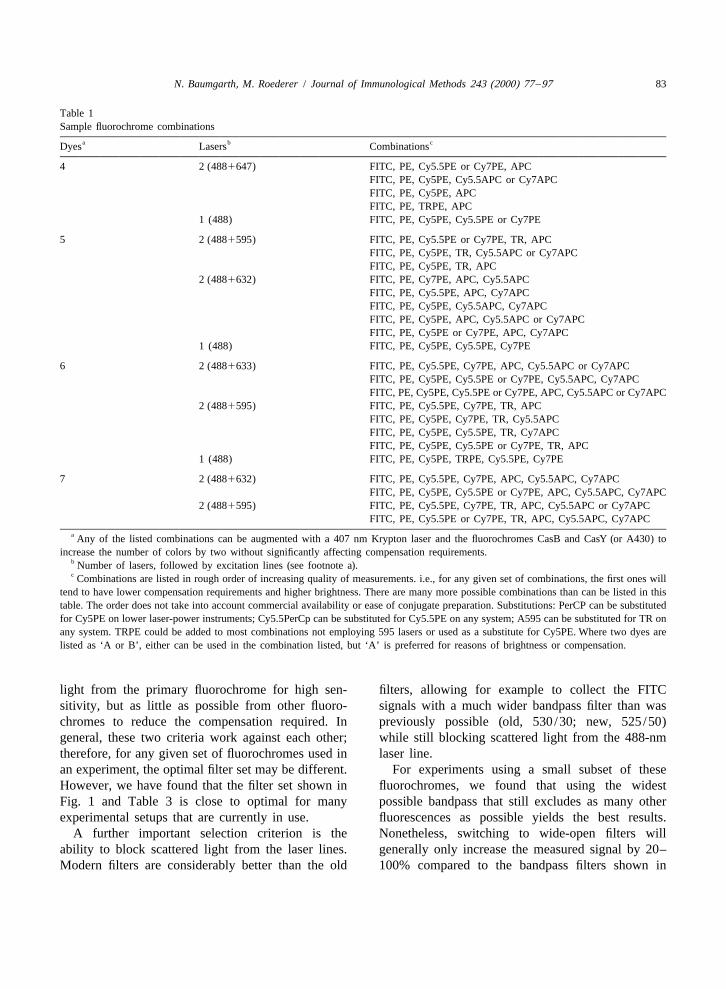
Table 1Sample fluorochrome combinations
a b cDyes Lasers Combinations
4 2 (4881647) FITC, PE, Cy5.5PE or Cy7PE, APCFITC, PE, Cy5PE, Cy5.5APC or Cy7APCFITC, PE, Cy5PE, APCFITC, PE, TRPE, APC
1 (488) FITC, PE, Cy5PE, Cy5.5PE or Cy7PE
5 2 (4881595) FITC, PE, Cy5.5PE or Cy7PE, TR, APCFITC, PE, Cy5PE, TR, Cy5.5APC or Cy7APCFITC, PE, Cy5PE, TR, APC
2 (4881632) FITC, PE, Cy7PE, APC, Cy5.5APCFITC, PE, Cy5.5PE, APC, Cy7APCFITC, PE, Cy5PE, Cy5.5APC, Cy7APCFITC, PE, Cy5PE, APC, Cy5.5APC or Cy7APCFITC, PE, Cy5PE or Cy7PE, APC, Cy7APC
1 (488) FITC, PE, Cy5PE, Cy5.5PE, Cy7PE
6 2 (4881633) FITC, PE, Cy5.5PE, Cy7PE, APC, Cy5.5APC or Cy7APCFITC, PE, Cy5PE, Cy5.5PE or Cy7PE, Cy5.5APC, Cy7APCFITC, PE, Cy5PE, Cy5.5PE or Cy7PE, APC, Cy5.5APC or Cy7APC
2 (4881595) FITC, PE, Cy5.5PE, Cy7PE, TR, APCFITC, PE, Cy5PE, Cy7PE, TR, Cy5.5APCFITC, PE, Cy5PE, Cy5.5PE, TR, Cy7APCFITC, PE, Cy5PE, Cy5.5PE or Cy7PE, TR, APC
1 (488) FITC, PE, Cy5PE, TRPE, Cy5.5PE, Cy7PE
7 2 (4881632) FITC, PE, Cy5.5PE, Cy7PE, APC, Cy5.5APC, Cy7APCFITC, PE, Cy5PE, Cy5.5PE or Cy7PE, APC, Cy5.5APC, Cy7APC
2 (4881595) FITC, PE, Cy5.5PE, Cy7PE, TR, APC, Cy5.5APC or Cy7APCFITC, PE, Cy5.5PE or Cy7PE, TR, APC, Cy5.5APC, Cy7APC
a Any of the listed combinations can be augmented with a 407 nm Krypton laser and the fluorochromes CasB and CasY (or A430) toincrease the number of colors by two without significantly affecting compensation requirements.
b Number of lasers, followed by excitation lines (see footnote a).c Combinations are listed in rough order of increasing quality of measurements. i.e., for any given set of combinations, the first ones will
tend to have lower compensation requirements and higher brightness. There are many more possible combinations than can be listed in thistable. The order does not take into account commercial availability or ease of conjugate preparation. Substitutions: PerCP can be substitutedfor Cy5PE on lower laser-power instruments; Cy5.5PerCp can be substituted for Cy5.5PE on any system; A595 can be substituted for TR onany system. TRPE could be added to most combinations not employing 595 lasers or used as a substitute for Cy5PE. Where two dyes arelisted as ‘A or B’, either can be used in the combination listed, but ‘A’ is preferred for reasons of brightness or compensation.
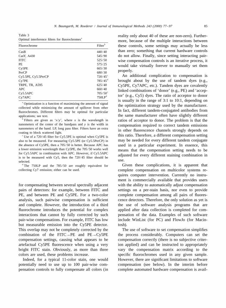
light from the primary fluorochrome for high sen- filters, allowing for example to collect the FITCsitivity, but as little as possible from other fluoro- signals with a much wider bandpass filter than waschromes to reduce the compensation required. In previously possible (old, 530/30; new, 525/50)general, these two criteria work against each other; while still blocking scattered light from the 488-nmtherefore, for any given set of fluorochromes used in laser line.an experiment, the optimal filter set may be different. For experiments using a small subset of theseHowever, we have found that the filter set shown in fluorochromes, we found that using the widestFig. 1 and Table 3 is close to optimal for many possible bandpass that still excludes as many otherexperimental setups that are currently in use. fluorescences as possible yields the best results.
A further important selection criterion is the Nonetheless, switching to wide-open filters willability to block scattered light from the laser lines. generally only increase the measured signal by 20–Modern filters are considerably better than the old 100% compared to the bandpass filters shown in
84 N. Baumgarth, M. Roederer / Journal of Immunological Methods 243 (2000) 77 –97
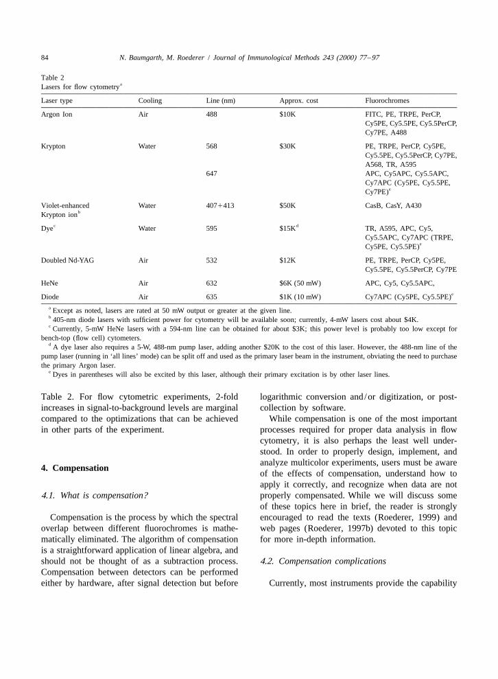
Table 2aLasers for flow cytometry
Laser type Cooling Line (nm) Approx. cost Fluorochromes
Argon Ion Air 488 $10K FITC, PE, TRPE, PerCP,Cy5PE, Cy5.5PE, Cy5.5PerCP,Cy7PE, A488
Krypton Water 568 $30K PE, TRPE, PerCP, Cy5PE,Cy5.5PE, Cy5.5PerCP, Cy7PE,A568, TR, A595
647 APC, Cy5APC, Cy5.5APC,Cy7APC (Cy5PE, Cy5.5PE,
eCy7PE)
Violet-enhanced Water 4071413 $50K CasB, CasY, A430bKrypton ion
c dDye Water 595 $15K TR, A595, APC, Cy5,Cy5.5APC, Cy7APC (TRPE,
eCy5PE, Cy5.5PE)
Doubled Nd-YAG Air 532 $12K PE, TRPE, PerCP, Cy5PE,Cy5.5PE, Cy5.5PerCP, Cy7PE
HeNe Air 632 $6K (50 mW) APC, Cy5, Cy5.5APC,eDiode Air 635 $1K (10 mW) Cy7APC (Cy5PE, Cy5.5PE)
a Except as noted, lasers are rated at 50 mW output or greater at the given line.b 405-nm diode lasers with sufficient power for cytometry will be available soon; currently, 4-mW lasers cost about $4K.c Currently, 5-mW HeNe lasers with a 594-nm line can be obtained for about $3K; this power level is probably too low except for
bench-top (flow cell) cytometers.d A dye laser also requires a 5-W, 488-nm pump laser, adding another $20K to the cost of this laser. However, the 488-nm line of the
pump laser (running in ‘all lines’ mode) can be split off and used as the primary laser beam in the instrument, obviating the need to purchasethe primary Argon laser.
e Dyes in parentheses will also be excited by this laser, although their primary excitation is by other laser lines.
Table 2. For flow cytometric experiments, 2-fold logarithmic conversion and/or digitization, or post-increases in signal-to-background levels are marginal collection by software.compared to the optimizations that can be achieved While compensation is one of the most importantin other parts of the experiment. processes required for proper data analysis in flow
cytometry, it is also perhaps the least well under-stood. In order to properly design, implement, andanalyze multicolor experiments, users must be aware
4. Compensationof the effects of compensation, understand how toapply it correctly, and recognize when data are not
4.1. What is compensation? properly compensated. While we will discuss someof these topics here in brief, the reader is strongly
Compensation is the process by which the spectral encouraged to read the texts (Roederer, 1999) andoverlap between different fluorochromes is mathe- web pages (Roederer, 1997b) devoted to this topicmatically eliminated. The algorithm of compensation for more in-depth information.is a straightforward application of linear algebra, andshould not be thought of as a subtraction process. 4.2. Compensation complicationsCompensation between detectors can be performedeither by hardware, after signal detection but before Currently, most instruments provide the capability
N. Baumgarth, M. Roederer / Journal of Immunological Methods 243 (2000) 77 –97 85
Table 3 reality only about 40 of these are non-zero). Further-aOptimal interference filters for fluorochromes more, because of the multiple interactions between
bFluorochrome Filter these controls, some settings may actually be lessthan zero; something that current hardware controlsCasB 440/40
CasY, A430 545/90 do not allow. Finally, since setting interacting pair-FITC 525/50 wise compensation controls is an iterative process, itPE 575/25 would take virtually forever to manually set themCy5PE 665/30
properly.PerCP 680/30c An additional complication to compensation isCy5.5PE, Cy5.5PerCP 720/45dCy7PE 785/45 brought about by the use of tandem dyes (e.g.,
TRPE, TR, A595 625/40 Cy5PE, Cy7APC, etc.). Tandem dyes are covalentlyAPC 660/40 linked combinations of ‘donor’ (e.g., PE) and ‘accep-cCy5.5APC 705/50
d tor’ (e.g., Cy5) dyes. The ratio of acceptor to donorCy7APC 750LPis usually in the range of 3:1 to 10:1, depending on
a Optimization is a function of maximizing the amount of signal the optimization strategy used by the manufacturer.collected while minimizing the amount of spillover from other
In fact, different tandem-conjugated antibodies fromfluorochromes. Different filters may be optimal for particularthe same manufacturer often have slightly differentapplications; see text.
b Filters are given as ‘x /y’, where x is the wavelength in ratios of acceptor to donor. The problem is that thenanometers of the center of the bandpass and y is the width in compensation required to correct tandem emissionsnanometers of the band. LP, long pass filter. Filters have an extra in other fluorescence channels strongly depends oncoating to block scattered light.
c this ratio. Therefore, a different compensation settingUse of a 720/45 filter for Cy5.5PE is optimal when Cy5PE ismay be needed for every different tandem conjugatealso to be measured. For measuring Cy5.5PE (or Cy5.5PerCP) in
the absence of Cy5PE, then a 705/50 is better. Because APC has used in a particular experiment. In essence, thisa lower emission wavelength than Cy5PE, the 705/50 works well means that the compensation setting needs to befor Cy5.5APC in combination with APC. However, if Cy5.5APC adjusted for every different staining combination inis to be measured with Cy5, then the 720/45 filter should be
use.selected.d Given these complications, it is apparent thatThe 750LP and the 785/50 are roughly equivalent for
collecting Cy7 emission; either can be used. complete compensation on multicolor systems re-quires computer intervention. Currently no instru-ment is commercially available that provides users
for compensating between several spectrally adjacent with the ability to automatically adjust compensationpairs of detectors: for example, between FITC and settings on a per-stain basis, nor even to providePE, and between PE and Cy5PE. For a two-color complete compensation among all pairs of fluores-analysis, such pairwise compensation is sufficient cence detectors. Therefore, the only solution as yet isand complete. However, the introduction of a third the use of software analysis programs that arefluorochrome introduces the potential for complex applied after data collection is completed for com-interactions that cannot by fully corrected by such pensation of the data. Examples of such softwarepair-wise compensations. For example, FITC has low include WinList (for PC) and FlowJo (for Macin-but measurable emission into the Cy5PE detector. tosh).This overlap may not be completely corrected by the The use of software to set compensation simplifiescombination of the FITC→PE and PE→Cy5PE the process considerably. Computers can set thecompensation settings, causing what appears to be compensation correctly (there is no subjective criter-artefactual Cy5PE fluorescence when using a very ion applied) and can be instructed to appropriatelybright FITC stain. Obviously, as more than three vary the compensation matrix according to thecolors are used, these problems increase. specific fluorochromes used in any given sample.
Indeed, for a typical 11-color stain, one would However, there are significant limitations to softwarepotentially need to use up to 100 pair-wise com- compensation (see below). In the interim beforepensation controls to fully compensate all colors (in complete automated hardware compensation is avail-

86 N. Baumgarth, M. Roederer / Journal of Immunological Methods 243 (2000) 77 –97
able, the optimal solution is to combine both hard- Examples for significant inter-laser compensationsware and software compensation to minimize the are shown in Table 4. Potential signal degradationlimitations of each. due to inter-laser compensation can occur with dye
combinations that are found at high levels on the4.3. Limitations of compensation same cell.
The principal limitation of software compensationBoth available hardware and software compensa- is caused by the inaccurate conversion from linear to
tion have significant limitations that place restrictions logarithmic signals by the electronics of the flowon the utility of PFC. Hopefully, some of these cytometer. Since this conversion is typically per-limitations will soon be obviated by new technology formed by analog circuitry, it is merely an approxi-developments, which would enable the easier set-up mate conversion. The software, however, treats theof new reagent combinations for multicolor flow data values as if they were an exact conversion,cytometric measurements. Until then, it is important which can result in compensated values that areto understand these limitations and how they may be significantly inaccurate. The major inaccuracy in thebest addressed. electronic linear to log conversion is due to the fact
One limitation that is shared by both hardware and that the dynamic range of the output is not exactlysoftware compensation is due to the low precision of four decades, as the software (and the user) is told tomeasurements in the far-red channels, Cy7APC, expect, but can range from 3.5 to 4.3 decades. TheCy5.5APC, and to a certain extent APC. In these effects of this error are insidious, in that data can bechannels, for dimly stained cells, the actual number properly compensated at a particular intensity, under-of photoelectrons detected by the PMT can be verylow, due to the inefficiency of the currently availablePMT to detect light at this wavelength. Even the
Table 4a‘deep red-sensitive’ PMTs now available do not Significant intralaser compensations
overcome this problem. The unavoidable countingFluorochrome Detector
errors inherent in the measurement can lead to theTR APC Cy5.5APC Cy7APCintroduction of significant artefacts. For example, if
only five photoelectrons are collected, the counting First→second laser compensation]Œ PE 1.4 0.68 0.17error is 5, or 45%. This error propagates to other
Cy5PE 0.32 9.0 6.8 1.0channels that require compensation from this detec-Cy5.5PE 0.11 1.3 13.3 2.8
tor. The result is that the ‘broadening’ of populations Cy7PE 1.1that occurs due to compensation (Roederer, 1997b,
b1999) is significantly worse for the far-red emitting Second→first laser compensationCy5PE Cy5.5PE Cy7PEfluorochromes.
Hardware compensation operates on the analog TR 2.2 1.1APC 4.6 1.3 0.38signals collected from the PMT, before further signalCy5.5APC 0.91 5.1 2.9processing is done. For detectors with sufficientCy7APC 1.4 0.9 14signal, this value is both precise and accurate,
a Significant spillovers are those above 0.1%. Values are givenrendering compensation close to exact. Problemsas the percentage of the signal level in the primary detector for aarise when compensation is required between signalsgiven fluorochrome that is found in the detector listed in each
propagated from different lasers, since the signals column. Only interlaser spillovers are shown; intralaser spilloversmust be electronically delayed so that they can be are significantly higher but can be more easily compensated bypresented to the analog compensation circuitry at the hardware. Values are given for a 488-nm first laser and a 595-nm
second laser. Different laser lines can give qualitatively similar butsame time. This electronic delay is difficult toquantitatively different results. Spillover values are system depen-perform precisely, without changing the shape of thedent and determined by optics, laser power, and gain of detector
electronic pulse generated by the PMT. Therefore, (PMT type, voltage, amplifier) on each channel pair.bhardware-based interlaser compensation is typically There are no significant spillovers into the FITC or PE
much less accurate than intra-laser compensation. channels from these dyes.
N. Baumgarth, M. Roederer / Journal of Immunological Methods 243 (2000) 77 –97 87
compensated at lower (or higher) intensities, and excessive concentrations of the conjugated antibodyovercompensated at higher (or lower) intensities. The are used, or when conjugates are used that wereless compensation that needs to be done by the prepared with a non-optimized ratio of fluorochromesoftware the smaller the extent of this problem is. to antibody. Titration of reagents and optimization ofThis again points to the importance of appropriate reagent conjugations are therefore particularly im-reagent /color choices, as described below. portant when attempting to scale up a one- or two-
Given the respective limitations of hardware and color stain to a stain that contains three or moresoftware compensation, our approach has been to use colors. Some reagents, such as Cy5PE, also exhibit ahardware compensation for as many pairs of signals certain degree of ‘stickiness’; in the case of Cy5PEas possible (Bigos et al., 1999). Currently we use 14 this is seen particularly with B cells. Finally, thehardware controls, dedicated to signals that require amount of cellular autofluorescence varies signifi-the largest amount of intralaser compensation. Soft- cantly in different areas of the spectrum. In par-ware compensation is used following data acquisition ticular, autofluorescence overlaps greatly with FITC,to complete the compensation process. It is critical to CasB, and CasY. Therefore, highly autofluorescentset the pair-wise settings conservatively, i.e., to cells such as macrophages might appear as ‘dullunder-compensate considerably where necessary. positive’ in these detectors, despite the fact that theyThere are two reasons for this: inadvertent over- might not stain specifically with any of the reagentscompensation cannot be corrected in software and used.thus causes irretrievable data error; and, given the The choice of fluorochrome/conjugate used formultiple interactions, proper compensation between identifying a given marker will depend on the levelsone pair of fluorochromes can lead to overcompensa- of expression and the cell types on which the markertion of a different pair. For example, proper com- is expressed. If this information is not at hand, it ispensation of TR into APC often results in (unrecov- advisable to use one of the ‘bright’ fluorochromeserable) overcompensation of Cy5PE into APC. such as PE, Cy5PE or APC, and use the duller stains
for identification of surface markers that are known,such as the lineage markers for lymphocyte subset
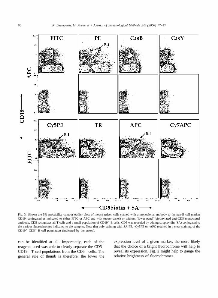
5. Choosing the right fluorochrome staining. As an example, we show in Fig. 3 variousdual-color staining combinations of splenic mouse
As outlined above, fluorochromes differ considera- cells for CD19, a lineage marker for B cells, andbly in relative ‘brightness’. Conjugation of the CD5. CD5 is expressed at high levels on all T cellsvarious fluorochromes to the same antibody and and at low levels on a small subset of B cells in the
1staining of the same population of cells can therefore spleen (Kantor and Herzenberg, 1993). CD5 Bresult in large differences in resolution of positive cells express somewhat higher levels of CD19. Asand negative events (Fig. 2). For single-color stain- apparent from the figure, resolution of the dully
1ing for a highly expressed marker such as CD8 on T stained CD5 B cells is achieved only when thecells (Fig. 2), the differences are merely cosmetic biotinylated CD5 antibody is revealed with strep-and no alterations in the frequency of negative and tavidin (SA) conjugated to PE, Cy5PE or APC (seepositive events are found after staining with different also Kantor and Roederer, 1997). Staining with otherconjugates. However, when staining for surface SA-fluorochrome conjugates does not reveal a dis-
1markers that exhibit high and low levels of expres- tinct CD5 B cell population. It is also useful to notesion, for example the expression of CD28 or FAS on that staining with SA-CasB resulted in an apparentT cells, a relatively ‘dull’ fluorochrome can result in staining of a small population of CD19-dull cells.an underestimation of the size of the positive popula- However, this ‘staining’ is in fact due to the auto-tion (false-negatives). fluorescence of macrophages which appears as a dull
False-positive results are seen particularly with positive stain in many channels (data not shown).dyes and conjugates that have high ‘background Hence, simply the choice of fluorochrome usedstaining’. High background staining can occur when will determine whether this small B cell population
88 N. Baumgarth, M. Roederer / Journal of Immunological Methods 243 (2000) 77 –97
Fig. 3. Shown are 5% probability contour outlier plots of mouse spleen cells stained with a monoclonal antibody to the pan-B cell markerCD19, conjugated as indicated to either FITC or APC and with (upper panel) or without (lower panel) biotinylated anti-CD5 monoclonal
1antibody. CD5 recognizes all T cells and a small population of CD19 B cells. CD5 was revealed by adding streptavidin (SA) conjugated tothe various fluorochromes indicated to the samples. Note that only staining with SA-PE, -Cy5PE or -APC resulted in a clear staining of the
1 1CD19 CD5 B cell population (indicated by the arrow).
expression level of a given marker, the more likelycan be identified at all. Importantly, each of the1 that the choice of a bright fluorochrome will help toreagents used was able to clearly separate the CD5
2 2 reveal its expression. Fig. 2 might help to gauge theCD19 T cell populations from the CD5 cells. Therelative brightness of fluorochromes.general rule of thumb is therefore: the lower the
N. Baumgarth, M. Roederer / Journal of Immunological Methods 243 (2000) 77 –97 89
6. Choosing the right combinations of chosen for the other conjugates that do not stain thefluorochromes cells identified by the APC stain.
The other crucial lesson from this experiment isWhat are the criteria for choosing the right that isotype controls are only appropriate when used
combination of fluorochromes? As outlined above, in the context of every other antibody stain. That is,one major criterion is the expected levels of expres- a sample that was stained with isotype controls on allsion for the markers of interest. Markers such as channels would show a background of only 0.2 onCD5 on B cells can only be identified when a bright the APC channel, considerably less than the back-conjugate is used; but identifying CD5 on T cells can ground observed on this channel given a full stainingbe accomplished with any fluorochrome. An un- set minus the APC stain (4.1). This is why falseknown surface molecule is initially best studied with positives will be identified without the use of appro-a conjugate that uses one of the ‘brightest’ fluoro- priate controls.chromes, PE, Cy5PE, or APC. Other considerations In the example shown in Fig. 3, i.e., measuring the
1include the spectral overlaps that exist between the low levels of CD5 on CD19 cells, one shouldvarious dyes in the staining combinations. Although choose to label CD19 (well-expressed on all B cells)hardware or software compensation can eliminate with a color that does not have a large compensationsome of the associated problems, the careful choice component with the fluorochrome used for CD5. Onof reagents can minimize the need for compensation. the other hand, CD8 (part of the ‘dump’ channel)
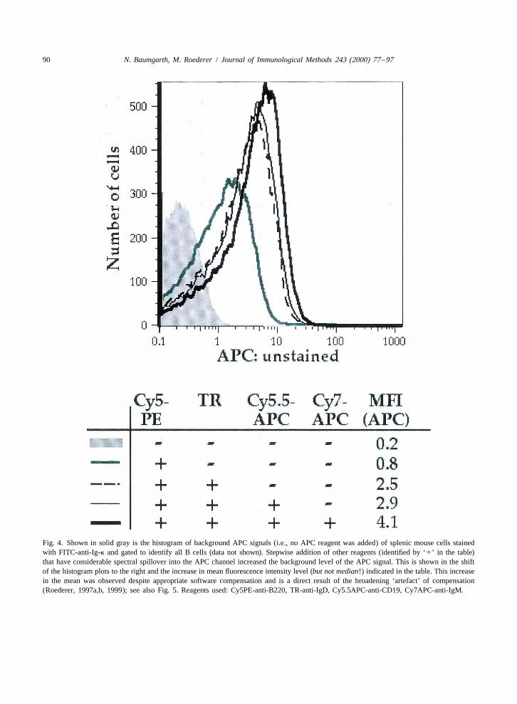
Fig. 4 shows an example of how the APC signal is could be assigned to such a color even though CD8affected by using one to four very brightly staining is extremely highly expressed. This is because CD8
1markers that all have significant spillover into the is not expressed on the cells of interest (CD19this channel. This figure therefore shows an example cells) and thus compensating CD8 fluorescenceof how best not to design an experiment. In this would have no impact on the quantitation of CD5 onexample we chose to add antibodies to B cell B cells.markers conjugated to Cy5PE, TR, Cy5.5APC and The dyes excited by the violet-enhanced kryptonCy7APC to the staining mix and then looked at the laser are particularly useful for multicolor stainingbackground APC levels. Remember that APC is one combinations. Although they appear rather ‘dull’of the dyes that are in general ‘bright’ (see Figs. 2 mainly due to the large autofluorescence backgroundand 3) and is therefore a good choice for identifying in these channels (Fig. 2), they have minimal overlapdimly expressed markers such as CD5 on B cells. As with any of the other fluorochromes and are easilywe add staining reagents to the cells, the background compensated against each other. Therefore anylevels of the APC signal increase significantly, from combination with a 407-nm line-excited dye shoulda mean fluorescence intensity of 0.2 to 4.1 (Fig. 4). work without problems, as long as the marker chosenThis increase in mean fluorescence intensity at the for the CasB or CasY conjugation is expressed atAPC detector is observed despite the fact that sufficiently high levels to separate the stained fromsoftware compensation was applied to the samples the unstained cell population. In many cases, signifi-and each compensation pair seemed to have been cant compensation can also be avoided by using dyescompensated appropriately (not shown; see Fig. 5 for excited by different lasers (however, see Table 4 forexplanation). Therefore, after addition of the Cy5PE, dyes that do require significant interlaser compensa-TR, Cy5.5APC and Cy7APC stains, a dim positive tion).staining with a reagent conjugated to APC could no In summary, the appropriate choice of reagentslonger be distinguished from the increased back- and reagent combinations requires certain knowledgeground staining; the effective ‘brightness’ of APC is of the characteristics of the individual fluorochromes,reduced 20-fold. Since a number of colors that are such as ‘brightness’ and ‘spillover’ into other chan-currently used for PFC show large overlaps into the nels, to avoid the use of inappropriate or uselessAPC detector, the usefulness of this detector is staining combinations. Typically, multiple differentsubstantially compromised unless reagents are combinations of fluorochrome/antibody pairings
90 N. Baumgarth, M. Roederer / Journal of Immunological Methods 243 (2000) 77 –97
Fig. 4. Shown in solid gray is the histogram of background APC signals (i.e., no APC reagent was added) of splenic mouse cells stainedwith FITC-anti-Ig-k and gated to identify all B cells (data not shown). Stepwise addition of other reagents (identified by ‘1’ in the table)that have considerable spectral spillover into the APC channel increased the background level of the APC signal. This is shown in the shiftof the histogram plots to the right and the increase in mean fluorescence intensity level (but not median!) indicated in the table. This increasein the mean was observed despite appropriate software compensation and is a direct result of the broadening ‘artefact’ of compensation(Roederer, 1997a,b, 1999); see also Fig. 5. Reagents used: Cy5PE-anti-B220, TR-anti-IgD, Cy5.5APC-anti-CD19, Cy7APC-anti-IgM.

N. Baumgarth, M. Roederer / Journal of Immunological Methods 243 (2000) 77 –97 91
Fig. 5. The effect of random noise added to an autofluorescence distribution. The process of fluorescence compensation introduces additionalerror into the distribution of measured fluorescence. Histograms in this figure represent a mathematical modeling of this process. (A) A typicaldistribution of cellular autofluorescence. The fluorescence distribution is log-normal, i.e., it is a Gaussian when viewed on a logarithmic axis.The distribution is shown only for the first 400 (of 1024) channels of a typical measurement parameter; this represents the first 1.5 decades offluorescence. (B) The distribution in (A), after instrumentation boundary conditions are imposed. Since there is no ‘zero’ on a logarithmic axis,the low end of the fluorescence scale is arbitrary. Any events with fluorescence below the lowest value (0.1 here) are automatically assigned tochannel zero and given a fluorescence value of 0.1. This results in an artificial peak at channel zero, the sum of all events with fluorescence lessthan this (shown in white bars). This same artefact occurs in two-dimensional histograms used for contour plots or density plots, causing theappearance of an artificial ‘peak’ (multiple contour lines) on the axis. This peak is an artefact of the measurement and visualization processes,and importantly, it does not represent a population of events distinct from those adjacent to it. (C) Effects of compensation. To model the errorintroduced by compensation, a random amount of fluorescence (averaging 060.4 fluorescence units) was added to each event in the distributionshown in (B). The fluorescence of about 25% of all events is now shifted to below 0.1 units (open bins), consequently these events are forcedinto the single point at channel zero. Note the scale change of the vertical axis. The reason for the asymmetric effect on the distribution (i.e., theright edge of the histogram increased very little, but the left tail increased greatly, and the mode of the distribution has increased) is aconsequence of the logarithmic scaling: a noise distribution that is linearly distributed was added to the log-normal distribution. Thus, as shownby the arrows, adding 0.4 units of fluorescence to an event at 0.45 (to 0.85) moves it much less ‘distance’ (in the log domain) than subtractingthe equivalent amount (to 0.05). Compensation introduces errors that are symmetrically distributed in the linear domain. Thus, one of theeffects of compensation is to alter the autofluorescence distribution from log-normal to that shown in (C). It is important to note that the medianfluorescence intensity in (C) is exactly the same as it is in (B), but the mean fluorescence has increased, and the mode (above channel 0) hasincreased even more. Because of our internal bias to use the mode (peak) to estimate a population’s center rather than the median or mean,which is virtually impossible to estimate visually, the distribution appears to have moved substantially. Finally, note that placement of a‘positive’ gate at, for example, 1 fluorescence unit would be appropriate for the ‘uncompensated’ parameter (B), but not for the compensatedparameter (C), even though both represent unstained distributions.

92 N. Baumgarth, M. Roederer / Journal of Immunological Methods 243 (2000) 77 –97
must be evaluated in order to select the panel that of CD5 on a small B cell subpopulation in the spleenprovides the most information. (Fig. 3), it would be difficult to accurately determine
the frequency of these cells using this two-colorstain. In Fig. 6 we outline how the simultaneous use
7. Enhanced accuracy of measurement with of four (Fig. 6B) and six (Fig. 6C) colors enhancesmulticolor flow cytometry the accuracy of the measurement and the amount of
information that can be obtained from one stain (Fig.Despite the relatively good resolution that the use 6A). In this figure we compare the staining pattern of
of SA-PE, Cy5PE and APC afforded for the staining spleen cells isolated from wild-type mice and gene
Fig. 6. Shown are 5% probability contour outlier plots of (A) two-, (B) four- and (C) six-color stains of single cell suspensions preparedfrom mouse spleens derived from normal wild-type C57BL/6 mice and mice deficient in the signaling molecule vav (vav2 / 2). (A) CD19
1 2 1 1and CD5 staining of splenic B cells shows a reduction in the number of CD5 CD19 T cells and CD19 CD5 B cells. (B) Addition of adump channel (reagents to CD3, CD4, CD8 and F4/80) and a second marker (CD43) expressed on mouse ‘B-1’ cells identifies the lack of
1B-1 cells among the overall normal frequency of CD19 B cells in vav2 / 2 mice compared to their wild-type controls. (C) Addition ofanti-IgM, anti-IgD and anti-CD21 in the staining cocktail reveals alterations in IgM and IgD expression of follicular B cells and the
hi neg lo negappearance of normal frequencies of marginal zone (CD21 CD43 ) and immature (CD21 CD43 ) B cells and the absence of B-1 cellslo 1 hi lo(CD21 CD43 ) among IgM IgD cells.
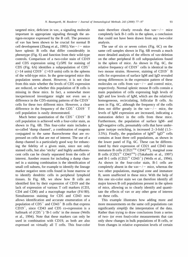
N. Baumgarth, M. Roederer / Journal of Immunological Methods 243 (2000) 77 –97 93
targeted mice, deficient in vav, a signaling molecule stain therefore clearly reveals that vav2 /2 miceimportant in appropriate signaling through the an- completely lack B-1 cells in the spleen, a conclusiontigen-receptor expressed by the B cell. The presence that could not have been drawn from any two-colorof vav has been shown to be crucial for normal B analysis.cell development (Zhang et al., 1995).Vav2 /2 mice The use of six or seven colors (Fig. 6C) on thehave splenic B cells that differ considerably in same cell samples shown in Fig. 6B reveals a muchphenotype (Fig. 6) and function from their wild-type more detailed analysis of the effects of vav-deletioncontrols. Comparison of a two-color stain of CD19 on the other peripheral B cell subpopulations foundand CD5 expression using Cy5PE for staining of in the spleen of mice. As shown in Fig. 6C, the
1 1CD5 (Fig. 6A) identifies a subpopulation of CD5 relative frequency of CD19 cells is identical in the1 1(.14 units) CD19 (.0.9 units) cells in the spleens two mouse strains. However, staining of CD19 B
of the wild-type mice. In the gene-targeted mice this cells for expression of surface IgM and IgD revealedpopulation seems absent. However, it is not clear strong differences in the expression pattern of thesefrom this stain whether the levels of CD5 expression molecules on cells from vav2 /2 and control mice,are reduced, or whether this population of B cells is respectively. Normal splenic mouse B cells contain amissing in these mice. In fact, a somewhat more main population of cells expressing high levels ofinexperienced investigator might not even see a IgD and low levels of IgM, which are the relatively
1difference in the CD5-staining patterns of the CD19 homogeneous, recirculating, follicular B cells. Ascells for these two different mice. However, a clear seen in Fig. 6C, although the frequency of the cells
1 2difference in the frequency of the CD5 CD19 T does not differ greatly in the vav2 /2 mice, thecell population is easily demonstrated. levels of IgM expression are increased, indicating a
1 1Much better quantitation of the CD5 CD19 B maturation defect in the cells from these mice.cell population is achieved with a four-color stain, as Furthermore, the population of surface IgM- andshown in Fig. 6B. This four-color stain includes a IgD-negative cells, presumably cells that have under-so-called ‘dump channel’, a combination of reagents gone isotype switching, is increased 2–3-fold (1.5–
hi loconjugated to the same fluorochrome that are ex- 3.5%). Finally, the population of IgM IgD cellspressed on cells that are not of interest. The use of a contains at least three cell populations, as shown indump channel is a particularly good way for enhanc- the lower panel of Fig. 6C. These can be differen-ing the fidelity of a given stain, since not only tiated by their expression of CD21 and CD43 into
neg negstained cells, but also ‘sticky’ and highly autofluores- immature B cells (CD21 CD43 ), marginal zonehi negcent cells can be clearly separated from the cells of B cells (CD21 CD43 ) (Takahashi et al., 1997)
lo 1interest. Another reason for including a dump chan- and B-1 cells (CD21 CD43 ) (Wells et al., 1994).nel in a staining combination is the identification of As shown in the four-color stain, B-1 cells aresmall cell subsets, for example to identify the lineage completely absent in the vav2 /2 mice, whereas themarker negative stem cells found in bone marrow or two other populations, marginal zone and immatureto identify dendritic cells in peripheral lymphoid B, seem unaffected in these mice. With the help oftissues. In Fig. 6B, we show how B cells are this one six-color stain we can therefore identify allidentified first by their expression of CD19 and the major known B cell populations present in the spleenlack of expression of various T cell markers (CD3, of mice, allowing us to clearly identify and quanti-CD4 and CD8) and a macrophage marker (F4/80). tate the effects of vav or any other gene of interestSimultaneous staining for CD5 and CD43 now on these cells.allows identification and accurate enumeration of a This example illustrates how adding more and
1 1population of CD5 and CD43 B cells that express more measurements on the same cell populations can1CD19 , since CD43 and CD5 co-expression is a significantly simplify the interpretation of the data.
1hallmark of (CD5 ) ‘B-1 cells’ in the mouse (Wells Rather than trying to draw conclusions from a serieset al., 1994). Note that these markers can only be of two- (or even four)-color measurements that canused in combination with CD19, as both are also only show changes in bulk populations of B cells, orexpressed on virtually all T cells. This four-color from changes in relative expression levels of certain

94 N. Baumgarth, M. Roederer / Journal of Immunological Methods 243 (2000) 77 –97
markers on any of several indistinguishable cell tandem dyes that are prepared in the laboratory,types, the use of PFC allows us to pinpoint precisely stains that use tandem dyes from the same lot shouldthe defects in B cell development of these mice. suffice. Thus, careful inventory of conjugates in a
laboratory that includes the lot number of a tandemdye for each antibody conjugate can greatly reduce
8. Staining controls the number of compensation control samples foreach experiment.
Control stains are important for all experimentsusing flow cytometry, but they become particularly 8.2. Isotype or unstained controlscritical for complex multicolor stains. The higher thenumber of fluorochromes and antibodies used in each The use of isotype control antibodies is usually ofstain the greater the risk for artifacts introduced by little value, since each antibody and antibody conju-compensation errors and/or reagent interactions. In gate has very different characteristics in terms ofgeneral, two types of controls should be included and background staining, ‘stickiness’, etc. If nonspecificdata collected with every experiment: compensation staining is suspected, the use of an ‘Fc-block’, suchcontrols and staining controls. as anti-Fc receptor (CD16/CD35) antibodies (for
It will become apparent from the discussion below staining mouse cells) might be useful. These anti-that the number of control tubes might often exceed bodies are usually applied for 10 or 15 min beforethe number of sample tubes in a given experiment. It the stains are added.is necessary to invest this significant effort in the Since completely unstained cells have very differ-controls in order to ensure that the valuable sample ent levels of background than cells that have beenthat is stained with so many different markers does stained with multiple reagents (see Fig. 4 and itsnot suffer from experimental artefacts, or become discussion), the best control for any given marker ofuninterpretable. interest in a multicolor staining combination is a
stain that contains all reagents but the one of interest.8.1. Compensation controls For example, a five-color combination control would
include one or more four-color combinations, eachFor each fluorochrome used in an experiment, one leaving out one of the reagents in the complete stain.
should include a ‘compensation control’, i.e., a single In this way, positive versus negative gating can becolor stain, for which data are collected. It is not more rigorously determined and applied. In someappropriate to skimp on the number of tubes col- cases, this gate may not be a one-dimensionallected by trying to design stains that can control for (histogram gate), but may require a sloping line inmultiple compensation settings in a single tube. two dimensions because of the ever-increasingIdeally, the reagent used for the compensation sam- broadening imposed by compensation as signalple should be the same as that used in the staining intensity increases.cocktail. For the non-tandem dyes (FITC, PE, TR,APC), however, a reagent that stains brightly on agood number of cells is sufficient and can be used 9. Advantages of PFCinstead of a reagent that might only stain a verysmall subpopulation of cells. In general the best Most of the advantages of increasing the numberresult is obtained by using the brightest possible of colors in flow cytometry experiments are readilyreagent as the compensation control. In contrast, and apparent. First, the information content increasesas outlined above, the tandem-dyes (Cy5PE, Cy7PE, geometrically with the number of parameters simul-Cy7APC, etc.) may exhibit great lot-to-lot variations. taneously analyzed. Second, and just as important,As lot-numbers of the fluorochromes are not usually information can be obtained from multicolor experi-supplied with the conjugate if bought from a com- ments that is not available in any other way. Formercial source, each different staining combination example, no combination of one-color stains canshould have its own compensation control. For accurately enumerate or be used to isolate
N. Baumgarth, M. Roederer / Journal of Immunological Methods 243 (2000) 77 –97 95
1 1CD3 CD4 CD82 T cells (excluding, for example, cytometry can result in the unambiguous identifica-1 1 1 1CD3 CD4 CD8 T cells and small CD4 mono- tion of rare cell subsets. For example, using eight-
cytes). Third, detailed immunophenotyping can be color PFC we were able to identify a small popula-combined with functional assays (DNA/Cell cycle, tion of follicular B cells in the mouse, with an as yetmetabolic activity, cytokine assays, apoptosis assays, unknown function, that expresses high levels of theetc.) to provide a distribution of activities across nonclassical MHC molecule CD1 (Amano et al.,highly defined cell subsets. Fourth, by staining for 1998).multiple markers in one cocktail, far fewer samples Other examples include the use of 10-color PFCneed to be prepared for each experiment. This is for the identification of antigen-specific lymphocytes.particularly important when analyzing precious sam- Multimers of soluble MHC-class I molecules loadedples (e.g., pediatric samples, leukocytes isolated from with a peptide of known origin are now often used to
1biopsies, rare antigen-specific lymphocytes, mouse identify antigen-specific CD8 T cells in humantissues that yield small numbers of cells). And fifth, blood or various tissues in the mouse (Altman et al.,less total reagents are used, since less duplication of 1996). As the number of antigen-specific T cells isthe same reagent among multiple tubes is required. often very small, even a small amount of contamina-
A number of technical developments have also tion can lead to considerable errors in the quantita-allowed the increasing use of flow cytometry for tion of these cells which makes further analysisextensive functional studies on cells. Increasing the inaccurate. We recently showed that the use of PFCnumber of simultaneously measured parameters is allows the identification and functional characteriza-particularly useful for these applications, since one tion of gd T cells specific for a nonclassical MHCcan combine detailed immunophenotyping with func- molecule T22. gd T cells constitute roughly 1% oftional studies. One example is the use of cytoplasmic the splenic cell population of which about 0.5%, orcytokine staining for the identification of cytokine 0.005% of all spleen cells, bind to the T22 tetramerprofiles among bulk populations of lymphocytes. By (Crowley et al., 1999). Using multicolor cytometrycombining antibodies against a number of cell we could show not only that those cells bind to thesurface markers together with antibodies specific for MHC multimer, but also that they alter their surfacecytokines, we recently demonstrated that populations expression of activation markers such as CD69 andof human peripheral blood T cells classified by their CD62L after binding the MHC multimer (Crowley etdifferential expression of various ‘activation al., 1999).markers’ differ in their relative frequencies of IL-4 In a separate study, we also used the MHCand IFN-g producers. Importantly, the relative fre- multimer approach to identify potentially tumor-reac-quencies of these phenotypically and functionally tive cytotoxic T cells in patients with metastaticdistinct T cell subpopulations in the blood differed melanoma (Lee et al., 1999). Because of the rarity of
25 23among people with various disease states (Mitra et these cells (10 to 10 of PBMC), standardal., 1999). In the future, the use of additional techniques are unable to derive much informationantibodies against chemokine- and homing-receptors about these cells. We were able to quantitate thewill likely lead to the identification of further T cell expression of nearly 40 different cell surface an-subsets. When combined with antibodies against a tigens (to accurately pinpoint the T cell subset tonumber of cytokines, this is likely to yield further which the antigen-specific T cells belong), as well asimportant and novel information regarding the rela- determine their cytokine profile, showing that thesetionship between T cell phenotype, function and the cells are functionally anergic (whereas viral antigen-migration pattern of these cells. Eventually, this specific T cells from the same patient samples weremight lead to the identification of correlates of normally functional). We were even able to performdisease pathogenesis that will provide important serological T cell receptor repertoire measurement toinformation on which to base therapeutic interven- demonstrate that, in one patient, these cells (con-tions. stituting 2% of all CD8 T cells) expressed only a
The increased quality of the information obtained single Vb gene.with PFC compared with two- or three-color flow Although in routine use for only 2 years, 11-color
96 N. Baumgarth, M. Roederer / Journal of Immunological Methods 243 (2000) 77 –97
PFC has proven to be a most valuable tool for the and Susan Wormsley, to PharMingen and to Bectonstudy of different aspects of cellular immunology. Dickinson for significantly supporting our reagentThese successes are almost certainly a harbinger of development program; and to both Becton Dickinsonthe new age of cytometry that we will enter as and Cytomation for instrumentation support. Theinstrumentation to support true multicolor flow cyto- vav2 /2 mice were provided by the Crabtree labora-metric measurements become commercially avail- tory at Stanford. Finally, we wish to thank theable. This new age will encompass considerably Herzenberg laboratory and the flow cytometry com-more complex experiments, data analysis, and hur- munity at large for their support, enthusiasm, anddles. In the end, however, these complex analyses valuable ideas.will yield a more complete understanding of biology,especially immunobiology. Ultimately, PFC is a toolthat will simplify complex biological processes so Referencesthat we may better comprehend their interactions,their functions, and their role in biology. Altman, J.D., Moss, P.A.H., Goulder, P.J.R., Barouch, D.H.,
McHeyzer-Williams, M.G., Bell, J.I., McMichael, A.J., Davis,We hope that with this brief outline on theM.M., 1996. Phenotypic analysis of antigen-specific T lym-advantages and pitfalls of multicolor flow cytometryphocytes. Science 274, 94–96.
we can encourage and provide some guidance to an Amano, M., Baumgarth, N., Dck, M.D., Brossay, L., Kronenberg,increasing number of users for whom flow cytometry M., Herzenberg, L.A., Strober, S., 1998. CD1 expressionpresents a new technology that allows them to defines subsets of follicular and marginal zone B cells in the
spleen: b2-microglobulin-dependent and independent forms. J.address previously unanswerable questions.Immunol. 161, 1710–1717.
Bigos, M., Baumgarth, N., Jager, G.C., Herman, O.C., Nozaki, T.,Stovel, R.T., Parks, D.R., Herzenberg, L.A., 1999. Nine color
Acknowledgements eleven parameter immunophenotyping using three laser flowcytometry. Cytometry 36, 36–45.
Crowley, M.P., Fahrer, A.M., Baumgarth, N., Hampl, J.,The development of multicolor flow cytometryGutgemann, I., Teyton, L., Chien, L.-h., 1999. A population ofspanned more than half a decade and represents themurine gd T cells that recognize a nonclassical MHC class I
efforts of a large number of individuals. We thank in molecule. Science 287, 314.particular Drs. Leonard and Leonore Herzenberg in Kantor, A., Roederer, M., 1997. FACS analysis of leukocytes. In:whose laboratory both fluorescence-activated flow Herzenberg, L.A., Weir, D.M., Herzenberg, L.A., Blackwell,
C. (Eds.), Handbook of Experimental Immunology, 5th Edi-cytometry and Polychromatic Flow Cytometry weretion. Blackwell Science, Cambridge, pp. 49.1–49.13.born. The work, continuous drive and implementa-
Kantor, A.B., Herzenberg, L.A., 1993. Origin of murine B celltion of technical improvements by Dr. David Parks, lineages. Annu. Rev. Immunol. 11, 501–538.Marty Bigos, Richard Stovel, Tom Nozaki, Wayne Lee, P.P., Yee, C., Savage, P.A., Fong, L., Brockstedt, D., Weber,Moore, and Adam Treister have led to the develop- J.S., Johnson, D., Swetter, S., Thompson, J., Greenberg, P.D.,
Roederer, M., Davis, M.M., 1999. Characterization of circulat-ment of both the hardware and software toolsing T cells specific for tumor-associated antigens in melanomanecessary to make this technology work. We thankpatients. Nat. Med. 5, 677–685.
Drs. Alan Waggoner, Aaron Kantor, Rachel Gerstein, Mitra, D.K., Rosa, S.C.D., Luk, A., Balamurugan, A., Khaitan,Michael Anderson, and Richard Haugland for their B.K., Tung, J., Mehra, N.K., Terr, A.I., O’Garra, A., Herze-collaborative efforts in the development of many of nberg, L.A., Herzenberg, L.A., Roederer, M., 1999. Differen-
tial representations of memory T cell subsets are characteristicthe new fluorochromes we employ, as well as Drs.of polarized immunity in Leprosy and atopic diseases. Int.Randy Hardy and Alan Stall, who developed theImmunol. 11, 1801–1810.
conjugation protocols that we routinely use. We also Roederer, M., 1997a. Methods for fluorescent conjugation oflike to acknowledge Drs. Stephen De Rosa, Dipendra monoclonal antibodies. http: / /www.drmr.com/abcon.Mitra, and Nobukazu Watanabe, who spearheaded Roederer, M., 1997b. Compensation in flow cytometry: a perspec-
tive. http: / /www.drmr.com/compensation.many of the early research projects that helped focusRoederer, M., 1999. Compensation. In: Robinson, J.P., Darzynk-the development efforts, and Gina Jaeger, Ometa
iewicz, Z., Dean, P.N., Dressler, L.G., Rabinovitch, P.S.,Herman, and Iwan Tjioe for expert technical assis- Stewart, C.C., Tanke, H.J., Wheeless, L.L. (Eds.), Currenttance. We are especially grateful to Dr. Ernie Huang Protocols in Cytometry. Wiley, New York, in press.

N. Baumgarth, M. Roederer / Journal of Immunological Methods 243 (2000) 77 –97 97
Roederer, M., DeRosa, S., Gerstein, R., Anderson, M., Bigos, M., levels during the development of autoimmunity in MRL/ lprStovel, R., Nozaki, T., Parks, D., Herzenberg, L., Herzenberg, mice. J. Immunol. 159, 1557–1569.L., 1997. 8-Color, 10-parameter flow cytometry to elucidate Wells, S.M., Kantor, A.B., Stall, A.M., 1994. CD43 (S7) expres-complex leukocyte heterogeneity. Cytometry 29, 1–12. sion identifies peripheral B cell subsets. J. Immunol. 153,
Shapiro, H.M., 1994. In: Practical Flow Cytometry. Wiley-Liss, 5503–5515.New York. Zhang, R., Alt, F.W., Davidson, L., Orkin, S.H., Swat, W., 1995.
Takahashi, K., Kozono, Y., Waldschmidt, T.J., Berthiaume, D., Defective signaling through the T- and B-cell antigen re-Quigg, R.J., Baron, A., Holers, V.M., 1997. Mouse comple- ceptors in lymphoid cells lacking the vav proto-oncogene.ment receptors type 1 (CR1;CD35) and type 2 (CR2;CD21). Nature 374, 470–473.Expression on normal B cell subopulations and decreased