A Case of Mycosis Fungoides Occurring after Primary Gamma ...
3
Brief Report Vol. 30, No. 5, 2018 635 Received October 31, 2017, Revised November 20, 2017, Accepted for publication November 30, 2017 Corresponding author: Dong-Youn Lee, Department of Dermatology, Samsung Medical Center, Sungkyunkwan University School of Medicine, 81 Irwon-ro, Gangnam-gu, Seoul 06351, Korea. Tel: 82-2-3410-3543, Fax: 82-2-3410-3869, E-mail: [email protected] ORCID: https://orcid.org/0000-0003-0765-9812 This is an Open Access article distributed under the terms of the Creative Commons Attribution Non-Commercial License (http://creativecommons.org/ licenses/by-nc/4.0) which permits unrestricted non-commercial use, distribution, and reproduction in any medium, provided the original work is properly cited. Copyright © The Korean Dermatological Association and The Korean Society for Investigative Dermatology REFERENCES 1. Kamra HT, Gadgil PA, Ovhal AG, Narkhede RR. Steato- cystoma multiplex-a rare genetic disorder: a case report and review of the literature. J Clin Diagn Res 2013;7:166-168. 2. Jeong SY, Kim JH, Seo SH, Son SW, Kim IH. Giant steato- cystoma multiplex limited to the scalp. Clin Exp Dermatol 2009;34:e318-e319. 3. Liu Q, Wu W, Lu J, Wang P, Qiao F. Steatocystoma multiplex is associated with the R94C mutation in the KRTl7 gene. Mol Med Rep 2015;12:5072-5076. 4. Covello SP, Smith FJ, Sillevis Smitt JH, Paller AS, Munro CS, Jonkman MF, et al. Keratin 17 mutations cause either steatocystoma multiplex or pachyonychia congenita type 2. Br J Dermatol 1998;139:475-480. 5. Irvine AD, McLean WH. Human keratin diseases: the increasing spectrum of disease and subtlety of the pheno- type-genotype correlation. Br J Dermatol 1999;140:815-828. https://doi.org/10.5021/ad.2018.30.5.635 A Case of Mycosis Fungoides Occurring after Primary Gamma-Delta T-Cell Lymphoma Se Jin Oh, Hyun Jeong Byun, Seung Hwan Oh, Ji-Young Jun, Ji-Hye Park, Jong Hee Lee, Dong-Youn Lee, Joo-Heung Lee, Jun-Mo Yang Department of Dermatology, Samsung Medical Center, Sungkyunkwan University School of Medicine, Seoul, Korea Dear Editor: Mycosis fungoides (MF) represents the most common type of cutaneous T-cell lymphoma, characterized by epi- dermotropic proliferation of small-to medium-sized T-cells 1 . On the other hand, primary cutaneous gam- ma-delta T-cell lymphoma (PCGD-TCL) is composed of a clonal proliferation of mature, activated gamma-delta T-cells with a cytotoxic phenotype 1 . Here, we report a case of MF following PCGD-TCL. To our knowledge, this is the first reported case. A 52-year-old woman presented with a 2-year history of erythematous lesion with crust and erosion on the right pubic area. She had no particular medical history. A biop- sy specimen revealed infiltration of medium to large sized atypical lymphoid cells in the dermis and subcutis (Fig. 1). We received the patient’s consent form about publishing all photographic materials. The majority of atypical cells were CD3+, CD4−, CD8−, and CD56+. Also im- munostaining revealed the absence of T-cell receptor (TCR)-βF1. Laboratory examination revealed leukocytosis (9,900/μl; normal: 3,150∼8,630/μl). Positron emission tomography showed no internal organ involvement. The patient was diagnosed with PCGD-TCL. The patient re- ceived multidrug chemotherapy; 6 cycles of bortezomib, cyclophosphamide, adriamycin, vincristine, and predniso- lone. After 1 year PCGD-TCL recurred and the patient re- ceived radiotherapy after 6 cycles of chemotherapy. This resulted in complete response for PCGD-TCL. Three years later, the patient revisited with erythematous scaly patches on her extremities and trunk. A biopsy specimen showed epidermal and dermal infiltration of atypical lymphocyte (Fig. 2). Phenotypes of these cells were CD3+, CD4+,
Transcript of A Case of Mycosis Fungoides Occurring after Primary Gamma ...
Brief Report
Vol. 30, No. 5, 2018 635
Received October 31, 2017, Revised November 20, 2017, Accepted for publication November 30, 2017
Corresponding author: Dong-Youn Lee, Department of Dermatology, Samsung Medical Center, Sungkyunkwan University School of Medicine, 81 Irwon-ro, Gangnam-gu, Seoul 06351, Korea. Tel: 82-2-3410-3543, Fax: 82-2-3410-3869, E-mail: [email protected]: https://orcid.org/0000-0003-0765-9812
This is an Open Access article distributed under the terms of the Creative Commons Attribution Non-Commercial License (http://creativecommons.org/licenses/by-nc/4.0) which permits unrestricted non-commercial use, distribution, and reproduction in any medium, provided the original work is properly cited.
Copyright © The Korean Dermatological Association and The Korean Society for Investigative Dermatology
REFERENCES
1. Kamra HT, Gadgil PA, Ovhal AG, Narkhede RR. Steato-
cystoma multiplex-a rare genetic disorder: a case report and review of the literature. J Clin Diagn Res 2013;7:166-168.
2. Jeong SY, Kim JH, Seo SH, Son SW, Kim IH. Giant steato-
cystoma multiplex limited to the scalp. Clin Exp Dermatol 2009;34:e318-e319.
3. Liu Q, Wu W, Lu J, Wang P, Qiao F. Steatocystoma
multiplex is associated with the R94C mutation in the KRTl7 gene. Mol Med Rep 2015;12:5072-5076.
4. Covello SP, Smith FJ, Sillevis Smitt JH, Paller AS, Munro CS,
Jonkman MF, et al. Keratin 17 mutations cause either steatocystoma multiplex or pachyonychia congenita type 2.
Br J Dermatol 1998;139:475-480.
5. Irvine AD, McLean WH. Human keratin diseases: the increasing spectrum of disease and subtlety of the pheno-
type-genotype correlation. Br J Dermatol 1999;140:815-828.
https://doi.org/10.5021/ad.2018.30.5.635
A Case of Mycosis Fungoides Occurring after Primary Gamma-Delta T-Cell Lymphoma
Se Jin Oh, Hyun Jeong Byun, Seung Hwan Oh, Ji-Young Jun, Ji-Hye Park, Jong Hee Lee, Dong-Youn Lee, Joo-Heung Lee, Jun-Mo Yang
Department of Dermatology, Samsung Medical Center, Sungkyunkwan University School of Medicine, Seoul, Korea
Dear Editor:Mycosis fungoides (MF) represents the most common type of cutaneous T-cell lymphoma, characterized by epi-dermotropic proliferation of small-to medium-sized T-cells1. On the other hand, primary cutaneous gam-ma-delta T-cell lymphoma (PCGD-TCL) is composed of a clonal proliferation of mature, activated gamma-delta T-cells with a cytotoxic phenotype1. Here, we report a case of MF following PCGD-TCL. To our knowledge, this is the first reported case. A 52-year-old woman presented with a 2-year history of erythematous lesion with crust and erosion on the right pubic area. She had no particular medical history. A biop-sy specimen revealed infiltration of medium to large sized atypical lymphoid cells in the dermis and subcutis (Fig. 1). We received the patient’s consent form about publishing
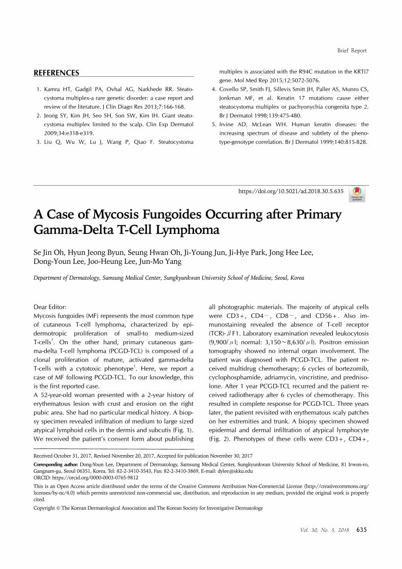
all photographic materials. The majority of atypical cells were CD3+, CD4−, CD8−, and CD56+. Also im-munostaining revealed the absence of T-cell receptor (TCR)-βF1. Laboratory examination revealed leukocytosis (9,900/μl; normal: 3,150∼8,630/μl). Positron emission tomography showed no internal organ involvement. The patient was diagnosed with PCGD-TCL. The patient re-ceived multidrug chemotherapy; 6 cycles of bortezomib, cyclophosphamide, adriamycin, vincristine, and predniso-lone. After 1 year PCGD-TCL recurred and the patient re-ceived radiotherapy after 6 cycles of chemotherapy. This resulted in complete response for PCGD-TCL. Three years later, the patient revisited with erythematous scaly patches on her extremities and trunk. A biopsy specimen showed epidermal and dermal infiltration of atypical lymphocyte (Fig. 2). Phenotypes of these cells were CD3+, CD4+,

Brief Report
636 Ann Dermatol
Fig. 2. (A) Erythematous scaly patches on the thigh and the lower leg. (B) Epidermal and dermal infiltration of atypical lymphocyte (H&E, ×200) (C) Immunohistochemistry for T-cell receptor (TCR)-βF1. Biopsy specimen shows TCR-βF1 positivity of lymphocytic infiltrate. (H&E, ×200).
Fig. 1. (A) Erythematous patch with ulcer and crust on the right pubic area. (B) Infiltration of medium to large sized atypical lymphoid cells in the dermis and subcutis (H&E, ×200).
and CD8−. Also immunostaining revealed TCR-βF1+ and TCR-cγM1−. The patient was diagnosed with MF following PCGD-TCL. She received 11 cycles of brentux-imab therapy. The different types of lymphomas coexisting in the same patient are classified into three categories according to the Working Formulation of non-Hodgkins lymphoma2. Discordant lymphomas are two histologically distinct lym-phomas at two different anatomic sites. Composite lym-phomas have two types of lymphoma within the same anatomic lesion. Secondary lymphoma is lymphoma which appeared later when two different lymphomas occur se-quentially2. PCGD-TCL was introduced as a definitive entity in the 2008 World Health Organization classification of lympho-
mas1. The tumor cells characteristically have a TCR-βF1−, CD3+, CD2+, and CD56+ phenotype and most cases lack both CD4 and CD81. On the other hand, MF shows epidermotropic proliferation of small-to medium-sized T-cells. Mostly, tumor cells have a mature CD3+, CD4+, CD45RO+, and CD8− T-cell phenotype1. MF is a risk factor for development of other hematological disorders such as lymphomatoid papulosis, CD30+ ana-plastic large cell lymphoma and B-cell lymphoma3-5. In addition, several cases were reported that molecular anal-yses revealed that the same neoplastic clone of T lympho-cytes was present in MF and associated lymphoma3. In this case, however, different lymphomas with independent findings of immunohistochemistry occurred sequentially, MF occurring after PCGD-TCL. There is no reported case

Brief Report
Vol. 30, No. 5, 2018 637
Received July 24, 2017, Revised November 15, 2017, Accepted for publication December 2, 2017
Corresponding author: Je-Ho Mun, Department of Dermatology, Seoul National University College of Medicine, 101 Daehak-ro, Jongno-gu, Seoul 03080, Korea. Tel: 82-2-2072-3274, Fax: 82-2-742-7344, E-mail: [email protected]: https://orcid.org/0000-0002-0734-2850
This is an Open Access article distributed under the terms of the Creative Commons Attribution Non-Commercial License (http://creativecommons.org/licenses/by-nc/4.0) which permits unrestricted non-commercial use, distribution, and reproduction in any medium, provided the original work is properly cited.
Copyright © The Korean Dermatological Association and The Korean Society for Investigative Dermatology
like our patient in the literature. One possible explanation could be proposed for this association. After receiving treatment of the primary lesion, the condition of patient with or without immunodeficiency is more likely to be vulnerable to secondary malignancy.In conclusion, we emphasize that when patients diag-nosed with cutaneous lymphoma present new skin le-sions, clinicians should consider skin biopsies and further immunostaining.
CONFLICTS OF INTEREST
The authors have nothing to disclose.
REFERENCES
1. Sabattini E, Bacci F, Sagramoso C, Pileri SA. WHO class-ification of tumours of haematopoietic and lymphoid tissues
in 2008: an overview. Pathologica 2010;102:83-87.2. National Cancer Institute sponsored study of classifications
of non-Hodgkin's lymphomas: summary and description of a
working formulation for clinical usage. The Non-Hodgkin's Lymphoma Pathologic Classification Project. Cancer 1982;
49:2112-2135.
3. Zackheim HS, Jones C, Leboit PE, Kashani-Sabet M, McCalmont TH, Zehnder J. Lymphomatoid papulosis associated
with mycosis fungoides: a study of 21 patients including
analyses for clonality. J Am Acad Dermatol 2003;49:620-623.4. Marschalkó M, Csomor J, Eros N, Szigeti A, Hársing J,
Szakonyi J, et al. Coexistence of primary cutaneous
anaplastic large cell lymphoma and mycosis fungoides in a patient with B-cell chronic lymphocytic leukaemia. Br J
Dermatol 2007;157:1291-1293.
5. Barzilai A, Trau H, David M, Feinmesser M, Bergman R, Shpiro D, et al. Mycosis fungoides associated with B-cell
malignancies. Br J Dermatol 2006;155:379-386.
https://doi.org/10.5021/ad.2018.30.5.637
Pigmented Onychomatricoma Showing a Longitudinal Melanonychia: A Case Report and Brief Review of Literature
Sung Cheol Jung1, Tae Min Lee1, Minsoo Kim2, Gwanghyun Jo3,4, Je-Ho Mun3,4
1Seoul National University College of Medicine, Seoul, Korea, 2Department of Medicine, University of Auckland, Auckland, New Zealand, 3Department of Dermatology, Seoul National University College of Medicine, 4Institute of Human-Environment Interface Biology, Seoul National University, Seoul, Korea
Dear Editor:Onychomatricoma is a rare benign fibroepithelial tumor of the nail unit. It typically presents as thickening of the nail plate, splinter hemorrhage, xanthonychia, and transverse
overcurvature, and generally arises in patients with pale skin. Onychomatricoma rarely manifests longitudinal mel-anonychia (LM). In this case, it is termed pigmented onychomatricoma. However, the occurrence of onycho-


![Significance of CD30 Expression by Epidermotropic T Cells ... · diagnosis included LyP, lymphomatoid pityriasis lichenoides and “pityriasis lichenoides-like” mycosis fungoides.[7,8]](https://static.fdocuments.in/doc/165x107/60223092b9e61714693c3a28/significance-of-cd30-expression-by-epidermotropic-t-cells-diagnosis-included.jpg)