A C-terminal amphipathic helix is necessary for the in ... · A C-terminal amphipathic helix is...
6
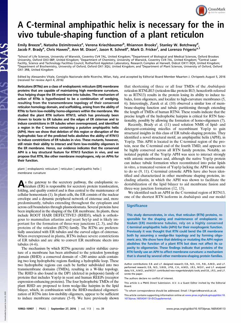
A C-terminal amphipathic helix is necessary for the in vivo tubule-shaping function of a plant reticulon Emily Breeze a , Natasha Dzimitrowicz a , Verena Kriechbaumer b , Rhiannon Brooks c , Stanley W. Botchway d , Jacob P. Brady e , Chris Hawes b , Ann M. Dixon c , Jason R. Schnell e , Mark D. Fricker f , and Lorenzo Frigerio a,1 a School of Life Sciences, University of Warwick, Coventry CV4 7AL, United Kingdom; b Department of Biological and Medical Sciences, Oxford Brookes University, Oxford OX3 0BP, United Kingdom; c Department of Chemistry, University of Warwick, Coventry CV4 7AL, United Kingdom; d Central Laser Facility, Science and Technology Facilities Council, Rutherford Appleton Laboratory, Research Complex at Harwell, Didcot OX11 0QX, United Kingdom; e Department of Biochemistry, University of Oxford, Oxford OX1 3QU, United Kingdom; and f Department of Plant Sciences, University of Oxford, Oxford OX1 3RB, United Kingdom Edited by Alessandro Vitale, Consiglio Nazionale delle Ricerche, Milan, Italy, and accepted by Editorial Board Member Maarten J. Chrispeels August 5, 2016 (received for review April 4, 2016) Reticulons (RTNs) are a class of endoplasmic reticulum (ER) membrane proteins that are capable of maintaining high membrane curvature, thus helping shape the ER membrane into tubules. The mechanism of action of RTNs is hypothesized to be a combination of wedging, resulting from the transmembrane topology of their conserved reticulon homology domain, and scaffolding, arising from the ability of RTNs to form low-mobility homo-oligomers within the membrane. We studied the plant RTN isoform RTN13, which has previously been shown to locate to ER tubules and the edges of ER cisternae and to induce constrictions in ER tubules when overexpressed, and identified a region in the C terminus containing a putative amphipathic helix (APH). Here we show that deletion of this region or disruption of the hydrophobic face of the predicted helix abolishes the ability of RTN13 to induce constrictions of ER tubules in vivo. These mutants, however, still retain their ability to interact and form low-mobility oligomers in the ER membrane. Hence, our evidence indicates that the conserved APH is a key structural feature for RTN13 function in vivo, and we propose that RTN, like other membrane morphogens, rely on APHs for their function. plant | endoplasmic reticulum | reticulon | amphipathic helix | membrane curvature A s the gateway to the secretory pathway, the endoplasmic re- ticulum (ER) is responsible for secretory protein translocation, folding, and quality control and is thus central to the maintenance of cellular homeostasis (1). In plant cells, the ER consists of the nuclear envelope and a dynamic peripheral network of cisternae and, more predominantly, tubules extending throughout the cytoplasm and across cell boundaries through plasmodesmata. Several proteins have been implicated in the shaping of the ER membrane. In plants, these include ROOT HAIR DEFECTIVE3 (RHD3), which is ortholo- gous to mammalian atlastins and yeast Sey1p and is likely im- portant for the formation of three-way junctions (2, 3), and the proteins of the reticulon (RTN) family. The RTNs are preferen- tially associated with ER tubules and the curved edges of cisternae. When overexpressed in planta, RTNs induce severe constrictions of ER tubules and are able to convert ER membrane sheets into tubules (4–6). The mechanism by which RTNs generate and/or stabilize curva- ture of a membrane has been attributed to the reticulon homology domain (RHD): a conserved domain of ∼200 amino acids contain- ing two long hydrophobic regions flanking a hydrophilic loop. These two hydrophobic regions can each be further subdivided into two transmembrane domains (TMDs), resulting in a W-like topology. The RHD is also found in the DP1 (deleted in polyposis) family of proteins that includes Yop1p in yeast and human REEPs (receptor expression-enhancing proteins). The four hydrophobic TMDs of the plant RHD are proposed to form wedge-like hairpins in the lipid bilayer, which, in combination with the RHD-mediated oligomeri- zation of RTNs into low-mobility oligomers, appear to be sufficient to induce membrane curvature (5–9). We have previously shown that shortening of three or all four TMDs of the Arabidopsis reticulon RTNLB13 (reticulon-like protein B13; henceforth referred to as RTN13) results in the protein losing its ability to induce tu- bules, form oligomers, and localize to high-curvature membranes (5, 6). Interestingly, Zurek et al. (10) observed a similar loss of mem- brane-shaping function and tubule partitioning through extending the length of TMDs of human RTN4. These results indicate that the precise length of the hydrophobic hairpins is critical for RTN func- tionality, possibly by allowing the formation of homo-oligomers (7). Recently, Brady et al. (11) used solution NMR of lipid- and detergent-containing micelles of recombinant Yop1p to gain structural insights in this class of ER tubule-shaping proteins. They identified a novel structural motif, an amphipathic helix (APH), in Yop1p. This APH is located in the C-terminal region of the pro- tein, near the C-terminal end of the fourth TMD, and appears to be highly conserved across all RTN family proteins. Notably, an isolated peptide of the Yop1p APH was seen to interact strongly with anionic membranes and, although the native Yop1p protein can induce tubule formation when reconstituted into polar lipids in vitro, a truncated version of Yop1p lacking the APH was unable to do so (9, 11). C-terminal cytosolic APHs have also been iden- tified and characterized in other membrane shaping proteins, in- cluding atlastin, in which the APH was shown to facilitate the destabilization of the lipid bilayer to aid membrane fusion and three-way junction formation (12, 13). We identified such an APH in the C-terminal region of RTN13, one of the shortest RTN isoforms in Arabidopsis and our model Significance This study demonstrates, in vivo, that reticulon (RTN) proteins, re- sponsible for the shaping and maintenance of endoplasmic re- ticulum (ER) membrane tubules, rely on a highly conserved C-terminal amphipathic helix (APH) for their morphogenic function. Previously it was thought that RTN could bend the ER membrane both by assuming a wedge-like topology and by forming oligo- meric arcs. We show here that deleting or mutating the APH region abolishes the function of a plant RTN but does not affect its ca- pacity to oligomerize. These findings indicate that proteins of the RTN family use an APH to affect membrane curvature: a mechanism that is shared by several other membrane-shaping protein families. Author contributions: E.B. and L.F. designed research; E.B., N.D., V.K., R.B., A.M.D., and L.F. performed research; E.B., N.D., S.W.B., J.P.B., C.H., A.M.D., J.R.S., M.D.F., and L.F. analyzed data; V.K., A.M.D., and M.D.F. contributed new reagents/analytic tools; and E.B., J.R.S., and L.F. wrote the paper. The authors declare no conflict of interest. This article is a PNAS Direct Submission. A.V. is a Guest Editor invited by the Editorial Board. 1 To whom correspondence should be addressed. Email: [email protected]. This article contains supporting information online at www.pnas.org/lookup/suppl/doi:10. 1073/pnas.1605434113/-/DCSupplemental. 10902–10907 | PNAS | September 27, 2016 | vol. 113 | no. 39 www.pnas.org/cgi/doi/10.1073/pnas.1605434113 Downloaded by guest on June 13, 2020
Transcript of A C-terminal amphipathic helix is necessary for the in ... · A C-terminal amphipathic helix is...
A C-terminal amphipathic helix is necessary for the invivo tubule-shaping function of a plant reticulonEmily Breezea, Natasha Dzimitrowicza, Verena Kriechbaumerb, Rhiannon Brooksc, Stanley W. Botchwayd,Jacob P. Bradye, Chris Hawesb, Ann M. Dixonc, Jason R. Schnelle, Mark D. Frickerf, and Lorenzo Frigerioa,1
aSchool of Life Sciences, University of Warwick, Coventry CV4 7AL, United Kingdom; bDepartment of Biological and Medical Sciences, Oxford BrookesUniversity, Oxford OX3 0BP, United Kingdom; cDepartment of Chemistry, University of Warwick, Coventry CV4 7AL, United Kingdom; dCentral LaserFacility, Science and Technology Facilities Council, Rutherford Appleton Laboratory, Research Complex at Harwell, Didcot OX11 0QX, United Kingdom;eDepartment of Biochemistry, University of Oxford, Oxford OX1 3QU, United Kingdom; and fDepartment of Plant Sciences, University of Oxford, OxfordOX1 3RB, United Kingdom
Edited by Alessandro Vitale, Consiglio Nazionale delle Ricerche, Milan, Italy, and accepted by Editorial Board Member Maarten J. Chrispeels August 5, 2016(received for review April 4, 2016)
Reticulons (RTNs) are a class of endoplasmic reticulum (ER) membraneproteins that are capable of maintaining high membrane curvature,thus helping shape the ER membrane into tubules. The mechanism ofaction of RTNs is hypothesized to be a combination of wedging,resulting from the transmembrane topology of their conservedreticulon homology domain, and scaffolding, arising from the ability ofRTNs to form low-mobility homo-oligomers within the membrane. Westudied the plant RTN isoform RTN13, which has previously beenshown to locate to ER tubules and the edges of ER cisternae and toinduce constrictions in ER tubules when overexpressed, and identifieda region in the C terminus containing a putative amphipathic helix(APH). Here we show that deletion of this region or disruption of thehydrophobic face of the predicted helix abolishes the ability of RTN13to induce constrictions of ER tubules in vivo. These mutants, however,still retain their ability to interact and form low-mobility oligomers inthe ER membrane. Hence, our evidence indicates that the conservedAPH is a key structural feature for RTN13 function in vivo, and wepropose that RTN, like other membrane morphogens, rely on APHs fortheir function.
plant | endoplasmic reticulum | reticulon | amphipathic helix |membrane curvature
As the gateway to the secretory pathway, the endoplasmic re-ticulum (ER) is responsible for secretory protein translocation,
folding, and quality control and is thus central to the maintenance ofcellular homeostasis (1). In plant cells, the ER consists of the nuclearenvelope and a dynamic peripheral network of cisternae and, morepredominantly, tubules extending throughout the cytoplasm andacross cell boundaries through plasmodesmata. Several proteins havebeen implicated in the shaping of the ERmembrane. In plants, theseinclude ROOT HAIR DEFECTIVE3 (RHD3), which is ortholo-gous to mammalian atlastins and yeast Sey1p and is likely im-portant for the formation of three-way junctions (2, 3), and theproteins of the reticulon (RTN) family. The RTNs are preferen-tially associated with ER tubules and the curved edges of cisternae.When overexpressed in planta, RTNs induce severe constrictionsof ER tubules and are able to convert ER membrane sheets intotubules (4–6).The mechanism by which RTNs generate and/or stabilize curva-
ture of a membrane has been attributed to the reticulon homologydomain (RHD): a conserved domain of ∼200 amino acids contain-ing two long hydrophobic regions flanking a hydrophilic loop. Thesetwo hydrophobic regions can each be further subdivided into twotransmembrane domains (TMDs), resulting in a W-like topology.The RHD is also found in the DP1 (deleted in polyposis) family ofproteins that includes Yop1p in yeast and human REEPs (receptorexpression-enhancing proteins). The four hydrophobic TMDs of theplant RHD are proposed to form wedge-like hairpins in the lipidbilayer, which, in combination with the RHD-mediated oligomeri-zation of RTNs into low-mobility oligomers, appear to be sufficientto induce membrane curvature (5–9). We have previously shown
that shortening of three or all four TMDs of the Arabidopsisreticulon RTNLB13 (reticulon-like protein B13; henceforth referredto as RTN13) results in the protein losing its ability to induce tu-bules, form oligomers, and localize to high-curvature membranes (5,6). Interestingly, Zurek et al. (10) observed a similar loss of mem-brane-shaping function and tubule partitioning through extendingthe length of TMDs of human RTN4. These results indicate that theprecise length of the hydrophobic hairpins is critical for RTN func-tionality, possibly by allowing the formation of homo-oligomers (7).Recently, Brady et al. (11) used solution NMR of lipid- and
detergent-containing micelles of recombinant Yop1p to gainstructural insights in this class of ER tubule-shaping proteins. Theyidentified a novel structural motif, an amphipathic helix (APH), inYop1p. This APH is located in the C-terminal region of the pro-tein, near the C-terminal end of the fourth TMD, and appears tobe highly conserved across all RTN family proteins. Notably, anisolated peptide of the Yop1p APH was seen to interact stronglywith anionic membranes and, although the native Yop1p proteincan induce tubule formation when reconstituted into polar lipidsin vitro, a truncated version of Yop1p lacking the APH was unableto do so (9, 11). C-terminal cytosolic APHs have also been iden-tified and characterized in other membrane shaping proteins, in-cluding atlastin, in which the APH was shown to facilitate thedestabilization of the lipid bilayer to aid membrane fusion andthree-way junction formation (12, 13).We identified such an APH in the C-terminal region of RTN13,
one of the shortest RTN isoforms in Arabidopsis and our model
Significance
This study demonstrates, in vivo, that reticulon (RTN) proteins, re-sponsible for the shaping and maintenance of endoplasmic re-ticulum (ER) membrane tubules, rely on a highly conservedC-terminal amphipathic helix (APH) for their morphogenic function.Previously it was thought that RTN could bend the ER membraneboth by assuming a wedge-like topology and by forming oligo-meric arcs. We show here that deleting or mutating the APH regionabolishes the function of a plant RTN but does not affect its ca-pacity to oligomerize. These findings indicate that proteins of theRTN family use an APH to affect membrane curvature: amechanismthat is shared by several other membrane-shaping protein families.
Author contributions: E.B. and L.F. designed research; E.B., N.D., V.K., R.B., A.M.D., and L.F.performed research; E.B., N.D., S.W.B., J.P.B., C.H., A.M.D., J.R.S., M.D.F., and L.F. analyzeddata; V.K., A.M.D., andM.D.F. contributed new reagents/analytic tools; and E.B., J.R.S., and L.F.wrote the paper.
The authors declare no conflict of interest.
This article is a PNAS Direct Submission. A.V. is a Guest Editor invited by the EditorialBoard.1To whom correspondence should be addressed. Email: [email protected].
This article contains supporting information online at www.pnas.org/lookup/suppl/doi:10.1073/pnas.1605434113/-/DCSupplemental.
10902–10907 | PNAS | September 27, 2016 | vol. 113 | no. 39 www.pnas.org/cgi/doi/10.1073/pnas.1605434113
Dow
nloa
ded
by g
uest
on
June
13,
202
0
protein for the plant RTN family (4–6). Here we show that thispredicted APH, although not required for oligomer formation, isessential for the membrane-shaping function of RTN13 in vivo.Deletion of the APH or disruption of its hydrophobic face resultsin a loss of tubule-forming activity in vivo but does not affect theability of the protein to form homotypic interactions, or to lo-calize to the ER.
Results and DiscussionArabidopsis has 21 predicted RTN proteins, of which RTN13 isone of the smallest (206 amino acids), consisting of the RHDplus short N- and C-terminal extensions. We have previouslyshown that RTN13 is localized to the tubular cortical ER and,when ectopically expressed, is capable of inducing constrictionsin ER tubules and to convert ER sheets into tubules, as dem-onstrated for other, longer RTN isoforms (4–6). As such, RTN13can be considered to be the minimal functional plant RTNprotein, and is thus a useful model for studying the relationshipbetween the structural motifs and topology of RTNs, and theirER morphogenic properties.To investigate whether an APH akin to the one identified in
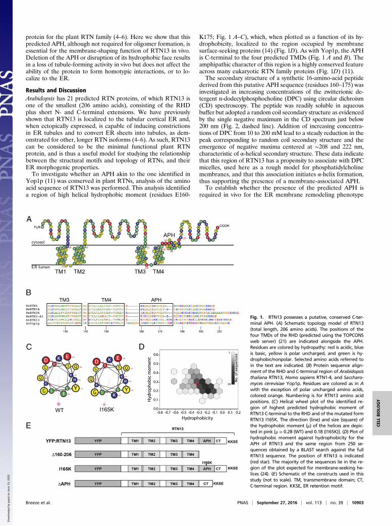
Yop1p (11) was conserved in plant RTNs, analysis of the aminoacid sequence of RTN13 was performed. This analysis identifieda region of high helical hydrophobic moment (residues E160-
K175; Fig. 1 A–C), which, when plotted as a function of its hy-drophobicity, localized to the region occupied by membranesurface-seeking proteins (14) (Fig. 1D). As with Yop1p, the APHis C-terminal to the four predicted TMDs (Fig. 1 A and B). Theamphipathic character of this region is a highly conserved featureacross many eukaryotic RTN family proteins (Fig. 1D) (11).The secondary structure of a synthetic 16-amino-acid peptide
derived from this putative APH sequence (residues 160–175) wasinvestigated in increasing concentrations of the zwitterionic de-tergent n-dodecylphosphocholine (DPC) using circular dichroism(CD) spectroscopy. The peptide was readily soluble in aqueousbuffer but adopted a random coil secondary structure as evidencedby the single negative maximum in the CD spectrum just below200 nm (Fig. 2, dashed line). Addition of increasing concentra-tions of DPC from 10 to 200 mM lead to a steady reduction in thepeak corresponding to random coil secondary structure and theemergence of negative maxima centered at ∼208 and 222 nm,characteristic of α-helical secondary structure. These data indicatethat this region of RTN13 has a propensity to associate with DPCmicelles, used here as a rough model for phosphatidylcholinemembranes, and that this association initiates α-helix formation,thus supporting the presence of a membrane-associated APH.To establish whether the presence of the predicted APH is
required in vivo for the ER membrane remodeling phenotype
Fig. 1. RTN13 possesses a putative, conserved C-ter-minal APH. (A) Schematic topology model of RTN13(total length, 206 amino acids). The positions of thefour TMDs of the RHD (predicted using the TOPCONSweb server) (21) are indicated alongside the APH.Residues are colored by hydropathy: red is acidic, blueis basic, yellow is polar uncharged, and green is hy-drophobic/nonpolar. Selected amino acids referred toin the text are indicated. (B) Protein sequence align-ment of the RHD and C-terminal region of Arabidopsisthaliana RTN13, Homo sapiens RTN1-4, and Saccharo-myces cerevisiae Yop1p. Residues are colored as in Awith the exception of polar uncharged amino acids,colored orange. Numbering is for RTN13 amino acidpositions. (C) Helical wheel plot of the identified re-gion of highest predicted hydrophobic moment ofRTN13 C-terminal to the RHD and of the mutated formRTN13 I165K. The direction (line) and size (square) ofthe hydrophobic moment (μ) of the helices are depic-ted in pink [μ = 0.28 (WT) and 0.18 (I165K)]. (D) Plot ofhydrophobic moment against hydrophobicity for theAPH of RTN13 and the same region from 250 se-quences obtained by a BLAST search against the fullRTN13 sequence. The position of RTN13 is indicated(red star). The majority of the sequences lie in the re-gion of the plot expected for membrane-seeking he-lices (24). (E) Schematic of the constructs used in thisstudy (not to scale). TM, transmembrane domain; CT,C-terminal region. KKSE, ER retention motif.
Breeze et al. PNAS | September 27, 2016 | vol. 113 | no. 39 | 10903
CELL
BIOLO
GY
Dow
nloa
ded
by g
uest
on
June
13,
202
0

induced by overexpression of RTN13, we first generated C-ter-minal truncations of RTN13, tagged at the N terminus with YFP(4), by inserting a stop codon immediately before the predictedAPH (Δ160–206; Fig. 1E), and by excising the sequence encodingthe predicted APH (ΔAPH; Fig. 1E).When coexpressed transiently in Nicotiana benthamiana epi-
dermal cells with the ER luminal marker, GFP-HDEL, all mu-tants retained their ability to localize to tubular ER (Fig. 3).Overexpression of full-length YFP-RTN13 caused the typical,marked constriction of ER tubules (Fig. 3A), as previouslyreported. Tubule constrictions displace the luminal marker todiscrete, punctate regions of the ER network, thereby resultingin a significant reduction in the degree of colocalization of GFP-HDEL with YFP-RTN13 (Fig. 3E). Deletion of the APH plusC-terminal region (Δ160–206) and of the region encoding theAPH alone (ΔAPH) abolished this phenotype (Fig. 3 B and D)such that the luminal marker GFP-HDEL colocalized with thetagged RTN13 throughout the ER network (Fig. 3E), indicatingthat these mutants, although correctly locating to the ER, are in-capable of inducing constrictions.To further investigate whether the putative APH alone was
solely responsible for the protein’s tubule forming capacity, anisoleucine residue in the center of the predicted hydrophobicface of the APH was mutated to a lysine residue (I165K). Thismutation introduces a positive charge within the hydrophobicregion of the APH, which decreases the magnitude of the hy-drophobic moment by a third relative to WT, effectively abol-ishing amphipathicity in the helix (Fig. 1C). The ER tubuleconstriction phenotype was not observed on transient expressionof this construct (Fig. 3C). The steady-state protein expressionlevels of ΔAPH were comparable to full-length YFP-RTN13,whereas I165K and Δ160–206 mutant protein expression wasslightly lower (Fig. S1A). Infiltration with serial dilutions ofAgrobacterium tumefaciens (OD600) carrying full-length YFP-RTN13, which resulted in progressively lower amounts of de-tectable protein (Fig. S1B), did not affect the appearance of theER constriction phenotype even at the lowest agrobacterial titer(Fig. S1C). Thus, we conclude that this phenotype is unlikely tobe a function of subtle differences in the protein concentrationsachieved within our standard agroinfiltration procedure.
Taken together, these data indicate that the presence of anintact C-terminal APH is necessary for the membrane-shapingactivity of RTN13 in vivo.In addition to the tubule constriction phenotype shown in Fig.
3, we attempted to assess the effect of full-length RTN13 and ofthe APH mutants on the diameter of ER tubules. We imaged theER lumen (labeled by GFP-HDEL) in the presence of eitherfull-length or mutated YFP-RTN13, using the Airyscan detector
Fig. 2. RTN13 residues 160–175 form an α-helix in the presence of detergentmicelles. CD spectra of the putative APH peptide (EYGDQIQKHLGSLKDK, resi-dues 160–175) dissolved in 25 mM sodium phosphate buffer (pH 7.0, dashedline), as well as increasing concentrations of buffer-solubilized micelles. Thepeptide attained a maximum helical content of 31% in 200 mM DPC (as esti-mated by fitting of the data as described in Materials and Methods). CD datafitting summary: helix, 30.6%; strand, 21.3%; turns, 19.6%; disordered, 27.8%.
Fig. 3. Deletion of the APH or disruption of its hydrophobic face impairsRTN13 function. N. benthamiana epidermal cells were coinfiltrated withA. tumefaciens carrying plasmids encoding the indicated constructs, togetherwith the soluble ER marker, GFP-HDEL. Representative confocal images of leavestransfected with (A) full-length, (B) Δ160–206, (C) I165K, and (D) ΔAPH. (Scalebar, 5 μm.) (E) Average Pearson’s correlation coefficients (PCCs) of cells coex-pressing GFP-HDEL and full-length YFP-RTN13 or APH mutants, where a PCC of+1 indicates perfect colocalization, 0 indicates no colocalization, and −1 indicatesnegative colocalization. Error bars indicate the SEM; n = 8–9 cells. Asterisks in-dicate a significant difference between full-length RTN13 and the relevant APHmutant (Student t test; P < 0.001).
10904 | www.pnas.org/cgi/doi/10.1073/pnas.1605434113 Breeze et al.
Dow
nloa
ded
by g
uest
on
June
13,
202
0

of a Zeiss LSM 880 confocal microscope, which is able to achievea lateral resolution of around 140 nm. We collected images from10 expressing cells per construct (representative images areshown in Fig. 4A, Left), and subjected each image to networkanalysis. The tubular ER network was automatically extracted togive a single-pixel-wide skeleton. The local image intensity nor-mal to the skeleton was integrated to provide an estimate of therelative amount of GFP-HDEL present for every point alongeach tubule. Each integrated intensity profile was then scannedto detect intensity peaks, corresponding to bulges in the tubules,and intensity troughs, corresponding to constriction sites (Fig.4A, Right). Although the tubules are at or below the resolutionlimit, the relative intensities can be used as a proxy for the widthof the ER tubules, with the additional assumption that GFP-HDEL is evenly distributed in the ER lumen.We thus estimated the width of the tubules in unperturbed ER
(GFP-HDEL alone) and in the presence of full-length or mutatedYFP-RTN13. Our analysis showed that constrictions can be regu-larly detected even in unperturbed ER tubules (Fig. 4A, Bottom, andB). However, we observed that, in the presence of full-length YFP-RTN13, the mean tubule width and the width at the constrictionsites were significantly narrower than in unperturbed ER (Fig.4B). In contrast, in the presence of the RTN13 APH mutants, themean width of the ER tubules was not significantly different tothat observed in the unperturbed network. These data indicate thatectopic expression of RTN13 negatively affects the width of ERtubules and that this property requires the presence of an APH.Because the RTN proteins are known to form both hetero-
and homo-oligomers, which create immobile, arc-like scaffolds inthe tubular ER membrane, it is possible that the removal ormutation of the APH affects the ability of RTN13 to oligo-merize. Previous work indicated that altering the length of theTMDs within the RHD abolished RTN function, but this wasconcomitant with a loss of the protein’s capacity to homo-oli-gomerize (5). To test whether our APH mutants had similarlylost the ability to form low mobility homo-oligomers and/or tointeract with WT RTN13, we used sucrose sedimentation velocitygradients, fluorescence recovery after photobleaching (FRAP)analysis, and Förster resonance energy transfer (FRET), measureddirectly by fluorescence lifetime imaging (FLIM).We isolated microsomes from agroinfiltrated N. benthamiana
leaves, solubilized the membranes with 1% digitonin, and resolvedthe extracts on a continuous 5–30% (wt/vol) sucrose gradient, withcomparison against known molecular weight marker proteins.Fractions were analyzed by SDS/PAGE and immunoblotted withanti-GFP antiserum. Fig. 5 shows that both full-length YFP-RTN13and the APH mutants form high-molecular-weight oligomeric com-plexes that have comparable sedimentation properties, peakingaround a molecular mass of ∼200–250 kDa. This result indicates thatthese complexes may contain four to five monomers, which iscomparable to that observed for yeast and mammalian reticulons (7).FRAP of a region of the tubular ER expressing YFP-RTN13
appears relatively slow in comparison with non–membrane-shapingER membrane proteins, as observed previously (5, 7) (Fig. S2). Incomparison, our previously described mutant ΔTM4, where the fourTMDs of RTN13 were each shortened to a length of 17 residues (5),shows markedly higher mobility (Fig. S2B) (4), with a FRAPprofile that is significantly different to that observed in full-lengthRTN13 (χ2 test for trend; P = 0.001; Fig. S2E). In contrast, boththe YFP-RTN13 mutant lacking the C-terminal region (Δ160–206;Fig. S2C) and the I165K mutant (Fig. S2D) displayed recovery ki-netics that are comparable with those of full length (P = 0.181 and0.245, respectively). These data indicate that mutations affectingthe putative APH regions do not affect the protein’s capacity toform homo-oligomers.We also used FRET-FLIM analysis to test whether the above
mutants were still able to interact with full-length RTN13. YFP(donor) was fused to the mutant RTN13 proteins and mCherry
(mCh; acceptor) to the full-length RTN13 sequence. Donor andacceptor constructs were coinfiltrated into N. benthamiana epider-mal cells and FRET-FLIM used to assess protein–protein interac-tions. The lifetime values for each RTN13 pair is shown in Table 1.Fig. S3 shows fluorescence lifetimes of 35S:YFP:RTN13 expressedalone or in combination with 35S:mCh:RTN13, as representativeexamples of the results obtained from FRET-FLIM analyses.FRET-FLIM has been used extensively in planta to confirm
protein–protein interactions identified through a range of other
Fig. 4. The APH is necessary for the action of RTN13 on the diameter of ERtubules. (A) N. benthamiana leaf cells were coinfiltrated with A. tumefacienscarrying plasmids encoding the indicated constructs. Cells were imaged witha Zeiss Airyscan detector (Left) and subjected to ER network analysis (Right).(B) Comparison of average ER tubule width and constrictions resulting fromthe expression of full-length YFP-RTN13 or the APH mutants. Asterisks in-dicate significant differences (Student t test; P < 0.001).
Breeze et al. PNAS | September 27, 2016 | vol. 113 | no. 39 | 10905
CELL
BIOLO
GY
Dow
nloa
ded
by g
uest
on
June
13,
202
0
techniques (6, 15). A reduction in the lifetime of donor fluores-cence by as little as 0.2 ns in the presence of an acceptor fluo-rophore has been shown to be indicative of a direct interactionbetween two proteins such that they are in close enough proximity(1–10 nm) for quenching to occur (15–17). Here, all of the com-binations tested displayed at least a 0.35-ns decrease in lifetimewhen expressed as a donor/acceptor pair compared with expres-sion of the donor alone (Table 1 and Fig. S3). Hence, removal ofthe APH or disruption of its hydrophobic face does not affect theability of the protein to homo-oligomerize or to interact with thefull-length RTN13 isoform. This observation is in agreement withprevious studies of RTN13 and other RTN interactions, where atruncated version of RTN13 lacking the two C-terminal TMDs wasstill shown to form homotypic interactions (6).Although the role of APHs in sculpting membranes has been
well documented for several classes of proteins including Sar1,epsin, endophilin, and ArfGAP1 (refs. 18 and 19 and referencestherein), the apparent absence of an APH in proteins of the RTNfamily suggested that their membrane-sculpting action arosefrom a combination of wedging, due to their membrane topology,and scaffolding, resulting from their ability to form immobileoligomers (7, 20). Indeed, altering the length of the TMD, both byshortening and extension, resulted in a loss of function. AlteringTMD length, however, also impacted on the ability of the proteinto oligomerize, making it impossible to assess the relative
contribution of the two mechanisms described above. Our dataindicate that the newly identified, conserved APH at the C terminusof RTN13 is necessary for its ability to shape ER tubules in vivo.Loss of the APH or mutations that disrupt its amphipathic naturedo not affect RTN13’s ability to form oligomers but abolish its ca-pacity to constrict ER tubules. Previously, Shibata et al. (7) showedthat mutants of ScRTN1p with defects in oligomerization were stillable to induce ER tubule formation. In the light of the resultspresented here, we now suggest that this is likely due to theC-terminal APH region remaining unaffected in those mutants. Inconclusion, although it is likely that the RHD alone is sufficient toguarantee reticulon oligomerization, its main role may be to providean environment for the correct insertion and interaction of thecrucial membrane-shaping APH.
Materials and MethodsIn Silico analysis of RTN13. A consensus prediction of the membrane topologyof AtRTN13 was performed using TOPCONS (21) with further refinement ofthe position of the TMDs based on sequence alignments with the humanRTNs and experimental evidence described previously (5, 11). The predictedtopology of RTN13 was plotted using the TEXtopo package in LaTeX (22).Based on the alignment, a putative 16-amino-acid APH in the region C-ter-minal to the TMD was identified in AtRTN13 using the HELIQUEST webserver algorithm (23). The membrane-seeking properties of the AtRTN13APH and the corresponding region in 250 sequences obtained from a BLASTsearch against the full AtRTN13 sequence were evaluated as described byEisenberg et al. (24).
Peptide Analysis and CD. These methods are described in the SI Materialsand Methods.
Generation of RTN13 Constructs. A complete list of all primers used in thisstudy is given in Table S1. All PCRs were performed using high-fidelity DNApolymerase (Accuzyme; Bioline). The coding region of RTN13 was cloned andfused to enhanced yellow fluorescent protein (eYFP) as previously described(4) and used to generate a YFP:RTN13 Gateway Entry clone in pDONRZeo(ThermoFisher Scientific). A point mutation in the entry clone (I165K) wascreated through site directed mutagenesis PCR with overlapping forwardand reverse primers containing the appropriate sequence mismatch, fol-lowed by digestion of the methylated template with 10 U DpnI (New EnglandBiolabs) for 1 h at 37 °C and transformation into DH5α chemically competentEscherichia coli cells. A mutated version of RTN13 in which the APH alone wasexcised (ΔAPH) was similarly created using overlapping primers homologousto the sequence before and after the desired deletion, in a two-step process(deletion of E160-L169, followed by deletion of G170-K175). The native ormutated YFP:RTN13 in pDONRZeo was subsequently used as a PCR template tocreate an amplicon with XbaI and SacI recognition sites, which were thendigested (XbaI and SacI; New England Biolabs), gel purified (QIAquick Gel Ex-traction kit; Qiagen), and ligated (T4 DNA ligase; ThermoFisher Scientific) intothe corresponding sites in pVKH18-EN6 expression vector (25). A C-terminaltruncation of RTN13 in which a stop codon was inserted after E159 (RTN13Δ160–206) was similarly generated. For FRET-FLIM analysis, a mCherry:RTN13 fusionwas created by fusion PCR in which PCR fragments (10–50 ng each) of mCherry
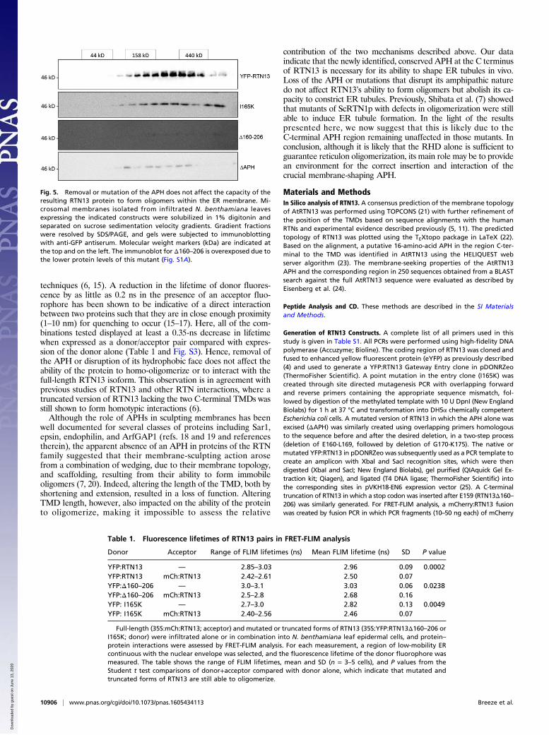
Fig. 5. Removal or mutation of the APH does not affect the capacity of theresulting RTN13 protein to form oligomers within the ER membrane. Mi-crosomal membranes isolated from infiltrated N. benthamiana leavesexpressing the indicated constructs were solubilized in 1% digitonin andseparated on sucrose sedimentation velocity gradients. Gradient fractionswere resolved by SDS/PAGE, and gels were subjected to immunoblottingwith anti-GFP antiserum. Molecular weight markers (kDa) are indicated atthe top and on the left. The immunoblot for Δ160–206 is overexposed due tothe lower protein levels of this mutant (Fig. S1A).
Table 1. Fluorescence lifetimes of RTN13 pairs in FRET-FLIM analysis
Donor Acceptor Range of FLIM lifetimes (ns) Mean FLIM lifetime (ns) SD P value
YFP:RTN13 — 2.85–3.03 2.96 0.09 0.0002YFP:RTN13 mCh:RTN13 2.42–2.61 2.50 0.07YFP:Δ160–206 — 3.0–3.1 3.03 0.06 0.0238YFP:Δ160–206 mCh:RTN13 2.5–2.8 2.68 0.16YFP: I165K — 2.7–3.0 2.82 0.13 0.0049YFP: I165K mCh:RTN13 2.40–2.56 2.46 0.07
Full-length (35S:mCh:RTN13; acceptor) and mutated or truncated forms of RTN13 (35S:YFP:RTN13Δ160–206 orI165K; donor) were infiltrated alone or in combination into N. benthamiana leaf epidermal cells, and protein–protein interactions were assessed by FRET-FLIM analysis. For each measurement, a region of low-mobility ERcontinuous with the nuclear envelope was selected, and the fluorescence lifetime of the donor fluorophore wasmeasured. The table shows the range of FLIM lifetimes, mean and SD (n = 3–5 cells), and P values from theStudent t test comparisons of donor+acceptor compared with donor alone, which indicate that mutated andtruncated forms of RTN13 are still able to oligomerize.
10906 | www.pnas.org/cgi/doi/10.1073/pnas.1605434113 Breeze et al.
Dow
nloa
ded
by g
uest
on
June
13,
202
0

with a 3′ overhang to RTN13, and RTN13 with a 5′ overhang to mCherry,were mixed and PCR amplified.
Transient Expression Analysis. All RTN13 constructs were transformed by heat-shock into A. tumefaciens strain GV3101 and were transiently expressed inN. benthamiana leaf epidermal cells by agroinfiltration at an OD600 of 0.5, aspreviously described (26). In addition, serial dilutions of A. tumefacienscarrying the 35S:YFP:RTN13 construct were made from OD600 0.5–0.05 foragroinfiltration.
Protein Resolution and Detection. Total proteins were isolated from infiltratedN. benthamiana leaves by homogenizing in a protein extraction buffer(0.2 M NaCl, 1 mM EDTA, 2% (vol/vol) 2-mercaptoethanol, 0.2% TritonX-100, 0.1 M Tris·HCl, pH 7.8, complete protease inhibitor mixture tablet;Roche Diagnostics). For sucrose gradient analysis, ER microsomes were iso-lated from infiltrated leaves as previously described (27). Microsomal pelletswere resuspended in 100 μL TKMG lysis buffer [50 mM Tris·HCl, pH 7.0,150 mM KCl, 2 mM MgCl2, 10% (vol/vol) glycerol, 1 mM EDTA, 1 mM PMSF,1 mM 4-(2-aminoethyl)benzenesulfonylfluoride hydrochloride] containing1% digitonin. Solubilized lysate was centrifuged for 10 min at 12,000 × g toseparate out any remaining leaf debris. Microsomes were loaded onto continuous5–30% (wt/vol) sucrose gradients and ultracentrifuged at 166,000 × g for 4.5 hat 25 °C in a Beckman SW55Ti rotor. Twenty gradient fractions were col-lected and subjected to SDS/PAGE on 15% (vol/vol) acrylamide gels; 500 μgeach of ferritin (440 kDa), aldolase (158 kDa), and ovalbumin (44 kDa) (GEHealthcare) were used as molecular weight standards and stained withCoomassie Brilliant Blue R-250 following electrophoresis. Proteins were trans-ferred onto PVDF membranes and subjected to immunoblotting with anti-GFP-HRP antisera (Miltenyi Biotec). Immunoblots were developed by enhancedchemiluminescence (Promega) using a ImageQuant LAS4000 imaging system(GE Healthcare).
Confocal Microscopy. Freshly excised leaf samples were mounted in water andimaged on a Leica TCS SP5 confocal microscope with a 63× oil immersionobjective lens. GFP and eYFP were excited at 488 and 514 nm, respectively.
GFP was detected in the 494- to 513-nm range and eYFP in the 524- to554-nm range. Simultaneous detection of GFP and YFP was performed bycombining the settings indicated above using the sequential scanning facility ofthe microscope, as instructed by the manufacturer. For FRAP analysis, sampleswere imaged with a Zeiss LSM880 equipped with a 63× oil immersion objective.YFP was excited at 514 nm and detected in the 520- to 560-nm range. For ERnetwork analysis, samples were imaged using a 100×/1.46 NA oil immersionobjective on a Zeiss LSM880 equipped with an Airyscan detector. Images wereacquired using a defined region of interest (ROI) with an average of four, with2,048 × 2,048 pixels of image size and 8-bit image depth, taking care that everypart of each image remained fully within the dynamic range of pixel intensity.
Colocalization Analysis. Colocalization analysis was completed using theImageJ-based open source processing package Fiji v1.49r (28). First a medianfilter (radius = 0.5 pixels) was used to reduce salt and pepper noise and arolling ball filter (radius = 20 pixels) was used to reduce the backgroundnoise in each image. The RGB image was then split into 8-bit red and greenchannel images, and a ROI for each image was selected. Saturation level wasset at 0.1% to normalize the images. Minimum pixel intensity was set to0 for each channel. To measure the Pearson’s coefficient, the CoLoc_2 pluginwas used (point spread function = 3px and Costes iterations = 50). A mini-mum of eight images, corresponding to eight different cells, was used foreach RTN construct.
ER Network Analysis. The detailed analysis method is described in SI Materialsand Methods. All of the image analysis routines were implemented inMATLAB (The Mathworks) and are available in a standalone package or aMATLAB app from https://www.markfricker.org.
ACKNOWLEDGMENTS. We thank Philip Young for help with FRAP analysis.We thank the Biotechnology and Biological Sciences Research Council(BBSRC) for a Midland Integrative Biosciences Training Partnership (MIBTP)studentship (to N.D.). This work was funded by BBSRC European ResearchArea - Coordinating Action in Plant Sciences (ERA-CAPS) Grant “Plant Endo-plasmic Reticulum And Seed Productivity (PER ASPERA)” (to L.F. and C.H.).
1. Hawes C, Kiviniemi P, Kriechbaumer V (2015) The endoplasmic reticulum: A dynamicand well-connected organelle. J Integr Plant Biol 57(1):50–62.
2. Zhang M, et al. (2013) ROOT HAIR DEFECTIVE3 family of dynamin-like GTPases me-diates homotypic endoplasmic reticulum fusion and is essential for Arabidopsis de-velopment. Plant Physiol 163(2):713–720.
3. Chen J, Stefano G, Brandizzi F, Zheng H (2011) Arabidopsis RHD3 mediates the gen-eration of the tubular ER network and is required for Golgi distribution and motilityin plant cells. J Cell Sci 124(Pt 13):2241–2252.
4. Tolley N, et al. (2008) Overexpression of a plant reticulon remodels the lumen of thecortical endoplasmic reticulum but does not perturb protein transport. Traffic 9(1):94–102.
5. Tolley N, et al. (2010) Transmembrane domain length is responsible for the ability of aplant reticulon to shape endoplasmic reticulum tubules in vivo. Plant J 64(3):411–418.
6. Sparkes I, et al. (2010) Five Arabidopsis reticulon isoforms share endoplasmic re-ticulum location, topology, and membrane-shaping properties. Plant Cell 22(4):1333–1343.
7. Shibata Y, et al. (2008) The reticulon and DP1/Yop1p proteins form immobile oligo-mers in the tubular endoplasmic reticulum. J Biol Chem 283(27):18892–18904.
8. Voeltz GK, Prinz WA, Shibata Y, Rist JM, Rapoport TA (2006) A class of membraneproteins shaping the tubular endoplasmic reticulum. Cell 124(3):573–586.
9. Hu J, et al. (2008) Membrane proteins of the endoplasmic reticulum induce high-curvature tubules. Science 319(5867):1247–1250.
10. Zurek N, Sparks L, Voeltz G (2011) Reticulon short hairpin transmembrane domainsare used to shape ER tubules. Traffic 12(1):28–41.
11. Brady JP, Claridge JK, Smith PG, Schnell JR (2015) A conserved amphipathic helix isrequired for membrane tubule formation by Yop1p. Proc Natl Acad Sci USA 112(7):E639–E648.
12. Liu TY, Bian X, Sun S, Hu X (2012) Lipid interaction of the C terminus and associationof the transmembrane segments facilitate atlastin-mediated homotypic endoplasmicreticulum fusion. Proc Natl Acad Sci USA 109(32):E2146–E2154.
13. Faust JE, et al. (2015) The Atlastin C-terminal tail is an amphipathic helix that perturbsthe bilayer structure during endoplasmic reticulum homotypic fusion. J Biol Chem290(8):4772–4783.
14. Eisenberg D, Schwarz E, Komaromy M, Wall R (1984) Analysis of membrane andsurface protein sequences with the hydrophobic moment plot. J Mol Biol 179(1):125–142.
15. Kriechbaumer V, et al. (2015) Reticulomics: Protein-protein interaction studies withtwo plasmodesmata-localized reticulon family proteins identify binding partnersenriched at plasmodesmata, endoplasmic reticulum, and the plasma membrane. PlantPhysiol 169(3):1933–1945.
16. Stubbs CD, Botchway SW, Slater SJ, Parker AW (2005) The use of time-resolvedfluorescence imaging in the study of protein kinase C localisation in cells. BMC CellBiol 6(1):22.
17. Osterrieder A, et al. (2009) Fluorescence lifetime imaging of interactions betweenGolgi tethering factors and small GTPases in plants. Traffic 10(8):1034–1046.
18. Shen H, Pirruccello M, De Camilli P (2012) SnapShot: Membrane curvature sensors andgenerators. Cell 150(6):1300–1300.e2, 1300.e1–1300.e2.
19. Suetsugu S, Kurisu S, Takenawa T (2014) Dynamic shaping of cellular membranes byphospholipids and membrane-deforming proteins. Physiol Rev 94(4):1219–1248.
20. Zimmerberg J, Kozlov MM (2006) How proteins produce cellular membrane curva-ture. Nat Rev Mol Cell Biol 7(1):9–19.
21. Tsirigos KD, Peters C, Shu N, Käll L, Elofsson A (2015) The TOPCONS web server forconsensus prediction of membrane protein topology and signal peptides. NucleicAcids Res 43(W1):W401–W407.
22. Beitz E (2000) T(E)Xtopo: Shaded membrane protein topology plots in LAT(E)X2e.Bioinformatics 16(11):1050–1051.
23. Gautier R, Douguet D, Antonny B, Drin G (2008) HELIQUEST: A web server to screensequences with specific alpha-helical properties. Bioinformatics 24(18):2101–2102.
24. Eisenberg D, Weiss RM, Terwilliger TC (1982) The helical hydrophobic moment: Ameasure of the amphiphilicity of a helix. Nature 299(5881):371–374.
25. Batoko H, Zheng HQ, Hawes C, Moore I (2000) A rab1 GTPase is required for transportbetween the endoplasmic reticulum and golgi apparatus and for normal golgimovement in plants. Plant Cell 12(11):2201–2218.
26. Sparkes IA, Runions J, Kearns A, Hawes C (2006) Rapid, transient expression of fluo-rescent fusion proteins in tobacco plants and generation of stably transformed plants.Nat Protoc 1(4):2019–2025.
27. Aggarwal C, et al. (2014) Blue-light-activated phototropin2 trafficking from the cy-toplasm to Golgi/post-Golgi vesicles. J Exp Bot 65(12):3263–3276.
28. Schindelin J, et al. (2012) Fiji: An open-source platform for biological-image analysis.Nat Methods 9(7):676–682.
29. Kovesi PD (1999) Image feature from phase congruency, Videre. J Comput Vis Res1(3):1–26.
30. Obara B, Grau V, Fricker MD (2012) A bioimage informatics approach to automaticallyextract complex fungal networks. Bioinformatics 28(18):2374–2381.
31. Kovesi PD (2000) MATLAB and Octave functions for computer vision and imageprocessing. Available at www.peterkovesi.com/matlabfns/. Accessed June 2, 2016.
32. Whitmore L, Wallace BA (2004) DICHROWEB, an online server for protein secondarystructure analyses from circular dichroism spectroscopic data. Nucl Acids Res 32(WebServer issue):W668–W673.
33. Whitmore L, Wallace BA (2008) Protein secondary structure analyses from circulardichroism spectroscopy: methods and reference databases. Biopolymers 89(5):392–400.
34. Sreerama N, Woody RW (2000) Estimation of protein secondary structure from cir-cular dichroism spectra: Comparison of CONTIN, SELCON, and CDSSTR methods withan expanded reference set. Anal Biochem 287(2):252–260.
Breeze et al. PNAS | September 27, 2016 | vol. 113 | no. 39 | 10907
CELL
BIOLO
GY
Dow
nloa
ded
by g
uest
on
June
13,
202
0