Chapter 4 Membrane Structure and Function. Phospholipid - amphipathic molecule.

Interaction of class A amphipathic helical peptides with phospholipid u nilamellar vesicles
Jeffrey A. Gazzara," Michael C. Phillips,+ Sissel Lund-Katz,+ Mayakonda N. Palgunachari,§ Jere P. Segrest,§ G. M. Anantharamaiah,§ and Julian W. Snow1**
Department of Chemistry,* Philadelphia College of Pharmacy and Science, Philadelphia PA 191 04; Department of Biochemistry,+ Allegheny University of the Health Sciences, Philadelphia, PA 19129; and Departments of Medicine and Biochemistry and Molecular Genetics and the Atherosclerosis Research Unit,$ University of Alabama at Birmingham Medical Center, Birmingham, AL 35294
Abstract The exchangeable apolipoproteins are important in determining the structure/function properties of lipopro- teins. These proteins typically contain varying amounts of am- phipathic helices. Five model peptides, 18A, Ac-18A-NHy, Ac- 18R-NH2, 37pA, and 37aA, have been designed to investigate variations of the amphipathic a-helix structural motif on their lipid-binding properties. These include the 18-residue p e p tides, 18A and Ac-18A-NHp, examples of class A helices, and Ac-18R-NH2, which has the positions of acidic and basic resi- dues interchanged relative to 18A. Three larger peptides were also studied: 36A, a dimer of 18A, 37pA and 37aA, dimers of 18A coupled by Pro (18A-Pro-18A) and Ala (18A-Ala-l8A), respectively. We report here the results of a thermodynamic characterization of the binding properties of these peptides to small unilamellar vesicles of POPC. Partition coefficients, K,,, were determined by fluorescence spectroscopy and bind- ing enthalpies, AH, by titration calorimetry. These parameters were used to obtain the free energies, AG", and entropies, AS", of binding. The results of this study indicate K,, values on the order of lo5, with interactions being enthalpically but not en- tropically favored in all cases. The presence of positively charged residues at the interface (18A and Ac-18A-NH2) en- hances binding but has little effect on the extent of bilayer penetration. The presence of tandem repeats decreases lipid affinities for these small, highly curved bi1ayers.a Our results are consistent with the idea that interaction appears to be con- fined largely to the surface, with some degree of penetration of the hydrophobic face of the helix into the interior of the bilayer.-Gazzara, J. A, M. C. Phillips, S. Lund-Katz, M. N. Palgunachari, J. P. Segrest, G. M. Anantharamaiah, and J. W. Snow. Interaction of class A amphipathic helical peptides with phospholipid unilamellar vesicles..[. Lipid Rps. 1997.38: 2134- 2 146.
amino acids ( l ) , is critical for the function of HDL in reverse cholesterol transport. An interesting aspect of the exchangeable plasma apolipoproteins, such as apoA-I, is that their lipid-binding properties appear to be modulated to a great extent by the amphipathic a- helix structural motif (2-4). The lipid affinity of these helices is influenced by such physicochemical proper- ties as the hydrophobicity of the amino acid side chains on the nonpolar face, the distribution of charged resi- dues around the helix axis, the magnitude of the helix hydrophobic moment, and the length of the helix (3, 4). ApoA-I contains eight 22-mer amphipathic helical domains, the majority of which are characterized as class A, having positively charged residues at the polar- nonpolar interface and negatively charged residues at the center of the polar face (3). The quantitative basis for the lipid affinities of proteins or peptides containing amphipathic a-helical domains is not well understood at present.
To develop a detailed molecular understanding of the lipid affinity of amphipathic helices and the ex- changeable apolipoproteins, several synthetic model peptides have been designed. The 18-residue model peptide, 18A, has the amino acid sequence Asp Trp Leu Lys Ala Phe Tyr Asp Lys Val Ala Glu Lys Leu L,ys Chi Ala Phe, characterizing it as class A ( 5 , 6) . 18A has the ability to activate the plasma enzyme lecithin : choles- terol acyltransferase (LCAT) , solubilize DMPC bilayers
Supplementary keywords fluorescence titration calorimetry circular dichroism
amphipathic helix phospholipid vesicles -~
Abbreviations: apo, apolipoprotein; (:D, circulai- dichi-oisni; DMPC, 1 ,'L-dimyristol-sn-glycero-3-phosphocholine; EDTA, ethylene- diamine-tetraacetate; e.u., entropy units; Gdn.HC1, guanidine hydro- chloride; HDL, high density lipoprotein; K,,, partition coefficient; LCAT, lecithin :cholesterol acyltransferase; MLV, multilamellar vrsi-
The structure and Of plasma lipoproteins are modulated by the apolipoproteins associated with
L I I
their surface. F ~ ; example, apo~ipoprotein A-I ( a p o ~ - de(s); NATA, N-acetyltryptophanamide; POPC, l-paltnitoyl-2-oleo).l- sn-glyrero-~-phosphocholine; PRS, phosphate-buffered saline; SUI',
I ) , the major protein of high density lipoproteins 5nlall unilanlellar vesicle(s,,
(HDL), consisting of a single polypeptide chain of 243 'To whom correspondentr qhoiild he nddresscd
2134 Journal of Lipid Research Volume 38, 1997
by guest, on July 12, 2018w
ww
.jlr.orgD
ownloaded from
to form discs (4), and compete with apoA-I for the sur- face of HDL (6, 7). The helical wheel representation of 18A shows four positively charged Lys residues at the nonpolar-polar interface of the amphipathic helix and a total of four negatively charged Asp and Glu residues at the center of the polar face (8). Results of I3C NMR studies with peptide-phospholipid complexes are con- sistent with the idea that charged residues are distrib uted about the polar face of the helix in this manner (9). The location of these charged residues has been stated to be significant for determining the free energy of association and depth of bilayer penetration of the helix ("snorkel hypothesis", 3, 4). The formal charges at the amino and carboxyl termini of 18A interact ad- versely with the helical dipole, resulting in a decrease in helicity that affects the ability of the peptide to mimic more closely the properties of apoA-I (6, 10). Derivati- zation of the amino (acetyl) and carboxyl (amide) ter- mini removes these charges, thereby stabilizing the he- lix in Ac-18A-NH2 by about 1.3 kcal per mole of residue compared to 18A, which results in large increases in he- licity both in solution and when bound to lipid (6).
To investigate the importance of charge distribution in 18A (and Ac-18A-NH2) and understand the role of interfacial Lys residues in lipid association, a modified version of Ac-18A-NH2, with the positions of the charged residues reversed, was synthesized (Ac-18R- NH,); the sequence is Ac-Lys Trp Leu Asp Ala Phe Tyr Lys Asp Val Ala Lys Glu Leu Glu Lys Ala Phe-NH,. The helical wheel diagram representation of this peptide shows four negatively charged amino acid residues at the polar-nonpolar interface, and positively charged residues at the center of the polar face. Also as apoA-I has tandem amphipathic helical repeats coupled by Pro residues, the model peptides 37pA and 37aA were de- signed. 37pA, a dimer of 18A linked by Pro (18A-Pro- 18A), mimics many properties of apoA-I (8, 7, 11). It has been shown that the linking of two amphipathic hel- ices by Pro increases the lipid-binding ability relative to the individual helices (8, 12). The model peptide 37aA is also a dimer of 18A, but linked by an Ala (18A-Ala- 18A). The Pro or Ala residues (37pA or 37aA) cause the polar-nonpolar interfaces of the fully helical, 18- residue, segments to be twisted 100" out of register, or almost perpendicular to each other (13). The structures of the particles formed by interaction of these mole- cules with phospholipid have been studied extensively by some of us (for reviews, see 3-5).
This study was performed with the synthetic phospholipid, l-palmitoyl-2-oleoyl-sn-glycero-3-phos- phocholine (POPC). This lipid was chosen because its gel-to-liquid crystalline transition temperature is below O"C, and also because its fatty acyl chains are similar to those of phospholipids of lipoproteins (14). The POPC was used to prepare small unilamellar vesicles ( S U V )
that were homogeneous in diameter (20 nm) as well as composition. Fluorescence spectroscopy was used to monitor the binding process. As the binding of peptides to fluid phospholipids occurs with an appreciable change in enthalpy, ultrasensitive titration calorimetry was utilized to determine the enthalpy of interaction for these model peptides at 25°C. We report here the re- sults of a quantitative thermodynamic characterization of the effects of varying the location of charged residues about the polar face, and of modifymg helix length on the lipid affinity and depth of bilayer penetration for model amphipathic a-helices.
MATERIALS AND METHODS
Materials
Peptides 18A, Ac-18A-NH2, Ac-18R-NH2, 36A, 37pA, and 37aA were synthesized by solid-phase methods and purified as described previously (6-8). 1-Palmitoyl- 2-oleoyl-sn-glycero-3-phosphocholine was purchased from Avanti Polar Lipids, Inc. (Alabaster, AL) and used without further purification. Sodium chloride, diso- dium ethylenediamine-tetraacetate (EDTA), sodium di- hydrogen phosphate monohydrate, anhydrous sodium hydrogen phosphate, potassium iodide, and sodium thiosulfate were purchased from Fisher Scientific Com- pany (Fair Lawn, NJ). Acrylamide (purity > 99.9%) and guanidine hydrochloride (Gdn.HC1, purity 2 99%) were obtained from Bio-Rad (Richmond, CA) and GIBCO BRL (Gaithersburg, MD), respectively. Indole, N-acetyltryptophanamide, and (1s)-( +)-10-camphor- sulfonic acid were purchased from Sigma Chemical Co. (St. Louis, MO).
Preparation of peptide solutions
The peptide solutions were prepared by dissolving the peptide in 6 M Gdn.HC1 and dialyzing extensively against phosphate-buffered saline (PBS, pH 7.4; NaH2P04. H 2 0 5.81 mM, NapHP04 9.19 mM, NaCl 145 mM) using 1000 MW cutoff dialysis bags. All the experi- ments described below were conducted with the same buffer system. Peptide concentrations were determined in buffer by measuring the absorbance at 280 nm
cm-'; eeRo(36A) = 13600 M-' cm-', (37aA, 3'7pA) = 13500 M - ' cm-I).
Preparation and characterization of phospholipid vesicles
SUV were prepared as follows: 16 mL of a solution of POPC in chloroform (20 mg/mL) was placed in a 15- mL Corex test tube in four 4-mL aliquots. After each
(E,NO( 18A, Ac-18A-NH2, Ac-18R-NH2) = 6800 M-'
Gazzuru et al. Interactions of amphipathic helical peptides with lipid 2135
by guest, on July 12, 2018w
ww
.jlr.orgD
ownloaded from

addition, the chloroform was removed under a stream of N, leaving a thin film of lipid on the walls of the tube. Any remaining solvent was removed by drying under vacuum at 40°C for 2 h. The dried lipid \vas hydrated with 5 mL of a solution containing 150 m M NaCI, 1 mM EDTA, then the suspension was vortexed to give a final lipid concentration of about 64 mg/mL. The lipid was dispersed by sonication with a Bransoii sonifier (model 2.50, equipped with a tapered microtip) under N2 in a11 ice bath keeping the lipid above the POP<: gel-liquid crystalline transition ( -5OC). The dispersion was soni- cated with an output of 80 watts in 5-mi11 intervals, sepa- rated by 2-min cooling periods, for 50 mill. The method of Barenholz et al. (15) for the isolation of' the SLY from the dispersion was used. The multilarnellar vesi- cles (MLV), undispersed lipid and titanium ~vei-c rc- moved by centrifuging at 40,000 rpin (106,000 ga t ft)r 2 h at 5"C, using a Reckman L,5-50B ultracentrifiige equipped with a Ti50 rotor. The upper one-third of the supernatant containing the SUV was carefully removed with a syringe and used for the experiments described below. '4 portion of the supernatant was subjected t o gel filtratioli on a calibrated Superose 6 Pharmacia column. Approximately 95% of the lipid \vas found in a single peak corresponding to SUV with a Stokes' radius ot xp- proximately 10.0 nm.
The molecular weight of the SUV was calculated us- ing the following formula (16)
where rC is the external radius in cm, d is the bilayer thickness (2.94 X lo-' cm), N is Avogadro's number, V, is the partial specific volume of egg phosphatidylcho- line (0.9986 cni"/g), and p is the specific volume ofwa- ter (1 cni"/g). The phospholipid concetitration was cle- termined spectrophotometrically by measuring the absorbance at 254 nm ( e y i l = 88.9 M ctii- ') with it
Perkin-Elmer Lambda-6 spectrophotometer. This spec- trophotometric assay was compared to a phosphorus assay (1 7) arid was found to agree with it. The concell- tration of S U V was calculated by dividing the conceritl'a- tion of lipid by the number of moles of POP<: per vesicle (mols of POPC/rrioI of SUV = 2.1 x { O " ) .
which peptide solutions at concentrations between 2- 3 PM were titrated with small aliquots of' SUV at concm- trations of about 1.5 p ~ . All spectra were corrected fbr light scattering from the added lipid dispersion by digi- tal subtraction of the appropriate SUV blanks. The Trp fluorescence intensities, F, , for each addition werc' taken as areas under the emission spectra. These values were then corrected for dilution and normalized with respect to the fluorescence intensity 01' the peptide in buffer, FII. The data were f i t to the partitioning niodcl described in the Appendix. Fluorescence quenching ex- perirneiits were performed with potassium iodide (KI) and acrylamide solutions (4 M each). The KI contained 1 rnM sodium thiosulfate (NayS20:,) to prevent I,, 1i)r- mation ( 18). To minimize absorptive screening by the acrylamide, an excitation wavelength of' 29.5 11111 was used fc)r these experiments. Aliquots of quenchel- ( 1.5 pL) were added to 3 mL of 6 prvi peptide solution and vortexed. After each addition of quencher the fluorcs- c'eiice intensity was measured at the emission imxirnuiii and corrected for dilution. For acrylamide quciiching, intensity values wer-e also corrected for the absorpti\.c screening effect with the following equation ( 15))
where f,.,, is the absorptive screening correction tictor arid A is the absorbance of quencher at the exciting wavelength (ey!,; = 0.25 M I cm I ) . The fluorescence quenching data were plotted as (F,,/F) - I versus [Q I where F,, and F are the fluorescence intensities in thc absence arid presence of qiiericher, respectively, ;ind [Q] is the quencher concentration. These plots werv analyzed according to the Stern-Volniei- equation [Or collisional quenching (20)
lG1. 3 )
where Ks\ is the Stern-Volmer constant for the colli- sional-quenching process. For plots that did not display considerable static quenching, the initial slopes were used to determine K,, . When considerable static quenching was observed, these data were analyzed x- cording to the inodified Stern-Volmer equation (20) where V is the static quenching constant.
~~ l+/. 1) Fluorescence spectroscopy
Elmer LS-5B Iiirninescence spectrometer equipped with a thermostatically controlled cell holder at 25"<;. Spectra were obtained with an excitation wavelength of 280 nm arid recorded from 290-450 nrn at 1 -nm inter- vals. The partition coefficients, Kp, for the model pep- tides were determined from results of experiments in
F'J = (K,] [Q] + I)e\lQ1 F Fluorescence studies were performed with a Perkin-
Circular dichroism
The far-UV C:D spectra were recorded wilh a coin- puter-interfaced J A X O .J-41 A spectropolarimeter, which was calibrated with (IS)-( +)-l O-carnphoi-siil- fonic acid (2 1 ) . The spectra were measured from 260-
2136 journal of Lipid Research Volime SX, 1997
by guest, on July 12, 2018w
ww
.jlr.orgD
ownloaded from

190 nm at 1-nm intervals and 1-nm bandwidth at 25°C. All CD spectra were baseline-corrected and signal-aver- aged by adding four scans each with 1 sec averaging per point and a scan speed of 100 nm/min. A 0.1-cm path- length cell was used to obtain the spectra with peptide concentrations at 50 PM. The mean-residue ellipticity, [e] (deg cm2 dmole-’), was calculated using
0(MRW) 1 0c1
le1 = Eq. 5 )
where, 8 is the observed ellipticity in degrees, MRW is the mean residue weight of the peptide, C is the concen- tration of peptide in g/mL, and 1 is the cell path length in cm. The following equation was used to estimate the percent helicity of the peptide (22).
+ 3000) x 100 E?. 6 ) %a - helicity = (36000 + 3000)
where, [e],,, is the mean residue ellipticity at 222 nm.
4.0 I--
3.5
3.0
._ 2. v) 5 2.5
2.0
+ - 0
0
i $ 1.5
1 .o
- L L
0.5
0.0
300 325 350 375 400 425 450
Wavelength (nm)
Titration calorimetry
Enthalpies of peptide binding to SUV were measured with a MicroCal OMEGA titration calorimeter (Micro- Cal, Northampton, MA) using acquisition software sup- plied by MicroCal. Samples were dialyzed simulta- neously and extensively against the same PBS buffer, using 1000 M W cutoff dialysis bags, to ensure buffer match, then were degassed under vacuum prior to use. Measurements were made at 25°C by titrating 10.0-pL aliquots of peptide solution, at concentrations between 350-400 p ~ , into the cell (1.3655 mL) containing an excess of lipid, at concentrations >16 mM, to ensure quantitative binding of peptide to vesicle. The concen- tration at which lipid is in excess was determined by injecting peptide into several S W concentrations. When SUV are in excess, all injections yield similar AH values. Mixing of reactants was accomplished by rotat- ing the syringe, whose end was fashioned into a paddle, at 400 RPM. The average enthalpies of binding for each peptide were obtained from six injections. These en- thalpies were corrected for enthalpies of peptide dilu- tion and dissociation at 25°C; these values were deter- mined by titrating peptide into buffer alone.
Treatment of data
Experimental parameters determined by either non- linear curve fitting or initial slope method were ob- tained using the Marquardt-Levenberg algorithm, with the SigmaPlot software package by Jandel Scientific (San Rafael, CA) . The corresponding standard devia- tions, CJ were calculated from the standard error of the fit and the number of data points, n, from CJ = standard error J n . Precision of parameters was confirmed by re-
Fig. 1. Effect of S W on the intrinsic Huorescencr of 18A f& POP(:/ peptide molar ratios of 0-40 at 25°C. The spectra are corrected f ix light scattering but not for dilution.
peating experiments no less than three times. Standard deviations of parameters calculated from experimental parameters were determined by calculating the propa- gated errors.
RESULTS
The 18-residue peptides 18A, Ac-18A-NH2, and Ac- 18R-NH2, each contain a single Trp at the no. 2 posi- tion; the 37-residue peptides, 37pA and 37aA contain 2 Trp residues at positions no. 2 and 21 (36A at no. 2 and 20). These Trp residues were incorporated in the peptides as probes of peptide-phospholipid interaction because of the known sensitivity of their fluorescence properties (h,,,,, and intensity) to the polarity of their microenvironment (23). Figure 1 demonstrates a dra- matic increase in fluorescence intensity and a blue shift from 348 to 334 nm for 18A with the addition of POPC SUV to the peptide solution, indicating an interaction of monomeric peptide with the hydrophobic interior of the vesicle bilayer (24). We determined that peptides were monomeric at the low concentrations used in the fluorescence studies, prior to addition of vesicles, in a separate study in which we subjected peptide so- lutions at 25 PM first to cross-linking with dimethyl suberimiddte, then to electrophoresis on calibrated, high-density polyacrylamide gels (M. Parmer and S.
Gnzzcim rt of. Interactions of amphipathic helical peptides with lipid 2137
by guest, on July 12, 2018w
ww
.jlr.orgD
ownloaded from

Lund-Katz, unpublished results). Additionally, the con- centration-dependence of the ellipticity at 222 nm indi- cated that peptides only self-associate at concentrations above 25 (6). 1 SA is capable of forming discs when coni- plexed with DMPC, as was mentioned above; however, with POPC we observed no differences in migration pat- terns between vesicles and peptide-vesicle complexes on calibrated, Superose 6 columns, indicating that disc formation was not occurring under conditions used in these studies (peptide concentrations less than 5 PM, data not shown). Variations of these control experi- ments were performed in which the migration of POPC SUV, using tritiated phospholipid, was followed on Sepharose CL-6B columns by monitoring radioactivity. No difference in migration was observed for SUV in the absence or presence of representative 18-residue (Ac- 18A-NH2) or 37-residue (37pA) peptides (data not shown). Additionally, electron microscopic examina- tion of SUV-peptide complexes with negative staining, using sodium phosphotungstate, showed no evidence of characteristically stacked or individual discs (data not shown).
Increases in fluorescence intensity and blue shifts were observed for all the peptides except 36A, which showed a decrease in intensity in the presence of vesi- cles, as was observed by others (13). A single measure of peptide-vesicle interaction was taken by determining the area under an emission spectrum (F, ) . This area was then normalized to the area obtained for the Same peptide solution prior to the addition of vesicles, yield- ing a unitless parameter, FI,/F13 (see Materials and Methods). This measure of binding indicates that it is saturable for all the peptides, except 36A (Fig. 2), which was not included in this study due to its failure to pro- duce an increase in fluorescence intensity in the pres- ence of vesicles. This was likely due to enhanced self-association because the nonpolar Faces of 36A mole- cules aligned next to each other with their helical axes parallel, are capable of interacting with each other along the entire length of the molecules (13).
Previous studies have reported the lipid affinities of apolipoproteins in terms of equilibrium binding con- stants (24-26). However, due to the absence of distinct binding sites on the vesicle surface, it seems more ap- propriate to treat the peptide-SUV interaction as an equilibrium partitioning between the aqueous and lipid phases (27,28). Accordingly, we have developed a parti- tion model that expresses the peptide-vesicle interac- tion in terms of a (unitless) partition coefficient, K, =
X!,/X,?, the ratio of peptide mole fractions iii the lipid and aqueous phases (see Appendix). This model has also been used to obtain stoichiometric parameters that characteriz,e the peptide-SUV interactions (Table 1).
m % i
/ !
2.5
2.0 1 I d
1.5
1 .o I I I l I I i - i -
0 50 100 150 200 250 300 350 400 450 500 550
[POPC] pM
Fig. 2. Equilihrium hinding isotherms for model peptides titrated with SUV at 25°C. The norinalized fluorescence intensities, F, /Fl,, for 18A (.),Ac-18A-NHy (A),Ac-18R-NH2 ( A ) , 37pA ( O ) , a n d 3 7 d (0) were plotted as a function of lipid concentration. Partition coef- ficients, K,,, for each peptide were determined by curve fitting thc respective binding isotherms (see Methods and Appendix).
The Kp values were converted to standard free energies of partitioning, AGO, according to AG" = -RTlnK,>. The KF values obtained by fitting the data of Fig. 2 to this model (Table 1) are large, on the order of lo', with corresponding standard free energies of partitioning ranging from -7.3 to -8.4 kcal/mol (Table 2).
The increases in FI /F13 for the unblocked peptides (18A, 37pA, and 37aA) are much larger than for both
TABLE 1. Partition coefficients and lipid t o peptide ralios for the partitioning of model peptides to POPC SUV, 25°C
"Results given -t SD; number of experiments 2 3. "This ratio, obtained under saturating conditions, is n ~ ~ n ~ e r i c a ~ ~ y
equal to [PI ] (POPC/SUV) / [POPC], whrre [PI] is the tliffercncc hc- twecn the total and free peptide concen(T;ltions, [PI - [PI] (scc A p pendix for calculation o f [Pa]).
'Thc nnfnerator is taken to he 60% of the total [k'ok'c], (60%' is the ratio of the area of the outer leaflet o f a sphere or20 niii tlianic- ter to the total area ofthe outer and inner Icaflets). The denominato~ is [P,] (SCC footnote 6 ) .
2138 Journal of Lipid Research Volume 38, 1997
by guest, on July 12, 2018w
ww
.jlr.orgD
ownloaded from

TABLE 2. Thermodynamic parameters for the partitioning of model peptides to POPC SW, 25OC
Peptide AGI' AH AS"
kcal/mol cal/(mol K ) 1 8A -7.6 2 0.2 -11.3 +- 0.4 -12 -+ 2 Ac-lSA-NH:, -8.4 t 0.2 -15.8 +- 0.3 -25 +- 1 Ac-1 8R-NHy -7.3 +- 0.3 -17.1 t 0.6 -33 +- 2 37pA -7.9 % 0.1 -25 ? 2 -56 ? 8 37aA -7.7 t 0.1 -21 +- 3 -45 ? 10 36A ND -12.6 t 0.8 ND
Results given as means 2 SD; ND, not determined.
blocked peptides (Ac-18A-NH2 and Ac-18R-NH2) at sat- uration (FL./FB = F,,,). These differences in F,,, values indicate that the Trp residues of the various peptides are exposed to environments of differing polarity, al- though it should not be inferred that these differences are directly related to differences in depth of penetra- tion of peptides into the bilayer. Somewhat qualitative measures of partitioning/penetration are the corre- sponding blue shifts in the emission maxima. These shifts, together with the absolute A,, in the absence and presence of lipid, are shown in Table 3. Also shown are data for a control, N-acetyltryptophanamide (NATA).
In order to obtain a more reliable measure of relative extents of bilayer penetration for each peptide, fluo- rescence quenching experiments were performed. Similar experiments have been performed by Mishra et al. (29) for Ac-18A-NH2 and Ac-18R-NH2 with 1,2-dim- yristoyl-.sn-glycero-3-phosphocholine (DMPC) MLV. The water-soluble quenchers iodide (I-) and acryl- amide were used for these experiments (20). Suscepti- bility to quenching by both should be inversely related to depth of penetration. Quenching by either I-, with a formal negative charge, or acrylamide, which is rela-
TABLE 3. Tlyptophan emission maxima of the model peptides and the model peptide-POPC complexes
Emission h,,,.,"
POP(. Pept1dc ' Buffer Complex Blue Sh~ft POPC/Peptide
nm nm mol/mol 1 SA 348 334 14 550 Ac-18A-NHz 360 336 24 190 Ac-lSR-NHz 348 334 14 550 37pA 335 332 3 340 37aA 335 332 3 340 NATA" 350 346 4 550
"Peptide Concentration was 6 FM. "Emission maxima were measured using an excitation wavelength
'Ratios determined from total [POPC] and [PI J at saturation (see
"N-acetyltrvpt~,phanaInide.
of 295 nm.
footnote c, Table 1).
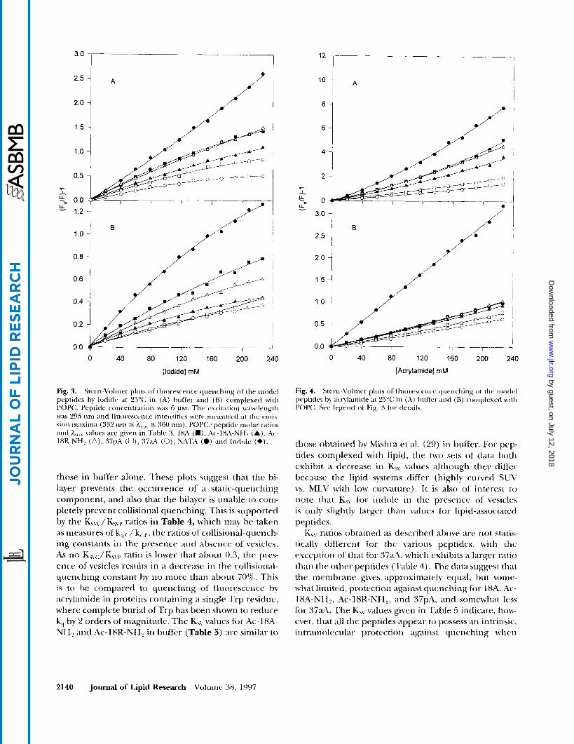
tively large and polar, is known to be limited to surface Trp (30); neither is capable of quenching fluorescence from buried Trp residues in globular proteins The quenching data are expressed as Stern-Volmer plots, which present fluorescence intensities as a function of quencher concentration. The experimental parameter derived from a typical Stern-Volmer plot is KsLr the Stern-Volmer constant (see Materials and Methods). For dynamic quenching the parameter of interest is k,, the bimolecular rate constant for quenching, which is related to Ksv by Ksv = k,T,,, where T,, is the fluores- cence lifetime in the absence of quencher. Assuming T, to be constant for the various peptides, Ks\, is thus a convenient measure for determining relative differ- ences in quencher accessibility to Trp when the pep- tides are partitioned into the bilayer. As KsLj is directly proportional to k,,, smaller values of KS\. indicate greater penetration into the bilayer. Typically, when static quenching is observed in addition to dynamic quench- ing, the contribution of each can be determined with the modified Stern-Volmer equation (Eq. 4, Materials and Methods). The Stern-Volmer plots for the five p e p tides are shown in Fig. 3 and Fig. 4, along with those for indole. It has been observed that only 10-40% of the nonpolar surface of indole is buried in the bilayer, suggesting its location at the hydrocarbon-water inter- face (31). The indole experiments then constitute a control which should be useful in gauging relative depths of penetration.
The most straightforward interpretation of a linear Stern-Volmer plot for an efficient quencher is in terms of a collisional-quenching mechanism, in which case the slope, Ksv, is a measure of the collisional-quenching constant (see Eq. 3) (20). However, most of the curves in Figs. 3 and 4 exhibit at least some degree of either upward or downward curvature. Stern-Volmer plots for I- quenching, with few exceptions, exhibit downward curvature (Fig. 3 ) , which is typical for this quencher (23). The deviation from linearity is not great, however, and approximate KsL7 values were determined from ini- tial slopes. The quenching of NATA fluorescence by acrylamide has been studied by others (32). Upward curvature in this case has been interpreted as the pres- ence of a static-quenching component, in addition to a collisional component, which at higher concentra- tions is due to acrylamide molecules being adjacent to the fluorophore at the moment of excitation, but which does not involve formal complexation. If this interpreta- tion is correct, then analysis of the upward curvature in the plots of Fig. 4A according to the modified Stern- Volmer equation is appropriate (20, 23). The Stern- Volmer plots for quenching by acrylamide in the pres- ence of vesicles (Fig. 4B) exhibit less curvature than
Gazzaru et al. Interactions of amphipathic helical peptides with lipid 2139
by guest, on July 12, 2018w
ww
.jlr.orgD
ownloaded from
2.5
2.0
1.5
1 .o
0.5
r - k 0.0
1.2
0.8 ,
0.6
I
0.4
0.2
00 0 40 80 120 160 200 240
[Iodide] mM
Fig. 3. Stern-Volrner plots of fluoresce-rice qiieric-hing ot tlir modcl peptides by iodide at 25°C; in (A) brrffer antl ( H ) complexed with POPC;. Peptide c o ~ ~ ~ ~ i i t r a t i ( i n was 6 P M . Thv excitation wavelength was 295 iirn illid lluor.esccrice intensities \+we incasiired a t the crni\- sioii maxima (332 iirn 5 h,,,,, 5 360 nnij. PC>P(:/peptitlc niolar ratios and h,,,,,, values arc given in TdhlC 3. 18A (W) , Ac-18A-Ncf2 (Aj, ik- IXK-NH, (A), :37pA (U), 37aA (0). NATA (0) ;111tl Inrlolc (e).
those in buffer alone. These plots suggest that the bi- layer prevents the occurrence of a static-quenching component, and also that the bilayer is unable to coin- pletely prevent collisional quenching. This is supported by the Ks\,JKSvl9 ratios in Table 4, which may be taken as measures of k,, ( , / k,,,,,, the ratios ofcollisional-quencli- ing constants in the presence aiid absence of vesicles. As no Ks\.c:/K5\rl, ratio is lower that about 0.3, the pres- ence of vesicles results in a decrease in the collisioiial- quenching constant by no more than about 70%. This is to be compared to quenching of' fluorescence by acrylamide in proteins containing a single Tip residue, where complete burial of Trp has been shown to reduce k,, by 2 orders of magnitude. The KhV values for Ac-18A- NH? antl Ac-lSK-NH, in buffer (Table 5 ) are similar t o
" 4 A
6
2.0 1 1.5 ~ 1
80 120 160 200 240 0 40
[Acrylamide] mM
those obtained by Mishra et al. (29) iii buffer-. For pep- tides complexed with lipid, the two sets of data both exhibit a decrease i n Ksy values although they differ because the lipid systems differ (highly cuivetl SUV vs. MLV with low curvature). It is also of interest to
note that K5\ for indole in the presence of' vesicles is only slightly larger than values for lipid-associated peptides.
K5v ratios obtained as described above are not statis- tically different for the \a-ious peptides, with thc exccptioti of' that for 37aA, which exhibits a larger ratio than the other peptides (Table 4). 'The &ita suggest that the membrane gives approximately equal, but some- what limited, protection against quenching for 1XA, Ac- 18A-NH2, Ac-lSR-NH,, and 37pA, and somewhat less for 3 7 d . The K,, values given i n 'Fable 5 indicate, how- ever, that all the peptides appear to possess an intrinsic, intl-amolcciilar protectioii against quenching whcn
2140 Journal of Lipid Research Voluinc. 98, 1997
by guest, on July 12, 2018w
ww
.jlr.orgD
ownloaded from
TABLE 4. Stern-Volnier quenching constants of peptide-POPC complexes relative to Stern-Volmer quenching constants of
peptides in buffer
K\\< ''/K\\,."
Prptidr Iodide' Actylamidc'
18A 0.52 t 0.06 0.35 f 0.07 Ac-l8A-N H, 0.46 2 0.09 0.29 2 0.01 Xc-1 HR-NH, 0.47 2 0.07 0.31 2 0.02 37pA 0.39 t 0.14 0.39 2 0.04 R7a-2 0.60 ? 0.12 0.56 2 0.02 NATA 0.64 2 0.08 0.68 2 0.10
"K,,, is the Stern-Volmer quenching constant of peptide-POPC
"K,,,, is the Stern-Volmer quenching constant of peptide in
'Values calculated from data in Tdbk 4, 2 SD.
complex.
huftcr.
compared to the control, NATA (Figs. 3A, 4). This self-protection effect is greatest for the 37-residue pep- tides and least for 18A. A useful parameter for learning about protection conferred by the bilayer, hence about depth-of-penetration, should be a ratio of Ksv obtained in the presence of vesicles (Ksvc:) to that obtained in buffer alone (Ksvr.), which effectively normalizes infor- mation about bilayer protection to the self-protection effect. These ratios are given in Table 4.
Calorimetric studies were conducted in order to com- plete the thermodynamic analysis. For these experi- ments peptides at initial concentrations between 350 and 4 0 0 ~ ~ were injected from a syringe in 10-pl ali- quots into a 1.365-m1 cell containing an excess of lipid to ensure essentially complete binding of peptide. In the syringe, prior to injection, peptides (P) are self-asso-
TABLE 5. Stern-Volmer quenching constant5 of the model peptides in buffer and the model peptide-POPC complexes
Iodide Acylamidr
POPC POP(: Peptide'' H u f k r <:omplex' Buffer Complex'
18A 8.0 2 0.4 4.2 t 0.4 1 1 2 2 3.9 i 0.2 Ac-18A-NH, 5.7 5 0.3 2.6 f 0.5 12.6 2 0.3 3.7 k 0.1 Ac-lSK-NH, 7.2 5 0.9 3.4 t 0.3 13 2 1 4.0 i 0.1 37pA 4.6 2 0.5 1.8 f 0.6 7.9 2 0.3 3.1 -+ 0.3 37aA 3 . 5 5 0 . 3 2 . 1 5 0 . 4 5 . 9 k 0 . 2 3 . 3 t o . 1 NATA 11.2 2 0.7 7.2 5 0.8 21 2 3 14.3 t 0.5 Indole ND" 2.5 5 1 ND 5.2 2 0.6
"Peptide concentration was 6 PM. ' 2 SD, number of experiments = 3. 'POPC/peptide molal- ratios are given in Tdhk 3. "Not determined.
6.4
6.0
5.6
8 8 5.2 I
4.8
4.4
4.0
0 200 400 600 800 1000 1200 1400
Time (sec)
Fig. 5. Thermograms for the dissociation of 1XA and binding of 18A to SUV at 5°C. The endotherms ( - - - ) for the dissociation werc oh- tained by injecting 10 pL aliquots of a 360 p~ solution of 18A into PBS. The exotherms (-) for the binding to SUV were obtained by injecting 10 WL aliquots of a 360 VM solution of 18A into an excess of lipid ( > I f " , see Materials and Methods).
ciated (P,,). It is assumed that peptide aggregates must dissociate before partitioning into the membrane oc- curs, or
hq. 7)
Figure 5 shows a typical thermogram for the parti- tioning of 18A into POPC S U V , together with a control in which peptide was titrated into buffer only. As pep- tide concentrations in the calorimeter cell were similar to those in the fluorescence experiments, at which pep- tides are monomeric, as mentioned previously, controls yield the heat of dissociation of peptide aggregates to monomers. Dissociation is seen to be an endothermic process, whereas the enthalpy for the entire process de- picted by Eq. 7 is exothermic. The enthalpy of parti- tioning, AH, can be calculated from AH = -
AH,,,,, where AH,,,,, is the observed enthalpy of parti- tioning and AH,,,,, the enthalpy of dissociation of self- associated peptides. The standard entropy, AS", of parti- tioning was obtained by using AS" = -(AG" - AH")/ T. It is assumed that AH') is approximately equal to AH. These thermodynamic parameters are summarized in Table 2.
In order to determine the secondary structure of the peptides their far-UV CD spectra were recorded in
Gazzam d al. Interactions of amphipathic helical peptides with lipid 2141
by guest, on July 12, 2018w
ww
.jlr.orgD
ownloaded from

TABLE 6. a-Helix content of model peptidea in buffer and complexed with POPC, 25°C:
r.nl/mol K kr.nl/mol 18A 1 3 49 36 ti.5 -26.6 -8.4 75 Ac-18A-NHp 38 5ti 18 3.2 -15.1 -4.2 26 Ac-1XR-NH, 14 62 48 8.6 ?L)A -11.2 6.5 37pA 26 12 I6 5.9 -24.2 -7.7 31 37aA 28 qY 21 7.8 -32.0 - 10. I 48
- < r
"AS",,,,,,, = -4.1 e.u./r-esidue (increase in number of helical residues), see Disciission. "AH,,,,,,, = - 1.3 heal/ (mol of rrsidue) (increase in nunibrr of helical residues), w e 1)iscussion
buf'fer and when complexed with POPC S W . These re- sults are summarized in Table 6.
DISCUSSION
The amphipathic helices formed by the 18-residue peptides, Ac- 18A-NH2 and Ac-18R-NH can form helices of length sufficient to cover the fatty acyl chains at the edge of a bilayer disc comprised of DMPC. As discoidal particles have been suggested to be the nascent form of HDL, and the preferred substrate for LCAT, studies of the interaction of the 18-residue peptides with phos- pholipids are of particular interest. The 37-residue pep- tides, 37pAand 37aA, which are dimers of 18A linked by either a Pro (37pA) or Ala (37aA), are of added interest because tandem duplication of amphipathic helices ex- ists in the exchangeable apolipoproteins, as do twists between polar and nonpolar faces and turns from one helical segment to another (4). Previous studies have shown that 18A and 18R (unblocked version of Ac-18R- NH,) both interact superficially with the bilayer surface of POPC MLV (33). Also, both 37pA and 37aA are capa- ble of partially penetrating a lecithin monolayer, both to a significantly greater extent than 36A (13). 37pA interacts optimally with large, DMPC MLV relative to both 36A and 37aA (13).
In the present study, a combination of isothermal ti- tration calorimetry and fluorescence spectroscopy has enabled us to obtain a comprehensive set of thermody- namic parameters that characterize the interaction of the 18- and 37-residue peptides with POPC S U V . These parameters, together with results from circular dichro- ism and fluorescence quenching studies, provide con- siderable insight regarding the molecular nature of the interaction.
Fluorescence studies show that lipid affinities are high, with partition coefficients on the order of 10" (Td-
ble 1). A comparison of these values indicates that af- finities, for S W , increase in the order Ac-lSR-NH? < 18A < 37aA < 37pA < Ac-18A-NH2. The K,, value for Ac-18A-NH2, with the greatest lipid affinity, is 7 times larger than that for Ac-18R-NH2, with the least. It is of interest to note that the K, value for the interaction of 18A with POPC S W is almost 50 times greater than the value obtained previously for binding of 18A to large, MLV, also comprised of POPC (33), thereby indicating the importance of vesicle size in determining lipid af- finity. The circular dichroism studies indicate that an increase in a-helical content occurs for all peptides upon interaction with the bilayer, with the increase be- ing greatest for Ac-18A-NH2 (48%) and least for 37pA (1696, Table 6). Massey, Gotto, and Pownall (34) have shown that the interaction of apoA-I1 and apoC-I11 with a variety of phospholipids occurs with a release of - 1.3 kcal per mole of residue converted from a random coil to or-helix. This is comparable to values determined by Chon and Scheraga ( 3 5 ) and Scholtz et al. (36) for simi- lar conversions. This indicates that approximately -11.2 kcal/mol (65% of'the total AH of partitioning) and -7.7 kcal/mol (31% of the total AH of parti- tioning) can be attributed to an increase in helicity of Ac-18R-NH2 and 37pA, respectively, upon int.eraction with vesicles.
The thermodynamic data indicate that peptidc- vesicle interactions are exothermic in all cases and ac- companied by relatively small, negative entropy changes (Table 2). As the calorimetric data were corrected for self-association, the enthalpies in Table '2 correspond to monomeric peptides interacting with vesicles. It might appear that peptide-vesicle interaction is driven by the removal of the hydrophobic residues of monomeric peptides from exposure to water. However, such a clas- sic hydrophobic event would be entropically driven with t:ery little enthalpy change (37). I n contrast, interac- tions between these peptides and highly curved bilayer surfaces are enthalpically driven, a phenomenon that
2142 Journal of Lipid Research Volume 38, 1997
by guest, on July 12, 2018w
ww
.jlr.orgD
ownloaded from

has been observed elsewhere for interaction of other amphipathic molecules with highly curved membranes (38,39). The negative entropy changes indicate the ab- sence of a large net release of water from hydration shells that would occur in a classic hydrophobic event, but rather are likely due to a combination of other fac- tors, such as the statistical effect of decreasing the num- ber of independent particles in the system, thereby de- creasing the entropy; an increase in peptide helicity likewise decreases entropy. This contribution to the de- crease in entropy has been estimated (40), as -4.1 en- tropy units (e.u., cal/(mol K)) per residue. Using this number, together with the circular dichroism results, the change in entropy due to increase in helicity varies from about -13 e.u./peptide for Ac-18A-NH2 to about -35 e.u./peptide for Ac-18R-NH2.
As was mentioned previously, a number of small am- phiphiles, in addition to the peptides in this study, have been observed to interact with bilayer surfaces in enthal- pically driven processes. A general model of interaction has been published by Pownall et al. (41). Features unique to the systems studied here are similar for all the peptides. Amounts of a-helical content vary for free peptides in the aqueous phase. The partitioning process consists of an increase in helical content, accompanied by partial penetration of the amphipathic helices be- yond the polar head groups into the hydrophobic inte- rior of the bilayer. That there is some interaction with the hydrophobic interior of the membrane is supported by the observation that the affinity of 18A for large, POPC MLV is 1-2 orders of magnitude smaller (33) than it is for smaller, more highly curved POPC S U V in which the isothermal lateral compressibility is greater and the accessibility to the fatty acyl chains correspond- ingly greater. However, the existence of only partial pro- tection by the bilayer against quenching by the water- soluble quenchers I- and acrylamide to all the peptides and to indole is consistent with the idea that peptide- membrane interaction is largely confined to the sur- face, with only partial penetration of the peptides beyond the polar head groups into the hydrophobic re- gion of the bilayers. The extent of penetration is about the same for the 18-residue peptides and 37pA, but somewhat less for 37aA. The relative lack of protection for all the peptides may be due partly to the accessibility of water to the interior of the bilayer (42). This may explain, in part, why partitioning of the peptides is not an entropically driven, classic hydrophobic event. The interaction is enthalpically driven, with a significant por- tion of the driving force due to increased peptide helicity that occurs upon partitioning. The increase in peptide helicity is likely due to the replacement of water-peptide backbone hydrogen bonds with intrapeptide hydrogen
bonds as the nonpolar side chains encounter the hy- drophobic interior of the bilayer. The majority of the re- mainder of the enthalpy is probably due to van der Waals interactions between the phospholipid acyl chains and the nonpolar faces of the peptides (38).
The lipid affinities are high for all the model pep- tides. It appears that the arrangement of charge in a class A amphipathic helix (Ac-18A-NH2) leads to greater lipid affinity than the arrangement in which charges are reversed (Ac-18R-NH2). However, there is little difference between Ac-18A-NH2 and Ac-18R-NH2 with respect to bilayer penetration. The structural basis for the difference in lipid affinity is not clear. Table 2 indicates that the enthalpies of interaction for both Ac- 18R-NH2 and Ac-18A-NH2 are similar, while the mag- nitude of entropy decrease is about 30% greater for Ac-18R-NH2. The CD results indicate a much larger in- crease in helicity upon interaction with vesicles for Ac- 18R-NH2 (65%) than for Ac-l8A-NH2 (26%) (Table 6). According to estimates given above, enthalpic and en- tropic contributions to free energies due to changes in helical content essentially cancel each other. When en- thalpies and entropies in Table 2 are corrected for esti- mated contributions due to increases in helicity, bilayer partitioning is seen to be enthalpically driven for Ac- 18A-NH2 but is favored both enthalpically and entropi- cally for Ac-18R-NH2, with entropy making the larger contribution.
The 37-residue peptides, 37pA and 37aA, individual segments of which form class A amphipathic helices, have KP values comparable to each other, but less than that for Ac-18A-NH2 and greater than that for Ac-18R- NH,. The presence of either a Pro (37pA) or Ala (37aA) link between helical segments would impart about a 100" twist between the interfacial planes if each seg- ment were fully helical, as mentioned above. Several res- idues at the center of these peptides, therefore, are probably non-helical in the presence of lipid, allowing the interfaces of the remaining helices to align tangen- tially to the bilayer surface, thereby minimizing contacts between the hydrophobic faces and the aqueous me- dium. This idea is supported by the CD results, which indicate a lower helical content for 37-residue peptide- vesicle complexes (less than 50%) than for 18-residue peptide-vesicle complexes. Although there is very little difference in lipid affinity between 37pA and 37aA for the highly curved surface of S U V , it is interesting to note that results of a previous study indicated that 37pA has a greater affinity for large, DMPC MLV than 37aA does (13). The importance of vesicle curvature in de- termining lipid affinity has already been mentioned above and will be emphasized in the accompanying pape r . I
Garrara rt al. Interactions of amphipathic helical peptides with lipid 2143
by guest, on July 12, 2018w
ww
.jlr.orgD
ownloaded from

APPENDIX
The binding of model peptides (P) to lipid has been treated as an equilibrium partitioning between the aqueous and lipid phases
The partition coefficient, K,,, is defined as
Eq. 9)
where XL is the mole fraction of P in the lipid phase and Xi! is the mole fraction of P in the aqueouq phase.
Conservation requires that
[PI = [PI1 + [PI1
where [PI is the total P concentration and [P,] and [PI ] are the concentrations of P in the aqueous and lipid phases respectively. The fraction of P 111 the aqueous phase, is
hq. 10)
Ki4. 1 I )
I t has been shown that the intrinsic fluorescence in- tensity of model peptides in buffer, F,,, increases when partitioned into lipid (Fig. 1). The increased fluorcs- cence intensity, F, , is directly proportional to X;,. The total intensity is the sum of FI, and the fluorescence in- tensity of P partitioned into lipid. When the fluores- cence data are plotted as F1 /FI$ lipid concentration, [ I , ] , a hyperbolic curve is obtained (Fig. 2). This binding isotherm can be expressed as
I.,. 12)
where C,, and C, are proportionality constants corrc- sponding to P in the aqueous and lipid phases, respec- tively. For reasons discussed above, when [L] = 0 must equal 1. At saturation cp = 0 and C;,, is equal to F ,,,., ~, where F ,,,,, ~ is the value of FJF,, tinder saturating conditions. Substitution of these values for C,, and CI into Eq. 12 gives the following equation for F, /Fl,
F1 - - cp(1 - F,,,,,,) Kq. I 3) Fl,
Experimentally, FH is determined initially for a vol- ume, V,,, of P solution in the absence of lipid. Lipid at a concentration [L,i] is then added, typically in aliqiiots of 50-200 pL, to give a total volume of added lipid, which is used to calcalate the total d u m e of solution, V, = i'<, + \',,, and the number of moles of lipid, r i l =
[L,,,]V,,. Assuming that the nurnber of moles of P i n thc
aqueous phase, ti;, is negligible compared to the nuin- her of moles of water, II,,, X> becomes
x; = !$ I:!/. 14) [I\\
Using Eqs. 9, 10, and 14, the following quadratic equa- tion for [P,] can he obtained
[55.6] [PI i Kl, V, i KP 55 6 r i l 0 = [P,]! - + -- + [PI [P,] + L 2 - p
I:(/. 1 5 )
where [55.6] is the concentration of water. Substituting [I,] for 11, /V, and using the quadratic formula t o wl\c for [P,] gives
+ [L] + [PI - -\in [ 5.5.61
K,, ___-
- k,q. 16n) 2
[P,l =
where D is the discriminant, given by
D = (M + [I.] + [ P l y - 4[55.6] [PI K.1. KI,
k/ 160)
The negative dD term in Eq. 16a is used to satisfy the coiidition 0 5 [P,] 5 [PI. Dividing Eq. 16a by [PI and substituting into Eq. 13 gives an expression for the bind- ing isotherms in tvrms o f K13.
Kq. 17)
The KIJ value for each model peptide was obtained by curve fitting the respective binding isotherm to Eq. 17.
This rescwch WAS supported i n part by National Institutes of. Health Program Prqjects HL 22633 and HL, 34343. ~ ~ ~ o n l o c r ' l ~ ~ l ~rrrlrrrri 20 ~~rtr171brr 19% mnd ;?I l f71 iWd / i j l l n 2 ~ , ~ f o r r /L/yi.
REFERENCES
Brewer, H. B., Jr., T. Fairwcll, A. LaRue, K. Konan, A. Hauser., and T. J. BronLert. 1978. The amino arid sc- quence of' human apoA-I, an apolipoprotein isolated from high density lipoproteins. BiocIirm. Wiofi/qs. Rev. ( h n t -
Segrest, J. P., K. L.. Jackson, J. I). Morrisett, and A. M . Gotto, Jr. 1974. A molecular theory of lipid-protein inter- actions in the plasma lipoproteins. I+,XS fM. 3 8 2-47- 253. Srgrcst,.l. P., H. l k I.,oof., J. (;. I)olilrnail, (:. (;. Bt-ouillrttc'.
T I I U ~ Z . 80: 623-630.
2144 Journal of Lipid Research Volumr 38, I997
by guest, on July 12, 2018w
ww
.jlr.orgD
ownloaded from

and G. M. Anantharamaiah. 1990. Amphipathic helix mo- tif: classes and properties. Proteins. 8: 103-117.
4. Segrest, J. P., M. K. Jones, H. De Loof, C. G. Brouillette, Y. V. Venkatachalapathi, and G. M. Anantharamaiah. 1992. The amphipathic helix in the exchangeable apoli- poproteins: a review of secondary structure and function. /. l i p i d Res. 33: 141-166.
5. 'Anantharamaiah, G. M., M. K. Jones, and J. P. Segrest. 1993. The Amphipathic helix. R. M. Epand, editor. CRC Press, Boca Raton, FL. 109-142.
6. Venkatachalapathi, Y. V., M. C. Phillips, R. M. Epand, R. F. Epand, E. M. Tytler, J. P. Segrest, and G. M. Anan- tharamaiah. 1993. Effect of end-group blockage on the properties of a class A amphipathic helical peptide. Pro- trinJ. 1 5 349-359.
7. Chung, B. H., G. M. Anantharamaiah, C. G. Brouillette, T. Nishida, a n d J . P. Segrest. 1985. Studies of synthetic peptide analogs of the amphipathic helix: correlation of structure with function. ,/. Bid . <:hem. 260: 10256-10262.
8. Anantharamaiah, G. M., J. L. Jones, C. G. Brouillette, C. F. Schmidt, B. H. Chung, T. A. Hughes, A. S. Bhown, and J. P. Segrest. 1985. Studies of synthetic peptide ana- logs of the amphipathic helix. Correlation of structure with function. ,/. Biol. Chrm. 260: 10248-10255.
9. Lund-Katz, S. L., M. C. Philips, V. K. Mishra,J. P. Segrest, and G. M. Anantharamaiah. 1995. Microenvironments of basic amino acids in amphipathic alpha helices bound to phospholipids: '"C NMR studies using selectively labeled peptides. Biochemistry. 92: 9219-9226.
10. Fairman, R., K. R. Shoemaker, E. J. York, J. M. Stewart, and K. L. Baldwin. 1989. Further studies of the helix di- pole model: effects of a free alpha-NH -,+ or apha-COO- group on helix stability. Protrins. 5: 1-7.
11. Jorgenson, E. V., G. M. Anantharamaiah, J. P. Segrest, .J. T. Gwynne, and S. Handwerger. 1989. Synthetic amphi- pathic peptides resembling apolipoproteins stimulate the rdease of human placental lactogen. J. Biol. Chrm. 264: 92 15-92 19.
12. Nakagawa, S. H., H. S. H. Lau, F. J. Kezdy, and E. T. Kai- scr. 1985. The use of polymer-bound oximes for the syn- thesis of large peptides usable in segment condensation: synthesis of a 44 amino acid amphiphiic peptide model of apolipoprotein A-I. J. A m . Chrm. Soc. 107: 7087-7092.
13. Mishra, V. K., M. M. Palgunachari, S. Lund-Katz, M. C. Phillips, J. P. Segrest, and G. M. Anantharamaiah. 1995. Ef'fects o f the arrangement of tandem repeating units of class A amphipathic a-helices in lipid interaction. ,I. Bid. {;hrm. 270: 1602-161 1.
14. Roseman, M. A,, B. R. Lentz, B. Sears, D. Gibbes, and T. E. Thompson. 1978. Production of large unilamellar vesicles by a rapid extrusion procedure. (X". Phy.~. 1,ip ids. 21: 205-222.
15. Barenhob, Y., D. Gibbes, B. J . Litman, J. Goll, T. E. Thompson, and F. D. Carlson. 1977. A simple method for the preparation of homogeneous phospholipid vesicles. Hiochrmikhy. 16: 2806-2810,
16. McLean, L. K., and M. C. Phillips. 1982. Cholesterol de- sorption from clusters of phosphatidylcholine and choles- trrol in unilamellar vesicle bilayers during lipid transfer or exchange. 13iochrmistry. 21: 4053-4059.
17. Sokoloff, I,., and G. H. Rothblat. 1974. Sterol to phospho- lipid molar ratios of L cells with qualitative and quantita- tive variations of cellular sterol. Proc. Soc. Exp. B i d . Mrrl. 146: 1166-1 172.
18. Lehrere, S. S. 1971. Solute perturbation of protein fluo- rescence. The quenching of the tryptophyl fluorescence of model compounds and of lysozyme by iodide ion. Rio- chemist?. 10: 3254-3263.
19. Calhoun, D. B., J. M. Vanderkooi, and S. W. Englander. 1983. Penetration of small molecules into proteins stud- ied by quenching of phosphorescence and fluorescence. Biochrmistry. 22: 1533-1539.
20. Eftink, M. R., and C. A. Ghiron. 3981. Fluorescence quenching studies with proteins. Anal. Biochrm. 114 199- 227.
21. k'dng,J. T., C. S. C. Wu, and H. M. Martinez. 1986. Calcula- tion of protein conformation from circular dichroism. Methods Enzymol. 130: 208-269.
22. Morrisett, J. D., J. S. K. David, H . J . Pownall, and A. M. Gotto, Jr. 1973. Interaction of an apolipoprotein (apo- LP-alanine) with phosphatidylcholine. Riochrmistq~. 12:
23. Lakowicz, J. K. 1983. Principles of Fluorescence Spectros- copy. Plenum Press, New York. 187-2 I 1.
24. Trauble, H., G. Middelhoff, and V. W. Brown. 1974. Inter- action of a serum apoliprotein with ordered and fluid lipid bilayers. Correlation between lipid and protein structure. 1C%7S Let t . 49: 269-275.
25. Weinberg, R. B., and M. K. Jordan. 1990. Effects of phos- pholipid on the structure of human apolipoprotein A- N . /. Bid . Chrm. 265: 8081-8086.
26. 'Weinberg, R. B., M. K. ,Jordan, and A, Steinmetz. 1990. Distinctive structure and function of human apolipopro- tein variant apoA-IV-2. ,/. H i d . Chrvn. 265: 18372-18378.
27. Seelig,J., S. Nebel, P. Ganz, and G. Bruns. 1993. Electro- static and nonpolar peptide-membrane interactions. Lipid binding and functional properties of somatostatin in analogues of charge z = + 1 to z = +3. Riochrmislry. 32: 9714-9721.
28. Matsuzaki, K., 0. Murase, H. Tokuda, S. Funakoshi, N. Fyjii, and K. Miyajir. 1994. Orientational and aggrega- tional states of magainin 2 in phospholipid bilayers. Hio- chrmist9. 33: 3342-3349.
29. Mishra, V. R, M. M. Palgunachari, J. P. Segrest, and A. M. Anantharamaiah. 1994. Interactions of synthetic peptide analogs of the class A amphipathic helix with lipids. ,/. Bid. Chrm. 269 7185-7191.
30. Kurzban G. P., G. Gitlin, E. A. Bayer, M. Wilchek, and P. M. Horowitz. 1989. Shielding of tryptophan residues of avidin by the binding of biotin. niochPmz.slry. 28: 8537- 854'2.
31. U'imley, W. C., and S. H. White. 1993. Membrane parti- tioning: distinguishing bilayer effects from the hydropho- bic effect. Biochemistry . 32: 6307-631 2.
32. Eftink, M. R., and C. A. Ghiron. 1976. Fluorescence quenching of indole and model micelle systems. ,/. Phys. Chrm. 80: 486-493.
33. Spuhler, P., G. M. Anantharamaiah, J. P. Segrest, a n d J . Seelig. 1994. Binding of apoliprotein A-I model peptides to lipid bilayers. ,/. R i d . Chon. 269: 23904-23910.
34. Massey, J. B., A. M. Gotto, Jr., and H. J. Pownall. 1979. Contribution of a-helix formation to their enthalpy of as- sociation with phospholipid. ,/. H i d . Chrm. 254 9359- 9361.
35. Chou, P. Y., arid H. A. Scheraga. 1971. Calorimetric mca- surement of enthalpy change in the isothermal helix-coil transition of poly-I.-lysine in aqueous solution. Rio/mly- m m . 10: 657-689.
1290-1299.
Gazzara et nl. Interactions of amphipathic helical peptides with lipid 2145
by guest, on July 12, 2018w
ww
.jlr.orgD
ownloaded from
36. Scholtz, J . M., S. Marqusee, R. Baldwin, E. J. York, J. M. Stewart, M. Santoro, and P. W. Boleri. 1991. Calorimetric determination of the enthalpy change for the a-helix to coil transition of an alanine peptide in water. Proc. Natl. Acud. Sci . USA. 88: 2854-2858.
37. Tanford, C. H. 1973. The Hydrophobic Effect: Formation of Micelles and Biological Membranes. Wiley, New York. 1-3.
38. Seelig, J. P., and Ganz. 1991. Nonclassical hydrophobic effect in membrane binding equilibria. Biochemistry. 30: 9354-9359.
39. Beschiaschvili, G., and J. Seelig. 1992. Peptide binding to
lipid bilayers. Nonclassical hydrophobic effect and mein- brane-induced pK shifts. Biochemist9 . 31: 10044- 10053.
40. Tanford, C . 1962. Contributions of hydrophobic interac- tions to the stability of the globular conformations of pro- teins. J. Am. Chem. Soc. 84: 4240-4247.
41. Pownall, H. J., J. B. Massey,J. T. Sparrow, and A. M. Gotto, Jr. 1987. Plasma Lipoproteins. A.M. Gotto, Jr., editor.
42. Jacobs, R. E., and S. H. White. 1989. The nature of hy- drophobic binding of small peptides at the bilayer inter- face: implications for the insertion of transhilayer helices. Biochemistly. 28: 3421-3437.
l % a ~ i ~ . 95- 127.
2146 Journal of Lipid Research Volume 38, 1997
by guest, on July 12, 2018w
ww
.jlr.orgD
ownloaded from