7/31/2017 Compound Light Microscopy · PDF fileCompound Light Microscopy ... appear brighter...
7
7/31/2017 1 Microscopy Chapter 3 BIO 440 Compound Light Microscopy • This is what we use in the laboratory • A series of finely ground lenses form a magnified image • Specimen is illuminated with visible light Fig. 3.1 Things to remember . . . • Magnifying power – ratio of the object’s apparent size when viewed through a lens compared to the appearance of the object when viewed with the naked eye from a distance of 25 cm • How to calculate the total magnification of a specimen
Transcript of 7/31/2017 Compound Light Microscopy · PDF fileCompound Light Microscopy ... appear brighter...
7/31/2017
1
Microscopy
Chapter 3
BIO 440
Compound Light Microscopy
• This is what we use in the laboratory
• A series of finely ground lenses form a
magnified image
• Specimen is illuminated with visible light
Fig. 3.1
Things to remember . . .
• Magnifying power – ratio of the object’s
apparent size when viewed through a lens
compared to the appearance of the object
when viewed with the naked eye from a
distance of 25 cm
• How to calculate the total magnification of a
specimen
7/31/2017
2
Things to remember . . .
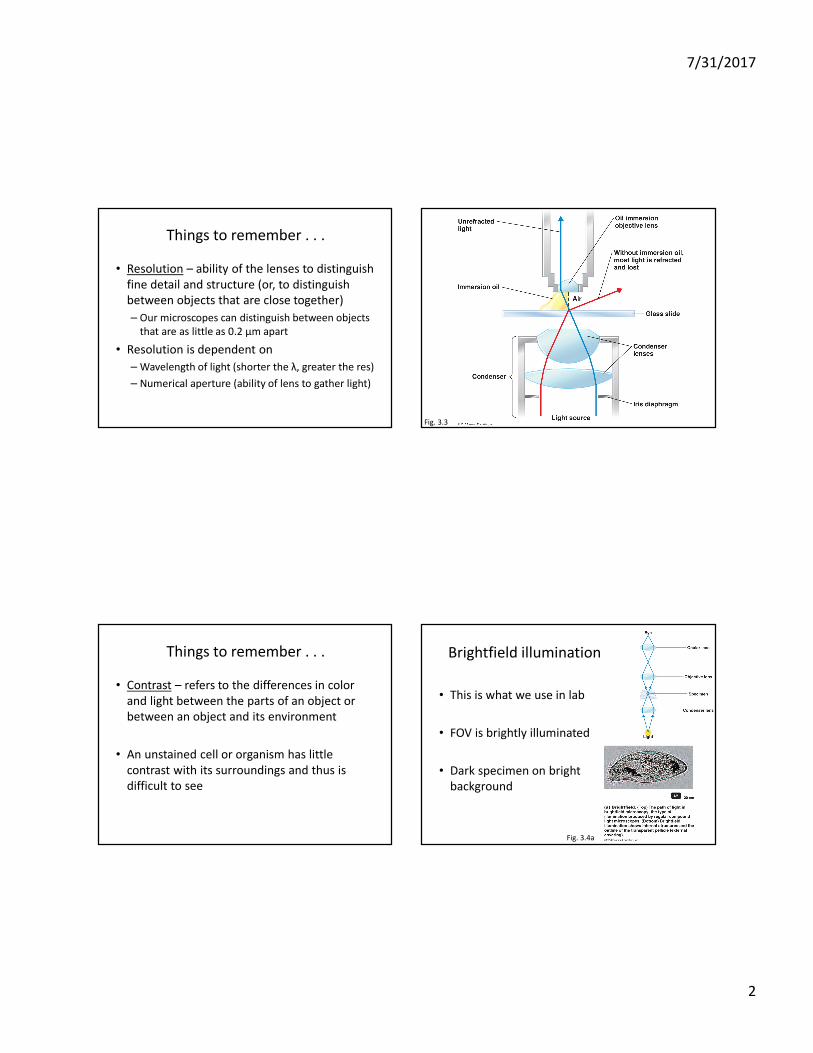
• Resolution – ability of the lenses to distinguish
fine detail and structure (or, to distinguish
between objects that are close together)
– Our microscopes can distinguish between objects
that are as little as 0.2 μm apart
• Resolution is dependent on
– Wavelength of light (shorter the λ, greater the res)
– Numerical aperture (ability of lens to gather light)
Fig. 3.3
Things to remember . . .
• Contrast – refers to the differences in color
and light between the parts of an object or
between an object and its environment
• An unstained cell or organism has little
contrast with its surroundings and thus is
difficult to see
Brightfield illumination
• This is what we use in lab
• FOV is brightly illuminated
• Dark specimen on bright
background
Fig. 3.4a

7/31/2017
3
Darkfield Microscopy
• Specimen appears light on
a dark background
• Handy in situations where
we want to see live cells,
cells are hard to see in
light or do not stain well
• Treponema pallidum –
causative agent of syphilis
Fig. 3.4b
Phase-Contrast
• Not necessary to fix or stain specimens, so allows us to see living microbes
• Separates illuminating light from light refracted off of specimen
• Better cellular detail
• Light travels faster through thinner areas and structures appear brighter
• Light travels slower through thicker areas and structures appear darker
Fig. 3.4c
Differential Interference Contrast (DIC)
• Uses two beams of light which pass through
prisms
• Resolution better than standard phase-
contrast
• Image brightly colored and appears 3D
Fig. 3.5
Fluorescence Microscopy
• Takes advantage of fluorescence, the ability of substances to absorb short wavelengths of light (UV) and give off light at a longer spectrum
• Some organisms fluoresce naturally under UV light, those that do not can be stained with fluorochromes
– (i.e. fluorescein isothiocyanate - FITC)
• Bright microbes on dark background
• Fluorescent – antibody (FA) technique (aka immunofluorescence)
7/31/2017
4
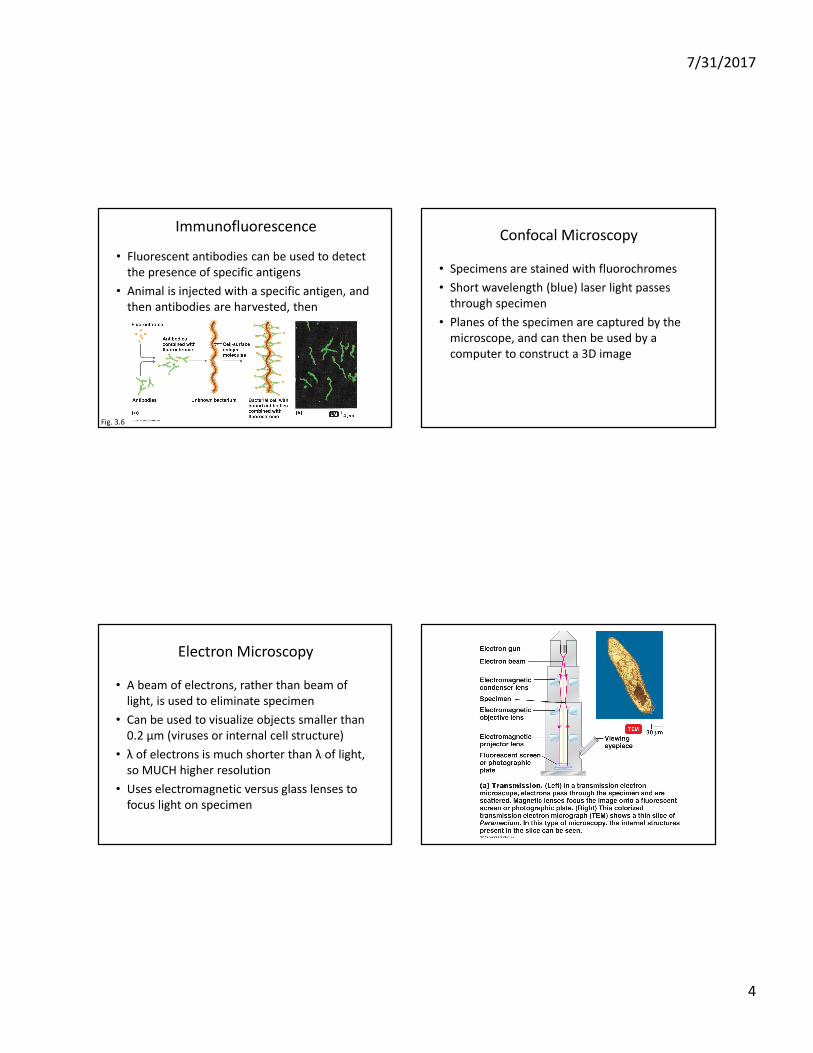
Immunofluorescence
• Fluorescent antibodies can be used to detect
the presence of specific antigens
• Animal is injected with a specific antigen, and
then antibodies are harvested, then
Fig. 3.6
Confocal Microscopy
• Specimens are stained with fluorochromes
• Short wavelength (blue) laser light passes
through specimen
• Planes of the specimen are captured by the
microscope, and can then be used by a
computer to construct a 3D image
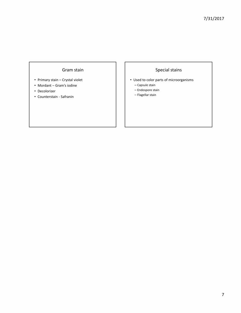
Electron Microscopy
• A beam of electrons, rather than beam of
light, is used to eliminate specimen
• Can be used to visualize objects smaller than
0.2 μm (viruses or internal cell structure)
• λ of electrons is much shorter than λ of light,
so MUCH higher resolution
• Uses electromagnetic versus glass lenses to
focus light on specimen

7/31/2017
5
Transmission electron microscopy
• Visualize viruses or internal intrastructure
• Ultrathin sections of specimen (100 nm)
• Image produced on fluorescent screen or
photographic plate
• 10,000X – 100,000X magnification
• High resolution (10 pm)
• Can use stains (mineral salts) to enhance
contrast
• Black and white images, 2D
Scanning electron microscopy
• Visualize surface features of intact cells and
viruses
• Image produced on viewing screen or
photographic plate
• 1,000X – 10,000X magnification
• High resolution (10 nm)
• Black and white images, 3D
Preparation of microscopic specimens
• Staining helps us localize and visualize small,
colorless microbes on microscope slides
• But before any staining occurs, we must first
“fix” the specimen
• Fixing the specimen
– Adhesion of the specimen to the slide
– Kills the specimen
– Preserves specimen with minimal distortion

7/31/2017
6
Heat fixation
• Make a smear
• Air dry/slide warmer
• Pass the slide (specimen side up) through the
flame of a Bunsen burner several times
• Once the heat fixation is completed, you can
go ahead and apply stain to the slide
Stains
• Stains are salts that are composed of a cation
and anion
• One of those ions is colored, and is referred to
as the chromophore
• Basic dyes are cationic, meaning that the
chromophore has a positive charge
– Crystal violet, methylene blue, safranin
• Acidic dyes are anionic, meaning that the
chromophore has a negative charge
– Eosin, nigrosin
Simple stains
• An aqueous or alcohol solution of a single
basic dye
• Good for visualizing cell shape and
arrangement
Differential stains
• Use more than one stain to differentiate
cellular components
• Used to visual structural differences between
different types of bacteria
– Gram stain
– Acid-fast stain
• Mycobacterium tuberculosis
• Mycobacterium leprae
7/31/2017
7
Gram stain
• Primary stain – Crystal violet
• Mordant – Gram’s iodine
• Decolorizer
• Counterstain - Safranin
Special stains
• Used to color parts of microorganisms
– Capsule stain
– Endospore stain
– Flagellar stain