2014 Characterization of thermally stable gamma alumina fibres ...
28
1 Characterization of thermally stable gamma alumina fibres biomimicking sisal Mónica Benítez-Guerrero a * , Luís A. Pérez-Maqueda b , Pedro E. Sánchez-Jiménez b and José Pascual- Cosp a . a Departamento de Ingeniería Civil, Materiales y Fabricación, Universidad de Málaga, C. Dr. Ortiz Ramos s/n, 29071 Málaga, Spain. b Instituto de Ciencia de Materiales de Sevilla, CSIC-Universidad de Sevilla, C. Américo Vespucio 49, 41092 Sevilla, Spain * Corresponding author, Address: Departamento de Ingeniería Civil, Materiales y Fabricación, Universidad de Málaga, C. Dr. Ortiz Ramos s/n, 29071 Málaga, Spain. Tel.: +34951952594. E-mail address: [email protected] Abstract Mesoporous gamma alumina fibres of high surface area, stable up to 1000 ºC, were synthesized by bioreplica technique using sisal fibres as templates. Alumina formation during pyrolysis and calcination of fibres infiltrated with aluminium chloride solution has been studied, paying special attention to the interaction between the precursor and sisal fibres, using several experimental techniques such as ATR-FTIR, coupled TG-FTIR and thermo-XRD analysis. The morphology and microstructure of the resulting alumina fibres were characterized using SEM and TEM. The crystallographic analysis of the alumina sample performed by electron and X- ray diffraction suggests that fibres are constituted by and -Al 2 O 3 crystallites, whose chemical structure was confirmed by ATR-FTIR and Al 27 -MAS-NMR. The specific surface area and porosity of ceramic fibres were determined by N 2 and CO 2 adsorption-desorption measurements. Resulting alumina fibres retain high specific surface areas of 200 m 2 /g and 150 m 2 /g even after calcination at 1000 °C for 15 h in dry air and for 4 h in wet air, respectively. Keywords: biomorphic alumina; biomimetic alumina; inorganic salt infiltration; sisal fibre biotemplate.
Transcript of 2014 Characterization of thermally stable gamma alumina fibres ...

1
Characterization of thermally stable gamma alumina fibres biomimicking sisal
Mónica Benítez-Guerrero a *, Luís A. Pérez-Maqueda b, Pedro E. Sánchez-Jiménez b and José Pascual-
Cosp a.
a Departamento de Ingeniería Civil, Materiales y Fabricación, Universidad de Málaga, C. Dr. Ortiz Ramos s/n, 29071
Málaga, Spain.
b Instituto de Ciencia de Materiales de Sevilla, CSIC-Universidad de Sevilla, C. Américo Vespucio 49, 41092 Sevilla,
Spain
* Corresponding author, Address: Departamento de Ingeniería Civil, Materiales y Fabricación, Universidad de
Málaga, C. Dr. Ortiz Ramos s/n, 29071 Málaga, Spain. Tel.: +34951952594. E-mail address:
Abstract
Mesoporous gamma alumina fibres of high surface area, stable up to 1000 ºC, were
synthesized by bioreplica technique using sisal fibres as templates. Alumina formation during
pyrolysis and calcination of fibres infiltrated with aluminium chloride solution has been studied,
paying special attention to the interaction between the precursor and sisal fibres, using several
experimental techniques such as ATR-FTIR, coupled TG-FTIR and thermo-XRD analysis. The
morphology and microstructure of the resulting alumina fibres were characterized using SEM
and TEM. The crystallographic analysis of the alumina sample performed by electron and X-
ray diffraction suggests that fibres are constituted by and -Al2O3 crystallites, whose chemical
structure was confirmed by ATR-FTIR and Al27-MAS-NMR. The specific surface area and
porosity of ceramic fibres were determined by N2 and CO2 adsorption-desorption
measurements. Resulting alumina fibres retain high specific surface areas of 200 m2/g and 150
m2/g even after calcination at 1000 °C for 15 h in dry air and for 4 h in wet air, respectively.
Keywords: biomorphic alumina; biomimetic alumina; inorganic salt infiltration; sisal fibre
biotemplate.

2
1. Introduction
High surface area mesoporous aluminas with narrow pore distribution have been the
subject of extensive research employing different strategies [1]. However, the synthesis of
aluminas with hierarchically ordered pore structure attracts more and more attention [2, 3], due
to the highly effective transport phenomena and the easy diffusion of reactants and products
when pores of different sizes are interconnected. Generation of controllable hierarchical pores
has been fundamentally carried out through agents acting as porosity generators like organic
additives and surfactants [4, 5], foams, emulsions or masks based on replica procedure [6], as
well as by using others methods as hydrothermal treatment [7], and recently, nanocasting
technique [8]. In any case, most of the employed processes generally involve the use of
expensive porosity agents which must be finally removed by extraction or calcination.
A method of ceramic synthesis that has attracted significant attention in the scientific
community is the variant of the infiltration-replica technique which uses natural masks or bio-
templates [9-11]. Through bio-replica, it is possible to generate ceramic materials which
faithfully reproduce the morphology and structure of the starting preforms in an easy way, with
a porous architecture scaled from nano-, micro- to macro-sizes, with isotropic, anisotropic,
homogeneous or heterogeneous properties, which are either difficult or very costly to produce
by conventional methods [12]. The interest on porous bio-replicas based on metal oxides such
as Al2O3, TiO2 and ZrO2 has been increasing in the last decades for their interest in
photocatalysis and photovoltaic, specially for manufacturing of gas sensors, filters, catalysts
carriers, etc. [13,14]. Lignocellulosic masks stand out among the wide range of materials
suitable to be used as templates [15,16], due to their ecological and economical origin, obtained
from renewable and even residual resources, which make it interesting as valorization process
[17,18].
The textural and crystallographic stability of aluminas are of great importance for
processes involving high temperatures, such as regeneration of catalyst beds and membranes,
being the collapse of alumina structure a limiting factor of the process efficiency. High
temperatures and high residence times reduce the specific surface area of alumina due to the

3
transformation to alpha phase, which is accompanied by coalescence and sintering phenomena.
Thus, the attainment of thermally stable alumina keeping high specific surfaces areas is the goal
of many researches. Ordered mesoporous aluminas exhibit SBET values ranging between 200 and
500 m2/g after calcination at temperatures between 450 and 700 ºC [1], and exceptionally 700 -
800 m2/g [19,20]. The surface area diminishes considerably at temperatures as high as 1000 ºC.
It was described the cationic surfactant synthesis of -Al2O3 with a resulting surface of 130 m2/g
after a thermal treatment at 1000 ºC for 2 h [21]. At the same calcination temperature, a mixture
of ()-Al2O3 with 90 m2/g was obtained by nanocasting [22]. Recently, it was reported the
generation of gamma-alumina monoliths through gel-casting of mesoporous alumina powder
and polymerization additives, reaching 110 m2/g after calcination at 1000 °C for 10 h [23].
Different works have dealt with the synthesis of porous alumina employing cellulose
derived templates. The results obtained up to the moment consist of sintered alpha alumina
cellular ceramics [24-26] and mesoporous aluminas which, as the ones obtained by conventional
synthetic methods, do not maintain elevated specific surface area at high temperatures. Using
filter paper as cellulose support, Shigapov et al. [27] synthesized alumina with 322 m2/g after
calcination at 800 °C for 2 h, which decreases to 21 m2/g after treatment at 1050 °C for 12 h.
Patel and Padhi [28] obtained at 1000 °C alumina fibres with 56 m2/g by infiltration of jute
fibres. Fan et al. [29], through natural cotton fibres, attained -Al2O3 with surfaces areas of
127.6 and 125.1 m2/g after a calcinations period of 2 h at 800 and 1000 °C, respectively.
In the present paper, it is described the synthesis of thermally stable alumina fibres of
high specific surface area up to 1000 ºC, which reproduce the morphology of sisal fibres. In a
first part, changes experimented by the lignocellulosic template after their infiltration with the
inorganic precursor are presented, as well as thermal phenomena occurring during
transformation to alumina during pyrolysis and calcinations stages. In a second part, the
composition, morphology and structure of the resulting alumina ceramic fibres are
characterized.

4
2. Experimental section
2.1. Synthesis
Natural sisal fibres used in this work, named NS, were provided by Cayetano García del
Moral S. L. (Cabra de Santo Cristo, Jaén, Spain). The fibres, manually cut to 2-4 mm length,
were subjected to an alkalinization processes to conditioning the surface of the fibres before the
infiltration stage. The alkali treatment was performed for 2 h at room temperature with a 4%
NaOH solution (Panreac, 231686) , followed by exhaustive washing with distilled water. Acetic
acid drops were added in the penultimate washing step. Pretreated sisal fibres are designated as
PS.
An aqueous solution of aluminium trichloride was prepared using AlCl3·6H2O (Panreac,
141097), HCl (37% Panreac, 131020) and aluminium foil, with the following molar ratio
1.5:1:0.75. PS fibres were immersed in the precursor solution for 240 h, washed with absolute
EtOH (Panreac, 141086) and allowed to dry at ambient temperature. Dry infiltrated sisal fibres,
named IS, were thermally treated in tubular furnace (Thermolyne F59340CM). The process
consisted of a pyrolysis/carbonization stage under nitrogen for 1 h, immediately followed by an
oxidation stage under air for 15 h at 1000 °C. An additional heating cycle was conducted at
1000 °C for 4 h using moistured air saturated at room temperature. In order to test its thermal
stability, another sample was prepared under the same conditions but which is also subjected to
an additional heating cycle at 1000 °C for 4 h using humid air, water saturated at room
temperature. The resulting ceramic fibres are referred as AF and AFh respectively.
2.2. Characterization methods
SEM analyses were performed on JSM-6400 and SM-6490LV JEOL microscopes. The
latter is coupled to an Oxford Instruments INCAx-Sight EDAX system, used to analyze
infiltrated fibres.
ATR-FTIR spectra were collected, on a Bruker Vertex 77 spectrometer and a Specac
Golden Gate ATR accessory, after 32 scans for the range 4000 - 500 cm-1, with a resolution of 4
cm-1.

5
XRD measurements were conducted in a Philips X´Pert PRO MPD, with a X´Celerator
RTMS system, using Cu K (= 1.5406 Å) monochromatic radiation. The proportion of
ordered regions of cellulose fibrils was determined for alkalized and infiltrated fibres using the
crystallinity index (CI), calculated by Segal equation [30]:
%CI = (1 - (Iam/Imax))·100 (1)
where Imax is the intensity of the main lattice reflection of cellulose, corresponding to the (002)
plane located around 22º (2), and Iam is the intensity attributed to amorphous material, around
18º, for I type cellulose. Alumina crystallite size was estimated through the Scherrer equation
applied to (440) and (113) planes for -Al2O3 and -Al2O3 respectively.
The crystallographic evolution of two portions of infiltrated fibres was followed
dynamically by temperature programmed X-ray diffraction (TXRD), scanning each
diffractogram every 50 ºC for 40 min. A fraction of the sample was heated from room
temperature to 400 ºC under inert atmosphere with a rate of 5 °C/min. Another fraction was pre-
pyrolized at 500 ºC and, after being finely ground, it was heated from 500 to 1150 °C at 10
ºC/min. The analysis was performed under nitrogen atmosphere between 500 and 900 °C and
under oxidizing atmosphere between 950 and 1150 °C.
TG/DSC analyses were carried out in a SDT 2960 TA Instrument, under nitrogen and
air atmospheres with a rate of 10°C/min. The released gases were analyzed in a Bruker
VECTOR 22 infrared spectrometer coupled to the outlet port of the SDT. The transfer line and
the cell were kept at 300 °C. Spectra were recorded in the range 4000-500 cm-1 every 61
seconds, after 64 scans and with a resolution of 4 cm-1.
XRF semi-quantitative analyses were performed using energy-dispersive X-ray
instruments, Axios PW4400 from PANalytical and XGT-5000WR from Horiba.

6
TEM micrographs and SAED electron diffraction patterns were collected on a Philips
CM-200 microscope. Previously, alumina sample was dispersed in EtOH, stirred in an
ultrasonic bath and, finally, one drop was deposited on carbon-coated copper grid.
XPS analysis of ground alumina sample was performed in a PHI 5700 Physical
Electronics spectrometer, employing non-monochromatic Mg Ka radiation (1253.6 eV). The
photoemission peaks were calibrated respect to the signal C 1s for adventitious carbon (284.8
eV) prior to the fitting procedure.
Solid state 27Al MAS-NMR analysis was conducted on a Bruker DRX-400
spectrometer, under a magnetic field of 9.6 T and a pulse of 1.1 s. The powdered sample was
spun at a rate of 10 kHz and the chemical shifts were referenced to 1M AlCl3·H2O solution.
N2 and CO2 adsorption-desorption isotherms were measured with a Micromeritics
ASAP 2020 instrument, at 77 K (-196 ºC) and 273 K (0 ºC) respectively. Samples were first
degassed at 90 °C for 1 h and then at 150 °C for 8 h under vacuum. The total surface areas
(SBET) were determined using the BET equation [31]. The total pore volumes (Vsp) were
calculated from the amount adsorbed at a P/P0 value of 0.995. The average pore sizes (wp) were
determined approximating the pore geometry to a cylinder. The external surface areas (Stext),
micropore areas (Stmicro) and micropore volumes (Vtmicro) were estimated from t-plot method [32]
Mesopore distributions were determined applying the BJH method to the adsorption branch of
the isotherms [33]. The thickness t-equation employed is the one proposed by Harkins and Jura
[34,35]. The surface areas and volumes of narrow-neck micropores, SDR and VDR, were estimated
from the CO2 adsorption data using the DR equation [36].
3. Results and discussion
3.1. Pretreated and infiltrated sisal fibres
Micrographs of natural sisal fibres NS, pretreated PS and infiltrated IS are shown in Fig.
1. Typical remnants of the spongy parenchyma cells (Fig. 1a) and the hollow polygonal cells or
ultimate cells [37] (Fig. 1b), which constitute a bundle of longitudinal fibres, are visible in the
external surface and section of sisal fibre, respectively. The alkalization process is widely used

7
for conditioning the surface of the fibres. It cleans the walls by removing waxes and other
lipophilic components, and also extracts a fraction of hemicellulose and inorganic constituents.
After removing a part of the cementitious matrix, the defibration of longitudinal beam occurs, as
can be seen in PS fibres (Fig. 1c), increasing the outer surface. Alkali treatment also produces a
more hydrophilic surface due to the more exposed hydroxyl groups from cellulosic components,
which could facilitate the subsequent interaction and adsorption of the aqueous solution
precursor. The acidic media of the precursor solution generates morphological changes in the
surface of sisal fibre after the infiltration step. Thus, IS fibres show a broken and unpaged
surface, in which conductive vessels are visible (Fig. 1d).
Figure 1: SEM micrographs of natural sisal fibre NS (A), sisal cross-section (B), alkalized sisal fibre PS (C), infiltrated sisal IS (D).
Like any plant, sisal absorbs nutrients and different soil minerals, essential for its
growing, and which turn into constituents of sisal fibres, in addition to cellulose, hemicellulose,
lignin and other smaller carbohydrates. Table 1S presents the composition of the ashes of NS
and PS. Calcium is the most abundant element in sisal, being mainly found as hydrated calcium

8
oxalate secretions, which decreases after washing and alkali treatments, increasing consequently
the proportion of Na and Mg.
Table 1S. XRF compositional analysis of natural NS and alkalized PS sisal ashes, and AFh alumina sample.
NS PS AFh
% ash 0.67 0.71 %weight %weight
Ca 74.39 58.68 Al2O3 99.36 Mg 10.22 29.74 Ca 0.28
Si 3.60 2.63 Fe 0.27 Na 3.54 4.96 Mn 0.09 Fe 2.32 1.09 S 1.94 1.16 K 1.16 0.11
Al 1.14 0.48 P 0.65 0.06
Sr 0.42 0.14 Ti 0.17
Mn 0.16 0.20 Cu 0.10 0.56 Ba 0.09 Zn 0.05 0.13 Cl 0.05 0.06
After infiltration, sisal fibres are covered with an amorphous compound derived from
aluminium chloride. The EDX analyses done on different areas of IS fibre (Fig. 2) are shown in
Table 1.
Figure 2: Selected regions on infiltrated fibres for EDX analysis.

9
Table 1. EDX chemical analysis of the infiltrated sisal fiber, IS, shown in Fig. 2
% weight Spectrum 1 Spectrum 2 Spectrum 3 Spectrum 4 C 48.60 48.22 60.38 46.50 O 45.34 44.89 33.04 43.64
Na 0.66 0.36 Mg 0.13 0.23 Al 2.41 4.22 2.18 4.28 Si 0.56 Cl 3.15 2.46 3.61 4.17 K 0.17
Ca 0.08 0.14 Mn 0.13 0.10 Fe 0.04
A compositional heterogeneity can be observed, which would be indicative of a
different local reactivity towards the aluminium precursor. Thus, some regions show Al/Cl
ratios closed to those of dimeric and trimeric aluminium complexes, while other regions show
Al/Cl ratios closed to that of the polymer.
Moreover, infiltration modifies the lignocellulosic structure, as can be proved by XRD
and ATR-FTIR analysis. Therefore, the lowest intensity about 2 at 18° observed in the XRD
diffractogram for the IS fibres (Fig. 1S) corresponds to a decrease in the amorphous content of
sisal fibres, yielding a moderate increase of %CI, as estimated by Eq. 1, up to ≈ 79% for IS
fibre, while that of PS fibre was ≈76%.
Figure 1S: XRD diffractograms of alkalized PS (a) and infiltrated IS (b) sisal fibers.

10
ATR-FTIR spectra of PS and IS fibres are shown in Fig. 3. Primarily, the
hemicellulosic components of the cell walls are extracted during alkalization process [38]. The
pretreatment performed in the present work is not strong enough to eliminate hemicellulose
completely and a small fraction remains, as the presence of a weak band at around 1726 cm-1
(Fig. 3a), associated with the C = O stretching of acetyl and acid groups of the hemicellulose
side chains reveals. The occurrence of hemicellulose is also corroborated by the TG-DSC
analysis, discussed in section 3.2.
Figure 3: ATR-FTIR spectra of alkalized PS (a) and infiltrated IS (b) sisal fibres.
Infiltration generates several changes in IS fibre visible in the region 4000 - 800 cm-1
(Fig. 3b), which can be subdivided into:
i) Modifications attributed to the presence of hydrolysed aluminium species that interact
with the lignocellulosic fibre.
The hydration environment of these species would be the cause of the most significant
changes. Is the case of the new contribution around 3080 cm-1, which produces the broadening
of the OH stretching band. Furthermore, as a consequence of the infiltration, a band of low
intensity appears around 2500 cm-1, associated with OH stretching mode of Al-OH2 groups [39]

11
and short hydrogen bonds [40]. This suggests that the interaction between precursor and fibre
could occur between the hydroxyl groups of hemi/cellulosic components and the hydrolysed
aluminium species. Another effect observed is a slight displacement of the band associated to
OH deformation mode, from 1597 to 1610 cm-1. The appearance of a new band at around 1462
cm-1 may be associated with the interaction or coordination between aluminium cations and
carboxyl groups from components of lignocellulosic fibre, such as glucuronic acids of the
remaining hemicellulose side chains. It has been reported that the symmetrical COO- stretching
vibration corresponding to the carboxylate bridges established with aluminium centers of
different nature are located between 1470-1460 cm-1 [41,42]. The corresponding antisymmetric
stretching vibrations, located at wavenumbers over1550 cm-1, could contribute to the
displacement of the OH-deformation observed.
ii) Changes due to the extraction of components, as well as to the decomposition of
carbohydrates during the infiltration period in acid medium.
The decrease in the intensity of asymmetric CH deformation band situated around 1423
cm-1 is attributed to the extraction of carbohydrates. In the case of lignin this band is mainly
related to the CH deformation vibrations associated to aromatic skeleton, such as methoxyl (-
OCH3) or hydroxymethyl (-CH2OH) groups [43,44]. Also, the decrease of the area of the strong
band at 1020 cm-1 is associated to a partial hydrolysis of the polysaccharide chains, as the
remanent hemicellulose and cellulose, particularly of "amorphous" character which is
chemically more reactive than crystalline cellulose fibrils. It is known that a mild acidic
environment and the presence of inorganic salts such as aluminium trichloride, promote this
hydrolysis [45,46]. This extraction would increase the content of cellulose with a greater
structural order, which explains the increased intensity of the characteristic vibrations of
cellulose, at 1155, 1101 and 1051 cm-1, further corroborated by the higher crystallinity index of
the infiltrated fibre IS, as reported above.
Furthermore, alkenes, cycloalkanes and aromatic compounds could be generated as a
consequence of the decomposition of carbohydrates in the lignocellulosic fibre, whose CH

12
stretching vibrations around 3100-3010 cm-1, aromatic skeleton and C=C stretching modes in
the range 1690-1500 cm-1, and CH deformation vibrations in the range 1470-1430 cm-1, could
contribute to the new bands observed in these regions.
3.2. Thermal evolution of the infiltrated sisal fibres
The infiltration with the alumina precursor generates changes in the thermal
decomposition behaviour of sisal fibre, as it is revealed by TG/DTG-DSC thermograms of
alkalized fibres and infiltrated fibres shown in Fig. 4.
Figure 4: TG/DTG/DSC thermograms of alkalized sisal fibres PS, under nitrogen (A) and air atmospheres (B); the corresponding thermograms for infiltrated fibres IS, under nitrogen (C) and air (D).
While, fundamentally both in nitrogen or air, only three stages are observed for PS (Fig.
4a and Fig. 4b), five stages can be distinguished for the IS (Fig. 4c and Fig. 4d). For both
samples, at temperatures below 110 °C, a small weight loss in the TG signals and a
corresponding endothermic effect in the DSC curves are associated with moisture evaporation.
Specifically, for PS fibres a second stage is distinguished between 190 and 390 ºC, with a total

13
mass loss of about 70% respect to the dry fibre (% dry basis, d.b.) which corresponds to the
overlapping decomposition/pyrolysis of the remaining hemicellulosic components and to
cellulose decomposition in a greater proportion [47,48]. In this temperature range, the DSC
curve under inert atmosphere shows an endothermic effect centred at 347 °C, associated with
the heat required to break the glycosidic bonds of cellulose chains, whereas under oxidizing
atmosphere it shows an overall exothermic effect around 349 °C, due to the oxidation of volatile
compounds. The third step above 400ºC under inert atmosphere corresponds to the
reorganization, condensation and aromatization of the carbon skeleton, accompanied by a small
and slow mass loss process. While under air, it corresponds to the combustion of the
carbonaceous residue, accompanied by an intense exothermic effect at 475 °C with a mass loss
of 30% (d.b.).
The simultaneous TG-FTIR analysis of the released gas, shown in Fig. 5, help to clarify
the phenomena taking place during the thermal decomposition of IS fibres.
Figure 5: FTIR of gases evolved during decomposition of IS fibres under nitrogen atmosphere, at 52 (a), 150 (b), 293 (d) and 356 ºC (d).
Thus, the first stage below 110 °C of endothermic character is primarily due to the
evolution of H2O and CO2, as shows the FTIR spectrum in Fig. 5a, associated with moisture
desorption and probably with the early decomposition of labile organic compounds generated

14
due to the infiltration. For the second stage, between 110 and 190 °C is an endothermic process
with a mass loss of 12% (d.b), mainly due to H2O, CO2, and in less proportion HCl (Fig. 5b),
associated with the alumina precursor dehydration which leads to the release of chloride anions.
The third stage, between 190 and 320 °C, presents a mass loss between 32 and 33% (d.b.)
coincident with the release of CO2, CO, H2O, formic acid, methanol, and formaldehyde (Fig.
5c), gases from the pyrolysis of lignocellulosic biomass. Immediately after, it can be
distinguished the fourth stage by a lower mass loss, around 16% in air and 23% under nitrogen,
which is coincident with the maximum temperature rate of decomposition of cellulose observed
in PS fibres. Under inert atmosphere, the third and fourth stages occur through a series of exo
and endothermic processes whose energies are nearly compensated. However, an exothermic
effect around 355 ºC is clearly detected during the thermal decomposition of IS fibres in air,
which also occurs for PS fibres, associated with the oxidation of volatiles from cellulose
pyrolysis. The fifth stage in nitrogen corresponds to the carbonization process (> 400 °C) while
in air to the char combustion (> 380 °C), an exothermic process with maximum at 465 °C and a
mass loss of 34 % (d.b.).
Infiltration with acidic precursor solutions explains that pyrolysis of lignocellulosic
materials takes place through two different processes. Diverse studies have shown that the
impregnation of biomass with chemical species such as AlCl3, H3PO4, H2SO4, HCl, FeCl3,
ZnCl2 NH4Cl promotes dehydration and inhibits depolymerization of cellulose [49,50]. Thus,
for IS sample, the intense third stage around 290 °C can be associated with the decomposition
of cellulose through a dehydration mechanism, leading to the formation of char, while the less
intense fourth stage around 350 °C can be assigned to the cellulose depolymerization process,
observed in PS fibres, which mainly generates volatile products. The global energy balance
would be the result of exothermic carbonization and endothermic volatilization processes
[51,52].
3.3. Correlation between decompositon of biotemplate and crystallization of alumina

15
The formation of alumina phase is influenced by the decomposition of sisal fibre
biotemplate, as deduced from the structural evolution of the alumina precursor analyzed by
TDRX. At low temperatures the crystallization of aluminium hydroxides or oxyhydroxides is
not perceptible. From room temperature until 250 °C, only the reflections from cellulose are
observed in the range from 15 to 22 ° (Fig. 6.1). With increasing temperature, cellulose loses its
crystalline structure progressively, thus decreasing the crystallinity index (Fig. 6.2), being
completely amorphous from 250 °C, which is coincident with the main decomposition step of IS
fibre (third stage).
Figure 6: TXRD under inert atmosphere of IS fibres (A), at 30 (a), 50 (b), 100 (c), 150 (d), 200 (e), 250 (f), 300 (g), 350 (h) and 400 º C (i). Evolution of the corresponding crystallinity index (B).

16
Figure 7: TXRD evolution of IS fibres pre-pyrolized at 500 ºC, studied under inert atmosphere (grey lines) at 500 (a), 600 (b), 700 (c), 800 (d) and 900 ºC (e), under air (black lines) at 950 (f) y 1000 (g), 1050 (h), 1100 (i) and 1150º C (j). Reflexion peaks at 2 = 42.4, 51.5, 56, 65.4 and 67 ° correspond to -Al2O3.
Under inert atmosphere, as temperature rises above 500 °C two wide peaks emerge
around 21 and 43 º, as shown in Fig. 7(a-e), associated with pseudomorphic carbon of the sisal
template [25], specifically to (002) and (100) reflections of turbostratic carbon, which appears
graphitized at 1200 °C as shows Fig. 2S. When air is introduced to the system and the
combustion of carbonaceous material occurs, aluminium oxide develops rapidly and low-
temperature (-Al2O3) and high-temperature transition aluminas (-Al2O3, -Al2O3) emerge,
even -Al2O3 is detected at temperatures below 1100 °C, as is shown in Fig. 7(f-h).

17
Figure 2S: XRD diffractograms of infiltrated fibers carbonized at 1200 ºC. (*) Reflexions attributed to -Al2O3.
The fact that the crystalline phase of alumina corresponding to a given temperature do
not develop completely under inert atmosphere, could be attributed to carbonaceous skeleton of
the biotemplate, which hinders the nucleation and growth of aluminium oxide from the carbon-
alumina composite. Only when carbonaceous material is removed by combustion, the alumina
crystallites begin to develop freely. Recent studies show that the presence of carbon delays the
crystallographic progression of alumina [53]. It follows that the crystallographic development of
alumina must be different depending on whether the aluminium oxide is in contact with the
carbon network or if is isolated. Further, the mineral matter from the biotemplate and from the
precursor solution will influence the growth of crystalline alumina phases.
3.4. Alumina ceramic fibres
SEM micrographs of Fig. 8 clearly evidence the structure of the alumina ceramic fibres,
which perfectly reproduce the morphology of sisal fibres, where parenchymal cell fragments
(Fig. 8a), vascular tissues (Fig. 8b) and bundles of ultimate fibres (Fig. 8c and Fig. 8d) can be
observed.
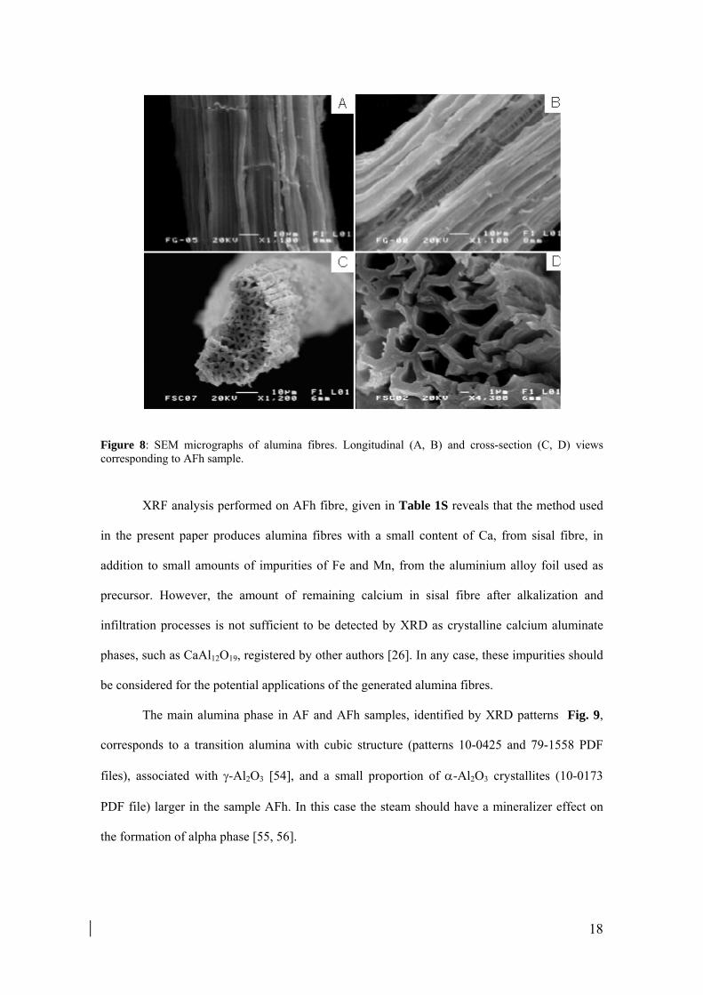
18
Figure 8: SEM micrographs of alumina fibres. Longitudinal (A, B) and cross-section (C, D) views corresponding to AFh sample.
XRF analysis performed on AFh fibre, given in Table 1S reveals that the method used
in the present paper produces alumina fibres with a small content of Ca, from sisal fibre, in
addition to small amounts of impurities of Fe and Mn, from the aluminium alloy foil used as
precursor. However, the amount of remaining calcium in sisal fibre after alkalization and
infiltration processes is not sufficient to be detected by XRD as crystalline calcium aluminate
phases, such as CaAl12O19, registered by other authors [26]. In any case, these impurities should
be considered for the potential applications of the generated alumina fibres.
The main alumina phase in AF and AFh samples, identified by XRD patterns Fig. 9,
corresponds to a transition alumina with cubic structure (patterns 10-0425 and 79-1558 PDF
files), associated with -Al2O3 [54], and a small proportion of -Al2O3 crystallites (10-0173
PDF file) larger in the sample AFh. In this case the steam should have a mineralizer effect on
the formation of alpha phase [55, 56].

19
Figure 9: XRD diffractograms of AF (a) and AFh (b) samples. The last is slightly moved along y-axis for better viewing. (■) -Al2O3.
The existence of gamma and alpha alumina at 1000 °C can be explained as follows. The
presence of transition phases may be related to the small size of aluminium oxide crystalline
seeds formed during the carbonization process, due to the inhibitory effect of the carbonaceous
scaffold, as commented in section 3.2. Moreover, the relatively low temperature of formation of
alpha phase with respect to the typical phase transition scheme proposed by Stumpf et al. [57]
and Tertian and Papée [58], could be due to various causes, such as the formation of a gel
composed of hydrolyzed polynuclear aluminium species during the infiltration step. It has been
described that aluminas generated from polyaluminium species, in particular from basic
aluminium chlorides, have lower transition temperatures than usual [59,60]. Furthermore, the
local temperature increase due to the combustion process of the carbonaceous matrix can help to
overcome the energy barrier necessary for the formation of alpha phase [61,62]. The distinction
between the transition aluminas Al2O3 and -Al2O3 is complex because they show very
similar crystal structures. In principle, the higher definition of the reflection associated to (222)
plane and the less intense reflection of the plane (111), at around 4.6 Å, suggest that the
transition phase corresponds to -Al2O3 [63]. The peak widths are indicative of the nano-sized
alumina crystallites, estimated at 4.1 and 5.5 nm for AF and AFh samples respectively.

20
ATR-FTIR spectrum of both alumina samples, presented in Fig. 10, does not discard
the presence of -Al2O3. The wavenumbers below 1000 cm-1 are related to the characteristics
Al-O vibration of aluminas. The region below 700 cm-1, associated with Al-O stretching
vibrations of octahedrally coordinated aluminium (v-AlO6), is more intense compared to the
region 900-750 cm-1, associated with Al-O stretching vibrations of aluminium in tetrahedral
coordination (v-AlO4), feature shared by low temperature transition aluminas, as and -Al2O3
phases [64].
Figure 10: ATR-FTIR spectra of AF (discontinuous line) and AFh (continuous line) samples.
27Al MAS-NMR analysis has been configured as a method for distinguishing the
coordination environments of aluminium, allowing to discern different phases of alumina in a
complementary manner. In the case of Al2O3 and Al2O3 their fractions of octahedrally
coordinated aluminium have been quantified about 65 ± 4% and 75 ± 4% respectively [65,66].
Two signals centred at 7 and 64 ppm are observable in the spectrum of AFh fibres shown in
Fig. 11, associated respectively with Al3 + in octahedral position (AlVI) and tetrahedral (AlIV),
not being perceptible the alpha phase contribution, characterized by an octahedral environment
around 13 ppm [40]. It has been estimated that the proportion of AlVI is close to 70%, which
suggests that both phases remain even after a reheating cycle with humid air.

21
Figure 11: 27Al-MAS-RMN of AFh sample (SSB: spinning side bands).
The infrared band centred at 3400 cm-1 and the shoulder around 3200 cm-1, shown in the
inset plot of Fig. 10, are associated with O-H stretching modes of hydroxyl groups located at the
surface of aluminium oxide, resulted from the moisture adsorption, which can be distinguished
by XPS analysis. Survey and multi-region XPS spectra of AFh fibre are shown in Fig. 12.
Figura 12: XPS spectra, survey (A), O1s region (B) and Al2p region (C), of AFh sample.

22
The binding energy of the photoelectron peaks O1s and Al2p are located respectively at
530.96 eV and 73.95 eV, and the kinetic energy of the Auger electron peak AlKLL is situated at
1387.64 eV, values which are characteristic for aluminium oxides [67]. Trough the fitting
procedure, the O1s band is found to be composed of three overlapping components, at 529.35,
530.79 and 532.36 eV with contributions close to 17, 56 and 27%, respectively. The lowest
energy component is associated to O2-, from oxygen combined with aluminium in the alumina
lattice, while the highest band energy component is due to physisorbed water [68]. The main
contribution is associated with hydroxyls groups due to water dissociation on the alumina
surface. It is known that the Al3+ cations of the alumina surface, as Lewis acid sites, are able to
dissociate the water molecules from ambient moisture [69].
The synthesized gamma-alumina fibres are mainly mesoporous, as revealed by nitrogen
adsorption-desorption analysis. The isotherms, shown in Fig. 13a, are of type IV and, in general
terms, exhibit a hysteresis loop close to H3 type according to the IUPAC classification [70].
Figure 13: Adsorption-desorption N2 isotherms for AF (■) and AFh (▲) samples (A). The first is moved 150 cm3/g from its original position for better viewing. The corresponding pore volume (B) and area (C) distributions.

23
AF sample is characterized by a continuous filling of pores as the pressure increases. It
is observed a slight restriction at P/P0 around 0.97 in which the adsorption and desorption
branches are close to overlap. In contrast, two different behaviours can be distinguished in AFh
sample between i) P/P0 = 0.6 to 0.75, where the adsorption is slow, the pore filling is continuous
and the hysteresis is close to H3 type, resembling to AF sample, and ii) P/P0 = 0.75 to 0.95,
where the adsorption capacity increases significantly and both branches are substantially
vertical and parallel, overlapping at high relative pressures, thus being the hysteresis loop closer
to H1 type. The isotherm of the recalcined sample AFh presents two peculiarities: a steeper
slope of the hysteresis loop and a plateau at high relative pressures. The first would be
indicative of a more uniform nanostructure and better pore connectivity [23,71] and the second
suggests a complete filling of pores, characteristics of a material with a porous structure more
organized and with a limited range of pore sizes. In fact, H1 hysteresis type is associated with
porous materials consisting of agglomerated particles with a certain compactness and even, it
has been associated with cylindrical mesopores open at both ends [72], while the H3 type,
without an adsorption limit at high P/P0, is typical of poorly aggregated materials or plate-like
particles living rise to slit-shaped pores [70]. The main results of the textural analysis performed
are listed in Table 2.
Table 2. Textural data of the ceramic fibers AF and AFh
a BET equation constant b Mean pore width, wp = 4V/S c Maximum pore size determined by BJH distribution from the adsorption branch
AF AFh SBET (m2/g) 200.8 153.9 C a 118.6 108.3 St Ext (m
2/g) 174.0 144.2 St micro (m
2/g) 26.8 9.6 SDR (m
2/g) 203.7 131.1 Vsp (cm3/g) 0.47 0.52 Vt micro (cm3/g) 0.0099 0.0028 VDR (cm3/g) 0.0816 0.0525 wp
b (Ǻ) 93 136 BJH max (dV/dD) c (Ǻ) 76 153 BJH max (dA/dD) c (Ǻ) 56 56

24
Alumina fibres present high external surface area StExt, very close to the total BET
surface area, mainly associated with the existence of mesopores in the range 3 - 40 nm, as
shown BJH distributions in Fig. 13b and Fig. 13c. The surface and volume of micropores
accessible to nitrogen, Stmicro and Vtmicro, are very low in AF sample and practically negligible
for AFh, while the surface and volume of those micropores accessible to CO2, SDR and VDR,
corresponding to narrow-neck pores, are still significant in AFh sample, values also lower than
those exhibited by AF.
The effects on the biomimetic alumina fibres generated by a second calcination step
under the presence of moisture consist essentially in modifying the pore volume and area
distributions, observed in Fig. 13b and Fig. 13c respectively. An increase in the average pore
size, from 9 nm to 14 nm, could explain the increment of about 11% in the mesopore volume,
Vsp, of AFh sample. This fact would be associated to the destruction of the smaller pores, as the
reduction of micropores accessible to N2 and CO2 , estimated around 72 and 36% for Vtmicro and
VDR, respectively (values derived from Table 2), indicates. The micro and nanostructural
reorganization experienced by alumina fibres during the recalcination process, resulting in a
pore size enlargement and/or in a better pore connectivity, also explains the decrease in specific
surface area, which is also consistent with the growh in crystallite size of -Al2O3 estimated by
the Scherrer equation. The reduction in SBET, Stmicro and SDR values, around 23, 64 and 36%
respectively (from Table 2), is associated especially to a decrease in the proportion of pores
with a diameter less than 12 nm, as deduced from Fig. 13b.
The morphology, nanostructure and crystallinity of the alumina particles of the AFh
fibre walls are analyzed by TEM images and SAED diffraction patterns, collected in Fig. 14.

25
Figure 14: TEM micrographs and SAED patterns of AFh sample.
Alumina fibres presents a spongy texture constituted by aggregated particles, of sizes
between 4 and 12 nm, the same order of magnitude as the crystallite estimated by the Scherrer
equation. The clusters of particles appear longitudinally arranged in elongated structures, with
widths of 120-200 nm, as seen in Fig. 14a. According to XRD, the SAED patterns show a
strong polycrystalline habit, where the most intense rings are those associated with the most
intense planes of the cubic phase of alumina, (440) and (400). At bigger magnifications,
different morphologies can be observed, as the wormhole type shown in Fig. 14b, with particle
sizes between 1 and 3 nm and micropores of average diameters between 1 and 2 nm.
Nevertheless, it should be noted that the dominant micromorphology consist of elongate
particles, around 15 - 30 nm of length and 3-6 nm of width, some of which appear twinned or
folded, generating slots of around 2 nm, as can be seen in Fig. 14b-c.

26
4. Conclusions
Mesoporous gamma alumina fibres with BET surface area of 200 m2/g were synthesized
through the replica method of sisal fibres, followed by a carbonization process and prolonged
calcination at 1000 °C. After a re-calcination stage in wet oxidizing atmosphere, the ceramic
fibres are maintained without a significant transformation to -Al2O3 phase, reducing specific
surface area by around 20%. This stage produces a reorganization of the porous architecture to a
more ordered structure, possibly with better connectivity between the pores, together with an
increase of the volume and the average size of mesopores, produced at the expense of the loss of
microporosity, which explains the decrease of specific surface area.
This paper analyzes the interaction of the aluminium inorganic precursor with the
lignocellulosic fibre and its thermal evolution. The interaction between fibre and precursor
could take place through the OH groups of the cellulosic components as well as through the
carboxylic groups of the glucuronic acid side chains of the remnant hemicellulose after the
alkalizing process. This effect will be studied in greater depth in a later investigation.
As temperature increases, the precursor evolves into amorphous aluminium
oxo/hydroxide, which not only covers the surface of the fibre, but becomes part of the walls,
due to the precursor infiltration process through the cell walls of the natural fibre, whose final
result are alumina ceramic fibres mimicking the natural masks. The slow transformation of the
aluminium oxide is associated with the difficulty of nucleation and growth processes during the
conformation of the ceramic structure, due to the impediment exerted by the carbonaceous
matrix, leading to the formation of low temperature transition alumina crystallites,
predominantly -Al2O3, stable at 1000 ºC.
Aknowlwdgments
The author M. Benítez Guerrero wishes to thank the Spanish Consejo Superior de
Investigaciones Científicas (CSIC) for the awarding of a JAE pre-doctoral grant. Support from the
“Laboratorio de Materiales y Superficies” (CSIC-Univ. Málaga) is acknowledged.

27
Financial support from projects CTQ2011-27626 (Spanish Ministerio de Economía y
Competitividad), TEP-7858 (Junta de Andalucía) and FEDER funds is acknowledged.
The authors express their gratitude to Dr. Pilar Pena Castro and to Dr. Antonio H. Aza, from the
Instituto de Cerámica y Vidrio (CSIC) in Madrid (Spain), to put at our disposal knowledge, skills and
means that have enabled the creation of the present work.
References
[1] C. Márquez-Álvarez, N. Zilkova, J. Pérez-Pariente, J. Cejka, Catal. Rev., 50 (2008) 222-286. [2] W. Cai, J. Yu, M. Jaroniec, J. Mater. Chem., 20 (2010) 4587-4594. [3] S.W. Bian, Y.L. Zhang, H.L. Li, Y. Yu, Y.L. Song, W.G. Song, Microporous Mesoporous Mater., 131 (2010) 289-293. [4] C. Lesaint, G. Kleppa, D. Arla, W.R. Glomm, G. ye, Microporous Mesoporous Mater., 119 (2009) 245-251. [5] Q. Wu, F. Zhang, J. Yang, Q. Li, B. Tu, D. Zhao, Microporous Mesoporous Mater., 143 (2011) 406-412. [6] Z.Y. Yuan, B.L. Su, J. Mater. Chem., 16 (2006) 663-677. [7] A. Stanislaus, K. Al-Dolama, M. Absi-Halabi, J. Mol. Catal. A: Chem., 181 (2002) 33-39. [8] Z. Wu, Q. Li, D. Feng, P.A. Webley, D. Zhao, J. Am. Chem. Soc., 132 (2010) 12042-12050. [9] A.H. Heuer, D.J. Fink, V.J. Laraia, J.L. Arias, P.D. Calvert, K. Kendall, G.L. Messing, J. Blackwell, P.C. Rieke, D.H. Thompson, A.P. Wheeler, A. Veis, A.I. Caplan, Science, 255 (1992) 1098-1105. [10] A.R. Studart, U.T. Gonzenbach, E. Tervoort, L.J. Gauckler, J. Am. Ceram. Soc., 89 (2006) 1771-1789. [11] P. Colombo, Philos. T. Roy. Soc. A, 364 (2006) 109-124. [12] T.X. Fan, S.K. Chow, D. Zhang, Prog. Mater. Sci., 54 (2009) 542-659. [13] J. Cao, C.R. Rambo, H. Sieber, Ceram. Int., 30 (2004) 1967-1970. [14] H. Zhou, T. Fan, D. Zhang, Chem. Sus. Chem., 4 (2011) 1344-1387. [15] H. Sieber, C. Hoffmann, A. Kaindl, P. Greil, Adv. Eng. Mater., 2 (2000) 105-109. [16] P. Greil, J. Eur. Ceram. Soc., 21 (2001) 105-118. [17] M. Singh, J. Martínez-Fernández, A.R. Arellano-López, Curr. Opin. Solid St. M., 7 (2003) 247-254. [18] M. Singh, B.M. Yee, J. Eur. Ceram. Soc., 24 (2004) 209-217. [19] F. Vaudry, S. Khodabandeh, M.E. Davis, Chem. Mater., 8 (1996) 1451-1464. [20] S. Valange, J.L. Guth, F. Kolenda, S. Lacombe, Z. Gabelica, Microporous Mesoporous Mater., 35-36 (2000) 597-607. [21] S. Acosta, A. Ayral, C. Guizard, L. Cot, J. Sol-Gel Sci. Techn., 8 (1997) 195-199. [22] Q. Liu, A. Wang, J. Xu, Y. Zhang, X. Wang, T. Zhang, Microporous Mesoporous Mater., 116 (2008) 461-468. [23] X. Yuan, S. Xu, J. Lü, X. Yan, L. Hu, Q. Xue, Microporous Mesoporous Mater., 138 (2011) 40-44. [24] J. Cao, C.R. Rambo, H. Sieber, J. Porous Mat., 11 (2004) 163-172. [25] C.R. Rambo, H. Sieber, Adv. Mater., 17 (2005) 1088-1091. [26] T.E. Andrade, C.R. Rambo, H. Sieber, A.E. Martinelli, D.M.A. Melo, J. Mater. Sci., 42 (2007) 5426-5430. [27] A.N. Shigapov, G.W. Graham, R.W. McCabe, H.K. Plummer Jr, Appl. Catal. A-Gen., 210 (2001) 287-300. [28] M. Patel, B.K. Padhi, J. Mater. Sci., 25 (1990) 1335-1343. [29] T. Fan, B. Sun, J. Gu, D. Zhang, L.W.M. Lau, Scripta Mater., 53 (2005) 893-897. [30] L. Segal, Text. Res. J., 29 (1959) 786-794. [31] S. Brunauer, P.H. Emmett, E. Teller, J. Am. Chem. Soc., 60 (1938) 309-319. [32] B.C. Lippens, J.H.d. Boer, J. Catal., 4 (1965) 319-323 [33] E.P. Barrett, L.G. Joyner, P.P. Halenda, J. Am. Chem. Soc., 73 (1951) 373-380. [34] W.D. Harkins, G. Jura, J. Am. Chem. Soc., 66 (1944) 1366-1373. [35] J.H.d. Boer, B.C. Lippens, B.G. Linsen, J.C.P. Broekhoff, A.v.d. Heuvel, T.J. Osinga, J. Colloid. Interf. Sci., 21 (1966) 405-414. [36] M.M. Dubinin, L.V. Radushkevich, Chem. Zentr., 1 (1947) 875-889.

28
[37] B.C. Barkakaty, J. Appl. Polym. Sci., 20 (1976) 2921-2940. [38] L.Y. Mwaikambo, M.P. Ansell, J. Appl. Polym. Sci., 84 (2002) 2222-2234. [39] D.A. Riesgraf, M.L. May, Appl. Spectrosc., 32 (1978) 362-366. [40] P. Brand, D. Müller, W. Gessner, Crys. Res. Tehcnol., 2 (1990) 951-956. [41] C.C. Landry, N. Pappé, M.R. Mason, A.W. Apblett, A.N. Tyler, A.N. MacInnes, A.R. Barron, J. Mater. Chem., 5 (1995) 331-341. [42] J. Van Den Brand, O. Blajiev, P.C.J. Beentjes, H. Terryn, J.H.W. De Wit, Langmuir, 20 (2004) 6308-6317. [43] R.H. Marchessault, Pure Appl. Chem., 5 (1962) 107-129. [44] O. Faix, Fourier Transform Infrared Spectroscopy, in: S.Y. Lin, C.E. Dence (Eds.) Methods in Lignin Chemistry, Springer-Verlag, New York, 1992, pp. 83-109. [45] Q.A. Nguyen, M.P. Tucker, Dilute acid/metal salt hydrolysis of lignocellulosics, U.S. Patent 6.423.154, 2002. [46] L. Liu, J. Sun, C. Cai, S. Wang, H. Pei, J. Zhang, Bioresource Technol., 100 (2009) 5865-5871. [47] P.E. Sánchez-Jiménez, L.A. Pérez-Maqueda, A. Perejón, J. Pascual-Cosp, M. Benítez-Guerrero, J.M. Criado, Cellulose, 18 (2011) 1487-1498. [48] P.E. Sánchez-Jiménez, L.A. Pérez-Maqueda, A. Perejón, J.M. Criado, Thermochim. Acta, 552 (2013) 54-59. [49] F. Suárez-García, A. Martínez-Alonso, J.M.D. Tascón, J. Anal. Appl. Pyrol., 62 (2002) 93-109. [50] P. Álvarez, R. Santamaría, C. Blanco, M. Granda, J. Anal. Appl. Pyrol., 74 (2005) 337-343. [51] R. Ball, A.C. McIntosh, J. Brindley, Combust. Theor. Model, 8 (2004) 281-291. [52] H. Yang, R. Yan, H. Chen, D.H. Lee, C. Zheng, Fuel, 86 (2007) 1781-1788. [53] H. Barzegar-Bafrooei, T. Ebadzadeh, Adv. Powder Technol., 22 (2011) 366-369. [54] Y. Repelin, E. Husson, Mat. Res. Bull., 25 (1990) 611-621. [55] M. Pijolat, M. Dauzat, M. Soustelle, Thermochim. Acta, 122 (1987) 71-77. [56] R.B. Bagwell, G.L. Messing, J. Am. Ceram. Soc., 82 (1999) 825-832. [57] H.C. Stumpf, A.S. Russell, J.W. Newsome, C.M. Tucker, Ind. Eng. Chem., 42 (1950) 1398-1403. [58] R. Tertian, D. Papée, J. Chem. Phys., 55 (1958) 341-353 [59] P. Brand, R. Troschke, H. Weigelt, Cryst. Res. Technol., 24 (1989) 671-675. [60] T. Yamaguchi, N. Mukouyama, T. Fujita, S. Taruta, K. Kitajima, Ceram. Int., 37 (2011) 201-206. [61] R.N. Das, A. Bandyopadhyay, S. Bose, J. Am. Ceram. Soc., 84 (2001) 2421-2423. [62] P. Wang, C. Wang, L. Lin, Y. Zhu, Y. Xie, J. Am. Ceram. Soc., 89 (2006) 2744-2748. [63] S. Zhou, R.L. Snyder, Acta Cryst. B, 47 (1991) 617-630. [64] C. Morterra, G. Magnacca, Catal. Today, 27 (1996) 497-532. [65] C.S. John, N.C.M. Alma, G.R. Hays, Appl. Catal., 6 (1983) 341-346. [66] C. Pecharromán, I. Sobrados, J.E. Iglesias, T. González-Carreño, J. Sanz, J. Phys. Chem. B, 103 (1999) 6160-6170. [67] J.F. Moulder, W.F. Stickle, P.E. Sobol, K.D. Bomben, Handbook of X-ray Photoelectron Spectroscopy, Perkin-Elmer Corporation, Physical Electronics Division, 1992. [68] M.P. Casaletto, G. Mattogno, M.A. Massucci, Appl. Surf. Sci., 211 (2003) 216-226. [69] M. Trueba, S.P. Trasatti, Eur. J. Inorg. Chem., (2005) 3393-3403. [70] K.S.W. Sing, D.H. Everett, R.A.W. Haul, L. Moscou, R.A. Pierotti, J. Rouquérol, T. Siemieniewska, Pure Appl. Chem., 57 (1985) 603-619. [71] S. Handjani, J. Blanchard, E. Marceau, P. Beaunier, M. Che, Microporous Mesoporous Mater., 116 (2008) 14-21. [72] J.L. Mohanan, S.L. Brock, Chem. Mater., 15 (2003) 2567-2576.