201 7 SUMM ER MEETING
117
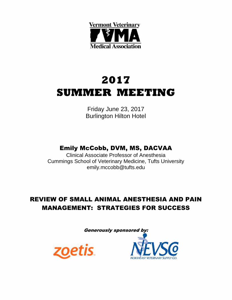
2017 SUMMER MEETING Friday June 23, 2017 Burlington Hilton Hotel Emily McCobb, DVM, MS, DACVAA Clinical Associate Professor of Anesthesia Cummings School of Veterinary Medicine, Tufts University [email protected] REVIEW OF SMALL ANIMAL ANESTHESIA AND PAIN MANAGEMENT: STRATEGIES FOR SUCCESS Generously sponsored by:
Transcript of 201 7 SUMM ER MEETING
2017
SUMMER MEETING
Friday June 23, 2017 Burlington Hilton Hotel
Emily McCobb, DVM, MS, DACVAA Clinical Associate Professor of Anesthesia
Cummings School of Veterinary Medicine, Tufts University [email protected]
REVIEW OF SMALL ANIMAL ANESTHESIA AND PAIN
MANAGEMENT: STRATEGIES FOR SUCCESS
Generously sponsored by:
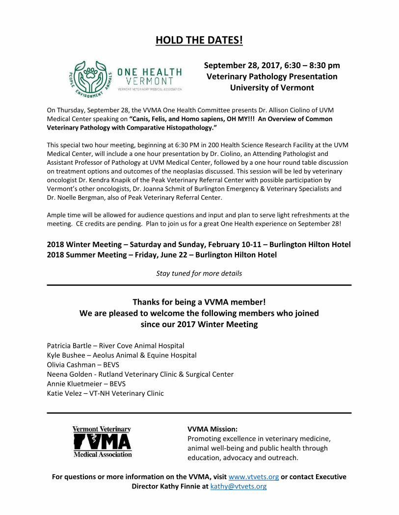
HOLD THE DATES!
September 28, 2017, 6:30 – 8:30 pm Veterinary Pathology Presentation
University of Vermont
On Thursday, September 28, the VVMA One Health Committee presents Dr. Allison Ciolino of UVM Medical Center speaking on “Canis, Felis, and Homo sapiens, OH MY!!! An Overview of Common Veterinary Pathology with Comparative Histopathology.” This special two hour meeting, beginning at 6:30 PM in 200 Health Science Research Facility at the UVM Medical Center, will include a one hour presentation by Dr. Ciolino, an Attending Pathologist and Assistant Professor of Pathology at UVM Medical Center, followed by a one hour round table discussion on treatment options and outcomes of the neoplasias discussed. This session will be led by veterinary oncologist Dr. Kendra Knapik of the Peak Veterinary Referral Center with possible participation by Vermont’s other oncologists, Dr. Joanna Schmit of Burlington Emergency & Veterinary Specialists and Dr. Noelle Bergman, also of Peak Veterinary Referral Center. Ample time will be allowed for audience questions and input and plan to serve light refreshments at the meeting. CE credits are pending. Plan to join us for a great One Health experience on September 28!
2018 Winter Meeting – Saturday and Sunday, February 10-11 – Burlington Hilton Hotel 2018 Summer Meeting – Friday, June 22 – Burlington Hilton Hotel
Stay tuned for more details
Thanks for being a VVMA member! We are pleased to welcome the following members who joined
since our 2017 Winter Meeting Patricia Bartle – River Cove Animal Hospital Kyle Bushee – Aeolus Animal & Equine Hospital Olivia Cashman – BEVS Neena Golden - Rutland Veterinary Clinic & Surgical Center Annie Kluetmeier – BEVS Katie Velez – VT-NH Veterinary Clinic
VVMA Mission: Promoting excellence in veterinary medicine, animal well-being and public health through
education, advocacy and outreach.
For questions or more information on the VVMA, visit www.vtvets.org or contact Executive Director Kathy Finnie at [email protected]

2017 Summer Meeting Exhibitors and Sponsors Thank you for your support of our Meeting!
4 Legs & A Tail Tim Hoen [email protected] Abaxis Thomas Dodd [email protected] Addie Stiles [email protected] Aesculight Rich Noss [email protected] Boehringer-Ingelheim Mary Kathryn Edwards [email protected] Blue Buffalo Jeff Libby [email protected] Michael Reynolds [email protected] Burlington Emergency & Vet. Specialists Whitney Durviage [email protected] Ceva Animal Health Tiffany Lewis [email protected] Tim Jolicoeur [email protected] Christian Veterinary Mission Dr. Amy St. Denis [email protected] Companion Therapy Laser by LiteCure Kevin Gouvin [email protected] Elanco Animal Health Elizabeth Hall [email protected] Henry Schein Animal Health Martha Rose [email protected] Jazz Heath [email protected] Hill’s Pet Nutrition, Inc. Dr. Andrew Hagner [email protected] Idexx Laboratories Ben Enger [email protected] Integra-Miltex Arthur Lubniewski [email protected] Jorgensen Laboratories Marnie Ciquera [email protected] K-Laser USA Gary Britkco [email protected] MWI Animal Health Paige Willson [email protected] Nestle Purina Lauren Koron [email protected] *NEVSCo Butch Roeder [email protected] Nutramax Laboratories Devon Hankins [email protected] Patterson Veterinary Supply George White [email protected] Peak Veterinary Referral Center Deborah Unica [email protected] Dr. Heather York [email protected] PENRO Specialty Compounding Neal Pease, R. Ph [email protected] Roadrunner Pharmacy Kevin Kerbert [email protected] Sedecal USA Matt Strauss [email protected] Trupanion Matthew Roach [email protected] Universal Imaging Michael McElinny [email protected] USDA, Food Safety Inspection Service Dr. Paul Cole [email protected] Dr. Emily Buskey [email protected] VT-CAN! Pamela Krausz [email protected] Vermont Disaster Animal Response Team Barry Londeree [email protected] Vermont Humane Federation Pamela Krausz [email protected] VetCor Jessica Bird [email protected] Bryan Brackett [email protected] Vetri Science Laboratories Jana Lafayette [email protected] Wilcox Pharmacy Tina Rotella [email protected]
*VVMA speaker sponsor

158 Hurricane Lane, Williston, VT 05495 P: 802-878-2022 F: 802-878-1524 email:[email protected] www.peakveterinaryreferral.com
Vermont Veterinary CardiologyDon Brown, DVM, PhD, Diplomate
ACVIM - CardiologyJenny Garber, DVM
Internal MedicineMarielle Goossens, DVM, Diplomate
ACVIMTim Bolton, DVM, Diplomate
ACVIM
OncologyKendra Knapik, DVM, Diplomate
ACVIM - OncologyNoelle Bergman, DVM, Diplomate
ACVIM - Oncology
Physical RehabilitationNancy Zimny PT, CCRT
Vermont Veterinary Eye CareSarah Hoy, DVM, MS, Diplomate
ACVO
NeurologyPhil March, DVM Diplomate
ACVIM - Neurology
SurgeryKurt Schulz, DVM, MS, Diplomate
ACVSKristian Ash, DVM
BehaviorPam Perry DVM, Practice Limited to
Behavior
DermatologyEd Jazic, DVM, Diplomate ACVD
COMPASSION EXPERTISE
TRUST

McCobb, 2017, all rights reserved
Veterinary CE for Vermont VMA
June 23, 2017
Dr. Emily McCobb
Review of Small Animal Anesthesia and Pain Management: strategies for success
Topics covered:
Anesthetic risks and patient assessment
Protocol design and review of drugs for sedation and anesthesia
Anesthesia for the geriatric patient
Monitoring and Trouble Shooting anesthesia to avoid complications
Acute Pain Management: innovative techniques
Description:
This CE will review the basics of assessing individual patients and designing a safe protocol for
them. It will also cover avoiding and addressing the most common anesthetic complications.
There will be plenty of time for discussion and practitioners are encouraged to bring questions.
In addition, we will review newer anesthetic and analgesic drugs and techniques.
Guide to talks:
1) Anesthetic Risks and Patient Assessment
Goal: To understand anesthetic risks for small animal patients
Summary: The anesthetic risk for small animal patients in general practice has been
examined in several studies and has decreased over the past several decades. The risk of
death for a healthy canine patient is about one in every two thousand anesthesia cases,
whereas the risk of death for cats is higher at about one in one thousand. The risk for
smaller species like rabbits is higher yet with death rates as high as 5 or 6 out of every one
thousand anesthesia cases.
The most recent large scale evaluation of anesthetic risk in small animals was done by
Dr. David Brodbelt and his team from the UK and was published in several articles. The
findings revealed several factors which appear to increase anesthetic risk. In general, since
the risk of anesthetic morbidity or mortality is highly correlated with patient health status
careful pre-anesthetic evaluation of the patient is warranted. Each patient should have his
or her risk factors individually assessed and this information should be conveyed to pet
owners to guide in decision making about the course of treatment.
McCobb, 2017, all rights reserved
Learning Objectives:
1) Be able to accurately assess the anesthetic risk for a small animal patient (dog, cat or
other small mammal) and to be able to discuss an individual’s risk factors with a pet
owner.
Selected references:
Brodbelt DC, D. Flaherty and GR Pettier. Anesthetic risk and informed consent. In Veterinary
Anesthesia and Analgesia, The Fifth Edition of Lumb and Jones, Wiley Blackwell, Eds: Grimm KA, Lamont
LA, Tranquilli, Greene SA, Robertson S., pp. 11-20.
Brodbelt DC, Blissitt, KJ, Hammond RA, et al. The risk of death: the confidential enquiry into
perioperative small animal fatalities. Vet Anaesth Analg 2008;35:365–73.
Brodbelt, D. "Feline anesthetic deaths in veterinary practice." Topics in companion animal
medicine 25.4 (2010): 189-194
2) Protocol design and review of drugs for sedation and anesthesia
Goal: To be able to create customized anesthetic protocols for sick and healthy canine and
feline patients.
Summary: There are a wide variety of safe and effective drugs and techniques appropriate anesthesia for small animal patients. Each clinician must determine the best protocol based on information about the patient’s health status and anesthetic risk. Each patient should be evaluated individually in order to determine the appropriate protocol. In many cases a standard protocol will be acceptable- more challenging is to identify patients for whom the routine protocol is contraindicated.
Patient preparation begins at admission and includes appropriate fasting times, based on species and age of the patient. Special considerations for pediatric patients are needed. From an anesthetic stand point, the pre-operative examination should focus on the patients’ temperament and cardiovascular fitness. When higher risk patients are identified, the protocol should be adjusted and this information conveyed to the pet owner. Pre-anesthetic testing is not generally not indicated for young and healthy patients but is helpful for patients with higher ASA status.
Balanced anesthesia is a key concept and refers to using multiple anesthetic agents to achieve anesthesia with minimal physiologic impairment for the patient. Selecting the appropriate anesthetic protocols depends on many factors including the patient status, the procedure to be performed and the availability of drugs as well as clinician comfort level. Criteria crucial for determining optimum protocol include: the provision of analgesia; stress reduction or anxiolysis; immobility and muscle relaxation; and safe, controlled, reversible depression of the CNS resulting in unconsciousness. Numerous cost-effective protocols
McCobb, 2017, all rights reserved
combining multiple anesthetic and analgesic drugs, including injectable and inhalant agents, exist for achieving balanced anesthesia in pediatric and adult patients. Anticholinergics may or may not be used according to veterinarian preference but should be available in case of an emergency. Drug doses should be calculated on a mg/kg basis and accurate weights should be obtained for dosing.
Analgesia is absolutely required for surgical patients and will be discussed further in a following lecture. Analgesic agents that may be used include opioid medications, ketamine, alpha 2 agonists, local anesthetics and NSAIDs. Many clinics employ the use of a single injection (total intramuscular anesthesia) to achieve sedation, analgesia and ultimately a surgical plane of anesthesia.
Learning Objective:
1) Describe patient features that are relevant to the anesthetic plan
2) List the components of an anesthesia protocol
3) Understand indications, contraindications and safe use principles for common
sedatives and anesthetics.
4) Be able to design an appropriate protocol for a healthy and a sick dog and cat using
drugs available in your practice.
Selected References:
AAHA fluid guidelines
Harold Davis, BA, RVT, VTS (ECC), Tracey Jensen, DVM, DABVP, Anthony Johnson, DVM,
DACVECC, Pamela Knowles, CVT, VTS (ECC), Robert Meyer, DVM, DACVAA, Renee Rucinsky,
DVM, DAVBP (Feline), Heidi Shafford, DVM, PhD, DACVAA. 2013 AAHA/AAFP Fluid Therapy
Guidelines for Dogs and Cats. J Am Anim Hosp Assoc 2013; 49:149–159.
AAHA Anesthesia Guidelines
Richard Bednarski, MS, DVM, DACVA (Chair), Kurt Grimm, DVM, MS, PhD, DACVA, DACVCP,
Ralph Harvey, DVM, MS, DACVA, Victoria M. Lukasik, DVM, DACVA, W. Sean Penn, DVM, DABVP
(Canine/Feline), Brett Sargent, DVM, DABVP (Canine/Feline), Kim Spelts, CVT, VTS, CCRP
(Anesthesia). AAHA Anesthesia Guidelines for Dogs and Cats. J Am Anim Hosp Assoc 2011;
47:377–385.
Barletta, M, Austin, BR, Ko, JC, Payton, ME, Weil, AB and T. Inoue. Evaluation of dexmedetomidine and ketamine in combination with opioids as injectable anesthesia for castration in dogs. JAVMA 2011; 289 (9): 1159-67.
Bednarski RM. Anesthesia and Analgesia for Domestic Species: Dogs and Cats. In Veterinary
Anesthesia and Analgesia, The Fifth Edition of Lumb and Jones, Wiley Blackwell, Eds: Grimm KA,
Lamont LA, Tranquilli, Greene SA, Robertson SA., pp 819-826
McCobb, 2017, all rights reserved
3) Anesthesia for the geriatric patient
Goal: To understand how to safely anesthetize older canine and feline patients for routine procedures.
Summary: the pet population is aging, and geriatric patients are increasingly
anesthetized as part of their routine care. The older patient may have altered
physiology and reduced ability to metabolize and clear drugs. In general, physiologic
reserves are decreased in these patients and the chance of co-existing disease is
increased. With a few modifications and attentive care, these patients can be
anesthetized safely and with good outcomes.
Learning Objectives:
1) Be able to describe relevant features of geriatric animal physiology and
pharmacology that affect the anesthetic protocol.
2) Be able to accurately discuss anesthetic risk for older patients with pet
owners.
3) Be able to design an appropriate anesthesia protocol for an older patient and
describe appropriate peri-anesthetic considerations for them.
Selected References:
Baetge, C. and N. Mathews. Anesthesia and Analgesia for Geriatric Veterinary Patients.
Veterinary Clinics of North America 2014
Neiger-Aeschbacher G. Geriatric patients. In: Seymour C, Due-Novakovski, editors. BSAVA
manual of canine and feline anaesthesia and analgesia. 2nd edition. Gloucester (UK):
BSAVA; 2010. p. 303–9.
Grape S, Ravussin P, Rossi A, et al. Postoperative cognitive dysfunction. Trends Anaesth Crit
Care 2012. DOI: http://dx.doi.org/10.1016/j.tacc.2012.02.002
4) Monitoring and Trouble Shooting anesthesia to avoid complications
Goal: To understand how to avoid and address common anesthetic complications through careful patient monitoring and anticipating likely problems. Summary: Basic Monitoring
Since subject physiology and homeostasis are altered by all anesthetic drugs monitoring is vital for safe anesthetic practice. The purpose of monitoring is to provide information on

McCobb, 2017, all rights reserved
basic body system function that will minimize the decrement of organ function, especially in patients with preexisting disease. Monitoring patient vital signs during anesthesia is thought to reduce anesthetic morbidity. Tracking physiologic variables provides early warning signs to changes in patient status leading to the opportunity to intervene in time to prevent more serious problems.
Both the ACVAA (American College of Veterinary Anesthesiologists) and the AAHA (American Animal Hospital Association) require an anesthetic record to be kept as a legal record of anesthetic related events. Record keeping also prompts the anesthetist to observe and evaluate the patient at regular intervals. A complete anesthesia record should include: documentation of all drugs administered during the peri-anesthetic period (from time of pre-medication and into recovery), recording of monitored variables every five to ten minutes as well as documentation of any untoward events or unusual circumstances.
ACVAA recommendations for monitoring are available on the organizations website (see below). Basic monitoring should encompass five areas: state of consciousness or anesthetic depth, adequacy of tissue blood flow (circulation), adequacy of blood oxygenation, adequacy of ventilation and body temperature. Each of these areas can be monitored invasively or non-invasively. If possible, more than one parameter should be monitored per body system. In addition, a great deal of information can be obtained by simple palpation and observation. Manual assessment of the patient should always be performed and is considered more reliable than any piece of monitoring equipment.
Depth of anesthesia can be assessed by eye position and ocular reflexes. Some agents may affect the ability of ocular change to be used to detect anesthetic depth. For example, dissociative anesthetics such as ketamine will produce a central eye position regardless of depth and opioids will affect pupil size. The corneal reflex should always be present during anesthesia as loss of the corneal reflex indicates an excessively deep level of anesthesia. Other indicators of anesthetic depth include the presence of withdrawal reflexes and degree of muscle relaxation. Jaw tone can be assessed as an indication of muscle relaxation. Autonomic responses (heart rate, blood pressure, respiratory rate) can be used as an indirect indication of patient depth. The end tidal concentration of anesthetic gases, if available, can help to determine depth because they will correspond to MAC multiples and suggest that an adequate level of anesthesia should be present. However, MAC values represent the average of many individuals and may or may not apply to the individual subject being anesthetized. The vaporizer setting or end tidal anesthetic agent values should thus be interpreted cautiously and in conjunction with other signs of patient depth.
According to ACVAA guidelines, minimum requirements for assessment of circulatory function should include assessment of heart rate and rhythm at least every 5 to 10 minutes along with gross assessment of peripheral perfusion using parameters such as pulse quality, mucous membrane color and capillary refill time. Other methods of ensuring circulatory function will depend on equipment available and the procedure being performed. Esophageal stethoscope probes can be placed to allow auscultation of the heart when the patient is draped off and inaccessible. Continuous audible monitors (pulse oximeters, dopplers) allow constant monitoring of heart rate and rhythm. ECG monitoring allows for the detection of cardiac arrhythmias. Blood pressure monitoring can be done via several methods including indirect methods such as oscillometric and Doppler ultrasonic flow
McCobb, 2017, all rights reserved
detectors or direct by means of an arterial catheter and a transducer or manometer. Mean arterial pressure reflects perfusion pressure and should remain above 60 mm Hg in order to ensure adequate perfusion of the kidneys and brain.
According to ACVAA guidelines, pulse oximetry should be used on all patients as a minimum safety standard to ensure that the oxygen concentration of arterial blood is adequate. Pulse oximeters non-invasively measure the hemoglobin saturation and also usually provide an audible detection of heart rate. The PCV must be at least 15%, indicating adequate levels of hemoglobin in order for the saturation to be measured. Because the oxygen-hemoglobin dissociation curve is sigmoid in shape the pulse oximeter reading is relatively insensitive at higher values (above 95 %). Pulse oximeter readings below 90 % indicated severe hypoxemia. Since this monitor depends on pulsatile blood flow it is also a good indicator of perfusion. Patients who are extremely vasoconstricted, hypovolemic or hypotensive may have erroneous pulse oximeter readings indicating that the patient’s perfusion may be inadequate. While the anesthesia machine safety standard for humans requires oxygen detectors to be present within the circuit, such a requirement does not exist for veterinary equipment and oxygen detectors or alarms are rarely used. Therefore, pulse oximetry readings are usually the first sign of a problem with the oxygen delivery equipment.
The adequacy of patient ventilation can be assessed either quantitatively or qualitatively. To qualitatively assess the adequacy of ventilation, observe thoracic wall motion or the movement of the rebreathing bag. Audible respiratory alarms are also useful. The gold standard for assessment of ventilation is the arterial carbon dioxide level. Normal carbon dioxide levels are 35 to 45 mm Hg. Capnography or measurement of the end tidal carbon dioxide level allows non-invasive but quantitative assessment of ventilation. Normal end tidal carbon dioxide levels are 5 to 10 mm Hg lower than arterial, except when the chest is open (thoracotomy). Finally, tidal volume can be measured using a respirometer.
Body temperature should be monitored regularly while patients are under anesthesia. Methods include rectal thermometers or rectal or esophageal temperature probes.
Patient Support:
While nearly all patients under anesthesia will tend to hypoventilate, in many cases canine and feline patients can be allowed to ventilate spontaneously without serious changes in monitored parameters. Anesthetic agents and patient positioning for surgery can result in decreased FRC and tidal volume. Moreover, agents such as inhalants and opioids decrease the ventilatory response to elevated carbon dioxide levels. While end tidal carbon dioxide levels in the 50s are probably not harmful for spontaneously breathing patients under anesthesia, higher levels indicate more severe hypoventilation and a need for mechanical ventilation. Hypercapnia may be particularly harmful for certain patients such as those with intracranial disease (due to effects of carbon dioxide levels on cerebral blood flow) or patients with pre-existing metabolic acidosis (due to the effect of hypercarbia on patient pH). Patients with intra cranial disease should have their end tidal CO2 maintained between 35 and 40 mm Hg and acidotic patients should be ventilated to prevent worsening of their pH with anesthesia. Mechanical ventilation may be required for certain protocols and procedures as well. Indications for mechanical ventilation include:
McCobb, 2017, all rights reserved
any dog or cat who can not adequately maintain their tidal volume or oxygenation with spontaneous ventilation, paralyzed patients and thoracic surgery. Apnea is considered an absolute indication for ventilation.
Since anesthetic depth depends on the alveolar concentration of inhaled anesthetic (which is determined by the delivered concentration minus tissue uptake) controlling alveolar ventilation through mechanical ventilation results in improved control of alveolar anesthetic concentration. Therefore, there will be a decrease in the magnitude of the difference between the inspired anesthetic concentration and the end alveolar anesthetic concentration when patients are mechanically ventilated. Clinically, this means that patients can often be maintained on a lower vaporizer setting when they are placed on a ventilator and it is often easier to achieve a stable plane of anesthesia when the patient is ventilated.
It is important to remember that positive pressure ventilation can have negative effects on cardiovascular performance. Increased intra-thoracic pressure will compress the heart and great vessels, thereby decreasing venous return and cardiac output. Decreased patient blood pressure and dampening of the arterial pressure waveform (or plethysmograph) are commonly seen in mechanically ventilated patients. These effects are exaggerated in patients with decreased circulating volume and can be made worse by the use of prolonged inspiratory times or positive end expiratory pressures. To minimize the effects of mechanical ventilation on cardiovascular function patients should be adequately volume loaded. In some cases positive inotropic agents may also be necessary.
There are several mechanical ventilators available that are designed for use in veterinary patients. Anesthesia ventilators are designed to provide mechanical ventilation for patients who are being maintained with inhalant anesthesia. They consist of a bellows placed within closed housing that takes the place of the reservoir bag in the anesthetic circuit. The ventilator bellows will deliver a specific tidal volume or a specific inspiratory pressure at a pre-selected rate. Different ventilators and types of ventilator will allow varying degrees of control of these parameters. The anesthesia ventilator has two circuits, the driving gas which compresses the bellows and delivers the tidal volume and the patient gas circuit which originates in the anesthesia machine and travels inside the bellows to deliver the oxygen and anesthetic gas to the patient breathing system.
Parameters that can be directly or indirectly controlled on most anesthesia ventilator include: inspiratory time, expiratory time (or respiratory rate), inspiratory to expiratory time ratio and tidal volume. The tidal volume for dogs and cats should typically be set between 10 and 20 ml/kg. The inspiratory to expiratory time ratio should be set at 1:2 or less in order to minimize the effects on venous return and cardiovascular function. For animals with normal lung function peak inspiratory pressures should not exceed 20 cm of water. Respiratory rates for dogs are usually set at 8 to 12 breaths per minute (bpm) or 6 bpm for very large dogs. For cats, slightly faster rates of 10 to 14 bpm are appropriate.
All patients under general anesthesia (for longer than 15 minutes) should be given intravenous crystalloid fluids. Fluids are administered to maintain the patient’s circulating blood volume in the face of decreased cardiac output (caused by most if not all anesthetic agents) and to make up for sensible and insensible losses. The usual dose of fluids administered to canine and feline patients has recently been decreased from 10 to 5
McCobb, 2017, all rights reserved
ml/kg/hr (see AAHA guidelines for small animal anesthesia). The higher rate may still be indicated for hypovolemic patients or for patients with renal insufficiency. Fluid rates may be decreased further in patients with cardiac disease who are vulnerable to volume over load (such as patients with chronic valvular disease) or in patients who have anemia or hypoproteinemia. Blood loss during surgery should be monitored closely. Blood lost should be replaced with two to three times the volume lost of crystalloids. This difference is due to the fact that crystalloid fluids remain in the vascular space for an average of 20 minutes. Colloids may be used in place of crystalloids if a longer lasting effect on vascular expansion is desired. Blood losses in excess of 20% of blood volume (estimated at 50 ml/kg for cats and 60-90 ml/kg for dogs) should be replaced with transfused blood products if available.
While fluids have no direct vasopressive effect, patients who are hypovolemic and hypotensive may respond to fluid bolus administration. Since certain anesthetic drugs (particularly acepromazine and isoflurane) cause vasodilation, maintenance of circulating volume is important.
Most patients become at least mildly hypothermic under anesthesia. General anesthesia decreases metabolism and heat production. In addition, there are evaporative losses from open body cavities as well as convection and conduction of heat away from the patient to the operating room and surfaces. While there are reportedly some benefits of mild hypothermia (decreased cerebral oxygen consumption) and in some situations hypothermia may be clinically useful (ie cardioplegia) negative effects of hypothermia are many. Hypothermia decreases MAC and the patient’s needs for inhalant anesthesia will decrease markedly when they are cold.
There are several methods commonly used to maintain patient body temperature. Using minimum fresh gas (oxygen) flow rates will help to maintain patient temperature. Patients may be placed on recirculating warm water blankets but never on electronic heating pads. Forced hot air patient warming systems are particularly useful. Finally, heating lamps may be helpful as long as care is taken not to burn the patient or damage their eyes.
Hyperthermia is also possible under anesthesia. Malignant hyperthermia is thought to occur in dogs although extremely rarely. Heavily muscled breeds such as Greyhounds may be more susceptible. Hyperthermia can also be seen in cats, particularly after pure opioid agonist administration. To avoid hyperthermia, the water blanket and or forced hot air warmers should generally be turned off once the patient reaches 100Fo. In recovery, it is important that patients be monitored carefully to avoid over heating.
The goal during the recovery period is for the patient to return to a state of awareness smoothly and quietly, without disruption of homeostatic mechanisms. Like induction, the recovery period is a transitional period when many complications can occur and thus patients must be watched closely. After the procedure is completed, the patient should be moved to a recovery area that is warm and quiet. Dogs should be extubated as soon as they have been observed to swallow reliably or are chewing. Cats are prone to laryngospasm and coughing and generally should be extubated as soon as they have a good amount of jaw tone. It is not uncommon for cats and dogs to show vocalization, paddling or other signs of dysphoria or excitement during recovery. Gentle soothing of the animal and

McCobb, 2017, all rights reserved
small doses of sedatives may be required to calm them. Pediatric dogs and cats may be fed as soon as they are able to eat safely.
Complications that can occur during the recovery period include vomiting, respiratory distress, pain, hypothermia and hypoglycemia. Respiratory distress can be seen from many causes. Patients should be monitored to ensure that their mucus membranes remain pink and that respiration appears adequate. Some patients may benefit from oxygen therapy during the immediate post-operative period. The clinician must always be prepared to deal with airway obstruction. Patients must be kept warm and external heating (recirculating warm water blankets or heat lamps) provided until the rectal temperature is over 99Fo. If a patient’s recovery is prolonged despite adequate warming and sufficient time for anesthetic metabolism, other causes such as hypoglycemia should be ruled out.
Learning Objectives:
1) List body systems that must be monitored 2) Understand common anesthetic complications 3) Be able to set up practice emergency supplies and conduct readiness drills with staff 4) Understand the two best monitors for detecting anesthetic emergencies
Selected References:
ACVAA. ACVAA Monitoring Guidelines Update, 2009. http://acvaa.org. Mosley, C. Veterinary Anesthesia Apparatus Checkout Recommendations (table 3.4), Anesthesia Equipment (Chapter 3). In: Veterinary Anesthesia and Analgesia, The Fifth Edition of Lumb and Jones. Wiley Blackwell, Eds: Grimm KA, Lamont LA, Tranquilli, Greene SA, Robertson SA, p. 63 Haskins SC. Monitoring anesthetized patients. In: Veterinary Anesthesia and Analgesia, the Fifth edition of Lumb and Jones, Grim, KA, Lamont, LA, Tranquilli, WJ, Greene, SA and SA Robertson, Ed. Ames: Wiley Blackwell Publishing. 2015. pp 86-113 Moens, Y and Coppens, P. 2007. Patient monitoring and monitoring equipment, In : BSAVA manual of canine and feline anesthesia and analgesia, 2nd ed. Eds: Seymour C and Duke Novakovski, pp 61-78 Recover Initiative. Reassessment campaign on veterinary resuscitation (recover). http://acvecc-recover.org
5) Acute Pain Management: innovative techniques
Goal: to demonstrate how to create multi-modal patient protocols which provide analgesia for surgical cases and provide updates on some newer medications and techniques.
McCobb, 2017, all rights reserved
Summary: The minimization of procedural pain and stress starts with its anticipation and prevention by preemptive and multimodal approaches and technical skill. Timing of analgesic administration must mesh with monitoring for pain and observation for side effects. Continuous infusion of certain types of analgesics (opioids, local anesthetics, ketamine, alpha 2 agonists) avoids “peaks and valleys” in drug levels and may provide better coverage for moderate to severe pain. The route of administration is important. Epidural administration of opioids can result in analgesia of the hind and forelimbs that is as good, or better than systemic administration with many fold lower doses. Local administration of analgesics can be used during surgery, but may be difficult to repeat postoperatively. Oral administration of some analgesic agents may be feasible or not. Generally, adequate analgesia for major surgery in dogs and cats will require intermittent dosing by injection, it is difficult to design totally “hands off” administration methods. Moreover, intermittent handling of all but the most difficult individuals is to be encouraged, because not until you interact with the animal can you truly assess their pain.
Because there is no universal objective measure of pain, subjective ones are used. Many experts agree that observation of behavior is the best way to assess animal pain. Detecting abnormal behavior indicative of pain in animals requires a willingness to look and learn, and a basic level of familiarity with the species. Ideally, each patient should be systematically evaluated for pain using a standardized assessment and results recorded. Scoring systems can be problematic; without training and experience, caregivers will differ markedly in their score. A commonly used system is the Visual Analog Scale (VAS) and the evaluator indicates their rating on a number line (0 to 10, or 0 to 100 where 10 or 100 is the most pain possible, and 0 is no pain). These are not advocated for multiple observers, particularly inexperienced ones for rating animals, as interpretations can vary considerably. Another type of pain scale is called a numeric rating scale (NRS), or Composite Measure Pain Scale (CMPS). NRS and CMPS involve having the observer rate several categories on a numerical basis, the “scores” for each category are added to get a “total pain score
Upon recovery from anesthesia, if animals are resting and relaxed, then a subsequent evaluation can be done every hour or so until body temperature is normal and the animal appears to be aware of its surroundings. The next dose of pain medication should be given after the shortest interval given for that drug – i.e., if hydromorphone lasts 4 hours, then plan for the q. 4 hour dosing. If at the time for re-dosing, the animal appears to have unrelieved pain, or is too sedate, examine for source of pain and either increase the dose or decrease the interval or add another medication. Pain that is unrelieved can often be detected by gentle palpation. Gentle palpation of the skin overlying the site may elicit a wince, a glance at the site, tensing of muscles or vocalization. If the animal is vocalizing or struggling, but upon talking to and touching the patient, calming occurs, there is a chance that the problem is less pain and more anxiety. If analgesics have been given, and the exam reveals no pain upon palpation, and the animal still vocalizes then a trial dose of acepromazine or dexmedetomidine can be given. Severe pain when it is not expected should prompt a search for serious causes such as infection. Response to administration of a dose of analgesic, if pain is thought present, will usually result in diminution of the clinical signs. If some relief occurs, an additional increment may be added (ie opioids) until the animal appears comfortable. The best pain management practices involve an individualized approach to dosing according to patient needs. On the other
McCobb, 2017, all rights reserved
hand, avoid allowing “as needed” dosing strategies, unless there are criteria for evaluation scheduled often enough that pain can be recognized and treated before it becomes moderate to severe. Interventions can be triggered to be given if a certain set of criteria occur. Assessment of pain is a subjective process. Signs of moderate to severe pain for dogs:
-abnormal sitting or lying posture -restlessness, thrashing -splinting of abdomen, “prayer position” -whining, groaning, screaming -limping, unwilling to get up, unwilling to lie down -lack of appetite -trembling, increased respiratory rate, expiratory grunt -bulging eyes, dilated pupils -aggression, resents being touched, dull behavior, won’t wag tail -licking or biting at affected area, lack of grooming
Signs of moderate to severe pain for cats:
-similar signs as for dogs -abduction of hind limbs -writhing -grouchy -some cats actually purr when they are distressed or in pain -aggressive, frantic -tearing at bandage -vocalizing -withdrawn / hiding behavior - crouching in the back of the cage -curled body posture -unwilling/ unable to use the litter box
Treatment of surgical pain
If pain is expected to be in the severe category, or is of an unknown magnitude, then having a caregiver observe animals overnight is the best level of care. Most pain therapies for moderate to severe pain will not last longer than 8 hours. If overnight care in the hospital is not available, consider scheduling surgery early in the day, and also use of transdermal fentanyl patches (3 – 4 mcg/kg) placed prior to surgery. Patches are only effective in dogs and cats if the fur is shaved and the patch applied to clean, intact, dry skin and held in place for 2 minutes. One half of a 25 ug patch may be covered for use in small cats. The onset time for transdermal fentanyl is approximately 12 – 24 hours in dogs and 6 – 24 hours in cats, but it is possible that the level of analgesia will be insufficient, and additional doses of opioids will be needed. Simbadol ™, (longer acting buprenorphine) is a newer product that can provide 24 hours of analgesia for cats.
The use of a strong preventative and multimodal approach often precludes the need to continue opioid medications beyond the first day or two after surgery. A local anesthetic
McCobb, 2017, all rights reserved
technique that may prove useful in dogs is the use of wound soaker catheter. These devices are now available for the veterinary market. Sources and directions for use of wound soaker catheters are available online. The device resembles a drain except that it is sealed at one end. They may be implanted into a wound bed to facilitate infusion of local anesthetic solution. When combined with the use of an elastomeric pump, this technique will allow for continuous infusion of pain medication when no attendant is present. (Cats should not receive infusions of local anesthetics). Other up and coming strategies are the use of lidocaine patches and liposome encapsulated local anesthetic products for incisional pain.
Learning Objectives: 1) To review the principles of pain physiology in order to understand the anatomic
target and pathway target for medications. 2) To understand how to incorporate local anesthetic techniques into every surgical
procedure 3) To understand the principles of preventative and multimodal analgesia 4) To incorporate innovative and opioid-sparing analgesic techniques into routine
protocols when appropriate.
Selected References:
American College of Veterinary Anesthesiology. Position statement on treatment of pain in animals. Avail at: http://www.acvaa.org/docs/Pain_Treatment Epstein, ME, Rodanm, I, Griffenhagen, G, Kadrilk, J, Petty, MC, Robertson, SA and W. Simpson. 2015 AAHA/AAFP Pain Management Guidelines for Dogs and Cats. Journal of Feline Medicine and Surgery 2015, 17 (3): 251-72. Ko J. Acute Pain Management. In: Ko J. Small Animal Anesthesia and Pain Management.
London, UK: Manson Publishing Ltd; 2013: 275-294.
Sparkes AH, Heine R, Lascelles BD et al. ISM and AAFP consensus guidelines: long-term use of
NSAIDs in cats. J of Feline Med Surg 2010; 12: 521-538.
Source for wound soaker catheters: http://www.milainternational.com/index.php/products/wound-catheter/diffusion-catheter-wound-catheter.html
6/8/2017
1
Anesthetic Risk and Patient Assessment
Emily McCobb DVM MS DACVAA
Vermont Veterinary Medical Association June 23, 2017
Outline: •1) What is the risk of anesthesia in small animal patients?
•2) How do we accurately assess an individual’s risk? –ASA status–Patient Evaluation –Client communications
Anesthetic Risk •Anesthetic morbidity:-Incidents that may pose a risk of harm to the patient Examples:
–Cough, injury, pain, ocular damage, mental status change, infection, GI complication, aspiration pneumonia
•Report rates 2-10% of cases

6/8/2017
2
Anesthetic Risk
•Anesthetic Mortality:–Generally if death is w/i 48 hours of anesthesia &–Anesthesia or sedation can not be ruled out as a cause
•Around 0.1-0.2% for healthy dogs and cats
•Health status is associated with risk of death
ASA Physical Status ClassificationClass Description Example (Human
Med)
Example(Vet Med)
INo organic, physiologic or biochemical disturbances (otherwise healthy patient)
Hernia repair, lumpectomy
OHE, TPLO
II Mild to moderate systemic disturbances
Controlled hypertension, controlled diabetes, mild obesity
Well regulated diabetes, mild cardiac disease
IIISevere but not incapacitating systemic disturbance
Poorly controlled hypertension, diabetes, ARF on dialysis
Significant cardiac dysfunction, renal disease, anemia
IVSevere systemic disturbance, life threatening w/ or w/o sx
CHF, hepato-renal failure, ↑ICP, A Fib
CHF, liver or kidney failure, uremia or toxemia
VMoribund patient with little chance of survival, surgery is last resort
Cerebral bleed,
“crash” in cardiac cath lab
Advanced organ failure, profound shock
EEmergency operation required
Acute trauma, post-operative complication requiring surgery
GDV, acute abdomen, trauma
Patient Evaluation
•ASA Status:–Reliably predicts peri-operative morbidity and mortality–Mean ICU stay of 0.1 days for ASA class I v. 4 days for ASA class IV
(Menke et al, Int Surg, 1993)
–Does not consider risk and complexity of planned surgical procedure
6/8/2017
3
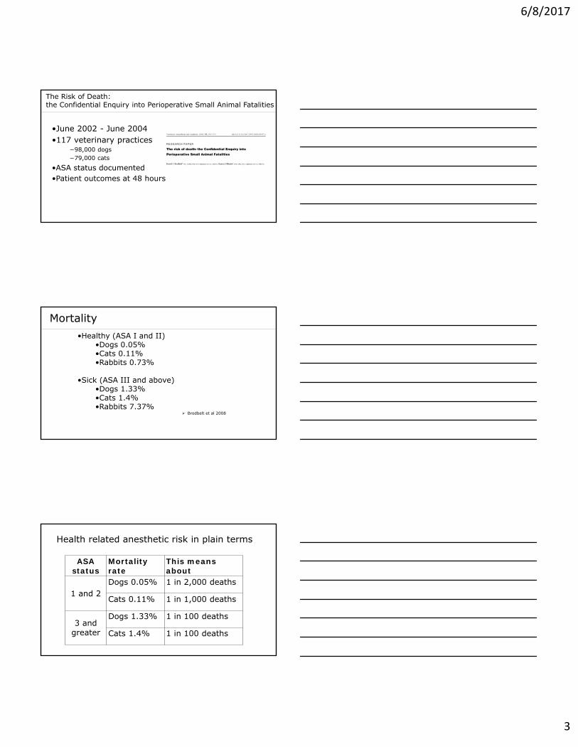
The Risk of Death: the Confidential Enquiry into Perioperative Small Animal Fatalities
•June 2002 - June 2004 •117 veterinary practices
−98,000 dogs−79,000 cats
•ASA status documented •Patient outcomes at 48 hours
Mortality •Healthy (ASA I and II)
•Dogs 0.05%•Cats 0.11%•Rabbits 0.73%
•Sick (ASA III and above)•Dogs 1.33%•Cats 1.4%•Rabbits 7.37%
Brodbelt et al 2008
Health related anesthetic risk in plain terms
ASA status
Mortality rate
This means about
1 and 2Dogs 0.05% 1 in 2,000 deaths
Cats 0.11% 1 in 1,000 deaths
3 and greater
Dogs 1.33% 1 in 100 deaths
Cats 1.4% 1 in 100 deaths
6/8/2017
4
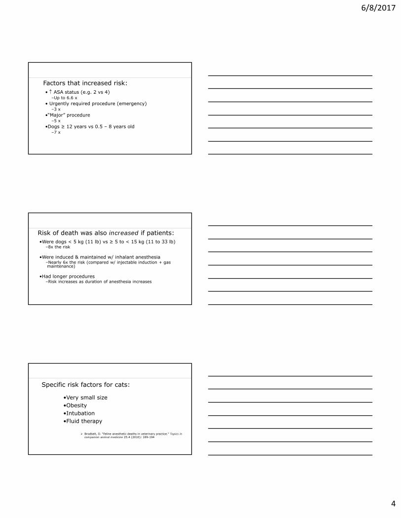
Factors that increased risk: • ASA status (e.g. 2 vs 4)
–Up to 6.6 x• Urgently required procedure (emergency)
–3 x•“Major” procedure
–5 x•Dogs ≥ 12 years vs 0.5 – 8 years old
–7 x
Risk of death was also increased if patients:•Were dogs < 5 kg (11 lb) vs ≥ 5 to < 15 kg (11 to 33 lb)
–8x the risk
•Were induced & maintained w/ inhalant anesthesia –Nearly 6x the risk (compared w/ injectable induction + gas maintenance)
•Had longer procedures –Risk increases as duration of anesthesia increases
Specific risk factors for cats:
•Very small size•Obesity•Intubation•Fluid therapy
Brodbelt, D. "Feline anesthetic deaths in veterinary practice." Topics in companion animal medicine 25.4 (2010): 189-194
6/8/2017
5
Specific risk factors for cats:
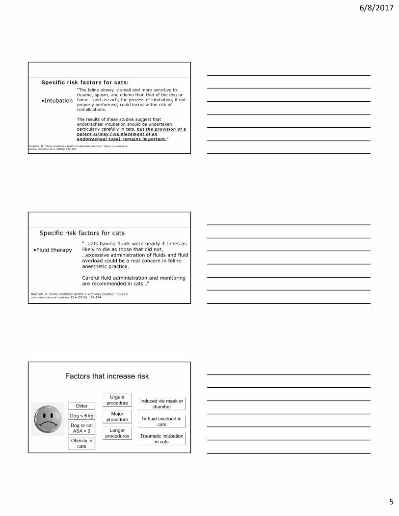
•Intubation
“The feline airway is small and more sensitive to trauma, spasm, and edema than that of the dog or horse… and as such, the process of intubation, if not properly performed, could increase the risk of complications.
The results of these studies suggest that endotracheal intubation should be undertaken particularly carefully in cats, but the provision of a patent airway (via placement of an endotracheal tube) remains important.”
Brodbelt, D. "Feline anesthetic deaths in veterinary practice." Topics in companion animal medicine 25.4 (2010): 189-194
Specific risk factors for cats
•Fluid therapy
Brodbelt, D. "Feline anesthetic deaths in veterinary practice." Topics in companion animal medicine 25.4 (2010): 189-194
“…cats having fluids were nearly 4 times as likely to die as those that did not, …excessive administration of fluids and fluid overload could be a real concern in feline anesthetic practice.
Careful fluid administration and monitoring are recommended in cats…”
Factors that increase risk
Dog < 5 kg
Dog or cat ASA > 2
Older
Urgent procedure
Major procedure
Longer procedures
Induced via mask or chamber
IV fluid overload in cats
Traumatic intubation in catsObesity in
cats

6/8/2017
6
Mortality is greater in cats •“That apparently healthy cats (ASA 1–2) had a twofold higher risk of death than healthy dogs, would suggest either preoperative assessment is poorer and more cats are misclassified as healthy when harbouringsignificant disease, or cats are at a greater risk of anaesthetic-related death. Cats are smaller than dogs in general and hence would be more prone to hypothermia, pre-disposing to prolonged recoveries and increased morbidity.”
(Brodbelt, 2010)
Addressing Anesthetic Risk
Careful patient assessment Preoperative stabilization Understand and properly maintain equipment Personnel training and supervision Monitor carefully during and after anesthesia Troubleshoot when problems occur Protocols and checklists Give owners information about risk
Patient Evaluation
•Procedure associated risk factors:–Risk of procedure itself can also be classified, independent of patient medical conditions
•Categories:–Low Risk = minimal physiologic stress–Medium Risk = moderate physiologic stress–High Risk = significant peri-operative physiologic stress
6/8/2017
7
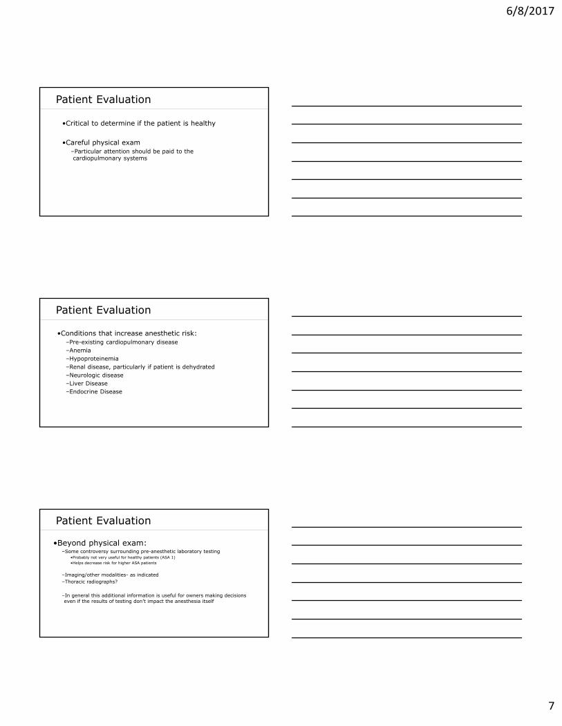
Patient Evaluation
•Critical to determine if the patient is healthy
•Careful physical exam–Particular attention should be paid to the cardiopulmonary systems
Patient Evaluation
•Conditions that increase anesthetic risk:–Pre-existing cardiopulmonary disease–Anemia–Hypoproteinemia–Renal disease, particularly if patient is dehydrated–Neurologic disease–Liver Disease–Endocrine Disease
Patient Evaluation
•Beyond physical exam:–Some controversy surrounding pre-anesthetic laboratory testing
•Probably not very useful for healthy patients (ASA 1)•Helps decrease risk for higher ASA patients
–Imaging/other modalities- as indicated –Thoracic radiographs?
–In general this additional information is useful for owners making decisions even if the results of testing don’t impact the anesthesia itself
6/8/2017
8
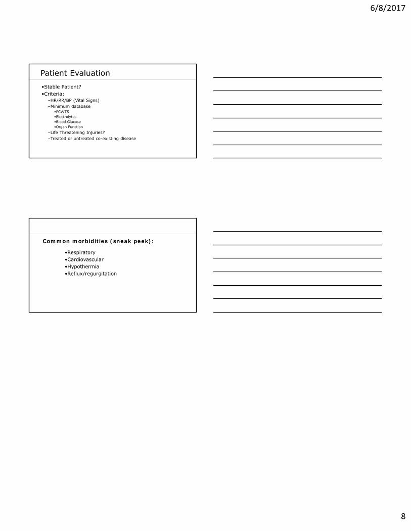
Patient Evaluation•Stable Patient?•Criteria:
–HR/RR/BP (Vital Signs)–Minimum database
•PCV/TS•Electrolytes•Blood Glucose•Organ Function
–Life Threatening Injuries?–Treated or untreated co-existing disease
Common morbidities (sneak peek):
•Respiratory •Cardiovascular•Hypothermia•Reflux/regurgitation
6/9/2017
1
Protocol Design Emily McCobb DVM MS DACVAA
Vermont Veterinary Medical Association June 23, 2017
Outline
•Features of the anesthetic plan •How to design the anesthetic protocol •The drug parade
–The most dense content of the day •Most important thing to remember:
– there is more than one way to spay a cat
•Putting it all together with cases and questions
Specifically….. –Basic principles/Definitions–“Pre-medication”
•Sedatives•Analgesics•Anticholinergics
–Chemical Restraint
–Adjunctive agents and techniques–Agents for induction and maintenance
–Putting it all together–Sample protocols/Cases
6/9/2017
2
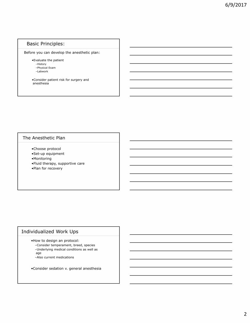
Before you can develop the anesthetic plan:
•Evaluate the patient–History–Physical Exam–Labwork
•Consider patient risk for surgery and anesthesia
Basic Principles:
The Anesthetic Plan
•Choose protocol•Set-up equipment•Monitoring •Fluid therapy, supportive care•Plan for recovery
Individualized Work Ups
•How to design an protocol:–Consider temperament, breed, species–Underlying medical conditions as well as age–Also current medications
•Consider sedation v. general anesthesia

6/9/2017
3
Elements of the Anesthetic Protocol
1) Choose a Pre-med (drugs and route)-based on patient health and planned procedure
2) Choose induction agent -based on availability, health status and clinician comfort
3) Maintenance -inhalant v. total injectable?
4) Other stuff: analgesics, adjuncts, supportive measures
Peri-Anesthetic Medications and Techniques:
•Any agent or technique that prevents or decreases harmful effects of anesthetics
–Anticholinergics, oxygen
•Or agents that improve the quality of anesthesia–Sedatives/analgesics
6/9/2017
4
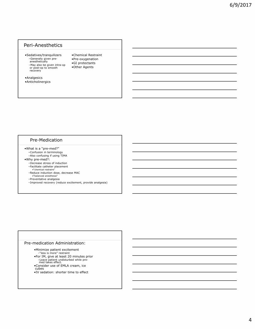
Peri-Anesthetics
•Sedatives/tranquilizers –Generally given pre-anesthetically
–May also be given intra-op or post-op to smooth recovery
•Analgesics•Anticholinergics
•Chemical Restraint•Pre-oxygenation•GI protectants•Other Agents
Pre-Medication
•What is a “pre-med?”–Confusion in terminology–Also confusing if using TIMA
•Why pre-med?:–Decrease stress of induction–Facilitate catheter placement
•“chemical restraint”–Reduce induction dose, decrease MAC
•“balanced anesthesia”–Preventative analgesia–Improved recovery (reduce excitement, provide analgesia)
•Minimize patient excitement–“less is more” restraint
•For IM, give at least 20 minutes prior –Leave patient undisturbed while pre-med takes effect.
•Consider use of EMLA cream, ice cubes•IV sedation: shorter time to effect
Pre-medication Administration:
6/9/2017
5
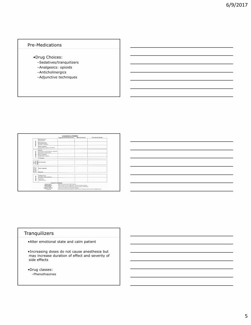
Pre-Medications
•Drug Choices:–Sedatives/tranquilizers–Analgesics: opioids–Anticholinergics–Adjunctive techniques
Tranquilizers•Alter emotional state and calm patient
•Increasing doses do not cause anesthesia but may increase duration of effect and severity of side effects
•Drug classes: –Phenothiazines

6/9/2017
6
Acepromazine
•Phenothiazine•Mechanism of action:
–Depression of reticular activating system and antidopaminergic actions w/i CNS
–Depression of CNS and catecholamines
•Results:–Calm patient–Anxiolytic –Lowers MAC
Acepromazine•Other useful affects:
–Antihistaminic–Anti-emetic (Anti-dopamine)–Chemical Restraint –Anti-arrhythmic (mild)
•Things to keep in mind: –higher dose increases effect time and side effects
–larger dose may be required in excited animals
–Long acting, non-reversible drug
Acepromazine•Cardiopulmonary side effects:•Vasodilation (α-1 adrenergic blockade) and decrease in BP
•Treatment is IV fluids, phenylephrine, not epinephrine
•May see reflex tachycardia•Dose dependent depression of myocardium and vascular smooth muscle
•Other side effects:–Mild resp. depression –decrease seizure threshold?–splenic enlargement –platelet effects–transient decrease in PCV–alters thermoregulation
Acepromazine
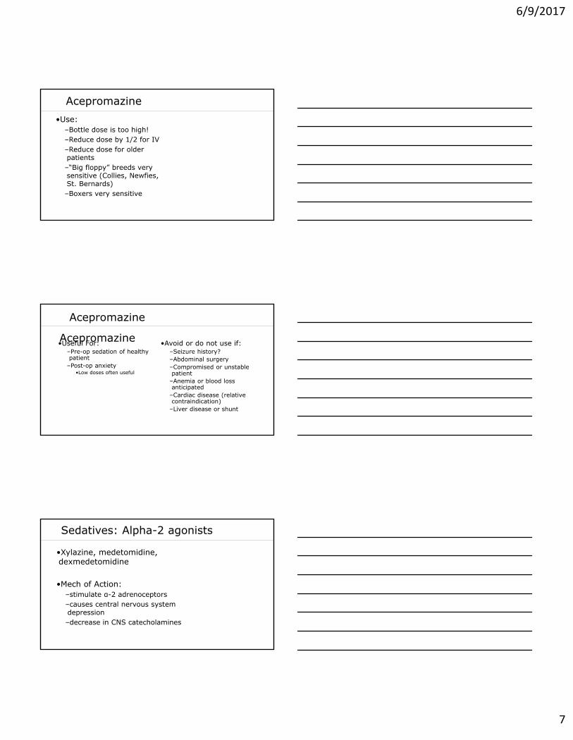
6/9/2017
7
•Use: –Bottle dose is too high!–Reduce dose by 1/2 for IV–Reduce dose for older patients–“Big floppy” breeds very sensitive (Collies, Newfies, St. Bernards)–Boxers very sensitive
Acepromazine
Acepromazine•Useful For:–Pre-op sedation of healthy patient
–Post-op anxiety•Low doses often useful
•Avoid or do not use if:–Seizure history?–Abdominal surgery–Compromised or unstable patient
–Anemia or blood loss anticipated
–Cardiac disease (relative contraindication)
–Liver disease or shunt
Acepromazine
Sedatives: Alpha-2 agonists
•Xylazine, medetomidine, dexmedetomidine
•Mech of Action: –stimulate α-2 adrenoceptors –causes central nervous system depression –decrease in CNS catecholamines

6/9/2017
8
Alpha-2 Agonists•Dose dependent sedation•Analgesic effects•Muscle relaxation•Higher doses needed in excited patients•Can cause vomiting
–Especially higher doses•Reversible (yohimbine, atipamezole)
–Not always desirable to reverse!
Alpha-2 Agonists
•Adverse Effects:–Profound cardiorespiratory depression–Vasoconstriction, biphasic blood pressure effect–Decrease cardiac output40-50%
–Cardiac Arrhythmias
Alpha-2 Agonists•Useful for:–chemical restraint and sedation (often profound) of young, healthy dogs and cats
–Pre-medication of young, healthy patients
–Post-op sedation, analgesia (“micro dose”)
•Contraindications:–Geriatric animals–Sick animals–Cardiac Disease
Alpha-2 Agonists
6/9/2017
9
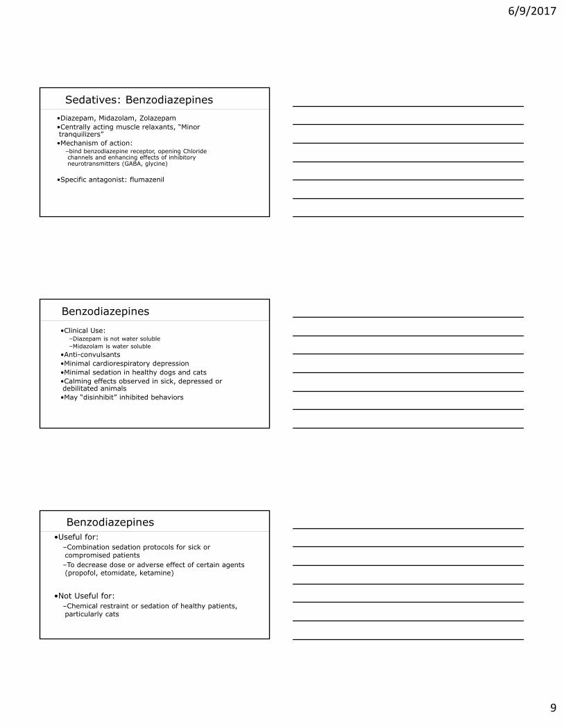
Sedatives: Benzodiazepines•Diazepam, Midazolam, Zolazepam•Centrally acting muscle relaxants, “Minor tranquilizers”•Mechanism of action:
–bind benzodiazepine receptor, opening Chloride channels and enhancing effects of inhibitory neurotransmitters (GABA, glycine)
•Specific antagonist: flumazenil
Benzodiazepines
•Clinical Use:–Diazepam is not water soluble–Midazolam is water soluble
•Anti-convulsants•Minimal cardiorespiratory depression•Minimal sedation in healthy dogs and cats•Calming effects observed in sick, depressed or debilitated animals•May “disinhibit” inhibited behaviors
Benzodiazepines•Useful for:
–Combination sedation protocols for sick or compromised patients –To decrease dose or adverse effect of certain agents (propofol, etomidate, ketamine)
•Not Useful for:–Chemical restraint or sedation of healthy patients, particularly cats

6/9/2017
10
Analgesics: Opioids•Used to provide preventative analgesia
–“peri-anesthetic adjuncts”•Also provide additional sedation
–May cause excitement in cats•Decrease MAC•Available choices:
–“pure” and “partial” agonists•Controlled substances
Partial or “mixed” agonist/antagonists•Butorphanol
–Mu antagonist–Kappa agonist–Sedation, cough suppression, mild analgesia
Opioids
•Buprenorphine–Partial mu agonist–Kappa agonist?–Moderate analgesia, minimal sedation
–Minimal respiratory depression but “sticky”
–Not for the unstable patient, hard to reverse
–Drug of choice for most cats (not for pre-med)
Butorphanol
•Useful for:–Mild to moderate sedation –Partial reversal of pure agonists
•Not useful for:–Painful procedures (unless very minimal)
Opioids
6/9/2017
11
Buprenorphine•Useful for:
–Mild to moderately painful procedures–Post-op analgesic, stable patients–Good post op choice for cats
•Not useful for:–Sedation–Intra-op (slow onset, not titratable)–Severe pain
Opioids
Pure Agonists
•Morphine, Hydromorphone, Oxymorphone, Fentanyl
–Mu agonists–*schedule II narcotics–Gold standard for analgesia–Moderately sedating in dogs
Opioids
Pure opioid agonists
•Adverse effects:–Vomiting (non-painful patients)–Respiratory depression –Bradycardia–Ileus, constipation –Urinary retention –Excitation (cats)–Hypothermia/Hyperthermia
Opioids

6/9/2017
12
Pure Agonists•Useful for:
–Pre-op sedation –Moderate to severe pain–Sick and fragile patients–Balanced anesthesia techniques
•Reduce MAC•Adjunctive analgesia•*fentanyl: short half life, good for CRIs
Opioids
Anticholinergics•Glycopyrrolate or Atropine
–Glycopyrrolate: slower onset, lasts longer•Mechanism of action:
–Competitively antagonize acetylcholine–Act at parasympathetic postganglionic neuroeffector sites (muscarinic)
•Sphincters•Smooth muscle
•Clinical Use–Prevent bradycardia or increase heart rate–Dry secretions
Anticholinergics
•Effects:–Speed heart rate (low doses cause bradycardia)
•Raise BP and CO, HR X SV = CO–Dry salivary and respiratory secretions–Tighten lower esophageal sphincter–Decrease GI motility–Weakly anti-emetic–Pupillary dilation (atropine)
6/9/2017
13
Anticholinergics
•Potential adverse effects:–Initial first or second degree AV block–Tachyarrhythmias:
•sinus tachycardia, other untoward arrhythmias, increasing myocardial oxygen consumption
Atropine
•Crosses BBB, may cause CNS effects•Rapid onset•Drug of choice for CPR
Anticholinergics
Glycopyrrolate•Synthetic, quaternary amine compound•Does not cross blood brain barrier or placenta•Slower onset, more gentle, drug of choice for most peri-anesthetic uses•More potent than atropine•Currently more expensive
Anticholinergics

6/9/2017
14
Indications:•Bradycardia, AV block
–Can be inherent to patient, drugs, or procedure –Reflex increases in vagal tone (laryngeal, ocular stimulation and vasovagal reflexes)
•Ocular surgery (oculocardiac reflex)•Brachycephalic or pediatric patients•Neck or back surgery
Anticholinergics
Contraindications:•Tachycardia
•Cardiac disease•Tachycardia•Geriatric patients
•Endoscopy (relative) •Prior administration of alpha-2
Anticholinergics
In summary…..
To choose a “pre-med”:1) Anticholinergic or no anticholinergic 2) Choose analgesic3) Choose sedative/tranquilizer4) Consider if additional chemical restraint needed
6/9/2017
15
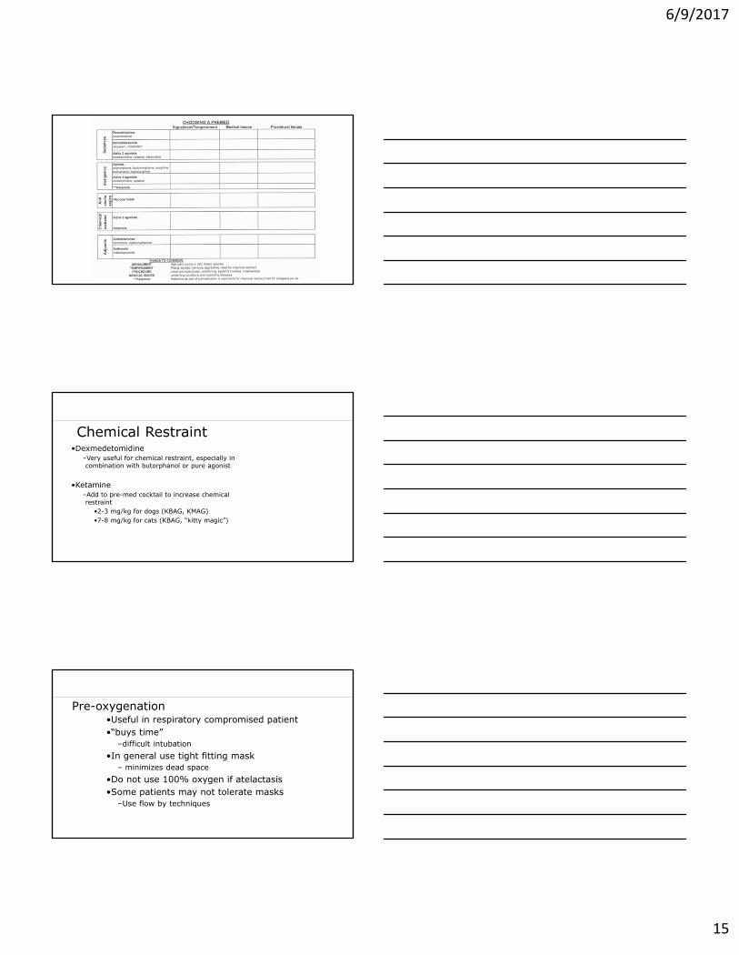
Chemical Restraint•Dexmedetomidine
–Very useful for chemical restraint, especially in combination with butorphanol or pure agonist
•Ketamine–Add to pre-med cocktail to increase chemical restraint
•2-3 mg/kg for dogs (KBAG, KMAG)•7-8 mg/kg for cats (KBAG, “kitty magic”)
Pre-oxygenation•Useful in respiratory compromised patient•“buys time”
–difficult intubation •In general use tight fitting mask
– minimizes dead space•Do not use 100% oxygen if atelactasis•Some patients may not tolerate masks
–Use flow by techniques
6/9/2017
16
Adjunctive Analgesic Techniques•Additional analgesia or MAC sparing may be needed intra-op or in recovery•Multimodal is optimal•Options:
–Additional bolus dose opioids–Epidural or local administration of opioids or local anesthetics–CRI of opioid, local anesthetics, ketamine–NSAIDs
Anesthetic Adjuncts•Antihistamines (benadryl)•Steroids•GI protectants/antacids•Antibiotics•Free radical scavengers•Anti-arrhythmics (beta blockers)
Sedation v. Induction
•Blurred boundaries: “conscious sedation”–useful for longer procedures or when more than simple sedation is required–careful monitoring is required

6/9/2017
17
Ok, so now how do I choose?•Consider patient characteristics and procedure
–Amount sedation/analgesia required–Determine if any drugs are specifically indicated or contraindicated
•Availability•Cost•Personal preference
–There are no safe anesthetics, only safe anesthetists
Induction Agents
•Available agents for IV induction:–Propofol–Ketamine–Ketamine/Propofol–Etomidate–Ketamine/Midazolam–Narcotic/Benzodiazepine–Alfaxalone
• Available agents for IM induction:−Telazol−Dexmedetomidine/Ketamine−Alfaxalone
Propofol•Non barbiturate sedative/hypnotic•Rapid metabolism
–Extrahepatic sites–Minimal to no cumulative effects
•Cardiovascular effects similar or more profound than pentothal
–Cardiac depression, hypotension–Short lived and generally well tolerated
•Does not increase intra-ocular or intracranial pressure
Induction Agents
6/9/2017
18
Propofol•Use
–Titrate slowly to effect (calculated dose over 90 seconds)•rapid administration can cause apnea
–Can cause methemoglobinemia in cats •with repeated administration
–Aseptic Technique (lipid/egg lecithin emulsion)•Can support bacterial growth
Propofol•Safe Handling:
–*should never be refrigerated–open vials must be used or discarded within 6 hours of opening–wipe bottle top with alcohol and use a new needle for each use
Propofol•Useful for:
–Conscious sedation, CRIs–Liver disease–Any case where rapid recovery is needed
•Not recommended:–Septic patients–Unstable patients–Patients who lack cardiovascular reserves–Wounds

6/9/2017
19
Alfaxalone/Alfaxan
Photo courtesy of Dr. Cheryl Blaze
Induction Agents
Alfaxalone•Alfaxalone (3 α-hydroxy- 5α-pregane- 11, 20-dione)
–Ultra short acting sedative hypnotic anesthetic
–Neuroactive steroid producing anesthetic effects through activity at the GABA-A receptor
•Similar molecular structure to progesterone
–Does not bind to sex hormone, glucocorticoid or mineralcorticoidreceptors
History and context •Work with steroid molecules and anesthesia goes back to the 1970s•Originally was formulated into a castor oil based surfactant
–Saffan- animals–Althesin- humans
•Initially doses much higher and lots of adverse effects (hyperemia/nose running of the paws and face for cats)
•Allergic reactions in dogs, cats and people due to cremaphor induced histamine release (vehicle)
•Now there are new non-toxic dilutents- cyclodextrins–Water soluble –Used in Africa, Europe and Australia for some time
Induction Agents: Alfaxalone

6/9/2017
20
What does it do?
•At an anesthetic dose this drug is similar to other sedative hypnotic agents that you are used to (thiobarturates and the non-barbiturate sedative hypnotics propofol and etomidate):
–Rapid loss of consciousness–Loss of swallowing reflex–Marked muscle relaxation
Induction Agents: Alfaxalone
Mechanism of Action •Alfaxalone induces anaesthesia through activity at the gamma amino butyric acid sub-type A receptor (GABAA)
• GABA is a major inhibitory neurotransmitter in the CNS•Alfaxalone enhances the effects of GABA at the GABAA receptors resulting in opening of channels into the cells and an influx of chloride ions.
•This causes hyperpolarization of the cells and inhibition of neural impulse transmission.
Alfaxalone
More thoughts on MOA
•Some subunit and receptor pool specificity•For example- unlike propofol, neuroactive steroids do not seem to also work through glycine channels
•Neurosteroids seem to act locally in a paracrine manner to modulate the receptor function
•Each drug thus has a unique effect profile (differences in effects at the GABA a subunit could explain the wide margin of safety for the drug)
Alfaxalone
6/9/2017
21
Alfaxan Solution • A clear, aqueous solution for intravenous injection• Registered for the induction and maintenance of
anesthesia in dogs and cats• 10 ml vial of 10 mg/ml alfaxalone solubilized in
hydroxypropyl β cyclodextrin• Iso-osmolar, sterile solution with a pH of 6.5-7
http//www.jurox.com
Alfaxalone
Alfaxane Formulation •Non-irritating if injected peri-vascularly•Supports bacterial growth but less than propofol•Beneficial Properties:
–Potent–Wide safety margin–Rapid metabolism and does not accumulate
Alfaxalone
•Metabolism/Pharmacokinetics:–Phase I and Phase II enzymes –unlike propofol,which is mostly phase II
•Pharmacodynamics:– Rapid onset–Duration of action (10 minutes – if un-premedicated)
–Up to 25 minutes if pre-medicated
–Good resp’ty & CV stability–HR, BP, C.O. – minimal changes
Alfaxalone
6/9/2017
22
Suggestions for use
•Administer slowly to decrease risk of apnea (over 60s)•Not analgesic (similar to propofol)•Rapid clearance is what can lead to rapid and ugly recoveries
–Good pre-med will help•Minimize light and sound stimulation during induction and recovery
Alfaxalone
Specifically for cats
•Metabolic profile may be more favorable than for propofol since there is less reliance on glucuronidation
•Does not cause oxidative damage like propofol, so might be better for cats who need a CRI or repeated dosing (ICU, RT) , especially if cat is anemic already
•Option for IM sedation a nice option, especially in the older cat or cat with cardiac disease
Alfaxalone
How we currently use this drug at Tufts •Once opened vial has a limited life span
–Try to use whole vial on day opened
•Induction Agent (1 to 3 mg/kg)
•IM as part of a combination protocol for cats –2 mg/kg alfaxane; 0.1 mg/kg oxymorphone, +/- 0.05 mg/kg acepromazine
• CRI dose: 0.07 mg/kg/min
Alfaxalone

6/9/2017
23
Alfaxane v. Propofol
•Similar CV impact•Can give alfaxalone IM (need higher dose)•Higher therapeutic index•Potential for rough recoveries with alfaxalone
Induction Agents
Ketamine•Dissociative
–Cateleptoid state•Useful to induce general anesthesia
–combine with a sedative –usually diazepam or midazolam
•Liver metabolism in dogs, renal excretion•In cats active metabolites get excreted
–prolonged recoveries possible with renal compromise
Induction Agents
Ketamine•Cardiovascular Effects:
–“sympathomimetic”–Stimulates cardiovascular system–Direct myocardial depression
•Respiratory Effects:–Apneustic breathing, bronchodilation
•Good somatic analgesic•Increases intraocular pressure•Can lower seizure threshold and increase intracranial pressure
6/9/2017
24
Ketamine
•Practical Use–Give one third of dose slowly then wait –Titrate additional drug to effect–May need to wait up to 90 minutes for full effect–Jaw tone and central eye position maintained–For best analgesic effect needs to be given pre-op,intra-op, post-op
Ketamine•Use for:
–Painful procedures–Unstable patients–Useful for almost all species
•Do not use for:–Urinary obstruction–Cats with HCM–Patients who may not tolerate tachycardia
Induction Agents
Ketamine Propofol Combo
Induction Agents

6/9/2017
25
Telazol
•Proprietary mixture of tiletamine and zolazepam•More potent
Induction Agents
Etomidate
•Features:–Non-barbiturate sedative/hypnotic
–Minimal cardiovascular and respiratory depression
–Liver metabolism–Rapid Recovery–Causes adrenocortical suppression
• Use: − Can cause excitement
and unsightly reactions at induction (seizures, vocalization, myoclonus, salivation and defecation)
− Patient must be well sedated first
Etomidate•Good For:
–Severe heart disease (heart failure)–Cardiovascular collapse–Liver disease
•Not recommended if:–Addison’s disease–Critically ill–Healthy patient
Induction Agents
6/9/2017
26
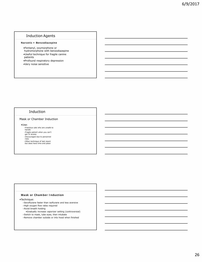
Narcotic + Benzodiazepine
•Fentanyl, oxymorphone or hydromorphone with benzodiazepine•Useful technique for fragile canine patients•Profound respiratory depression•Very noise sensitive
Induction Agents
Mask or Chamber Induction
•Use:–Fractious cats who are unsafe to handle
–Fragile patient when you can’t get IV access
–Discouraged due to personnel risks
–Often technique of last resort but does have time and place
Induction
Mask or Chamber Induction•Technique:
–Sevoflurane faster than isoflurane and less aversive –High oxygen flow rates required–Avoid breath holding
•Gradually increase vaporizer setting (controversial)–Switch to mask, lube eyes, then intubate–Remove chamber outside or into hood when finished
6/9/2017
27
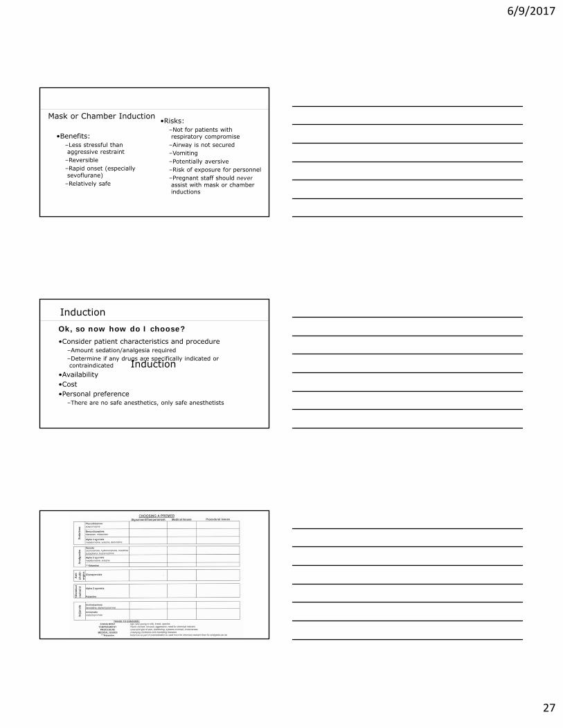
Mask or Chamber Induction
•Benefits:–Less stressful than aggressive restraint
–Reversible–Rapid onset (especially sevoflurane)
–Relatively safe
•Risks:–Not for patients with respiratory compromise
–Airway is not secured–Vomiting–Potentially aversive–Risk of exposure for personnel–Pregnant staff should never assist with mask or chamber inductions
Ok, so now how do I choose?•Consider patient characteristics and procedure
–Amount sedation/analgesia required–Determine if any drugs are specifically indicated or contraindicated
•Availability•Cost•Personal preference
–There are no safe anesthetics, only safe anesthetists
Induction
Induction

6/9/2017
28
Inhalants •Most common •Isoflurane v. Sevoflurane?
Maintenance
Inhalant v. Injectable
•Numerous protocols for TIVA and TIMA •Factors to consider
Maintenance
Mechanical Ventilation or not?
Maintenance

6/9/2017
29
Other Stuff for the Protocol
•Analgesics–Goals: multi-modal and preventative–CRIs commonly employed: lidocaine/ketamine (MLK, HLK, LK, etc.)
–Questions to ask: –What else can I give this pet? –Can the pet have an NSAID? –What local block could I do?
GI Meds
•Rationale:–Prevention of vomiting? –Prevention of GERD/Regurg–Standard for brachycephalics
•Options: –Metoclopramide–H2 Blockers
•Efficacy in dogs? –Maropitant (cerenia)
•Is also analgesic
Other Stuff
Plan for fluid therapy
Case Examples

6/9/2017
30
Case Examples
•Routine Dog Spay:–Butorphanol or pure agonist –Acepromazine–Glycopyrrolate depending on resting heart rate and breed
•Boston Terrier having cataract surgery:–Acepromazine–glycopyrrolate
Dog Pyometra•12 yo Pomeranian•Open Pyometra x 1 week•History of seizures- currently on phenobarbital BID and Baytril•BAR and no murmurs on PE•CBC/Chem W/NL
•Protocol: 120 mg Gabapentin PO, Hydromorphone 0.1 mg/kg and Midazolam at 0.2 mg/kg given IM and placed IV catheter; Propofol at 4 mg/kg, intubated and maintained on Isoflurane with LRS at 10 ml/kg/hr
Case Examples
•Young cat having a neuter:–Ketamine/dexmedetomidine/buprenorphine–Injecatable only–No intubation
•Older cat with heart disease having painful surgery or dental:–Oxymorphone or Hydrorphone–Midazolam–Alfaxalone if needed for restraint–Likely no glycopyrrolate–IV induction with propofol–Maintain on Isoflurane or sevoflurane–Watch blood pressue
Case Examples

6/9/2017
31
Questions?
McCobbAnesthesia of Geriatric Patient
6/6/2017
Vermont VMA 1
Anesthesia of the Geriatric Patient:
Guess What? Age IS a disease
Emily McCobb DVM MS DACVAAVermont Veterinary Medical Association
June 23, 2017
Outline
• Introduction
• Physiology and Pharmacology
• Clinical impacts
• Dos and Don’ts
The geriatric patient population
• Is increasing (human and animal)
• More commonly anesthetized
• Has special needs
McCobbAnesthesia of Geriatric Patient
6/6/2017
Vermont VMA 2
Why I say it’s a disease
• It’s a catchy title
• Age is a factor we need to pay attention to
• Increased risk of anesthetic death
Aging pets
• Have different medical and surgical needs• Need for dentistry or other minor procedures
• Underlying medical conditions
• Standard protocols may not apply
Is age relative?
• Geriatric = animal that has lived 75% of its expected life span
• Life span may depend on species, breed and size
• Also many individual variations

McCobbAnesthesia of Geriatric Patient
6/6/2017
Vermont VMA 3
Life Span by Size of Dog
years
Average adult body mass (kg)
Modified from Selman et al, Current Biology, V. 23
Basics to consider
• Perceived sensitivity and increased vulnerability to side effects
• Alterations in many physiologic parameters affect care
• Consider from a body systems perspective
Unavoidable effects of aging
• Basal function is relatively uncompromised by age alone
• HOWEVER‐• organ reserve are reduced
• ability to compensate for stress is ↓
• many geriatric patients also have diseased organs
McCobbAnesthesia of Geriatric Patient
6/6/2017
Vermont VMA 4
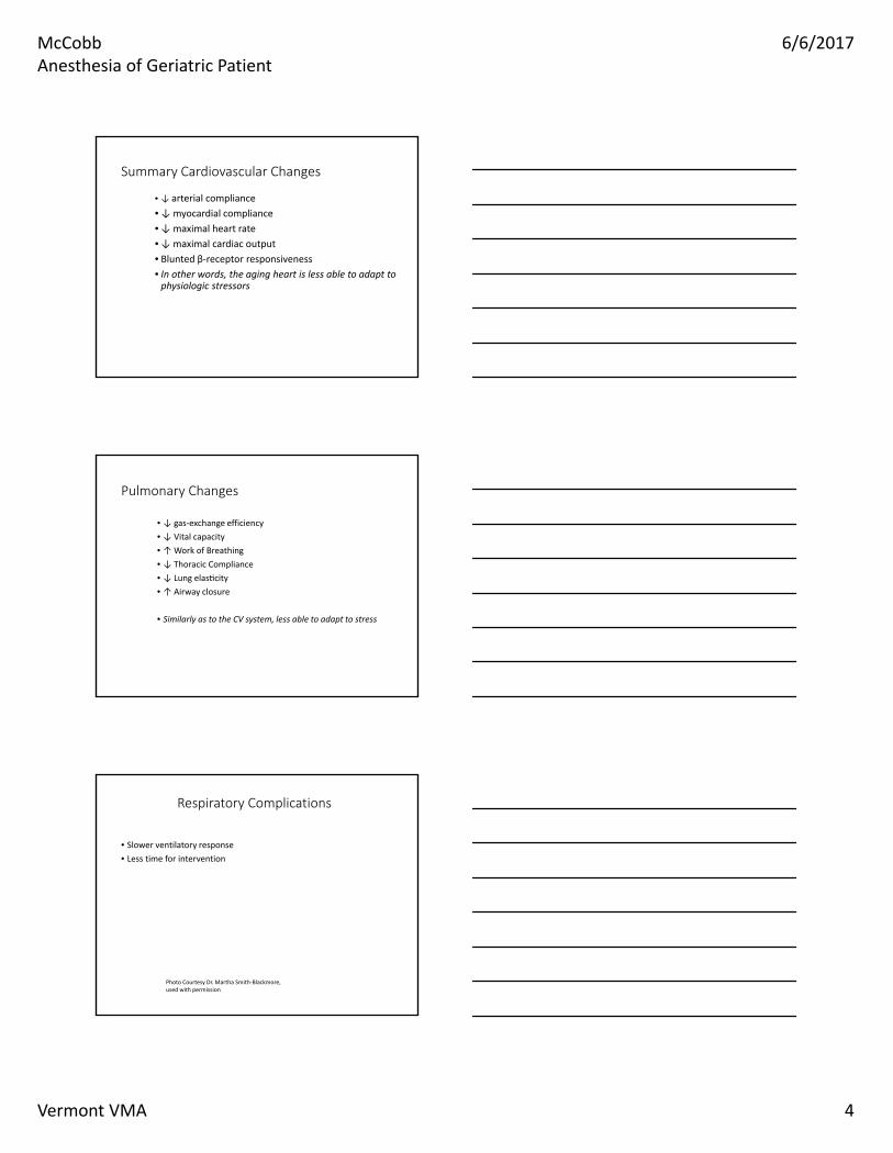
Summary Cardiovascular Changes
• ↓ arterial compliance
•↓ myocardial compliance
•↓ maximal heart rate
•↓ maximal cardiac output
• Blunted β‐receptor responsiveness
• In other words, the aging heart is less able to adapt to physiologic stressors
Pulmonary Changes
• ↓ gas‐exchange efficiency
• ↓ Vital capacity
• ↑ Work of Breathing
• ↓ Thoracic Compliance
• ↓ Lung elas city
• ↑ Airway closure
• Similarly as to the CV system, less able to adapt to stress
Respiratory Complications
• Slower ventilatory response
• Less time for intervention
Photo Courtesy Dr. Martha Smith‐Blackmore, used with permission
McCobbAnesthesia of Geriatric Patient
6/6/2017
Vermont VMA 5
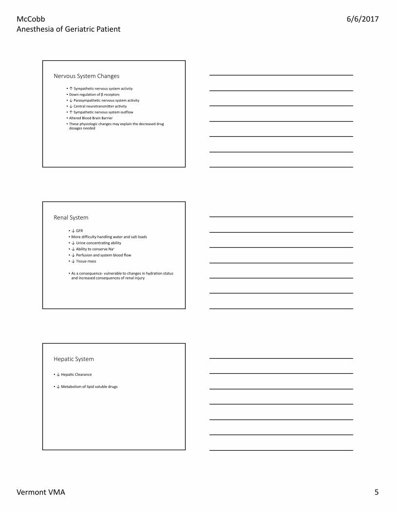
Nervous System Changes
• ↑ Sympathetic nervous system activity
• Down regulation of β receptors
• ↓ Parasympathe c nervous system ac vity
• ↓ Central neurotransmi er ac vity
• ↑ Sympathe c nervous system ou low
• Altered Blood Brain Barrier
• These physiologic changes may explain the decreased drug dosages needed
Renal System
• ↓ GFR
• More difficulty handling water and salt loads
• ↓ Urine concentra ng ability
• ↓ Ability to conserve Na+
• ↓ Perfusion and system blood flow
• ↓ Tissue mass
• As a consequence‐ vulnerable to changes in hydration status and increased consequences of renal injury
Hepatic System
• ↓ Hepa c Clearance
• ↓ Metabolism of lipid soluble drugs
McCobbAnesthesia of Geriatric Patient
6/6/2017
Vermont VMA 6
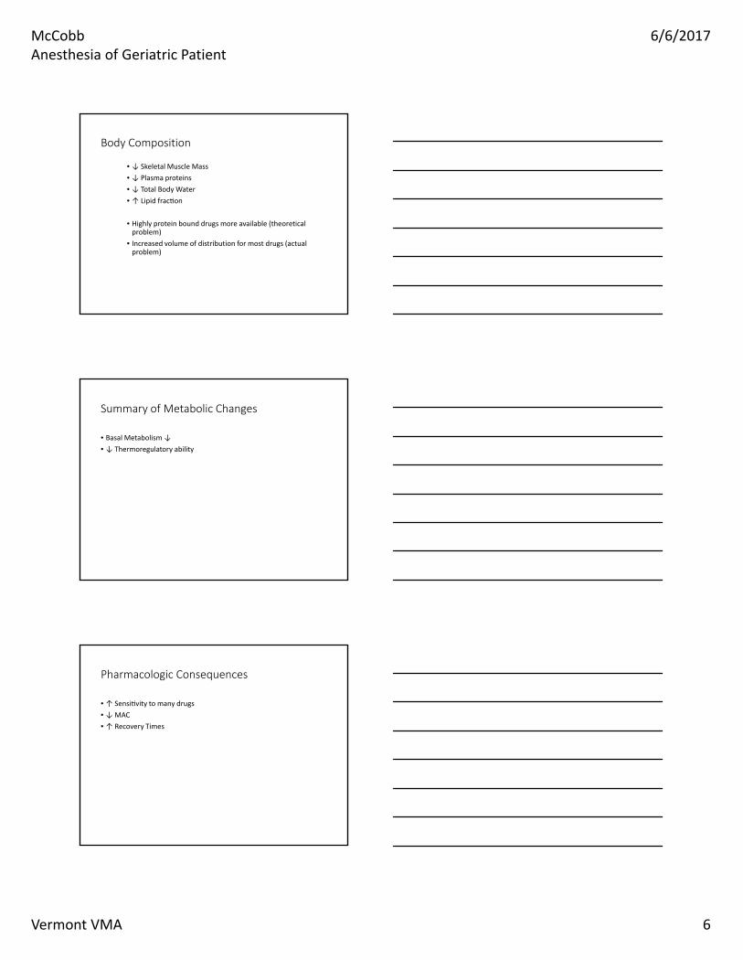
Body Composition
• ↓ Skeletal Muscle Mass
• ↓ Plasma proteins
• ↓ Total Body Water
• ↑ Lipid frac on
• Highly protein bound drugs more available (theoretical problem)
• Increased volume of distribution for most drugs (actual problem)
Summary of Metabolic Changes
• Basal Metabolism ↓
• ↓ Thermoregulatory ability
Pharmacologic Consequences
• ↑ Sensi vity to many drugs
• ↓ MAC
• ↑ Recovery Times
McCobbAnesthesia of Geriatric Patient
6/6/2017
Vermont VMA 7
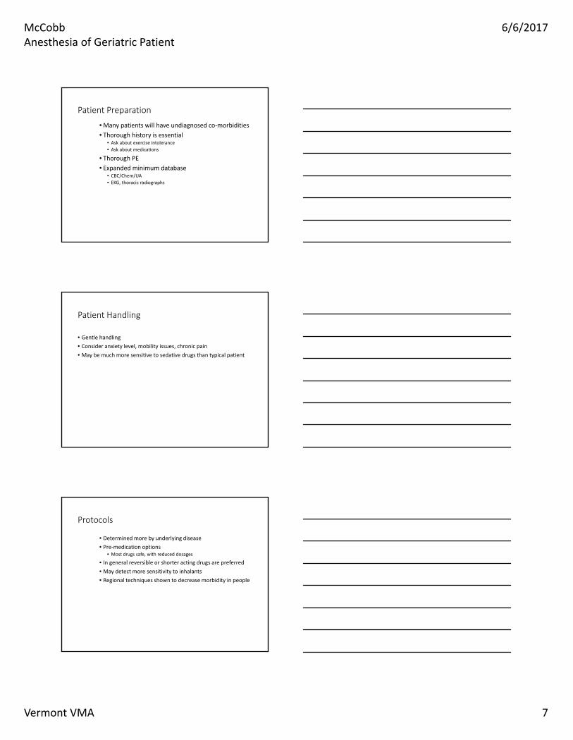
Patient Preparation
• Many patients will have undiagnosed co‐morbidities
• Thorough history is essential • Ask about exercise intolerance
• Ask about medications
• Thorough PE
• Expanded minimum database• CBC/Chem/UA
• EKG, thoracic radiographs
Patient Handling
• Gentle handling
• Consider anxiety level, mobility issues, chronic pain
• May be much more sensitive to sedative drugs than typical patient
Protocols
• Determined more by underlying disease
• Pre‐medication options• Most drugs safe, with reduced dosages
• In general reversible or shorter acting drugs are preferred
• May detect more sensitivity to inhalants
• Regional techniques shown to decrease morbidity in people

McCobbAnesthesia of Geriatric Patient
6/6/2017
Vermont VMA 8
• Consider sedation v. general anesthesia
Acepromazine
• Can cause hypotension
• Should reduce dose
• Effects can be long lasting
Benzodiazepines
• Minimal CV depression
• Good sedation in older patients
• Longer duration
McCobbAnesthesia of Geriatric Patient
6/6/2017
Vermont VMA 9
Dexmedetomidine
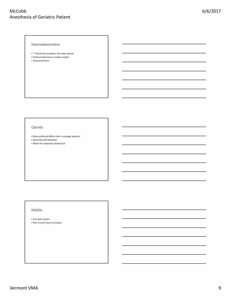
• ** Should be avoided in the older patient
• Profound decrease in cardiac output
• Vasoconstriction
Opioids
• More profound effects than in younger patients
• Generally well tolerated
• Watch for respiratory depression
NSAIDs
• Use with caution
• Risk of renal injury increased

McCobbAnesthesia of Geriatric Patient
6/6/2017
Vermont VMA 10
Propofol
• Rapid metabolism by hepatic and extra‐hepatic sites
• Cardiorespiratory depression
Monitoring
• Same as always‐ CV system, respiratory system and temperature
• Pulse OX, ETCO2, Blood Pressure, Temp
• Blood Pressure‐ critical
• May need to be more aggressive with pressors than typically as may not be able to handle fluids as well
• May require more heat support than typical
Post Anesthetic Management
• Anesthetists tend to have a short term mentality
• Most complications occur in the post op period

McCobbAnesthesia of Geriatric Patient
6/6/2017
Vermont VMA 11
Dos • Pre‐oxygenate patient
• Use pre‐medication
• Consider propofol (or alfaxalone)
• Reduce drug doses‐ up to 50 %
• Give IV fluids (5 to 10 ml/kg/hr)
• Monitor blood pressure
• Consider acepromazine for anxiety (low dose)
• Avoid anticholinergics
Pre‐oxygenation
• Helps avoid peri‐operative hypoxemia
More Dos
• Avoid hypothermia• Shivering ↑ myocardial oxygen demand
• Decreases coagulation and wound healing

McCobbAnesthesia of Geriatric Patient
6/6/2017
Vermont VMA 12
Don’t
• Handle patient roughly
• Use alpha‐2 agonists
• Be afraid of acepromazine
• Use NSAIDs (without extreme caution)• Possible role for Galiprant?
Complications
• More at risk than younger patients
• Hypotension, hypotension, hypotension • Best avoided by using a balanced anesthetic protocol
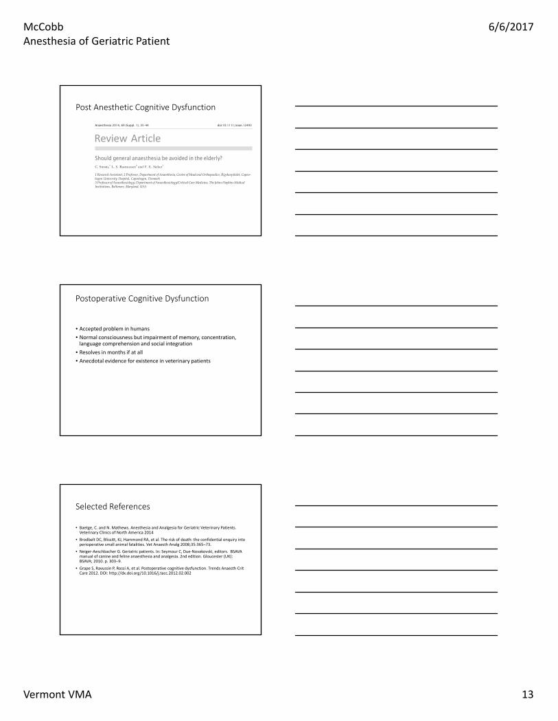
• Post Anesthetic Cognitive Dysfunction
Hypotension
• Generally need to decrease depth of anesthesia
• Add Fentanyl CRI to decrease inhalant
• Fluids if hypovolemic
• Dopamine at lowest possible dose
McCobbAnesthesia of Geriatric Patient
6/6/2017
Vermont VMA 13
Post Anesthetic Cognitive Dysfunction
Anaesthesia 2014, 69 (Suppl. 1), 35–44 doi:10.1111/anae.12493
Review Article
Should general anaesthesia be avoided in the elderly? C. Strøm,1 L. S. Rasmussen2 and F. E. Sieber3
1 Research Assistant, 2 Professor, Department of Anaesthesia, Centre of Head and Orthopaedics, Rigshospitalet, Copen- hagen University Hospital, Copenhagen, Denmark 3 Professor of Anaesthesiology, Department of Anaesthesiology/Critical Care Medicine, The Johns Hopkins Medical Institutions, Baltimore, Maryland, USA
Postoperative Cognitive Dysfunction
• Accepted problem in humans
• Normal consciousness but impairment of memory, concentration, language comprehension and social integration
• Resolves in months if at all
• Anecdotal evidence for existence in veterinary patients
Selected References
• Baetge, C. and N. Mathews. Anesthesia and Analgesia for Geriatric Veterinary Patients. Veterinary Clinics of North America 2014
• Brodbelt DC, Blissitt, KJ, Hammond RA, et al. The risk of death: the confidential enquiry into perioperative small animal fatalities. Vet Anaesth Analg 2008;35:365–73.
• Neiger‐Aeschbacher G. Geriatric patients. In: Seymour C, Due‐Novakovski, editors. BSAVA manual of canine and feline anaesthesia and analgesia. 2nd edition. Gloucester (UK): BSAVA; 2010. p. 303–9.
• Grape S, Ravussin P, Rossi A, et al. Postoperative cognitive dysfunction. Trends Anaesth CritCare 2012. DOI: http://dx.doi.org/10.1016/j.tacc.2012.02.002
McCobb 6/9/2017
Vermont VMA 2017 1
Patient Monitoring and Safety: Avoiding and Addressing
Common Anesthetic Complications
Emily McCobb DVM MS DACVAA
Vermont Veterinary Medical Association June 23, 2017
Outline•Preparation •Monitoring &Trouble-Shooting•Addressing common complications•Anesthetic Emergencies •Questions & Discussion
•Clinic•Patient•Team
Preparation
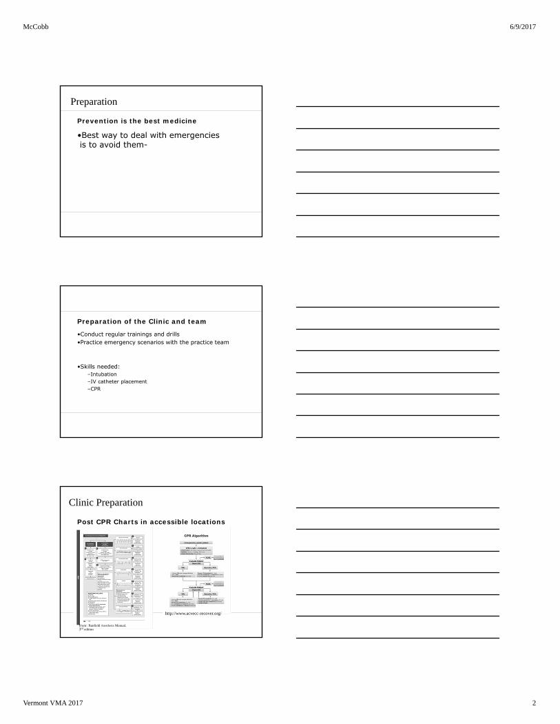
McCobb 6/9/2017
Vermont VMA 2017 2
Prevention is the best medicine
•Best way to deal with emergencies is to avoid them-
Preparation
Preparation of the Clinic and team
•Conduct regular trainings and drills•Practice emergency scenarios with the practice team
•Skills needed:–Intubation–IV catheter placement–CPR
Post CPR Charts in accessible locations
Clinic Preparation
From: Banfield Anesthesia Manual,3rd edition
http://www.acvecc-recover.org/

McCobb 6/9/2017
Vermont VMA 2017 3
Patient Selection and Preparation
•Identify and prepare for higher risk patients
Patient Preparation
•Patient History and Owner Consent•Fasting times
– Adults: 6-12 hours- Feral Cats
– Pediatrics- Feed small meal the morning of surgery- No more than 2 to 4 hours
• Water is not withheld
Patient Preparation
•Many patients will have undiagnosed co-morbidities
•Thorough history is essential (if possible!)•Thorough PE•Expanded minimum database
–CBC/Chem/UA –EKG, thoracic radiographs
McCobb 6/9/2017
Vermont VMA 2017 4
Pre-op Physical Exam
•Assess attitude and temperament•Identify high risk patients•Accurate weight•Cardiovascular fitness•Underlying conditions
Patient Preparation
1) Stabilize the unstable patient2) Patient optimization
-directed laboratory testing-diagnose and treat underlying diseases-adjust patient medications
3) Plan for intra-op and post-op needs
Patient Support
•Peri-operative Thermoregulation•Airway management •Hydration status•Special consideration:
–Pregnant patients–Geriatric–Pediatric–Brachycephalic Airway
McCobb 6/9/2017
Vermont VMA 2017 5
To intubate or not to intubate?
•Must always have oxygen and breathing circuit available
•Especially for cats and short surgery times intubation may not be needed and may be harmful
•Down side is increased waste gas exposure
•Dogs generally intubated
Airway Management
Fluid Therapy
Record Keeping and Monitoring
•Anesthesia Record:–Document affects of anesthetic drugs on patient
–Plan future anesthesia based on prior patient response
–In event of anesthetic related arrest, allows sequence of perioperative events to be reviewed
McCobb 6/9/2017
Vermont VMA 2017 6
What to record
•Species/breed/age/gender/wt/physical status•Procedure•All agents administered with dose in mg•Duration of anesthesia•Supportive measures•Difficulties encountered and means of correction•Vital readings?
Monitoring
•Importance of monitoring•Parameters to monitor:
–Depth–Cardiovascular System–Respiratory System–Temperature
•Objective means required
What tools do you have?
•Stethoscope•Esophageal Stethoscope•Pulse Oximeter•EKG•Blood Pressure •Capnograph

McCobb 6/9/2017
Vermont VMA 2017 7
What is the standard of care?
•ACVAA guidelines– www.acva.org
•ASV medical care guidelines for spay-neuter programs
-www.sheltervet.org
•AAHA Anesthesia Guidelines–https://www.aaha.org/professional/resources/anesthesia_guidelines_abstract.aspx
Monitoring Guidelines •Must monitor:
–Depth–Circulation–Oxygenation/ventilation–temperature
•Does not necessarily require specialized equipment
•Monitor every five minutes, record at least every ten
Doesn’t have to be fancy…
McCobb 6/9/2017
Vermont VMA 2017 8
Why monitor?
•Prevent anesthetic related morbidity and mortality
•Address trends in parameters before animal’s condition deteriorates
•Understand affect of anesthetic on our patient
•*Ultimate goal: ensure optimum anesthetic depth with minimal physiologic impairment
Assessment of anesthetic actions
•General Anesthesia:–Unconsciousness–Insensitivity to pain–Muscle relaxation–Absence of reflex responses
Who should monitor the patient?
•Responsible individual (doctor or technician) should be aware of the patient’s status at all times during anesthesia & recovery
•Be prepared to intervene when indicated or alert the veterinarian in charge of changes in the patient’s condition
•If responsible person can not be with the patient continually they should check the patient every five minutes and audible monitors should be used
McCobb 6/9/2017
Vermont VMA 2017 9
Patient Depth•Why does it matter?
Why do we care about patient depth?
•Too light:–Awareness–Recall–Pain –Movement
•Too deep:–Hypoventilation–Hypoxemia–Reduced cardiac output
–Hypotension–Inadequate tissue perfusion
–Hypothermia–Prolonged recovery
Depth of Anesthesia
•Can be particularly difficult to assess:–Anesthetic drugs that induce adequate anesthesia in one species or operation may not be sufficient in another species/situation
–Signs characterizing a continuum of progressive increases in CNS depression and analgesia may not occur with some drugs and drug combinations
McCobb 6/9/2017
Vermont VMA 2017 10
Depth of Anesthesia
•Examples of difficulties:–Ketamine: will not see ocular signs of increasing CNS depression (central pupil)
–Jaw tone also maintained
–Propofol: not analgesic at any concentration
Anesthetic Level
•Balance between amount of anesthetic administered and surgical stimulation (wakens patient)
•Patient’s requirements change over time (generally decrease)
•Anesthetic should be given “to effect”
Stages of General Anesthesia
•Describe levels of CNS depression•Not necessarily distinct but blend from one to the next
•Actual responses can vary between patients and patient condition
•Classically associated with inhalant anesthesia
McCobb 6/9/2017
Vermont VMA 2017 11
Planes of Surgical Anesthesia
•Light: until eyeball movement ceases•Medium: light plane of surgical anesthesia
–Stable respiration and pulse rate, abolished laryngeal reflexes, sluggish palpebral, strong corneal, muscle relaxation
•Deep: –Diaphragmatic respiration, profound muscle relaxation, weak corneal reflex, central, dilated pupil (too deep!)
Monitoring Depth
•Ocular Signs–Can be variable
•Jaw tone–Not always useful
•Vital Signs
Ocular Signs of Anesthesia
•Eye ball position and movement•Photomotor reflexes•Pupillary size•Lacrimation•Palpebral, corneal, and conjunctival reflexes

McCobb 6/9/2017
Vermont VMA 2017 12
Ocular Signs
•For dogs and cats at a light plane, the eyes are generally turned down and towards the nose
•Eyelids are closed and third eyelids are elevated•Palpebral reflex sluggish at a surgical plane•Pupillary size: altered by medications but generally are dilated, constricted, dilated
•No pupillary light reflex at a medium plane•No corneal reflex at deeper planes in dogs and cats
Mandibular muscle tone•Lots•Some •None
•Within context of the species and breed
•Not reliable in puppies
Other Signs
•Progressive decline in muscle tone
•Pedal reflex, ear pinch
•Loss of swallowing and vomiting reflex
Patient Depth

McCobb 6/9/2017
Vermont VMA 2017 13
Vital Signs
•Sudden change in heart rate or respiratory rate or blood pressure
•May indicate a change in anesthetic depth
•Usually trend upwards with a light plane and downwards with a deep plane
•Not reliable predictors
Cardiovascular System
•Circulation•Objective: to ensure adequate tissue blood flow
–Indirectly monitored: •pulse rate and quality•CRT•Bleeding at surgical site
–Directly monitored:•Blood pressure•Pulse oximeter
Circulation•Objective: ensure adequate blood flow to tissues
•Methods:–Palpation of peripheral pulse–Palpation of heartbeat through chest wall–Auscultation of heartbeat –Electrocardiogram (continuous)–Pulse oximeter–Non-invasive blood pressure monitor–Invasive blood pressure monitoring
McCobb 6/9/2017
Vermont VMA 2017 14
EKG
•Continuous monitoring of heart rate and rhythm
•Limited to electrical activity only
•May not reflect tissue perfusion
•Not recommended as sole monitor
Cardiovascular Monitoring•Heart Rate
–What is acceptable?
•Heart rate is too slow (bradycardia) when associated with low cardiac output, hypotension or poor tissue perfusion
•Tachycardia: generally a sign of an underlying problem
–can decrease cardiac output–can increase myocardial oxygen consumption
Vasomotor Tone•Regulates perfusion•Vasodilation
–improves perfusion–lowers blood pressure–causes: systemic inflammatory response, drugs, hyperthermia (isoflurane, acepromazine)
•Vasoconstriction–impairs perfusion, –raises blood pressure–causes: hypovolemia, heart failure, hypothermia, vasoconstrictors (dexmedetomidine)
McCobb 6/9/2017
Vermont VMA 2017 15
Arterial Blood Pressure
•Primary determinant of cerebral and coronary perfusion
•Maintain MAP above 60 mm Hg but below 140 mm Hg
•Subjective assessment of pulse quality reflects pulse pressure, not blood pressure (relates to stroke volume)
Cardiovascular Monitoring
Indirect Blood Pressure Measurement
•Sphygmomanometry–Occlusive cuff applied over an artery–Cuff should be 40% of circumference of the limb–Doppler ultrasound is use to hear the pulse–Reading typically recorded as the systolic blood pressure
Indirect Blood Pressure Measurement•Oscillometry
–Analyzes fluctuation of pressure in the cuff as it is slowly deflated
–provides a digital display of systolic, diastolic and mean blood pressures and heart rate
–Small patients and motion cause errors
McCobb 6/9/2017
Vermont VMA 2017 16
Interpreting Indirect Blood Pressure•All methods under estimate systolic blood pressure in cats
•All methods are least accurate when vessels are small, when blood pressure is low and when the vessels are constricted
•In general, indirect methods are good for monitoring trends in your patient but may be less accurate than direct methods
Respiratory System
•Oxygenation and ventilation
•Indirect monitoring:–Rate and depth of breathing–Depends on respiratory threshold to stimulation
•Decreased by barbiturates and opioids•Increased by surgical stimulation
Oxygenation•Objective: ensure adequate oxygenation of arterial blood
•Methods:–Observe mm color–Pulse oximetry–Blood-gas analysis
Respiratory System
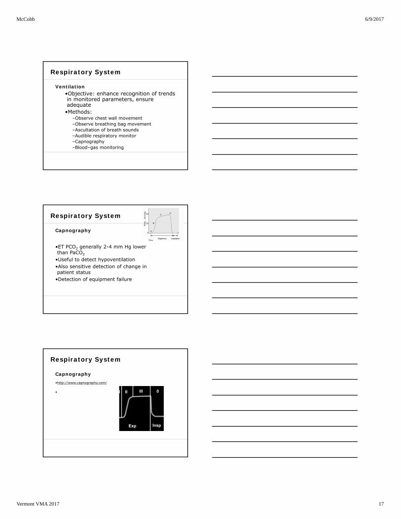
McCobb 6/9/2017
Vermont VMA 2017 17
Ventilation•Objective: enhance recognition of trends in monitored parameters, ensure adequate•Methods:
–Observe chest wall movement–Observe breathing bag movement–Ascultation of breath sounds–Audible respiratory monitor–Capnography–Blood–gas monitoring
Respiratory System
Capnography
•ET PCO2 generally 2-4 mm Hg lower than PaCO2
•Useful to detect hypoventilation•Also sensitive detection of change in patient status
•Detection of equipment failure
Respiratory System
Capnography•http://www.capnography.com/
•
Respiratory System

McCobb 6/9/2017
Vermont VMA 2017 18
Pulse Oximetry
•Should be 100%•Low values or errors can indicate problem•Not soley hypoxemia• Reduced perfusion
-vasoconstriction-too deep-too cold
• Very Sensitive
Temperature
•Hypothermia is common•Temperature should be maintained above 96° F
•Below this level anesthetic requirements are reduced and metabolic function can be disrupted
•Minimize intra-operative heat loss•Hyperthermia/Malignant Hyperthermia
Temperature Monitoring
•Esophageal or rectal thermistors attached to a continuously displayed thermometer
•Digital thermometer
•Temp checks post
McCobb 6/9/2017
Vermont VMA 2017 19
When do we stop monitoring?
•Continuous attention until extubation•Periodic checks throughout recovery
How do we know patient is ok?
•Not too deep (or too light)•MM pink, CRT below 2 sec•Reg RR with no effort•Easy to ascult heart and pulse palpates strong
•Temp ok
Monitors•Ongoing, automatic, audible monitors of organ function are mainstay
•Required by current guidelines •Single point in time measurement is meaningful when extremely abnormal
•Generally measurements are only meaningful in the context of trends
•But monitors have limitations
McCobb 6/9/2017
Vermont VMA 2017 20
Alarm Fatigue
If monitor alarms?
•First, check patient:–Listen to heart–Feel pulse
•Once confident patient is doing well then trouble shoot monitor
Complications:(Bad things that can happen)
Complications may be:
•Equipment related•Drug related•Patient related•Procedure related
OR……
•Some Combination of the above
McCobb 6/9/2017
Vermont VMA 2017 21
Equipment related
•Machine•Breathing Tube
Preventing Machine Related Complications
•Anesthesia machine check out
Common Anesthesia Machine Problems
•Stuck inspiratory/expiratory valves•Sodasorb issues •Leaks•Non-rebreather connected improperly•Improper plumbing
McCobb 6/9/2017
Vermont VMA 2017 22
ET tube problems
•Improper intubation•Problems with cuff•Kinking or disconnect•Biting through tube
•Post- anesthetic airway issues-
Other equipment considerations
•Closed pop off valve•Waste gas scavenging and monitoring
www.surgivet.com
Drug Related Complications
•Hypotension–Acepromazine–Isoflurane–Propofol
•Tachycardia–Ketamine–Glycopyrrolate
• Bradycardia– Opioids– Alpha 2 agonists
McCobb 6/9/2017
Vermont VMA 2017 23
Patient Related Complications
•Geriatric/Pediatric•Brachycephalic or difficult Airway•“sensitive” breeds•Occult disease underlying•Anxiety and stress of patient
Difficult Airway•Pre-oxygenate•Rapid induction with IV protocol•Intubate using visualization
Procedure Related Problems
Surgeon related
•Hemorrhage•Vagal responses
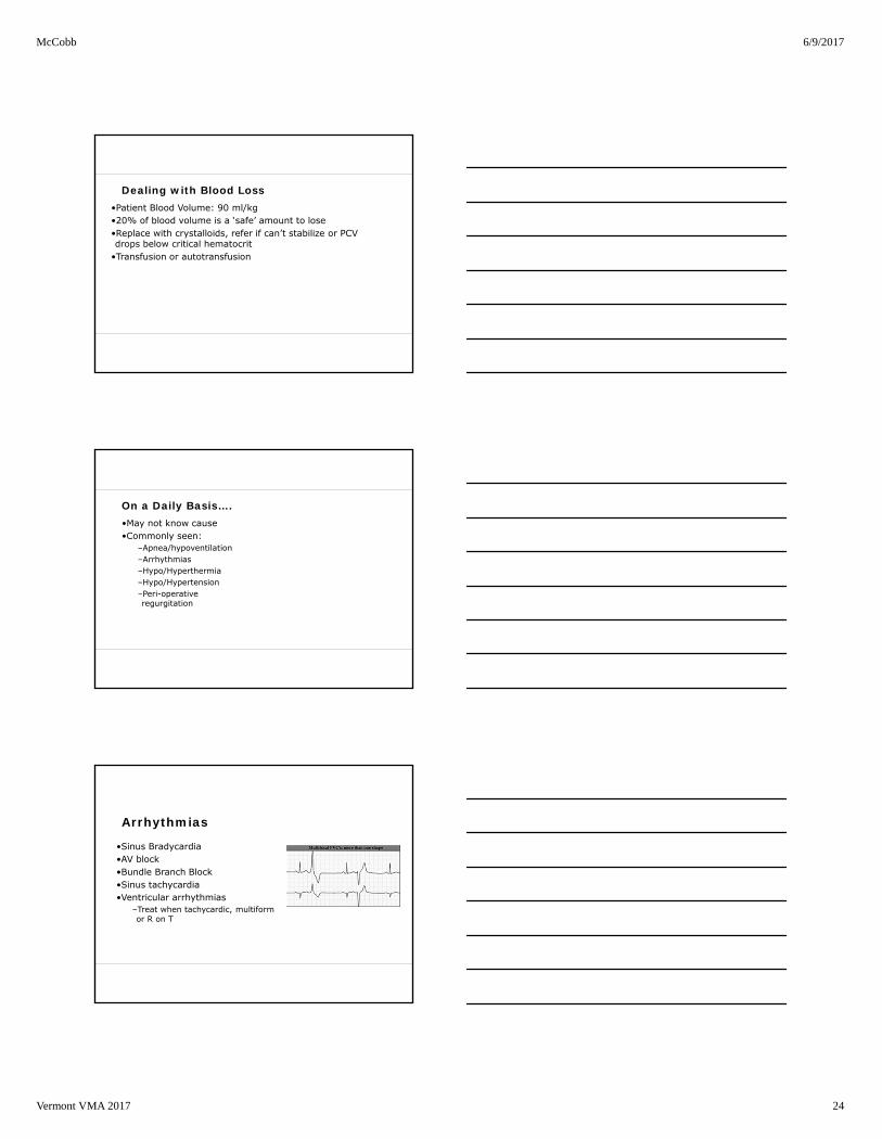
McCobb 6/9/2017
Vermont VMA 2017 24
Dealing with Blood Loss •Patient Blood Volume: 90 ml/kg•20% of blood volume is a ‘safe’ amount to lose•Replace with crystalloids, refer if can’t stabilize or PCV drops below critical hematocrit
•Transfusion or autotransfusion
On a Daily Basis….•May not know cause•Commonly seen:
–Apnea/hypoventilation–Arrhythmias–Hypo/Hyperthermia–Hypo/Hypertension–Peri-operative regurgitation
Arrhythmias
•Sinus Bradycardia•AV block•Bundle Branch Block•Sinus tachycardia•Ventricular arrhythmias
–Treat when tachycardic, multiform or R on T

McCobb 6/9/2017
Vermont VMA 2017 25
Causes of Bradycardia•Anesthetic Overdose•Opioids•А2-Agonists •Excessive vagal tone•Hypothermia•Hyperkalemia•Sick sinus syndrome•AV block•Metabolic failure•Hypoxia
Causes of Tachycardia•Anesthesia too light•Ketamine•Parasympatholytics•Sympathomimetics•Hypovolemia•Hyperthermia•Hypoxemia•Hypercapnia•Hypoglycemia•Individual variation•SVT•Pain•Pheochromocytoma
Vasomotor Tone•Regulates perfusion•Vasodilation
–improves perfusion–lowers blood pressure–causes: systemic inflammatory response, drugs, hyperthermia (isoflurane, acepromaz)
•Vasoconstriction–impairs perfusion, –raises blood pressure–causes: hypovolemia, heart failure, hypothermia, vasoconstrictors (dexmedetomidine)
McCobb 6/9/2017
Vermont VMA 2017 26
Complications: Hypotension
•Generally need to decrease depth of anesthesia
**Add Fentanyl CRI to decrease inhalant
•Fluids if hypovolemic**Dopamine at lowest possible dose
Hypothermia: negative effects
•↑ Stress Response•Impaired coagulation•Impaired tissue perfusion•Decrease MAC•Bradycardia•Delays recovery
•Best Cure is Prevention!!
Hyperthermia
•Avoid iatrogenic over heating•Metabolic and genetic hyperthermia syndromes•Greyhounds•Treatment:
–Rapidly identify (monitor temp)–Cooling, remove external warming devices

McCobb 6/9/2017
Vermont VMA 2017 27
Hyperthermia of the Cat
•retrospective study: Posner (NC state)•Implicated-
–Ketamine, hydromophone (any opioid?), intra-operative hypothermia
–Inflammatory mediators•What to do?
–Limited morbidity–Cool and monitor the patient–Reverse pure agonists–Give Acepromazine, NSAIDS?, fluids
Detecting Problems
•Monitor within window of “safe” parameters•Trouble shoot when values are outside of range•I.E.- HR> 140, < 60 for canines or < 100 for cats(keep protocol, individual patient in mind)
Using the Monitors
•Pulse Oximeter–the must have monitor–Hypoxia unlikely but should be addressed rapidly–Failure indicates problem with patient
•Capnography–excellent for detecting equipment problems
•These two monitors together should detect 99% of adverse anesthetic events

McCobb 6/9/2017
Vermont VMA 2017 28
Signs of a problem
•Patient not acting like they normally do•Hard time keeping them asleep•Lots of respiratory effort•Pulse ox alarming or pulse is hard to feel •Heart sounds far away•Sudden drop in ETCO2
Recovery •Monitor until patient is sternal and has good airway control
• Problems in Recovery
•Airway obstruction–Phenylephrine can be helpful
• Vomiting/Regurgitation +/- Aspiration
• Emergence delirium v. pain
McCobb 6/9/2017
Vermont VMA 2017 29
Adverse events
•Important to document and learn from •M and M logs and rounds•Culture of support and no blame
Top Five things I should tell you:
•1) have drills ahead of time•2) focus on high quality chest compressions•3) monitor end tidal co2•4) don't stop too soon in witnessed arrest•5) prevent!
Emergencies
RECOVER initiative •Latest update on CPR in veterinary medicine•Avail at: http://www.acvecc-recover.org/
Emergencies
McCobb 6/9/2017
Vermont VMA 2017 30
CPR
•High Quality Chest Compressions•Airway if not already capture•Ventilate and monitor CO2•Place EKG•Place IV•Reverse reversible drugs:
–Iso off!–Dexmedetomidine, benzodiazepines, opioids
•Keep track of time and interventions•Address underlying cause
Questions and Discussion

McCobbAcute Pain Management
6/6/2017
Vermont VMA 1
Acute Pain Management
Emily McCobb DVM MS DACVAA
Vermont Veterinary Medical Association
June 23, 2017
Outline
• Pain Physiology Quick Review
• Pain Assessment & Challenges
• Acute/Perioperative Pain
Pain Physiology
Diagram: Muir
From Compendium, 2003,
www.vetlearn.com
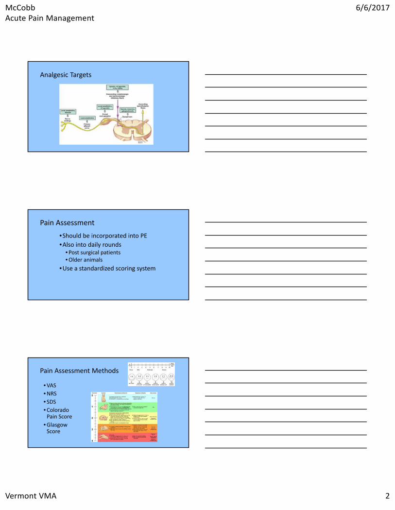
McCobbAcute Pain Management
6/6/2017
Vermont VMA 2
Analgesic Targets
Pain Assessment
•Should be incorporated into PE
•Also into daily rounds• Post surgical patients• Older animals
•Use a standardized scoring system
Pain Assessment Methods
• VAS
• NRS
• SDS
• Colorado Pain Score
• Glasgow Score
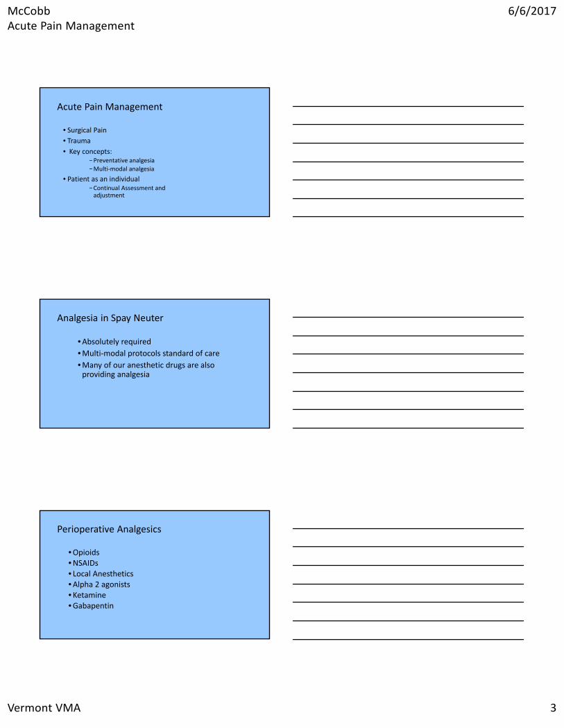
McCobbAcute Pain Management
6/6/2017
Vermont VMA 3
Acute Pain Management
• Surgical Pain
• Trauma
• Key concepts:−Preventative analgesia
−Multi‐modal analgesia
• Patient as an individual−Continual Assessment and
adjustment
Analgesia in Spay Neuter
• Absolutely required
• Multi‐modal protocols standard of care
• Many of our anesthetic drugs are also providing analgesia
Perioperative Analgesics
• Opioids• NSAIDs• Local Anesthetics• Alpha 2 agonists• Ketamine • Gabapentin
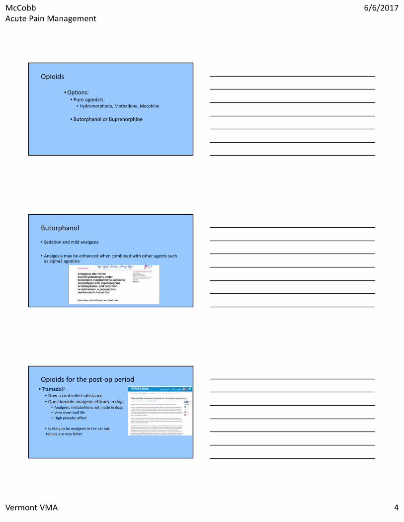
McCobbAcute Pain Management
6/6/2017
Vermont VMA 4
Opioids
• Options:• Pure agonists:
• Hydromorphone, Methadone, Morphine
• Butorphanol or Buprenorphine
Butorphanol
• Sedation and mild analgesia
• Analgesia may be enhanced when combined with other agents such as alpha2 agonists
Opioids for the post‐op period• Tramadol?
• Now a controlled substance
• Questionable analgesic efficacy in dogs• Analgesic metabolite is not made in dogs
• Very short half life
• High placebo effect
• Is likely to be analgesic in the cat but
tablets are very bitter
McCobbAcute Pain Management
6/6/2017
Vermont VMA 5
Buprenorphine for Post‐Op
• ‐widely used in cats and dogs
• ‐preferred dose in cats: 0.02 mg/kg, *SQ administration not recommended
• Problems with traditional formulation (Buprenorphine HCl):• duration of action (6‐8 hours?)
• COST $$$
Buprenorphine:Sustained Release
• Available from Zoo Pharm (Colorado) • 5 ml vials
• 3 mg/ml
• Dog dose: 0.03‐0.06 mg/kg q. 72 hours
• Cat dose: 0.12 mg/kg q. 72 hrs
• Concerns:• One case report of site reaction in a cat
• Compounding regulations?
Simbadol
• Zoetis product‐ available now
• 1.8 mg/ml
• Licensed for use in the cat
• 0.24 mg/kg every 24 hours, administer 1 hour before surgery
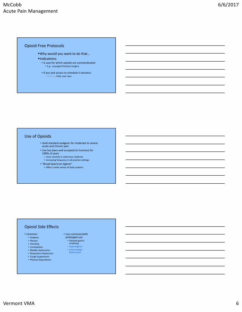
McCobbAcute Pain Management
6/6/2017
Vermont VMA 6
Opioid Free Protocols
Why would you want to do that…
Indications:
• A case for which opioids are contraindicated • E.g.. Laryngeal Paralysis Surgery
• If you lack access to schedule II narcotics • Shelters, field, over seas
Use of Opioids
• Gold standard analgesic for moderate to severe acute and chronic pain
• Use has been well accepted (in humans) for 1000s of years
• more recently in veterinary medicine
• increasing frequency in all practice settings
• “Broad Spectrum Agents”• Affect a wide variety of body systems
Opioid Side Effects
• Common:• Sedation
• Nausea
• Vomiting
• Constipation
• Bladder dysfunction
• Respiratory Depression
• Cough Suppression
• Physical Dependence
• Less common/with prolonged use:
• Delayed gastric emptying
• Hyperalgesia
• Immunologic Dysfunction

McCobbAcute Pain Management
6/6/2017
Vermont VMA 7
Balanced Anesthesia and Analgesia
• Goal for all patients should be to reduce dose and duration of opioid use
• For some patients an opioid free protocol may be indicated
• How do we accomplish this?• Multi‐modal and adjunctive techniques
Non‐opioid Analgesics
• Ketamine
• Local Anesthetics
• NSAIDs
• Alpha‐2 Agonists
• Gabapentin
A sample Opioid‐Free Protocol
• For Spay/Neuter:• Ketamine/dexmedetomidine
• For anesthesia induction & analgesia
• Local blocks
• Post‐op NSAIDs
• +/‐ Butorphanol• Side effects mostly associated with
µ
• Is it enough?

McCobbAcute Pain Management
6/6/2017
Vermont VMA 8
Lidocaine Patch
5 % Lidocaine
Approved for neuropathic pain in people
Shown to treat:• Constant aching/burning
• Intermittent sharp jabbing
• Allodynia/hyperpathia
Lidocaine Patch
Can be applied next to or directly over the incision
Proposed mechanism of action:• topical, not transdermal• lidocaine binds neuronal membrane receptors• stabilizes sodium channels, ↓ pain signal ini a on
and transmission • analgesic but not anesthetic
Applications: Humans
• Neuropathic Pain (shingles)• Knee osteoarthritis
• Reduction in pain intensity after 2 week treatment
• Theorized that lidocaine patch suppresses pain signals by local inhibition of abnormally functioning sodium channels within joint neurons
• Gammaitoni et al, 2004
• Post‐operative incisional pain
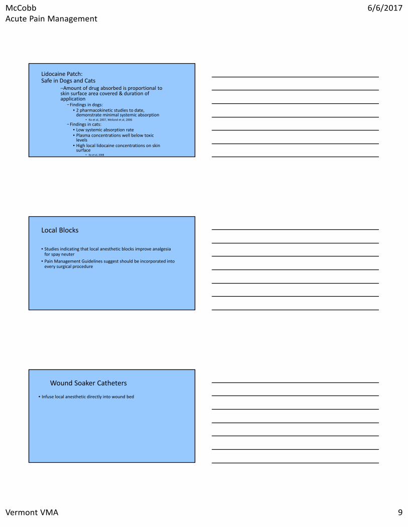
McCobbAcute Pain Management
6/6/2017
Vermont VMA 9
Lidocaine Patch: Safe in Dogs and Cats
**Amount of drug absorbed is proportional to skin surface area covered & duration of application
−Findings in dogs:• 2 pharmacokinetic studies to date,
demonstrate minimal systemic absorption• Ko et al, 2007, Weiland et al, 2006
−Findings in cats:• Low systemic absorption rate• Plasma concentrations well below toxic
levels• High local lidocaine concentrations on skin
surface• Ko et al, 2008
Local Blocks
• Studies indicating that local anesthetic blocks improve analgesia for spay neuter
• Pain Management Guidelines suggest should be incorporated into every surgical procedure
Wound Soaker Catheters
• Infuse local anesthetic directly into wound bed
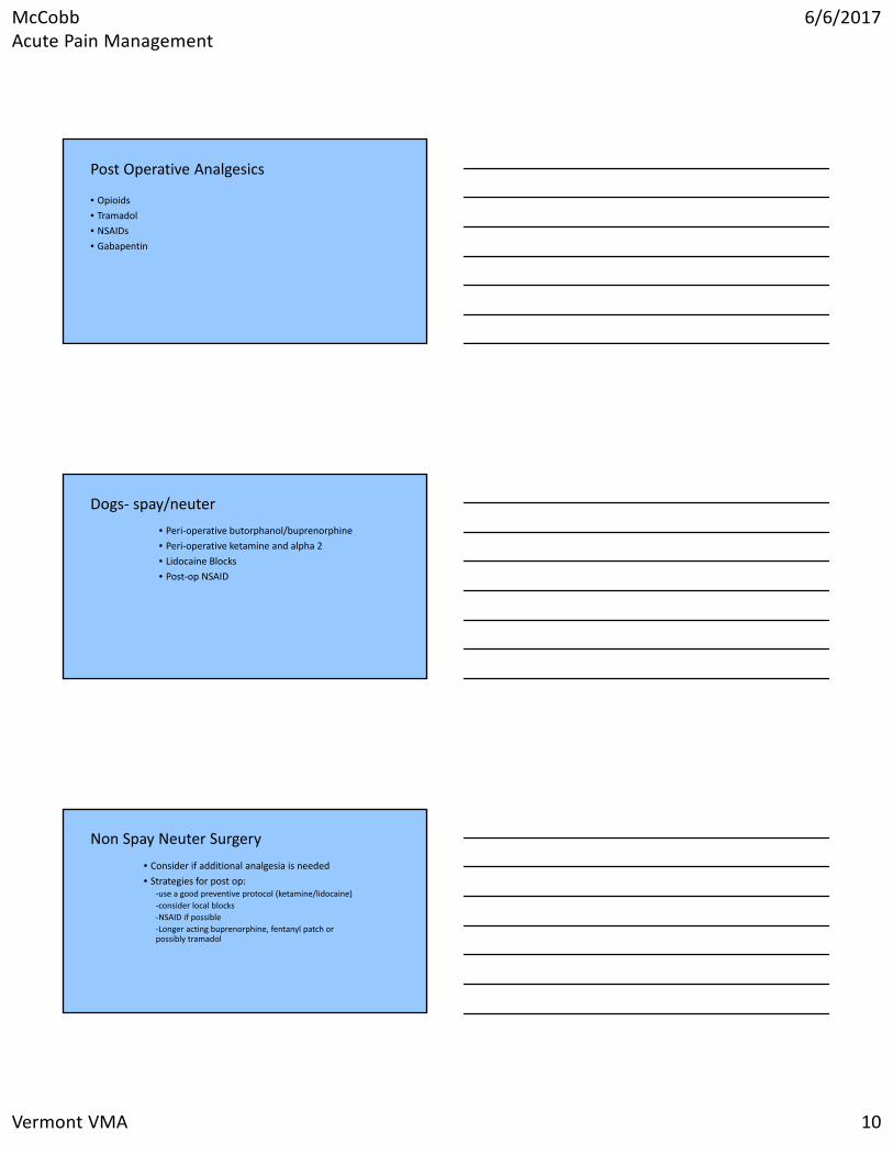
McCobbAcute Pain Management
6/6/2017
Vermont VMA 10
Post Operative Analgesics
• Opioids
• Tramadol
• NSAIDs
• Gabapentin
Dogs‐ spay/neuter
• Peri‐operative butorphanol/buprenorphine
• Peri‐operative ketamine and alpha 2
• Lidocaine Blocks
• Post‐op NSAID
Non Spay Neuter Surgery
• Consider if additional analgesia is needed
• Strategies for post op:‐use a good preventive protocol (ketamine/lidocaine)
‐consider local blocks
‐NSAID if possible
‐Longer acting buprenorphine, fentanyl patch or possibly tramadol
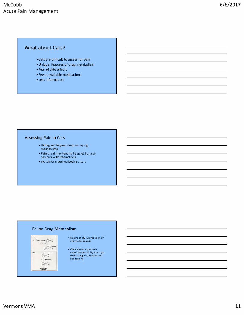
McCobbAcute Pain Management
6/6/2017
Vermont VMA 11
What about Cats?
• Cats are difficult to assess for pain
• Unique features of drug metabolism
• Fear of side effects
• Fewer available medications
• Less information
Assessing Pain in Cats
• Hiding and feigned sleep as coping mechanisms
• Painful cat may tend to be quiet but also can purr with interactions
• Watch for crouched body posture
Feline Drug Metabolism
• Failure of glucuronidation of many compounds
• Clinical consequence is exquisite sensitivity to drugs such as aspirin, Tylenol and benzocaine
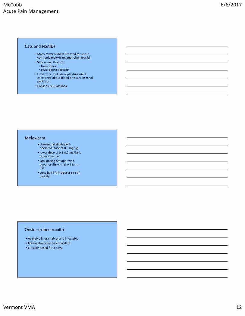
McCobbAcute Pain Management
6/6/2017
Vermont VMA 12
Cats and NSAIDs
• Many fewer NSAIDs licensed for use in cats (only meloxicam and robenacoxib)
• Slower metabolism• Lower doses
• Lower dosing frequency
• Limit or restrict peri‐operative use if concerned about blood pressure or renal perfusion
• Consensus Guidelines
Meloxicam
• Licensed at single peri‐operative dose at 0.3 mg/kg
• lower dose of 0.1‐0.2 mg/kg is often effective
• Oral dosing not approved, good results with short term use
• Long half life increases risk of toxicity
Onsior (robenacoxib)
• Available in oral tablet and injectable
• Formulations are bioequivalent
• Cats are dosed for 3 days
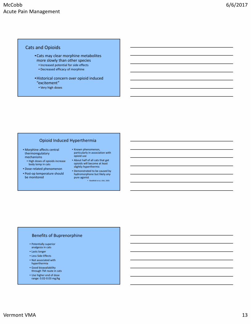
McCobbAcute Pain Management
6/6/2017
Vermont VMA 13
Cats and Opioids
•Cats may clear morphine metabolites more slowly than other species
• Increased potential for side effects• Decreased efficacy of morphine
•Historical concern over opioid induced “excitement”
• Very high doses
Opioid Induced Hyperthermia
• Morphine affects central thermoregulatory mechanisms
• High doses of opioids increase body temp in cats
• Dose‐related phenomenon
• Post‐op temperature should be monitored
• Known phenomenon, particularly in association with opioid use
• About half of all cats that get opioids will become at least slightly hyperthermic
• Demonstrated to be caused by hydromorphone but likely any pure agonist
• Niedfeldt et al, VAA, 2005
Benefits of Buprenorphine
• Potentially superior analgesia in cats
• Lasts longer
• Less Side Effects
• Not associated with hyperthermia
• Good bioavailability through TM route in cats
• Use higher end of dose range: 0.02‐0.03 mg/kg

McCobbAcute Pain Management
6/6/2017
Vermont VMA 14
Adjunctive Techniques for Pain Management in cats
• Perception of increased susceptibility to toxic effects of local anesthetics
• Tends to be less data
• Less amenable to techniques such as ice‐packing, TLC
Shelter Protocols for Cats
• Dexmedetomidine/Ketamine/Buprenorphine• Large variation in doses
• Alterations in dose primarily seems to effect duration
• Telazol based protocols
• Balanced Analgesia by definition
Non spay neuter protocol for a cat
• Dental?
• Mass removal?
McCobbAcute Pain Management
6/6/2017
Vermont VMA 15
Neuropathic Pain
• Pain resulting from structural changes within the peripheral and central nervous systems
• Primary lesion or dysfunction within the nervous system
• May be caused by poorly managed acute pain, chronic pain of any cause, or a primary lesion within the nervous system
Neuropathic Pain
• Complex etiology
• Often a feature of chronic pain
• Usually involves a behavior change• Emotional aspects of pain
• Dullness or unusual aggression
• Patients at risk:• Any patient with severe pain
• Injury to nervous system
Examples of Neuropathic Pain• Clinical conditions that may be associated with
neuropathic pain in veterinary patients:
• Pelvic fractures
• Amputation
• Lumbosacral lesions: cauda equina syndrome
• Spinal cord injury
• IVD disease
• FCE
• Feline interstitial cystitis
McCobbAcute Pain Management
6/6/2017
Vermont VMA 16
Treatment of Neuropathic Pain
• Attentive acute and peri‐operative pain management to reduce the incidence of peripheral and central sensitization
• Multi‐modal regimen
**Treatment may need to be life long
Neuropathic Pain: Treatment
• Novel analgesics• Gabapentin
• Amantadine
• Tricyclic anti‐depressants
• Local anesthetic infusions
• Acupuncture/electro‐acupuncture
• Other complementary techniques
Tramadol• Codeine analog
• Now controlled
• Synthetic, centrally acting analgesic
• Weak µ receptor agonist and serotonin reuptake inhibitor
• Useful in human patients with OA and allodynia
• Seems to be synergistic with NSAIDs
• Dose: 1‐5 mg/kg q 6 to 12 hours
• (1‐2 mg/kg in cats)
• Avoid concurrently with MAOIs (anipryl)
• Not good evidence for efficacy in canine patients…
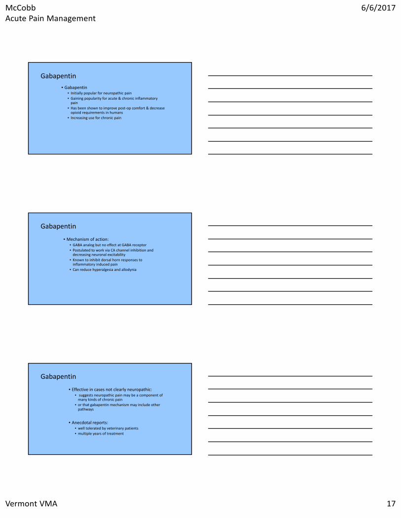
McCobbAcute Pain Management
6/6/2017
Vermont VMA 17
Gabapentin
• Gabapentin• Initially popular for neuropathic pain
• Gaining popularity for acute & chronic inflammatory pain
• Has been shown to improve post‐op comfort & decrease opioid requirements in humans
• Increasing use for chronic pain
Gabapentin
• Mechanism of action:• GABA analog but no effect at GABA receptor
• Postulated to work via CA channel inhibition and decreasing neuronal excitability
• Known to inhibit dorsal horn responses to inflammatory induced pain
• Can reduce hyperalgesia and allodynia
Gabapentin
• Effective in cases not clearly neuropathic:• suggests neuropathic pain may be a component of
many kinds of chronic pain
• or that gabapentin mechanism may include other pathways
• Anecdotal reports:• well tolerated by veterinary patients
• multiple years of treatment
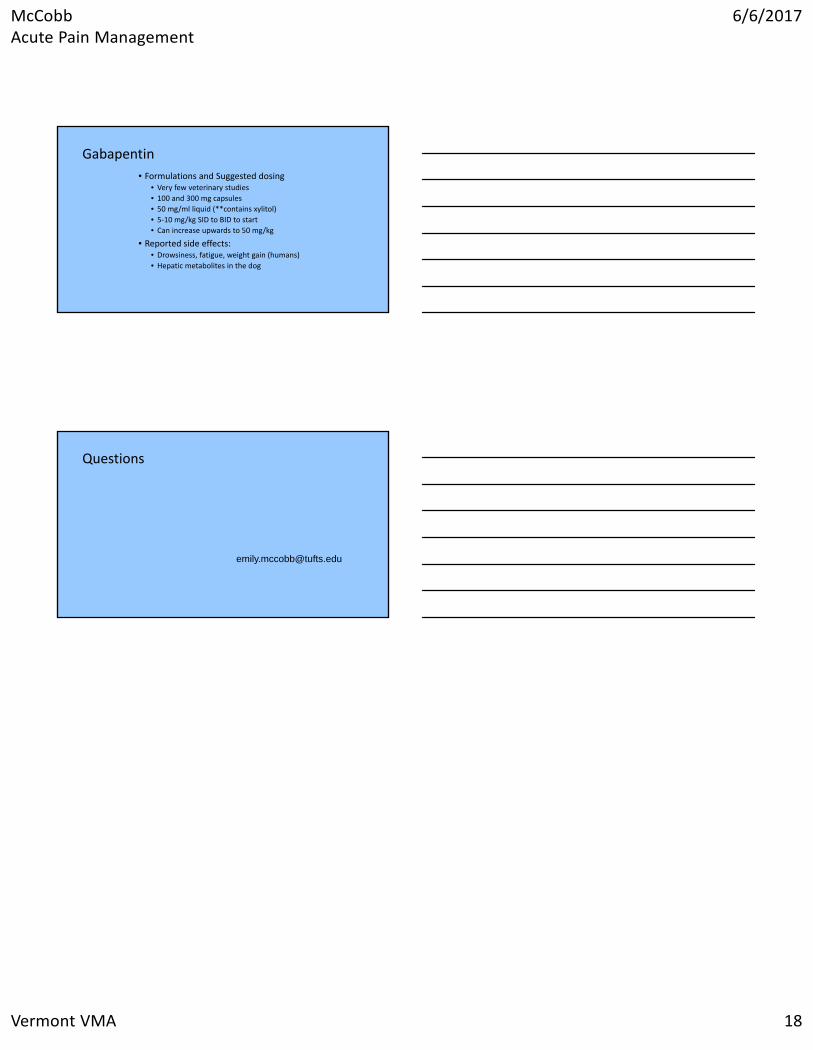
McCobbAcute Pain Management
6/6/2017
Vermont VMA 18
Gabapentin
• Formulations and Suggested dosing• Very few veterinary studies
• 100 and 300 mg capsules
• 50 mg/ml liquid (**contains xylitol)
• 5‐10 mg/kg SID to BID to start
• Can increase upwards to 50 mg/kg
• Reported side effects:• Drowsiness, fatigue, weight gain (humans)
• Hepatic metabolites in the dog
Questions