0001592897 793.chittkalab.sbcs.qmul.ac.uk/2013/Vukusic_Chittka_2013.pdf · Comp. by: Leela Stage:...
31
Comp. by: Leela Stage: Proof Chapter No.: 25 Title Name: CHAPMANSIMPSONANDDOUGLAS Date:10/7/12 Time:19:55:58 Page Number: 793 25 Visual signals: color and light production REVISED AND UPDATED BY PETER VUKUSIC AND LARS CHITTKA INTRODUCTION Insects generate a spectacular variety of visual signals, from multicolored wing patterns of butterflies, through metallic-shiny beetles to highly contrasting warning coloration of stinging insects and their defenseless mimics. Section 25.1 explains what colors are and the subsequent sections describe how insect colors result from a variety of physical structures (Section 25.2) and pigments (Section 25.3). Often, several pigments are present together, and the observed color depends on the relative abundance and positions of the pigments, as well as control signs generating color patterns (Section 25.4). The position of color-producing molecules relative to other structures is also important, and this may change, resulting in changes in coloration (Section 25.5). The many biological functions of color in insect signaling are covered in Section 25.6. Table 25.1 lists the sources of color in some insect groups. A small selection of insects also exhibit fluorescence or luminescence (Section 25.7). The Insects: Structure and Function (5 th edition), ed. S. J. Simpson and A. E. Douglas. Published by Cambridge University Press. © Cambridge University Press 2013.
Transcript of 0001592897 793.chittkalab.sbcs.qmul.ac.uk/2013/Vukusic_Chittka_2013.pdf · Comp. by: Leela Stage:...

Comp. by: Leela Stage: Proof Chapter No.: 25 Title Name: CHAPMANSIMPSONANDDOUGLASDate:10/7/12 Time:19:55:58 Page Number: 793
25 Visual signals: color and lightproduction
REV I S ED AND UPDATED BY PETER VUKUSIC AND
LARS CHITTKA
INTRODUCTION
Insects generate a spectacular variety of visual signals, from multicolored wing
patterns of butterflies, through metallic-shiny beetles to highly contrasting warning
coloration of stinging insects and their defenseless mimics. Section 25.1 explains what
colors are and the subsequent sections describe how insect colors result from a variety of
physical structures (Section 25.2) and pigments (Section 25.3). Often, several pigments are
present together, and the observed color depends on the relative abundance and positions
of the pigments, as well as control signs generating color patterns (Section 25.4). The
position of color-producing molecules relative to other structures is also important, and
this may change, resulting in changes in coloration (Section 25.5). The many biological
functions of color in insect signaling are covered in Section 25.6. Table 25.1 lists the
sources of color in some insect groups. A small selection of insects also exhibit
fluorescence or luminescence (Section 25.7).
The Insects: Structure and Function (5th edition), ed. S. J. Simpson and A. E. Douglas. Published by Cambridge University Press.© Cambridge University Press 2013.

Comp. by: Leela Stage: Proof Chapter No.: 25 Title Name: CHAPMANSIMPSONANDDOUGLASDate:10/7/12 Time:19:55:59 Page Number: 794
Table 25.1 Some of the principal causes of colors in different insects
Color Taxon Cause of color
Black* Homoptera, Aphididae Aphins
Coleoptera Melanin
Diptera Melanin
Lepidoptera (larvae) Melanin
Red Odonata Ommochromes
Hemiptera, Heteroptera Pterins and carotenoids
Hemiptera, Coccoidea Anthraquinones
Coleoptera, Coccinellidae Carotenoids
Diptera, Chironomidae (larvae) Porphyrin
Lepidoptera Ommochromes
Brown* Lepidoptera Ommochromes
Many orders – eye colors Ommochromes þ pterins
Orange Hemiptera, Heteroptera Pterins
Yellow Orthoptera, Acrididae Carotenoids, flavonoids
Hymenoptera Pterins
Lepidoptera, Papilionidae Papiliochromes
Brassy yellow Lepidoptera Interference color
Bronze Coleoptera, Scarabaeidae Interference color
Gold Coleoptera, Cassidinae Interference color
Lepidoptera, Danaidae (pupae) Interference color
Green Orthoptera Insectoverdin (carotenoid þ bilin)
Lepidoptera (caterpillars) Insectoverdin
Lepidoptera, Zygaenidae Interference color
Diptera, Chironomidae (adults) Bilin
Blue Odonata Interference color and ommochromes
Lepidoptera Interference color
Orthoptera, Acrididae Carotenoids
Ultraviolet Lepidoptera, Pieridae Interference color
794 Visual signals: color and light production
Comp. by: Leela Stage: Proof Chapter No.: 25 Title Name: CHAPMANSIMPSONANDDOUGLASDate:10/7/12 Time:19:56:00 Page Number: 795
25.1 The nature of color
Color is not an inherent property of objects; it is a
perceptual attribute that depends on illumination,
the spectral reflectance of an object and its
surroundings, as well as the spectral receptor types
and further neural processing in the animal in
question. Thus the same object might appear
differently colored to different viewing organisms.
A red poppy, for example, is red to human
observers, but appears as a UV-reflecting object to a
bee pollinator, which does not have a red receptor
and, like all insects studied to date, sees UV-A light
between 300 nm and 400 nm. For reasons of
simplicity, the color terminology in this chapter
specifies what a human observer will perceive under
daylight conditions. Information about UV is
provided separately where available.
Color is generated from white light incident upon
an insect when some of the incident wavelengths are
eliminated, usually by absorption in its
pigmentation, and the remainder are scattered. These
scattered wavelengths of the reflected or transmitted
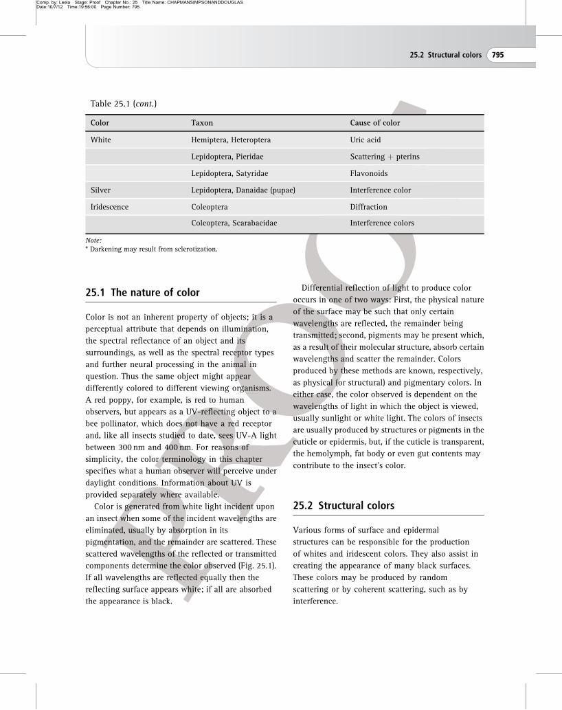
components determine the color observed (Fig. 25.1).
If all wavelengths are reflected equally then the
reflecting surface appears white; if all are absorbed
the appearance is black.
Differential reflection of light to produce color
occurs in one of two ways: First, the physical nature
of the surface may be such that only certain
wavelengths are reflected, the remainder being
transmitted; second, pigments may be present which,
as a result of their molecular structure, absorb certain
wavelengths and scatter the remainder. Colors
produced by these methods are known, respectively,
as physical (or structural) and pigmentary colors. In
either case, the color observed is dependent on the
wavelengths of light in which the object is viewed,
usually sunlight or white light. The colors of insects
are usually produced by structures or pigments in the
cuticle or epidermis, but, if the cuticle is transparent,
the hemolymph, fat body or even gut contents may
contribute to the insect’s color.
25.2 Structural colors
Various forms of surface and epidermal
structures can be responsible for the production
of whites and iridescent colors. They also assist in
creating the appearance of many black surfaces.
These colors may be produced by random
scattering or by coherent scattering, such as by
interference.
Table 25.1 (cont.)
Color Taxon Cause of color
White Hemiptera, Heteroptera Uric acid
Lepidoptera, Pieridae Scattering þ pterins
Lepidoptera, Satyridae Flavonoids
Silver Lepidoptera, Danaidae (pupae) Interference color
Iridescence Coleoptera Diffraction
Coleoptera, Scarabaeidae Interference colors
Note:* Darkening may result from sclerotization.
25.2 Structural colors 795
Comp. by: Leela Stage: Proof Chapter No.: 25 Title Name: CHAPMANSIMPSONANDDOUGLASDate:10/7/12 Time:19:56:00 Page Number: 796
25.2.1 Scattering
Light may be scattered (i.e., reflected in all
directions) by granules on, or irregularities in, a
surface. If these scattering centers are randomly
spatially distributed in or on the surface then
virtually all incident light is reflected diffusely and
the surface appears as a matt white. In some
Lepidoptera, such as Pieridae, this results from the
presence of densely packed arrays of pterin
granules across each white scale’s surface, the
effect of which is coupled with that of the structure
of the scale itself. In some Coleoptera, such as
Cyphochilus sp., a bright, diffuse white appearance
0
10
20
30
40
50
60
70
80
90
100
300 400 500 600 700
wavelength (nm)
refle
ctan
ce (
%)
(a)
(b)
1
2
3
4
0
10
20
30
40
50
60
70
80
90
100
300 400 500 600 700
wavelength (nm)
refle
ctan
ce (
%)
1
2
3
4
Figure 25.1 Representative reflectance
spectra of insect body surfaces. Colors
are produced by the absorbance of
certain wavelengths, for example by
pigments in the insect cuticle;
wavelengths that are not absorbed may
reach a viewer’s eye via reflectance or
transmission. (a) Reflectance spectra
from butterfly male forewings: (1) the
white (and UV-absorbing) wing of the
European cabbage white, Pieris rapae;
(2) the yellow brimstone, Gonepteryx
rhamni (Europe); (3) the purple (and
UV-reflecting) spots on the wings of the
Malayan jungleglory, Thaumantis
odana; and (4) green parts of the wings
of the Southeast Asian swallowtail
butterfly, Papilio lorquinianus.
(b) Reflectance of European bumble
bee workers: (1) the white (and
UV-reflecting) tip of the abdomen of
the large earth bumble bee, Bombus
terrestris dalmatinus; (2) the yellow
band on the thorax of B. t. dalmatinus;
(3) black parts of the abdomen of
B. t. dalmatinus; (4) the brownish-red
tip of the abdomen of the red-tailed
bumble bee, B. lapidarius.
796 Visual signals: color and light production
Comp. by: Leela Stage: Proof Chapter No.: 25 Title Name: CHAPMANSIMPSONANDDOUGLASDate:10/7/12 Time:19:56:00 Page Number: 797
is generated by scattering from interconnected
cuticular filaments that are randomly arranged
within each white-colored scale. Pearly whites, such
as those of Morpho sulkowski (Lepidoptera), are
produced by the addition of a degree of specular
(mirror-like) reflection in addition to the diffuse
scattering.
25.2.2 Interference in multilayers
Interference colors result from the reflection of light
from a series of neighboring interfaces that are
separated by distances comparable with a quarter of
the wavelength of light. As a result of wave
superposition from these reflections, some
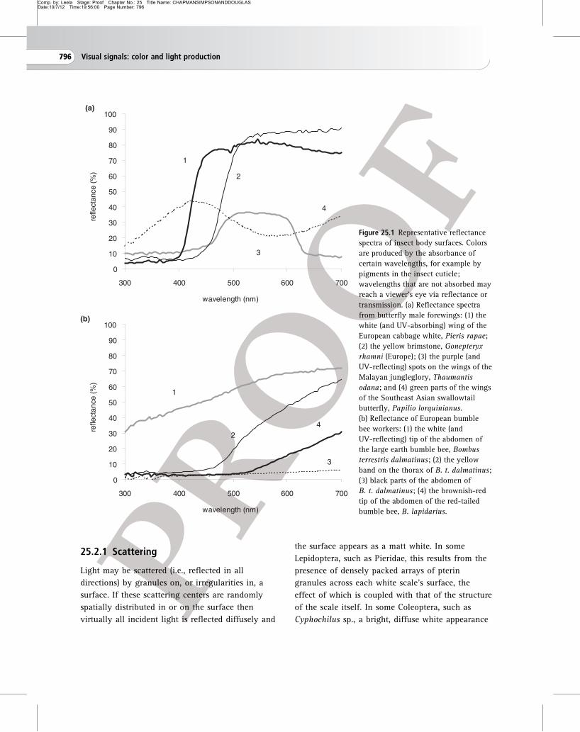
Scale ridges(a)
Scale ridge(b)
layers atregular 185 nmspacing
supporting column
Basal layer
Figure 25.2 Interference color production in different insects. Morpho butterfly wing scales. (a) Scanning electron
microscope image of two neighboring wing scales showing the ridging structures running the full length of each scale; scale
bar ¼ 50 mm. (b) Transmission electron microscope image of a cross-section of part of one of the scales; scale bar ¼ 1 mm.
The parallel horizontal layers of cuticle (with a regular spacing of 185 nm) and the air spaces between them are
responsible for the interference of the reflected blue color. Notice that the supporting columns are irregularly arranged so
only some are in the plane of the section (based on Vukusic et al., 1999).
25.2 Structural colors 797
Comp. by: Leela Stage: Proof Chapter No.: 25 Title Name: CHAPMANSIMPSONANDDOUGLASDate:10/7/12 Time:19:56:01 Page Number: 798
wavelengths are reflected or transmitted in phase and
are therefore reinforced, while others are out of phase
and are cancelled out. The net result is that only
certain wavelengths are reflected or transmitted and
the surface appears colored.
Interference colors are common in adult
Lepidoptera where the layers producing interference
are formed by modifications of the scales. Each of the
blue scales of the Morpho rhetenor butterfly, for
instance, consists of a flat basal plate carrying a large
number of near-parallel vertically aligned ridges that
run parallel with the length of the scale (Fig. 25.2).
Within each ridge are series of horizontal layers,
separated by air spaces. Collectively, the horizontal
layers in each adjacent ridge form a series of
reflecting surfaces, which are spaced such that a blue
color is produced by interference. Various optical
appearances are produced by a great variety of scale
modifications in many different Lepidoptera. For
instance, strong ultraviolet reflectance at a peak
wavelength of approximately 345 nm is generated in
the butterfly Colias eurytheme by horizontal layered
structures in its scale ridges that are 30–40% thinner
that those typical of iridescent Morpho butterflies.
Interference colors in other insects are produced by
reflection at the interfaces of layers in the cuticle
which differ in refractive index. The refractive
indices of the alternating layers in the pupa of the
danaid butterfly, Euploea, are 1.58 and 1.37. In jewel
beetles (Buprestidae) and tiger beetles (Cicindellidae),
these layers are in the exocuticle, but in tortoise
beetles (Cassidinae) and some butterfly pupae they
are in the endocuticle.
In some scarab beetles the reflecting surfaces are
layers of chitin/protein microfibrils with a common
orientation in the transparent exocuticle. Because
their arrangement changes progressively in
successive layers, creating helical orientation
(Section 16.2), any given orientation recurs at
intervals and, if the intervals are within the range of
wavelengths of light, interference colors are
produced. Due to this helical fibril orientation, the
reflected light is circularly polarized (CP). The
clockwise or anticlockwise orientation of fibrils
through the exocuticle determines the right-handed
or left-handed nature of the circular polarization. In
the majority of cases, left-handed CP light is
reflected from scarabs that exhibit this helical
structure. However, in very few cases, Plusiotis
resplendens for instance, both left- and
right-handed CP light are reflected concurrently due
to the presence of a multi-component helical
structure.
Whatever the nature or position of the reflecting
surfaces which form the layer interfaces, the
wavelength of the reflected light depends on their
spacing. Viewing the surface from a more oblique
angle is equivalent to increasing the optical path
length between successive surfaces so that the color
changes in a definite sequence as the angle of
viewing is altered. This change in color with the
angle of viewing is called iridescence and is a
characteristic of interference colors.
A constant spacing between the reflecting surfaces
produces relatively pure colors (Fig. 25.3a), as in the
scales of blue Morpho butterflies (Fig. 25.2), or the
moths Urania leilus and Chrysiridia rhipheus. In the
majority of insect multilayer examples, spectral
purity is enhanced when the reflecting layers are
backed by a layer of melanin-based pigment which
offers broad-band absorption to the light that is
transmitted through the multilayer. Color
conspicuousness is often enhanced by the presence of
a strongly black border surrounding the brightly
colored area, particularly so in some butterfly and
weevil color patterning.
Broad-band colors such as gold, silver and bronze,
by contrast, are produced if the spacing changes
systematically with increasing distance from the
surface of the cuticle. For example, in the endocuticle
of the pupa of Euploea, and the exocuticle of P.
resplendens, the thickness of the paired layers
changes systematically (Fig. 25.3b). This is referred to
as a chirped layer structure. It produces interference
over a broad range of wavelengths (Fig. 25.3b) and,
in the cases of these insects, creates a gold mirror
798 Visual signals: color and light production

Comp. by: Leela Stage: Proof Chapter No.: 25 Title Name: CHAPMANSIMPSONANDDOUGLASDate:10/7/12 Time:19:56:01 Page Number: 799
0300 400 500 600 700 800
θ
20
abso
lute
ref
lect
ivity
wavelength (nm)
40
80
100
(a)
60
0300 400 500 600
θ
700 800
20
abso
lute
ref
lect
ivity
wavelength (nm)
40
80
100
(b)
60
Figure 25.3 Theoretical modeling showing the spectral distribution of reflected intensities at multiple cuticular layers.
(a) Reflected intensities from 1, 2, 3, 5 and 10 layers of cuticle, respectively, resulting in the increasing reflectances shown.
In each case, air is the spacer layer and the normal incidence case is presented (dimensions used for modeling: cuticle layers
90 nm, and air layers 140 nm). Inset: a schematic diagram representing multiple reflections within a multilayer system
leading to constructive interference. This multilayer comprises a single set of layer dimensions and this leads to a relatively
narrow band of reflected wavelengths. (b) Reflection from a chirped multilayer (comprising ten cuticle layers of
consecutively increasing thickness, spaced by air) producing the appearance of metallic gold. Inset: schematic diagram of
multiple reflections in a chirped multilayer, showing the increasing thickness of cuticle layers through the system.
25.2 Structural colors 799

Comp. by: Leela Stage: Proof Chapter No.: 25 Title Name: CHAPMANSIMPSONANDDOUGLASDate:10/7/12 Time:19:56:02 Page Number: 800
appearance to human eyes. Silver interference colors
can be produced in the same way.
The peak reflected wavelength produced by
interference, and also to some extent the brightness
of the reflected colors, also depends on the refractive
indices of the materials forming the layers. The
horizontal layers in the ridges on blue Morpho wing
scales are uniformly separated by approximately
120 nm. Together with the thickness of the layers
themselves, approximately 80 nm, and the refractive
index of the cuticle material, approximately 1.56, the
system produces a bright blue interference color. The
paired layers in the exocuticle of the scarab beetle
Heterorrhina are of similar thickness, but the
interference color produced is green due to the
different refractive indices of the paired cuticular
layers compared with the air–cuticle interfaces in
Morpho.
The brightness of the reflected color significantly
increases with the number of reflecting layers.
Generally, the insects that exhibit higher layer
numbers are brighter in appearance, although this
additional brightness may be offset by the presence
of absorbing pigment within the layers in the system.
In Morpho rhetenor, for instance, there are 8–12
horizontal layers per ridge and in Morpho didius
there are 6–8 layers per ridge; quantitative
measurements indicate that M. rhetenor is
significantly brighter, reflecting 80% of incident blue
light, while M. didius reflects approximately 40%.
Similar numbers of layers are present in the cuticles
of Chrysochroa raja and other iridescent buprestid
beetles. By contrast, the metallic gold or silver
cuticles of pupae of danaid butterflies can have over
200 reflecting layers. They too reflect approximately
80% of the incident light over a broad band of
wavelengths (Fig. 25.3); the reflectivity is not higher
due to optical absorption by the layers’ constituent
materials.
Apart from colors of the human visible spectrum,
ultraviolet is also produced as an interference color.
This occurs in some butterflies of the Pieridae. In
Eurema, for example, certain scales possess
analogous scale structures to those of Morpho;
however, their reflecting layers are approximately
55 nm thick and are separated by air spaces of
approximately 80 nm. This system reflects light
with a peak wavelength of approximately 365 nm.
The brightness of a similar ultraviolet-reflecting
system in male Colias eurytheme butterflies has been
shown to be an indication of their fitness to
conspecific females. The number of their intra-ridge
reflecting layers, a variable which controls this
brightness, appears to be affected by larval food
quality.
Interference is responsible for the iridescence
of the membranous wings of many different
insects, particularly Odonata. Notable for their
intensity and spectral purity are the hindwings of
the dragonfly Neurobasis chinensis. In these
iridescent wings, an approximately 1 mm thick
multilayer on the upper side of the membrane is
backed by an approximately 2 mm layer of
melanin-based optical absorber for enhanced
color contrast.
25.2.3 Coherent scattering in otherstructures
While interference in mainly one-dimensional
multilayering is the origin for the majority of
structural colors in insects, there are examples of
other insect optical structures which have two- or
three-dimensional order. The brightly colored wing
scales of Parides sesostris and Teinopalpus imperialis
butterflies comprise good examples of such
three-dimensional structures, which is commonly
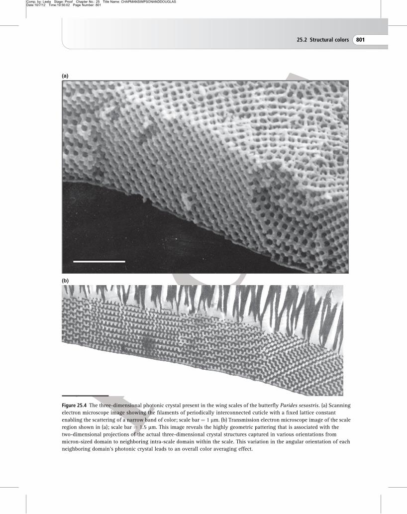
referred to as photonic crystals (Fig. 25.4). In their
scales, filaments of cuticle are interconnected
periodically in all three spatial dimensions, forming a
distinct crystalline geometry with a lattice constant
that is on a length-scale of 200–30 nm. The coherent
scattering conferred by this structure offers strong
light manipulation and, much like the multilayer
system in one dimension, prevents the propagation of
light through the crystal system. It is this which
800 Visual signals: color and light production
Comp. by: Leela Stage: Proof Chapter No.: 25 Title Name: CHAPMANSIMPSONANDDOUGLASDate:10/7/12 Time:19:56:02 Page Number: 801
(a)
(b)
Figure 25.4 The three-dimensional photonic crystal present in the wing scales of the butterfly Parides sesostris. (a) Scanning
electron microscope image showing the filaments of periodically interconnected cuticle with a fixed lattice constant
enabling the scattering of a narrow band of color; scale bar ¼ 1 mm. (b) Transmission electron microscope image of the scale
region shown in (a); scale bar ¼ 1.5 mm. This image reveals the highly geometric pattering that is associated with the
two-dimensional projections of the actual three-dimensional crystal structures captured in various orientations from
micron-sized domain to neighboring intra-scale domain within the scale. This variation in the angular orientation of each
neighboring domain’s photonic crystal leads to an overall color averaging effect.
25.2 Structural colors 801
Comp. by: Leela Stage: Proof Chapter No.: 25 Title Name: CHAPMANSIMPSONANDDOUGLASDate:10/7/12 Time:19:56:04 Page Number: 802
results in certain wavelengths being reflected,
producing a color appearance for the viewer. Many
colored weevils, such as Eupholus magnificus and
E. schoeneri, also exhibit this form of structure in
their elytral and body scales. Commonly, in the
lepidopteran and coleopteran scales that exhibit this
three-dimensional photonic crystal structure, small
grains of individual photonic crystal regions within
each scale are oriented in different directions so that
they reflect specific different colors. As an ensemble,
their overall effect is to create additive color mixing
and render the color as angle-independent rather
than truly iridescent.
25.3 Pigmentary colors
Pigments appear colored because they absorb
certain wavelengths of light, the unabsorbed
remaining light being scattered by various
nanostructures. The energy of absorbed light is
dissipated as heat. Which particular wavelengths
are absorbed depends on the molecular structure of
the compound. Particularly important in the
production of color are the number and arrangement
of double bonds, C¼C, C¼O, C¼N and N¼N.
Particular functional groups are also important. The
–NH2 and –Cl radicals, for example, shift the
absorptive region of a compound so that it tends to
absorb longer wavelengths. The color-producing
molecule, known as a chromophore, is often
conjugated with a protein molecule, forming a
chromoprotein. Insects are able to synthesize most
of their pigments, but not flavonoids or carotenoids
which are, consequently, acquired through diet. The
sources of some other pigments, found only in a few
insects, are unknown. The black or brown of
hardened cuticle often results from sclerotization
(Chapter 16). However, cuticular hardening and
darkening are not necessarily tightly linked. This is
illustrated by albino strains of the locust
Schistocerca, which have a hard but colorless
cuticle.
25.3.1 Pigments that are synthesized
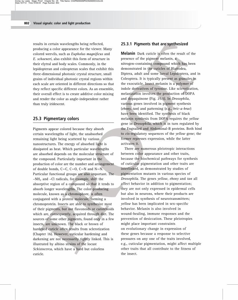
Melanin Dark cuticle is often the result of the
presence of the pigment melanin, a
nitrogen-containing compound which has been
demonstrated in the cuticles of Blattodea,
Diptera, adult and some larval Lepidoptera, and in
Coleoptera. It is typically present as granules in
the exocuticle. Insect melanin is a polymer of
indole derivatives of tyrosine. Like sclerotization,
melanization involves the production of DOPA
and dopaquinone (Fig. 25.5). In Drosophila,
various genes involved in pigment synthesis
(ebony, tan) and patterning (e.g., bric-a-brac)
have been identified. The synthesis of black
melanin synthesis from DOPA requires the yellow
gene in Drosophila, which is in turn regulated by
the Engrailed and Abdominal-B proteins. Both bind
to cis-regulatory sequences of the yellow gene; the
former represses expression, while the latter
activates it.
There are numerous pleiotropic interactions
between color appearance and other traits,
because the biochemical pathways for synthesis
of cuticular pigmentation and other traits are
interlinked, as demonstrated by studies of
pigmentation mutants in various species of
Drosophila. The genes yellow, ebony and tan all
affect behavior in addition to pigmentation;
they are not only expressed in epidermal cells
but also in neurons, where their products are
involved in synthesis of neurotransmitters;
yellow has been implicated in sex-specific
behavior. Melanin is also involved in
wound-healing, immune responses and the
prevention of desiccation. These pleiotropies
might place important constraints
on evolutionary change in expression of
these genes because a response to selective
pressures on any one of the traits involved,
e.g., cuticular pigmentation, might affect multiple
other traits that all contribute to the fitness of
the insect.
802 Visual signals: color and light production

Comp. by: Leela Stage: Proof Chapter No.: 25 Title Name: CHAPMANSIMPSONANDDOUGLASDate:10/7/12 Time:19:56:04 Page Number: 803
Pterins Pterins are nitrogen-containing
compounds, all having the same basic chemical
structure, a pyrazine ring and a pyrimidine ring, but
differing in the radicals attached to this nucleus. They
are synthesized from the purine guanosine
triphosphate, and this synthesis involves the rosy and
purple genes, which control eye color in Drosophila.
Not all pterins appear colored. Some pterins are
important metabolically as cofactors of enzymes
concerned with growth and differentiation and
may act as controlling agents in these processes.
They often occur together with pigments of
another group, the ommochromes, because they are
cofactors of the enzymes involved in ommochrome
synthesis. The vitamin folic acid also contains
a pterin.
Pterin pigments can be white (leucopterin and
isoxanthopterin, which absorb only in the
ultraviolet), yellow (e.g., xanthopterin or
dihydroxanthopterin, which absorb in the blue, but
not necessarily as strongly in the UV-A) and orange
to red (e.g., erythropterin, which absorbs blue, but
not as much in the UV-A). They are important
pigments in lepidopteran scales, where they are
concentrated in pigment granules located on the
crossribs. Leucopterin and xanthopterin are
common in the wings of Pieridae, supplementing
the structural white (Section 25.2.1). Males and
females of the Japanese butterfly, Pieris rapae
crucivora, both appear white to human observers,
but only males’ wings are adorned with beads,
resulting in a sexual dichroism when viewed by
conspecifics, such that only the males strongly
absorb ultraviolet. In the orange sulfur butterfly,
Colias eurytheme, pterin pigments interact with
iridescent structural coloration, effectively
amplifying the ultraviolet signal in males but not
females. The yellow of the brimstone butterfly,
Gonepteryx, is due to chrysopterin, the brighter
color of the male resulting from a higher pigment
concentration than is present in the female, while
the red of the orange-tip butterfly, Anthocharis, is
due to erythropterin. The yellows of Hymenoptera
are produced by granules of pterins in the
epidermis.
The pterins are also abundant in compound eyes,
occurring with ommochromes in the screening
pigment cells of the ommatidia. In this situation they
are sometimes components of granules, but are often
in solution. Several different pterins are present, not
all of them colored. Their functions in the eyes are
not fully understood, although some pterins are
engaged in circadian rhythm regulation. They
Abdominal B (abdomen)
Engrailed (wing spot)
YellowPO
blackmelanin
brownmelanin
yellowsclerotin
PO
PO
Tyrosine
Dopa
Dopamine
N-beta-alanyl-dopamine
TH
DDC
Ebony Tan
Figure 25.5 Melanin synthesis pathways in Drosophila.
Pigments and their precursors are shown in bold, enzymes
are shown in italics (DDC, dopadecarboxylase; PO,
phenyloxidases; TH, tyrosine hydroxylase). In different
cells different components of the pathway might be
expressed, resulting in different pigmentation of the
cuticle. Two proteins controlling expression of the yellow
gene by binding to its cis-regulatory sequences are shown
on top (underlined): in D. melanogaster males, the
Abdominal-B protein activates the yellow gene, resulting in
sex-specific pigmentation of the abdomen. In D. biarmipes,
males but not females have a conspicuous black spot on
the wing, and Engrailed protein represses the yellow gene
and thus the generation of wing spots. Note that dopa
is also a key intermediate in cuticular sclerotization and,
because of this, melanization and sclerotization
may sometimes be linked together (redrawn from
Wittkopp and Beldade, 2009).
25.3 Pigmentary colors 803
Comp. by: Leela Stage: Proof Chapter No.: 25 Title Name: CHAPMANSIMPSONANDDOUGLASDate:10/7/12 Time:19:56:05 Page Number: 804
accumulate with age in the eyes of higher
Diptera, and, because they are products of purine
degradation, they may provide a means of storage
excretion. The progressive accumulation provides a
means of aging these insects. However, no
comparable accumulation occurs in the eyes of the
moth Pectinophora, although changes do occur in the
pterins.
Ommochromes Ommochromes are yellow
(xanthommatin), red (ommatin) and brown
pigments usually occurring in granules coupled
with proteins. Ommochromes (and their precursors
such as kynurenin) typically absorb ultraviolet
and might thus have a photoprotective function.
The granules also contain accumulations
of calcium. Xanthommatin is the most
widely distributed ommochrome and is usually
present wherever ommochromes are found.
Ommochromes, principally xanthommatin, are
widely distributed in insects as screening pigments
in the accessory cells of the eyes, usually associated
with pterins. They are also present in the
photoreceptor cells. It is thought that this is their
original function in insects, and that its function in
integument coloration in several insect taxa is
derived. Yellow, red and brown body colors are
produced by ommochromes in the epidermis.
Xanthommatin turns from yellow to red upon
reduction. The pink of immature adult Schistocerca
is due to a mixture of ommochromes. Red Odonata,
and probably also the reds and browns of
nymphalid butterflies, are due to ommochromes,
while in blue Odonata a dark brown ommochrome
provides the background for the production of
structural blue believed to stem from coherent
scattering from quasi-ordered arrays of particles in
the endoplasmatic reticulum of pigment cells
underlying the cuticle. Epidermal ommochromes
sometimes directly underlie cuticular melanin,
and in these cases they do not contribute to the
insect’s color.
The ommochromes are a group of pigments
derived from the amino acid tryptophan via
kynurenine and 3-hydroxykynurenine (both of
which can also function as yellow pigments
themselves). The biochemical pathway leading to
their production involves the two enzyme genes
vermilion and cinnabar (coding for tryptophan 2,3
dioxygenase and kynurenine 3-monooxygenase,
respectively) and the ommochrome precursor
transporter gene white. Oxidative condensation of
3-hydroxykynurenine gives rise to the
ommochromes (Fig. 25.6). In larval Drosophila,
kynurenine production takes place primarily in the
fat body, but its conversion to
3-hydroxykynurenine occurs in the Malpighian
tubules, where it is stored. At metamorphosis, the
3-hydroxykynurenine is transported to the eyes,
where the ommochromes are formed. In the adult
fly, the whole process can take place in the cells of
the eye, although this is normally supplemented by
kynurenine and 3-hydroxykynurenine synthesized
elsewhere. The scale-forming cells on butterfly
wings also have the capacity to synthesize
ommochromes from tryptophan or from
3-hydroxykynurenine. Transport of ommochromes
in the hemolymph is achieved by specialized
binding proteins.
Ommochrome production is the only way in
which insects can remove tryptophan, which is
toxic at high concentrations such as may occur at
times of high protein turnover. A transitory increase
in tryptophan occurs at metamorphosis in
holometabolous insects, often followed by the
production of ommochromes. In Lepidoptera
ommochromes are accumulated in the meconium,
the accumulated waste products of the pupal period
which are voided immediately following eclosion.
They are responsible for its characteristic red/brown
coloration. Accumulation of ommochromes in the
integument causes the larva of the puss moth,
Cerura, to turn red just before pupation. Some of
the ommochrome produced at the time of pupation
contributes to the screening pigment in the eyes of
804 Visual signals: color and light production

Comp. by: Leela Stage: Proof Chapter No.: 25 Title Name: CHAPMANSIMPSONANDDOUGLASDate:10/7/12 Time:19:56:05 Page Number: 805
the adult. Red fecal pellets containing
ommochromes are produced by locusts during
molting or starvation. At these times, excess
tryptophan is likely to be liberated from proteins
that are broken down during structural
rearrangement or that are used for energy
production.
Tetrapyrroles There are two major classes of
tetrapyrroles: the porphyrins, in which the pyrroles
form a ring, and the bilins, which have a linear
arrangement of the pyrroles. The bilins are usually
associated with proteins to form blue
chromoproteins. Biliverdin occurs in many
hemimetabolous insects, but is also found in
Neuroptera and some Lepidoptera, although the latter
usually contain other types of bilins. Associated with
a yellow carotenoid, these pigments are responsible
for the greens of many insects. Sometimes the
pigments themselves are green. In Chironomus
(Diptera), bilins derived from the hemoglobin of the
larva accumulate in the fat of the adult and impart a
green color to the newly emerged fly. In Rhodnius,
the pericardial cells become green due to the
accumulation of bilins derived from ingested
hemoglobin.
A porphyrin having an atom of iron in its center is
called a heme molecule and this forms the basis of
tryptophan
formyl kynurenine
kynurenine
3-hydroxykynurenineka
r
3-hydroxykynurenine
kar
-
3-hydroxykynurenine
scarlet white
xanthommatin
pigmentgranule
cellcytosol
hemolymph
tryptophan 2,3 dioxygenase(vermilion)
kynurenine 3-hydroxylase(cinnabar)
Figure 25.6 Ommochromes synthesis in the eye of Drosophila melanogaster (and similarly in butterfly wing patterns).
Tryptophan is apparently moved into the cell by the amino acid transporter karmoisin (kar). The vermilion gene encodes for
tryptophan 2,3 dioxygenase, an enzyme that catalyzes tryptophan’s conversion to formyl kynurenine. Hydrolysis of
formyl kynurenine results in kynurenine, whose transformation into 3-hydroxykynurenine in turns requires the cinnabar
gene (and its product, kynurenine 3-hydroxylase). 3-hydroxykynurenine is also generated elsewhere in the body and
taken up directly from the hemolymph. White and scarlet are transporter molecules that form a dimer localized in the surface
of the pigment granules; they have been implicated in transporting ommatin precursors into the granule. Oxidative
condensation of 3-hydroxykynurenine generates the ommochromes. Synthesis of other ommochromes involves the same
pathway, differing in the final steps (after figure 1 in Reed and Nagy, 2005).
25.3 Pigmentary colors 805

Comp. by: Leela Stage: Proof Chapter No.: 25 Title Name: CHAPMANSIMPSONANDDOUGLASDate:10/7/12 Time:19:56:06 Page Number: 806
two important classes of compounds, the
cytochromes and the hemoglobins. In each case the
heme molecule is linked to a protein. All insects are
able to synthesize cytochromes, which are essential
in respiration, the different cytochromes differing in
the forms of their heme groupings. Normally they are
only present in small amounts and so produce no
color, but where they are present in high
concentrations, as in flight muscle, they produce a
reddish-brown color.
Some insects living in conditions subject to low
oxygen tensions contain hemoglobin in the
hemolymph, and these are colored red by the
pigment showing through the integument. In
Chironomus (Diptera) larvae the hemoglobin is in
solution in the hemolymph, while in the larva of
Gasterophilus (Diptera) it is in hemoglobin cells.
Many other insects contain hemoglobin in tracheal
tissue and the fat body. Hemoglobin serves a
respiratory function, but perhaps also serves as a
protein store and enables the aquatic hemipteran
Anisops to regulate its buoyancy (Section 17.9).
Papiliochromes Papiliochromes are yellow and
reddish-brown pigments known only from the
swallowtail butterflies, Papilionidae. Papiliochrome
II is pale yellow and is formed from one molecule of
kynurenine, derived from tryptophan, and one
molecule of b-alanine.
Quinone pigments The quinone pigments of
insects fall into two categories: anthraquinones
(violet, blue and green) and aphins (red or purple).
Both occur as pigments only among Homoptera
(Hemiptera), the former in coccids (Coccoidea) and
the latter in aphids only.
Anthraquinones are formed from the condensation
of three benzene rings. In the coccids they give the
tissues a red, or sometimes yellow, coloration. The
best known is cochineal from Dactylopius cacti. The
purified pigment is called carminic acid. It is present
in globules in the eggs and fat body of the female,
constituting up to 50% of the body weight. The larva
contains relatively little pigment.
Aphins are quinone pigments formed from the
condensation of three, in the monomeric forms, or
seven benzene rings, in dimeric forms. They are
found in the hemolymph of aphids, sometimes in
high concentration, and impart a purple or black
color to the whole insect. Neriaphin is a monomeric
form. Quinone tanning is known to produce dark
cuticle.
25.3.2 Pigments obtained from the food
Carotenoids Carotenoids are a major group of
pigments that are lipid soluble and contain no
nitrogen. They are built up from two diterpenoid
units joined tail to tail. In nearly all insect
carotenoids, the central chain contains 22 carbon
atoms with nine double bonds, and each of the end
groups contains nine carbon atoms. There are two
major groups of carotenoids: the carotenes, and their
oxidized derivatives, the xanthophylls.
Yellow, orange and red are commonly produced by
carotenoids, the color depending largely on whether
or not the terminal groups are closed rings and on
their degree of unsaturation. If the carotenoid is
bound to a protein, the color may be altered,
sometimes even resulting in a blue pigment. Insects
cannot synthesize carotenoids and consequently
must obtain them from the diet. Uptake from the food
is, at least to some extent, selective. Orthopteroids
preferentially absorb carotenes, while lepidopterans
favor xanthophylls. Some post-ingestive
modification of the carotenoids may occur.
Carotenoids can occur in many different tissues
and in all stages of development. A number of
structurally different carotenoids may be present in
one insect. The possible metabolic functions of
carotenoids in insects are not well understood. In
other organisms they protect cells from damage due
to photo-oxidation by light, but their importance to
insects in this regard is not known. They are also the
806 Visual signals: color and light production

Comp. by: Leela Stage: Proof Chapter No.: 25 Title Name: CHAPMANSIMPSONANDDOUGLASDate:10/7/12 Time:19:56:07 Page Number: 807
source of retinal in the insect’s visual pigments. Apart
from producing the reds and yellows of many insects,
in combination with a blue pigment, often a bilin,
they produce green. Green produced by a carotenoid
with a bilin is sometimes known as insectoverdin. In
some insects, the ability to sequester carotenoids
might have functions beyond pigmentation;
carotenoids also confer advantages with respect to
photo-oxidative stress induced by ultraviolet
radiation.
Flavonoids Flavonoids are heterocyclic
compounds commonly found in plants. In insects
they are mainly found among the butterflies and are
common in Papilionidae, Satyridae and Lycaenidae
as cream or yellow pigments. At least in some
species, flavonoids are present in all developmental
stages, including the egg, indicating that the
flavonoids are stored in internal tissues of the adult
as well as in the scales. Although some flavonoids
function as deterrents to herbivores, it is clear this is
not efficient for all insects. Where they occur in
insects, flavonoids are acquired exclusively from
herbivory, so the flavonoids present in their bodies
reflect what is present in the host plants. Some
post-ingestive modification of structure does occur
so that the flavonoids in the insect are not exactly the
same as those in the host. Perhaps these changes are
produced by the insects themselves, but it is possible
that their gut flora is responsible.
25.4 Color patterns
In general, insects do not have a uniform coloring,
but have specific and often finely detailed patterns.
The genetics and evolutionary developmental
biology have been especially well studied in
Drosophila and in the Lepidoptera. The development
of black spots on fruit fly wings as well as the
widespread lepidopteran eyespot patterns (with
concentric rings of different colors) is controlled by a
series of organizing centers from which morphogens
diffuse outwards, creating a concentration gradient.
Many of the wing-patterning genes first identified in
Drosophila, such as apterous, wingless, Distal-less,
engrailed and cubitus interruptus, are also involved
in lepidopteran eyespot formation, where they are
deployed in specific regions of the wings, often at
specific times during development. In butterflies the
wing pattern is produced by thousands of small
overlapping scales. Morphogens govern the
development of pigment in each scale-forming cell
and different concentrations induce different
pigments. Studies with tissue transplants have shown
that the size of the eyespot is determined primarily by
properties of tissue at the center, while the specific
colors in each ring depend on response thresholds
with which the host tissue responds to the chemical
signal diffusing from the center. As diffusion extends
uniformly outwards in all directions around an
organization center, a symmetrical pattern is
produced. It is also possible that a band of color
rather than a series of separate rings can be produced
by organization centers that are close together so that
the patterns they produce grade into each other. On
the forewings of many butterflies (Papilionoidea)
there are rows of organizing centers that produce
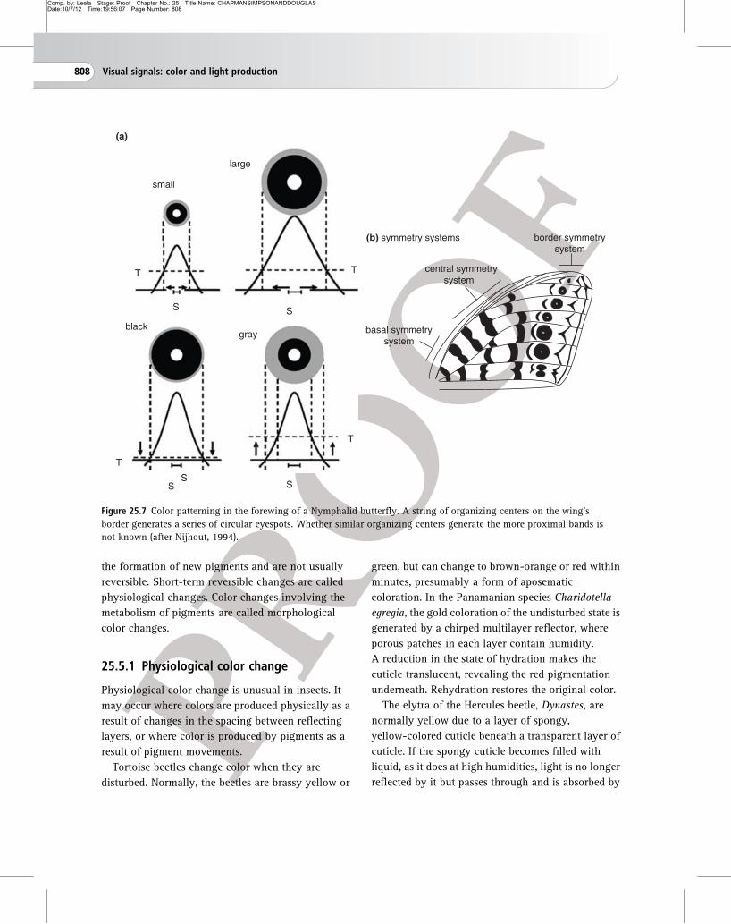
series of spots across the wing (Fig. 25.7). In general,
it appears that each scale contains only a single
pigment, although examples of scales with two
pigments are known. The pigment produced by each
scale-forming cell depends on synthesis within the
cell. Although some intermediates may be
manufactured elsewhere, scale color is not
determined by selective uptake of presynthesized
pigment. It seems, therefore, that the morphogens
moving between epidermal cells regulate the
synthetic machinery within each cell.
25.5 Color change
Color changes are of two kinds: short-term reversible
changes which do not involve the production of new
pigment, and long-term changes which result from
25.5 Color change 807
Comp. by: Leela Stage: Proof Chapter No.: 25 Title Name: CHAPMANSIMPSONANDDOUGLASDate:10/7/12 Time:19:56:07 Page Number: 808
the formation of new pigments and are not usually
reversible. Short-term reversible changes are called
physiological changes. Color changes involving the
metabolism of pigments are called morphological
color changes.
25.5.1 Physiological color change
Physiological color change is unusual in insects. It
may occur where colors are produced physically as a
result of changes in the spacing between reflecting
layers, or where color is produced by pigments as a
result of pigment movements.
Tortoise beetles change color when they are
disturbed. Normally, the beetles are brassy yellow or
green, but can change to brown-orange or red within
minutes, presumably a form of aposematic
coloration. In the Panamanian species Charidotella
egregia, the gold coloration of the undisturbed state is
generated by a chirped multilayer reflector, where
porous patches in each layer contain humidity.
A reduction in the state of hydration makes the
cuticle translucent, revealing the red pigmentation
underneath. Rehydration restores the original color.
The elytra of the Hercules beetle, Dynastes, are
normally yellow due to a layer of spongy,
yellow-colored cuticle beneath a transparent layer of
cuticle. If the spongy cuticle becomes filled with
liquid, as it does at high humidities, light is no longer
reflected by it but passes through and is absorbed by
SS S
SS
T
TT
T
small
large
(a)
blackgray
(b) symmetry systems
basal symmetrysystem
central symmetrsystem
y
border symmetrysystem
Figure 25.7 Color patterning in the forewing of a Nymphalid butterfly. A string of organizing centers on the wing’s
border generates a series of circular eyespots. Whether similar organizing centers generate the more proximal bands is
not known (after Nijhout, 1994).
808 Visual signals: color and light production

Comp. by: Leela Stage: Proof Chapter No.: 25 Title Name: CHAPMANSIMPSONANDDOUGLASDate:10/7/12 Time:19:56:07 Page Number: 809
black cuticle underneath, making the insect black. In
the field, these changes probably occur daily, so that
the insect tends to be yellow in the daytime, when it
is feeding among leaves, and dark at night, making it
less conspicuous.
Physiological color changes involving pigment
movements are known to occur in the stick insect
Carausius, in the grasshopper Kosciuscola and in a
number of blue damselflies (Zygoptera, Odonata). All
these insects become black at night due to the
movement of dark pigment granules to a more
superficial position in the epidermal cells (Fig. 25.8).
Kosciuscola is blue during the day as a result of
coherent scattering from small granules, less than
0.2 mm in diameter, believed to be composed mainly
of white leucopterin and uric acid. At night, the
blue is masked by the dispersal of larger pigment
granules among the small reflecting granules which
also disperse through the cell. Similar masking of
structurally scattered blues occurs in a number of
damselflies; in brown individuals of the stick insect
Carausius, ommochrome granules occupy a
superficial position at night, causing the insect to
become darker, while during the day they occupy a
proximal position in the epidermal cells, making the
insects paler. Green specimens of Carausius do not
change color because they lack ommochromes. The
granules move along the paths of microtubules which
may be responsible for the movements. Similar
microtubules are present in the epidermal cells of
Kosciuscola.
The color change in Kosciuscola and damselflies is
temperature dependent. The insects are always black
below a temperature which is characteristic of the
species, but usually about 15�C. At highertemperatures they tend to become blue, and, in some
species, the change to blue is enhanced by light. Here,
the epidermal cells are to some extent independent
longwavelengths
absorbed
whitelight
shortwavelengths
reflected
(a) blue
cuticle
smallgranules
largegranules
whitelight
(b) black
absorbed
Figure 25.8 Physiological color change
in Kosciuscola. The two diagrams have
the same numbers of large and small
granules (based on Filshie et al., 1975).
(a) At moderately high temperatures,
the larger granules are restricted to the
proximal parts of the epidermal cells.
Short wavelengths of light are
scattered by the small granules in the
more distal parts of the epidermal cell,
longer wavelengths travel further into
the cell and are absorbed. The insects
appear blue. (b) At lower temperatures,
the pigments are generally dispersed
through the cell. All the light is
absorbed by the larger granules and the
insects appear black.
25.5 Color change 809

Comp. by: Leela Stage: Proof Chapter No.: 25 Title Name: CHAPMANSIMPSONANDDOUGLASDate:10/7/12 Time:19:56:08 Page Number: 810
effectors, responding directly to stimulation. This is
true of the change from black to blue in the damselfly
Austrolestes, but the reverse change is controlled by
a secretion released from the terminal abdominal
ganglion. The significance of these changes is
unknown, but they may be thermoregulatory. Dark
insects absorb more radiation than pale ones so they
may warm up more rapidly in the mornings and
become active earlier than would be the case if they
remained pale.
25.5.2 Morphological color change
Changes in the amounts of pigments can occur in
response to a variety of external and internal factors.
Ontogenetic changes Many insects change color
in the course of development. For example, the eggs
of the plant-sucking bug Dysdercus nigrofasciatus
(Hemiptera) are white when laid, becoming yellow as
the embryo develops. The first-stage nymph is a
uniform yellow color when it hatches, but becomes
orange and then red. In the second instar, white
bands appear ventrally on some of the abdominal
segments (Fig. 25.9). These become more extensive
and white bands are also present dorsally in the later
stages. In the final larval instar, the red becomes less
intense, especially in the female, and the adults are
yellow with white stripes. The yellow and red colors
of this insect are produced by three pterins, the
proportions of which change through development to
produce the different colors. The white bands are
formed from uric acid.
It is also common for caterpillars to exhibit a
regular change in color during development. The
larva of the puss moth, Cerura, turns from green to
red just before pupation as a result of the production
of ommochrome in the epidermis. The early larva of
the swallowtail butterfly, Papilio demodocus, is
brown with a white band at the center, whereas the
late larva is green with purple markings and a white
lateral stripe.
In adult hemimetabolous insects, color change is
often associated with ageing and maturation. Adult
male Schistocerca change from pink to yellow as they
mature. The pink is produced by ommochromes in
the epidermis which decrease in amount as the insect
gets older, and the yellow is due to b-carotene, whichincreases with age. Color changes related to sexual
maturation also occur in some Odonata.
4
2
0
instar 2 instar 4 adult
erythropterin leucopterin isoxanthopterin
yellow
red white
pter
in (
μgm
g–1 )
Figure 25.9 Ontogenetic changes in pigmentation in Dysdercus nigrofasciatus. Underside of the abdomen of second- and
fourth-instar larvae and adult male. The change from red to yellow is accompanied by a reduction in the relative amount
of erythropterin present (expressed as mg of pterin per mg body weight). The white areas are due to uric acid in the
epidermal cells (data from Melber and Schmidt, 1994).
810 Visual signals: color and light production

Comp. by: Leela Stage: Proof Chapter No.: 25 Title Name: CHAPMANSIMPSONANDDOUGLASDate:10/7/12 Time:19:56:08 Page Number: 811
These changes are controlled by changes in
hormone levels associated with molting and sexual
maturation, but the effects may be very different in
different species. The changes in larval Cerura are
initiated by a low level of ecdysteroid in the
hemolymph, leading to the metabolism of
3-hydroxykynurenine to an ommochrome. Perhaps
the commitment peak which occurs before pupation
is normally responsible for the change (Section
15.4.2). Juvenile hormone leads to the accumulation
of xanthommatin in Bombyx caterpillars, but
prevents it in Manduca. Juvenile hormone,
concerned in sexual maturation in adult
Schistocerca, also regulates the accumulation of
carotenoids which make the insects yellow as they
become sexually mature. The hormone bursicon,
which controls sclerotization, probably also regulates
darkening occurring immediately after ecdysis.
Homochromy The colors of several insect species
change to match the predominant color of the
background. This phenomenon is called
homochromy. The changes may involve the basic
color of the insect or may involve a general
darkening. For example, larvae of the grasshopper
Gastrimargus tend to assume a color more or less
matching the background when reared throughout
their lives on that background (Fig. 25.10). Insects
0
25
50
75
100
% in
cla
ss
black background
0
25
50
75
100gray background
0
25
50
75
100white background
% in
cla
ss%
in c
lass
0
green background
yellow background
orange background
black gray yellow-gray
yellow orange-brown
insect colors
Figure 25.10 Homochromy in the
grasshopper Gastrimargus. Each graph
shows the ground color of insects in
the final larval stage after rearing from
the first stage in containers with
different colored backgrounds (after
Rowell, 1970).
25.5 Color change 811
Comp. by: Leela Stage: Proof Chapter No.: 25 Title Name: CHAPMANSIMPSONANDDOUGLASDate:10/7/12 Time:19:56:09 Page Number: 812
reared on black become black, those reared on white
are pale gray. On a green background, however, most
insects develop a yellowish coloration. The
differences are produced by different amounts of
black pigment, possibly melanin, in the cuticle and a
dark ommochrome in the epidermis, together with
yellow and orange pigments in the epidermis. The
darkening of the stick insect Carausius that occurs on
a dark background is also due to the accumulation of
two ommochromes, xanthommatin and ommin. In
this insect, changes occur at any time during the
course of a stadium, but in most other examples the
change is first seen at a molt. Ommochrome
deposition begins after about five days on the new
background, and the changes in other insects
generally require a similar period of exposure.
Caterpillars, too, may exhibit some degree of
homochromy. Larvae of the hawk moth Laothoe
populi may be almost white or yellow-green,
depending on the plants on which they are reared,
and the pupae of some butterflies that are not
concealed in a cocoon or in the soil may be green or
dark or pale brown, according to their surroundings.
An extreme example of homochromy occurs in
African grasshoppers after bush fires. Many species
become black within a few days of the fire, the
change occurring in adults as well as larvae, but only
in bright sunlight. In diffuse light the change is much
less marked.
All these changes depend on visual stimulation,
although the details may vary from species to species.
In Carausius darkening results from weak
stimulation, or the absence of stimulation of the
lower parts of the compound eye, such as would
occur when the insect is on a dark surface. In
Gastrimargus darkening occurs if a high incident
light intensity is associated with a low reflectance
background, and changes in the yellow and orange
pigments are dependent on the wavelength of light.
Homochromy in caterpillars and lepidopteran pupae
is also determined by the wavelength of light and the
contrasts in intensity reaching the different
stemmata.
Visual inputs affect the activity of neurosecretory
cells in the central nervous system. Homochromy in
pupae of the peacock butterfly, Inachis io, is
produced by changes in the relative amounts of
melanin and lutein (a carotenoid) in the cuticle. The
accumulation of both pigments is controlled by a
single neuropeptide, which is widely distributed in
the central nervous system before being released into
the hemolymph. A low hormone titer immediately
before pupation stimulates melanization, a higher
titer results in increased incorporation of lutein into
the pupal cuticle. In the grasshopper Oedipoda
miniata, the dark-color-inducing neurohormone
(DCIN) has been identified as (His[7])-corazonin.
The stink bug Nezara viridula (Hemiptera) changes
from green to brown during diapause when
overwintering under leaf litter or bark, and changes
back to green again in spring, presumably to aid
camouflage; these changes are induced in part by day
length; temperature can also have an effect in some
species.
Other factors affecting color Temperature is
important in pigment development and there is a
general tendency for insects reared at very high
temperatures to be pale, and those developing at low
temperatures to be dark. Humidity affects the color of
many Orthoptera. Green forms are more likely to
occur under humid conditions, and brown forms
under dry conditions.
Crowding influences color in some insects, the
most extreme examples being locusts. Locust larvae
reared in isolation are green or fawn, while rearing in
crowds produces yellow and black individuals. The
colors and patterns change as the degree of crowding
alters; under high-density conditions, locusts
consume toxic host plants, and predators learn the
aposematic yellow–black coloration as a predictor of
toxicity. The larvae of some Lepidoptera, such as
Plusia and the armyworms, undergo comparable
changes, some occurring in the course of a stadium,
but the most marked alterations occur only at
molting.
812 Visual signals: color and light production

Comp. by: Leela Stage: Proof Chapter No.: 25 Title Name: CHAPMANSIMPSONANDDOUGLASDate:10/7/12 Time:19:56:09 Page Number: 813
25.5.3 Color polymorphism
Insects of many orders exhibit a green–brown
polymorphism, tending to be green at the wetter
times of year and brown when the vegetation is dry.
The two forms are genetically determined, but
homochromy that develops when the insect moves to
a new background may be superimposed on this. In
green morphs the production of ommochromes in the
epidermis is largely or completely inhibited; if it
occurred, the green would be obscured.
In some insects there are marked differences in
color between successive generations, correlated with
seasonal changes in the environment. Such seasonal
polyphenism and its physiological regulation is
discussed in Section 15.5. Extreme cases of color
polymorphism occur in some butterflies in relation to
mimicry (Section 25.6.1).
25.6 Significance of color
Insect pigments are often the end products of
metabolic processes and may have evolved originally
as forms of storage excretion. Pterins, for example,
may be derived from purines, such as uric acid.
Similarly, melanin production might be a method of
disposing of toxic phenols ingested or arising from
metabolism and it may be significant that melanin in
the cuticle is often produced above metabolically
active tissue such as muscle. Tryptophan in high
concentrations reduces the rate of development of
Drosophila andOryzaephilus and it is noteworthy that
ommochrome production follows the appearance of
unusually high levels of tryptophan in the tissues.
However, it is clear that storage excretion is not the
sole, or even primary, function of pigments in most
insects. For example, most insects excrete most of the
end product of the biologically active hydrogenated
pterins and do not store them. Pieris, on the other
hand, synthesizes much larger amounts than would
result from normal metabolism and accumulates them
in the wings, where they contribute to the color.
In most cases, the colors of present-day insects are
ecologically important. Color is most frequently
involved as a defense against vertebrate predators
and is also important in intraspecific recognition. It
also has important consequences for body
temperature, and color changes may contribute to
thermoregulation (Chapter 19).
25.6.1 Predator avoidance
In many species, color and color patterns have
evolved as part of a strategy to avoid predation. The
color patterns may function in a variety of ways in
different insects: for concealment (crypsis), to startle
a predator (deimatic behavior), to deflect attack from
the most vulnerable parts of the body or to advertise
harmfulness or distastefulness (aposematism).
Crypsis Color often helps to conceal insects from
predators. Many insects are known to select
backgrounds on which they are least conspicuous.
Often, homochromy is associated with some
appropriate body form and behavior as in stick
insects and many mantids and grasshoppers which
may be leaf-like or twig-shaped according to the
backgrounds on which they normally rest. Protection
may also be afforded by obliterative shading. Objects
are made conspicuous by the different light
intensities which they reflect as a result of their form.
A solid object usually looks lighter on the upper side
and darker beneath because of the effect of shadows,
but by appropriate coloring this effect can be
eliminated. If an object is shaded in such a way that
when viewed in normal lighting conditions all parts
of the body reflect the same amount of light, it loses
its solid appearance. Such countershading is
well-known in caterpillars, where the surface toward
the light is most heavily pigmented and the side
normally in shadow has least pigment. To be
successful in concealing the insect, this type of
pigmentation must be combined with appropriate
behavior patterns; if the larva were to rest with the
heavily pigmented side away from the light it would
25.6 Significance of color 813

Comp. by: Leela Stage: Proof Chapter No.: 25 Title Name: CHAPMANSIMPSONANDDOUGLASDate:10/7/12 Time:19:56:10 Page Number: 814
become more, not less, conspicuous. Color may also
afford protection if the arrangement of colors breaks
up the body form. Such disruptive coloration is
typically most efficient when some of the color
components match the background and others
contrast strongly with it. The color pattern should
introduce high-contrast internal boundaries on the
body surface, which surpass the salience of the
outline. Disruptive coloration occurs in some moths
which rest on tree trunks.
Deimatic behavior Some insect species have
colored wings, or other parts of the body, which are
normally concealed, but are suddenly displayed
when a potential predator approaches. This
behavior, sometimes associated with the production
of a sound, has been shown, in a few cases, to startle
the predator. It is known as deimatic behavior. In
many insects exhibiting this behavior, the hindwing
is deep red or black. It is normally concealed
beneath the forewing, but is revealed by a sudden
partial opening of the wings. This occurs in a
number of stick insects and mantids, and in moths
that normally rest on vegetation. Moths in the
family Arctiidae have bright red or yellow
abdomens, often with black markings. When
disturbed, the abdomen is displayed by opening the
wings. The deimatic display of some mantids
involves the front legs, which have conspicuous
marks on the inside. Galepsus, for example, displays
the insides of the fore femora and coxae, which are
orange, at the same time exposing a dark mark in
the ventral surface of the prothorax.
Some Lepidoptera have a pattern of scales on the
hindwing forming an eyespot which is displayed
when the insect is threatened. One of the
best-known examples is the peacock butterfly,
Inachis io. It has one eyespot on the upper surface of
each wing. These eyespots are primarily black,
yellow and blue, surrounded by dark red. The
butterfly rests with its wings held up over its back,
the upper surfaces of the forewings juxtaposed so
that the eyespots are concealed. If the insect is
disturbed by visual or tactile stimuli it lowers the
wings so that the eyespots on the forewings are
displayed and then protracts the forewings to expose
the hindwing eyespots. At the same time the insect
makes a hissing sound with a timbal on the forewing
(Section 26.3.3). The forewings are then retracted
and partly raised and the sequence of movements
repeated, sometimes for several minutes. While
displaying, the body is tilted so that the wings are
fully exposed to the source of stimulation and at the
same time the insect turns so as to put the stimulus
behind it. This eyespot display causes flight behavior
in at least some birds. Some mantids also have a
large eyespot on the hind wing. It remains
controversial whether these eyespots obtain their
startling function by resembling eyes, or whether
their highly contrasting patterns present a more
general aposematic pattern (see below) or induce
neophobia.
Deflection marks Small eyespots, often present on
the underside of the hindwings of butterflies, appear
to deflect the attention of birds away from the head
of the insect. There is no sharp distinction between
eyespots used for deimatic behavior and those
concerned with deflection. In general, deflecting
spots are probably smaller than those used in
intimidation, but it is possible that some may serve
either function, depending on the nature and
experience of the predator. Some butterfly species (in
the families Lycaenidae, Riodinidae and
Nymphalidae) have appendages on the hind wings
that look like antennae or legs, paired with eyespot
patterns, creating the illusion of the insect’s head
(Fig. 25.11). Observations from predator-induced
wing damage indicate that these “false heads” are
indeed efficient in deflecting vertebrate attacks to
less vulnerable body parts, giving the butterfly a
chance to escape.
Aposematic coloration Many insects are
distasteful by virtue of chemicals they produce
themselves, or that are sequestered from their food;
814 Visual signals: color and light production

Comp. by: Leela Stage: Proof Chapter No.: 25 Title Name: CHAPMANSIMPSONANDDOUGLASDate:10/7/12 Time:19:56:10 Page Number: 815
other insects sting (Section 27.9). Such insects are
commonly brightly colored, and are usually red or
yellow combined with black. Such coloration is a
signal to predators that the potential prey is
distasteful and should be avoided. It is called
aposematic coloration. In ladybird beetles, for
example, the degree of carotenoid-based redness in
the elytra of individuals correlates with alkaloid
content. For such coloration to be effective, the
predator must exhibit an innate or a learned
avoidance response. Aposematic species occur in
many orders of insects (Table 25.2). Development of
aposematic coloration is dependent upon population
density in some species of locust and grasshopper.
Mimicry Predators learn to avoid distasteful insects
with distinctive colors. If the color patterns of some
species are similar to each other, learning to avoid
one species because it is distasteful also produces an
avoidance of the other. Resemblance of one species to
another is called mimicry. Mimicry takes two forms,
Mullerian and Batesian. Species exhibiting Mullerian
mimicry are all distasteful. Here, the advantage to the
insects is that predation on any one species is
reduced. For example, many social wasp species –
such as Vespa and Vespula, which all have a sting –
have the same basic black and yellow pattern; if a
predator learns to avoid one species, it is likely to
avoid others with a similar appearance. Sometimes
the mimics have different lifestyles. Cotton stainer
bugs of the genus Dysdercus usually have similar red
and black coloration and all the species are
distasteful. The predaceous reduviid bug
Phonoctonus lives with Dysdercus and preys on them.
It, too, is unpalatable to predators, and is a color
mimic of Dysdercus so that avoidance of one leads to
avoidance of both species. Mullerian mimicry is also
common among Lepidoptera, with the genus
Heliconius having been especially well studied. The
adaptive polymorphism in H. erato is orchestrated by
a precise temporal and spatial expression pattern of
the cinnabar and vermilion genes, coding for
enzymes in ommochrome production.
In Batesian mimicry, only one of a pair of species
is distasteful, the other is not. They are called the
“model” and “mimic,” respectively. Here, the
palatable species gains some advantage from a
resemblance to a distasteful species. In this type of
mimicry it is essential that the mimic is uncommon
relative to the model. If this were not the case a
predator might learn to associate a particular pattern
with palatability rather than distastefulness. This
limits the numbers or distribution of a mimetic form,
but the limit may be circumvented by the mimic
becoming polymorphic, with each of its morphs
resembling a different distasteful species. The
best-known example of such polymorphism is that of
the female Papilio dardanus (Lepidoptera), which has
Figure 25.11 Deflection marks on the hind wing of a
lycaenid butterfly. Two eyespots paired with false
“antennae” generate the impression of a head, deflecting
predator attacks away from body parts where a bite might
be fatal (figure 3 from Stevens, 2005).
25.6 Significance of color 815

Comp. by: Leela Stage: Proof Chapter No.: 25 Title Name: CHAPMANSIMPSONANDDOUGLASDate:10/7/12 Time:19:56:11 Page Number: 816
a large number of mimetic forms mimicking a series
of quite different-looking butterflies. It appears that
alleles of a single gene explain all the natural
variants of this species.
Because the unpalatability of an individual may be
affected by the nature of the food it eats, mimicry
may vary temporally and spatially. For example, the
viceroy butterfly (Limenitis archippus) and queen
butterfly (Danaus gilippus) of North America are
both distasteful and normally exhibit Mullerian
mimicry. However, sometimes larvae of the queen
butterfly feed on plants that are so low in the
cardenolides sequestered by the insect that the
resulting adults are not distasteful. It may be
supposed that the palatable queen butterflies now
depend for protection on their resemblance to other
members of the species that have sequestered
cardenolides, and on unpalatable viceroy butterflies.
A special case of Batesian (deceptive) mimicry occurs
where some insects mimic the visual appearance of
their own predators, such as jumping spiders.
25.6.2 Intraspecific recognition
Color is important in intraspecific recognition in
many diurnally active insects, and its role is most
fully understood in damselflies and dragonflies
(Odonata), and in butterflies. It often has two
Table 25.2 Aposematic insects; examples from different orders
Order and species Stage Color Basis of unpalatability
Orthoptera
Zonocerus variegatus Adult B/Y and R markings Pyrrolizidine alkaloids
Hemiptera
Aphis nerii All Bright Y Cardiac glycosides
Oncopeltus fasciatus Adult Y/B spots Cardiac glycosides
Coleoptera
Coccinella septemfasciata Adult R/B spots Alkaloids
Tetraopes oregonensis Adult R Cardiac glycosides
Hymenoptera
Vespula vulgaris Adult Y/B stripes Sting
Lepidoptera
Tyria jacobaeae Larva B/Y stripes Pyrrolizidine alkaloids
Tyria jacobaeae Adult B/R marks Pyrrolizidine alkaloids
Battus philenor Larva R/B spots Aristolochic acids
Danaus plexippus Larva W/B and Y stripes Cardiac glycosides
Zygaena filipendula Adult B/R spots Cyanogenic glycosides
Note:B, black; Y, yellow; R, red; W, white.
816 Visual signals: color and light production

Comp. by: Leela Stage: Proof Chapter No.: 25 Title Name: CHAPMANSIMPSONANDDOUGLASDate:10/7/12 Time:19:56:11 Page Number: 817
principal functions in male behavior: the recognition
of females and the recognition of conspecific males;
females also select males by coloration.
In some dragonflies, male color is important in
defending a territory against other males. For
example, the dorsal side of the abdomen of male
Plathemis lydia is blue. If a second male enters a
territory, the resident male faces the intruder and
displays the color by raising the abdomen toward the
vertical. This has an inhibiting effect on the intruding
male. In the presence of a female, the abdomen is
depressed so that the blue is not visible as the male
approaches her. The females of many species change
color as they become sexually mature and this is
associated with a change in male behavior toward
them. Among the damselflies, the females of some
species are dimorphic, with one of the morphs
resembling the male, and there are distinct
differences in the behavior of males toward these two
morphs.
In butterflies the general color of a female may be
more important than the details of pattern in
attracting males, although size and movement are
also important. For example, males of the African
butterfly, Hypolimnas misippus, are attracted by the
female’s red-brown wing color; the black and white
markings sometimes present on the forewings are
unimportant, although white on the hindwing may
have an inhibitory effect. Males of Colias eurytheme
are attracted by the yellow underside of the female
hindwing, which is exposed when the female is at
rest. In this species the male reflects ultraviolet from
the upper side of its wings. This signal is used by the
female in interspecific mate recognition, and it also
inhibits attraction by other males, reducing the
likelihood of further male intervention when a male
is copulating. Such female mate recognition can have
an important role in reproductive isolation between
species. Strength of pigmentation or structurally
based ultraviolet iridescence can depend on an
individual’s ability to acquire resources, and
therefore be indicative of phenotypic or genotypic
mate quality. In some social wasps, facial patterns
differ between members of the nest, and colony
members can remember the individual appearance of
a nest-mate.
25.7 Light production
Intrinsic luminescence, i.e., light produced by the
insects, is known to occur in various Coleoptera,
primarily in the families Lampyridae (fireflies),
Elateridae (click beetles) and Phengodidae (railroad
worms), as well as in a few Collembola, such as
Onychiurus armatus, in the homopteran Fulgora
lanternaria, some larval Diptera of the families
Platyuridae and Bolitophilidae.
The light-producing organs occur in various parts
of the body. The collembolan Onychiurus emits a
general glow from the whole body. In most beetles
the light organs are relatively compact, and are often
on the ventral surface of the abdomen. In male
Photuris (Coleoptera) there is a pair of light organs in
the ventral region of each of the sixth and seventh
abdominal segments. In the female the organs are
smaller and often only occur in one segment. The
larvae have a pair of small light organs in segment
eight, but these are lost at metamorphosis when the
adult structures form. Larvae and females of railroad
worms (Phengodidae, Coleoptera) have 11 pairs of
dorso-lateral light organs on the thorax and
abdomen and another on the head. In Fulgora
(Hemiptera) the light organ is in the head. The light
organs are generally derived from the fat body, but in
the glow worm fly Arachnocampa (Diptera) they are
formed from the enlarged distal ends of the
Malpighian tubules.
25.7.1 Structure of light-producing organsin Coleoptera
Each light organ of an adult firefly consists of a
number of large cells, the photocytes, which lie just
beneath the epidermis and are backed by several
layers of cells called the dorsal layer cells (Fig. 25.12).
25.7 Light production 817
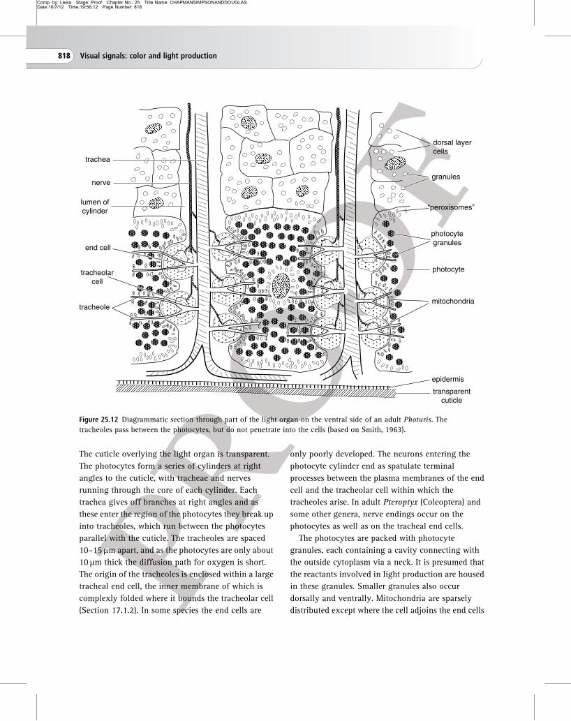
Comp. by: Leela Stage: Proof Chapter No.: 25 Title Name: CHAPMANSIMPSONANDDOUGLASDate:10/7/12 Time:19:56:12 Page Number: 818
The cuticle overlying the light organ is transparent.
The photocytes form a series of cylinders at right
angles to the cuticle, with tracheae and nerves
running through the core of each cylinder. Each
trachea gives off branches at right angles and as
these enter the region of the photocytes they break up
into tracheoles, which run between the photocytes
parallel with the cuticle. The tracheoles are spaced
10–15 mm apart, and as the photocytes are only about
10 mm thick the diffusion path for oxygen is short.
The origin of the tracheoles is enclosed within a large
tracheal end cell, the inner membrane of which is
complexly folded where it bounds the tracheolar cell
(Section 17.1.2). In some species the end cells are
only poorly developed. The neurons entering the
photocyte cylinder end as spatulate terminal
processes between the plasma membranes of the end
cell and the tracheolar cell within which the
tracheoles arise. In adult Pteroptyx (Coleoptera) and
some other genera, nerve endings occur on the
photocytes as well as on the tracheal end cells.
The photocytes are packed with photocyte
granules, each containing a cavity connecting with
the outside cytoplasm via a neck. It is presumed that
the reactants involved in light production are housed
in these granules. Smaller granules also occur
dorsally and ventrally. Mitochondria are sparsely
distributed except where the cell adjoins the end cells
dorsal layercells
granules
“peroxisomes”
photocytegranules
photocyte
mitochondria
epidermis
transparentcuticle
trachea
nerve
lumen ofcylinder
tracheole
end cell
tracheolarcell
Figure 25.12 Diagrammatic section through part of the light organ on the ventral side of an adult Photuris. The
tracheoles pass between the photocytes, but do not penetrate into the cells (based on Smith, 1963).
818 Visual signals: color and light production

Comp. by: Leela Stage: Proof Chapter No.: 25 Title Name: CHAPMANSIMPSONANDDOUGLASDate:10/7/12 Time:19:56:12 Page Number: 819
and tracheoles. The dorsal layer cells also contain
granules, generally understood to comprise urate
crystals, which form a reflecting layer at the back of
the light-producing region which directs the
luminescence more efficiently in the outward
direction. It is estimated that the two lanterns of
Photinus together contain about 15 000 photocytes,
forming some 6000 cylinders, each with 80–100 end
cells. The lanterns of larval fireflies contain the same
elements, but their organization is simpler. The
tracheal system is diffuse and there are no tracheal
end cells. Nerve endings occur on the photocytes, and
are not separated from them by the tracheal end cells
as in adult Photuris.
25.7.2 Mechanism of light production
Light is produced in organelles called peroxisomes,
noted as centers for enzymatic oxidation reactions.
A two-stage reaction occurs within these
peroxisomes. First, adenylation of substrate luciferin
(which is dependent on the presence of
magnesium-ATP) occurs under the catalytic action of
luciferase. The subsequent oxygenation of luciferyl
adenylate by molecular oxygen results in the
emission of light and the production of oxyluciferin
(Fig. 25.13).
The reaction is very efficient, some 98%of the energy
involved being released as light. Furthermore, in
fireflies the metabolic cost of the light-flash production
is low. It increases metabolic rates by approximately
40%, compared to 60% for walking activity.
In many insects the light produced by the
bioluminescence organs is yellow-green in color,
extending over a relatively narrow band of
wavelengths, 520–650 nm in Photinus and Lampyris
(Coleoptera). In larval and adult female railroad
worms (Coleoptera) such as Phrixothrix, the light
organs on the thorax and abdomen produce green to
orange light, depending on the species, in the range
530–590 nm. That on the head produces red light
with peak emission at 620 nm, but extending from
about 580 nm to over 700 nm. The light produced by
Arachnocampa (Diptera) is blue-green, that of
Fulgora (Hemiptera) white. In fireflies, despite
noticeable spectral differences in light production,
the luciferin substrate is identical and their DNA
similarity is 70�99%. The observed differences in
spectral output are caused by single amino acid
substitutions; namely, with variations in the
luciferase active site that controls the conformation
of the bound substrate.
25.7.3 Control of light production
The light organs of adult Photuris (Coleoptera) are
innervated by three and four dorsal unpaired median
(DUM) cells in the last two abdominal ganglia,
N
HO S N
S
H
COOH
luciferase+
ATP- Mg++
luciferin
adenylluciferin
N
HO S N
S
H
C - AMP
O
N
HO S N
S
C - AMP
O
O
N
HO S N
SO
O O
N
S N
S H
luciferase+O2
adenyloxyluciferin
oxyluciferin
light
Ol
OlOl
Figure 25.13 Basic reactions involved in light production
from luciferin.
25.7 Light production 819
Comp. by: Leela Stage: Proof Chapter No.: 25 Title Name: CHAPMANSIMPSONANDDOUGLASDate:10/7/12 Time:19:56:13 Page Number: 820
respectively, which release octopamine. Like other
DUM cells (Section 10.3.2), the axons from these cells
divide to send symmetrical branches to the lanterns
on each side. In most adult fireflies the axons
terminate on the tracheal end cells, but in larvae,
where there are no end cells, they innervate the
photocytes directly. The lantern end cells, nerve
endings and tracheolar cells form the “end organ”
complexes. These, together with the enclosed tracheal
channels, play a dominant role in the control of light
production since they are only observed in species
exhibiting flashing lanterns. No flashing lantern is
observed Photuris larva, but there is a slowly rising
and falling light production capacity. Peroxisomes
and mitochondria are also present in the larva but are
unsegregated and no specializations are observed in
the tracheal cell compartments.
In adult Photuris, light production appears to be
regulated by the availability of oxygen. As the DUM
neurons terminate on the tracheal end cells it is
presumed that neural activity causes a change in
these cells, facilitating the flow of oxygen to the
photocytes. Flash duration in adult fireflies is
generally very brief, a few hundred milliseconds,
with flashes following each other at regular
intervals. This implies that the oxygen supply to the
photocytes is closely regulated. It is believed that
hydrogen peroxide plays a role in this regulation:
The peroxisome oxidases use oxygen arriving at
open mitochondria to create hydrogen peroxide that
builds up explosively due to the shutdown of the
catalase. This completes the oxidation reaction and
triggers the flash. The precise temporal control of
firefly flashing is understood to be regulated by
nitric oxide (NO). NO synthase is localized near
synaptic terminals within the firefly lantern and
measurements indicate that externally added NO
stimulates bioluminescence production while the
addition of NO scavengers inhibits light production.
Furthermore, NO is known to control respiration by
photocyte mitochondria reversibly. The proposed
mechanism comprises neural stimulation resulting
in the transient release of NO that diffuses into the
periphery of adjacent photocytes. This inhibits
mitochondrial respiration and permits oxygen to
diffuse into the photocyte, which holds the
bioluminescence reactants.
Species of fireflies of the genus Pteroptyx
(Coleoptera), notably in Southeast Asia, form male
groups in which the individual insects flash in
synchrony. The males of these species, in isolation,
can flash regularly with almost constant intervals
between flashes. If an individual detects a flash
within a critical period of having produced its own
flash, it immediately flashes again. As the individuals
produce flashes at regular intervals, this resetting
rapidly results in synchronous flashing by all the
insects.
25.7.4 Significance of light production
Sexual communication is the principal role for
self-luminescence in most luminous insects. Light
signals are used in two basic ways in Lampyridae
(Coleoptera). In some species, such as Lampyris, the
female is sedentary and attracts the male to herself;
in other species, such as Photuris and Photinus, one
sex, usually the male, flies around flashing in a
specific manner. Flash duration and the interval
between flashes are often characteristic for the
species (Fig. 25.14) and flashing is associated with
distinct flight patterns. For instance, male Photinus
pyralis produce a flash lasting about 500ms at
six-second intervals. During the flash the male
climbs steeply and then hovers for about two
seconds. If a female flashes 1.5–2.5 seconds after the
male flash, he flies toward her and flashes again
three seconds later. He does not respond to flashes
occurring after different time intervals. Repeated
flashing sequences bring the male to the female.
Precise timing requires a very well-defined time
marker and flashes begin or end (sometimes both)
sharply; these transients probably provide the
820 Visual signals: color and light production

Comp. by: Leela Stage: Proof Chapter No.: 25 Title Name: CHAPMANSIMPSONANDDOUGLASDate:10/7/12 Time:19:56:13 Page Number: 821
temporal signals. Females of Pteroptyx are attracted
by the flashes of the male group.
After mating, the females of some Photuris species
change their behavior so their patterning of flashing
comes to resemble that of females of other species. In
this way they lure males of those species on which
they then feed. The luminescence of Arachnocampa
(Diptera) larvae also serves as a lure, attracting small
insects into networks of glutinous silk threads on
which they become trapped. The larvae then eat
them.
In railroad worms (Coleoptera) the lateral
light-producing organs may be suddenly illuminated
if the insect is attacked, and they possibly have a
defensive function. It is suggested that the red
head light provides these insects with
illumination, presumably when they are searching
for the millipedes on which they prey. The relatively
long wavelengths emitted by this organ may
not be visible to most other insect predators, but
the eyes of the railroad worms probably can detect
them.
The sensitivity of the dark-adapted compound
eyes of fireflies varies diurnally, as it does in at least
some other insects. It increases rapidly by about four
orders of magnitude at a time approximately
corresponding with the time at which the insects
normally flash.
Lampyris noctiluca
Phausis reticulata
Luciola aphrogeneia
Photuris lucicrescens
Photuris versicolor
Photinus pyralis
Photinus greeni
Photinus macdermotti
continuous glownot luminescent
long glow
50 10time (seconds)
malefemale
malefemale
malefemale
malefemale
malefemale
malefemale
malefemale
malefemale
Figure 25.14 Flash patterns of different fireflies (Lampyridae). The height of each symbol represents the intensity of the
flash; the shape shows intensity rising to a maximum and then declining. The dotted line in female Photuris lucicrescens
shows the interval during which the female flash may occur (based on Case, 1984).
25.7 Light production 821
Comp. by: Leela Stage: Proof Chapter No.: 25 Title Name: CHAPMANSIMPSONANDDOUGLASDate:10/7/12 Time:19:56:14 Page Number: 822
Summary
� There is a spectacular variety of insect color patterns, such as in the
butterflies, dragonflies and beetles. The ways in which butterflies, for
example, generate iridescence by physical structures shows nature’s
nanotechnology at its best. The way in which pigment patterns are
generated by the interaction of morphogens and response thresholds in flies
and butterflies has become a model system in evolutionary developmental
biology.
� Insects use visual color signals in a large variety of behavioral contexts, such as the
recognition of mates and conspecific competitors, but also to deter predators – and
signals are not always honest, such as in butterflies displaying eyespots and
harmless flies mimicking the body coloration of wasps.
� Some color signals are constitutively displayed, whereas in other species they are
only displayed when necessary, either to display to potential mates or to deter avian
predators with a sudden display of an eye-like pattern.
� Some species operating in dim light or darkness generate their own light,
mostly for purposes of mate attraction, but also to deter predators or indeed to
attract prey.
� In many other cases, insect body coloration serves not to attract attention, but the
opposite – to blend in with the substrate to minimize the chances of detection by
predators. Some insects can change color by remarkable physiological and
morphological mechanisms when necessary.
Recommended reading
Casas, J. and Simpson S. J. (eds.) (2011). Insect Integument and Colour, Advances in Insect
Physiology, vol. 38. London: Academic Press.
Ruxton, G. D., Sherratt, T. N. and Speed, M. P. (2004). Avoiding Attack. Oxford: Oxford
University Press.
Vukusic, P. and Sambles, J. R. (2003) Photonic structures in biology. Nature 424,
852–855.
Wittkopp, P. J. and Beldade, P. (2009). Development and evolution of insect pigmentation:
genetic mechanisms and the potential consequences of pleiotropy. Seminars in Cell &
Developmental Biology 20, 65–71.
822 Visual signals: color and light production

Comp. by: Leela Stage: Proof Chapter No.: 25 Title Name: CHAPMANSIMPSONANDDOUGLASDate:10/7/12 Time:19:56:15 Page Number: 823
References
Case, J. F. (1984). Vision in mating behavior of fireflies. Symposium of the Royal
Entomological Society of London 12, 195–222.
Filshie, B. K., Day, M. F. and Mercer, E. H. (1975). Colour and colour change in the
grasshopper, Kosciuscola tristis. Journal of Insect Physiology 231, 1763–1770.
Melber, C. and Schmidt, G. H. (1994). Quantitative variations in the pteridines during
postembryonic development of Dysdercus species (Heteroptera: Pyrrhocoridae).
Comparative Biochemistry and Physiology B 108, 79–94.
Nijhout, H. F. (1994). Symmetry systems and compartments in Lepidopteran wings: the
evolution of a patterning mechanism. Development (1994 Suppl.), 225–233.
Reed, R. D. and Nagy, L. M. (2005). Evolutionary redeployment of a biosynthetic module:
expression of eye pigment genes vermilion, cinnabar, and white in butterfly wing
development. Evolution & Development 7, 301–311.
Rowell, C. H. F. (1970). Environmental control of coloration in an acridid, Gastrimargus
africanus (Saussure). Anti-Locust Bulletin 47, 1–48.
Smith, D. S. (1963). The organization and innervation of the luminescent organ in a firefly,
Photuris pennsylvanica (Coleoptera). Journal of Cell Biology 16, 323–359.
Stevens, M. (2005). The role of eyespots as anti-predator mechanisms, principally
demonstrated in the Lepidoptera. Biological Reviews 80, 573–588.
Vukusic, P., Sambles, J. R., Lawrence, C. R. and Wootton, R. J. (1999). Quantified
interference and diffraction in single Morpho butterfly scales. Proceedings of the Royal
Society of London B 266, 1403–1411.
Wittkopp, P. J. and Beldade, P. (2009). Development and evolution of insect pigmentation:
genetic mechanisms and the potential consequences of pleiotropy. Seminars in Cell &
Developmental Biology 20, 65–71.
Summary 823