


Languages
Pages
Legal


www.eanm.org
Internal Dosimetry Task Force Report on:
Treatment Planning For Molecular Radiotherapy: Potential And Prospects European Association of Nuclear Medicine


Treatment Planning For Molecular Radiotherapy: Potential And Prospects Internal Dosimetry Task Force Report
www.eanm.org Page 3
CONTENTSAcronyms 4
Executive Summary 5
Introduction 6
Background to Radionuclide Therapy 7
Survey on the implementation of therapy and dosimetry procedures in Europe 9
Dosimetry for Therapy procedures 12131I NaI for the treatment of benign thyroid disease 13131I NaI for the treatment of differentiated thyroid cancer (DTC) with ablative
intent and in the case of recurrent disease 19131I mIBG for the treatment of neuroblastoma in children and young people adults 25131I mIBG for the treatment of neuroendocrine tumours in adults 29177Lu-DOTATATE for the treatment of neuroendocrine tumours 3390Y somatostatin analogues for the treatment of neuroendocrine tumours 37
Beta emitters for bone pain palliation 41223Ra dichloride for the treatment of bone metastases from castration resistant prostate cancer 45177Lu-PSMA ligands for the treatment of metastatic castration-resistant prostate cancer 4990Y microspheres for the treatment of primary and metastatic liver cancer 5390Y-ibritumomab tiuxetan for radioimmunotherapy of non-Hodgkin lymphoma 57
Radiosynovectomy 63
Resources requirements 66
Conclusion 66
Contributors 67
ACKNOWLEDGEMENTSWe are indebted to Sonia Niederkofler for her assistance in the organisation of this report.
Internal Dosimetry Task Force Report Treatment Planning For Molecular Radiotherapy: Potential And Prospects
www.eanm.orgPage 4
ACRONYMSBED – biologically effective dose
BSSD – Basic safety standards directive of Council directive 2013/59 Euratom
CR – Complete Response
CT – Computed Tomography
DTC – Differentiated Thyroid Cancer
EBRT – External Beam Radiation Therapy
EDTMP - ethylenediamine tetra methylene phosphonic acid
FDA – United States Food and Drug Administration
HCC – Hepatocellular Carcinoma
HEDP - Hydroxyethylidene Diphosphonic Acid
IDTF – EANM Internal Dosimetry Task Force
ICRP – International Commission on Radiological Protection
MAA – Macroaggregated albumin
MDP - Methylene diphosphonate
mIBG – Metaiodobenzylguanidine
MIRD – Medical Internal Radiation Dose
MRT – Molecular radiotherapy
NIS – sodium/iodide symporter
NTCP – Normal tissue complication probability
ORR – Overall Response Rate
PET – Positron Emission Tomography
PFS – Progression-Free Survival
PNET – Pancreatic Neuroendocrine Tumours
PRRT – Peptide Receptor Radionuclide Therapy
PSA – Prostate-Specific Antigen
PSMA – Prostate Specific Membrane Antigen
RBE – Relative Biological Effect
rhTSH – recombinant human TSH
RIT – Radioimmunotherapy
RNT – Radionuclide therapytherapy
SPECT – Single Photon Emission Computed Tomography
SNMMI – Society of Nuclear Medicine and Molecular Imaging
TARE – Transarterial Radioembolisation
TATE - (Tyr3)-octreotate
TCP – Tumour Control Probability
TOC – (Phe1-Tyr3)-octreotide
TOF – Time Of Flight
TSH – Thyroid-Stimulating Hormone

Treatment Planning For Molecular Radiotherapy: Potential And Prospects Internal Dosimetry Task Force Report
www.eanm.org Page 5
EXECUTIVE SUMMARY » Cancer and benign diseases have been treated with radiopharmaceuticals since the 1940s. A forthcom-
ing European council directive (council directive 2013/59 Euratom) mandates that treatments should be
planned according to the radiation doses delivered to individual patients, as is the case for external beam
radiotherapy. The directive also specifies that verification of the radiation doses delivered should be per-
formed.
» In recent years the number and range of radiotherapeutics available has expanded significantly. Many new
agents are in development or in early phase clinical trials. These will provide new treatment options for
many cancers, particularly following unsuccessful treatments with conventional chemotherapeutics or re-
lapse and will have a significant impact on the costs of healthcare.
» A survey of practice in Europe has shown a very wide range of practice in terms of treatment prescriptions,
not just between different centres but also between different centres in the same countries. Although the
Basic Safety Standards directive mandates the involvement of medical physics experts in therapeutic pro-
cedures, of those that responded this is not currently the case for 1 in 3 cases.
» In almost all therapeutic procedures considered, the ability to perform image-based patient-specific dosim-
etry has been demonstrated. This allows verification of the absorbed doses delivered to tumours, target vol-
umes and healthy organs. Patient-specific treatment planning is also feasible in all cases, either from tracer
studies with the therapeutic radionuclide, with surrogate imaging radionuclides as ‘companion diagnostics’,
or within an ‘adaptive planning’ strategy in the case of multiple administrations.
» Molecular radiotherapy (MRT) is a highly multidisciplinary area requiring a range of trained staff to provide
a comprehensive service. All therapy procedures have demonstrated the potential to be highly effective.
Dosimetry-based individualisation of treatment is likely to significantly improve this effectiveness, although
must be adequately resourced.

Internal Dosimetry Task Force Report Treatment Planning For Molecular Radiotherapy: Potential And Prospects
www.eanm.orgPage 6
INTRODUCTIONCancer and benign diseases have been treated with radiopharmaceuticals since the 1940s. Although internal dosimetry was initially investigated for benign and malignant thyroid disease with radioiodine, this was subsequently omitted and for over 60 years radiotherapeutic administrations have been primarily governed by fixed levels of activity, sometimes modified by patient weight or body surface area.
The aim of this report is to examine the potential for personalised, dosimetry-based treatment planning and
verification of the absorbed dose delivered. The main sections evaluate whether dosimetry is feasible for the
therapeutic procedures currently used, examine the evidence for absorbed dose-effect correlations, and spec-
ulate on how personalised treatment planning may be further developed. The results of a Europe wide survey
on current practice in MRT are also presented which serves to demonstrate the range of practices currently
offered and the need to promote European standardisation and optimisation. Finally, consideration is given to
the resources needed to deliver a comprehensive therapy service.
The European directive 2013/59/Euratom (1) is concerned with basic safety standards for protection against the
dangers arising from exposure to ionising radiation. Of particular relevance to medical procedures is the need for
justification of medical exposures and the recording and reporting of absorbed doses from medical procedures.
The general principle of optimisation is applied to radiotherapeutic procedures in terms of patient dosimetry:
Article 56 Optimisation‘For all medical exposure of patients for radiotherapeutic purposes, exposures of target volumes shall be indi-
vidually planned and their delivery appropriately verified taking into account that doses to non-target volumes
and tissues shall be as low as reasonably achievable and consistent with the intended radiotherapeutic purpose
of the exposure.’
The term ‘radiotherapeutic’ is specifically defined as ‘including nuclear medicine for therapeutic purposes’ (Defi-
nition 81).
This form of treatment has been known by many names. In general the most widely used term has been radionu-
clide therapy. In recent years the term ‘molecular radiotherapy’ (MRT) has gained acceptance to describe the use
of radiotherapeutics informed by patient-specific absorbed dose calculations, as this acknowledges that for any
given procedure, for any given patient, treatment outcome is dependent on the absorbed doses delivered to tu-
mours, target volumes and to healthy organs. However, it should be noted that not all therapy procedures employ
a molecular process, a notable exception being the use of 90Y microspheres for hepatocellular carcinoma and liver
metastases. In this respect, this generic term emulates that of ‘molecular imaging’ which is also widely applied to
functional imaging procedures. In this report ‘radionuclide therapy’ is used as a general term to refer to treatment
with radiopharmaceuticals, and the term ‘molecular radiotherapy’ is used where dosimetry is a key element.

Treatment Planning For Molecular Radiotherapy: Potential And Prospects Internal Dosimetry Task Force Report
www.eanm.org Page 7
BACKGROUND TO RADIONUCLIDE THERAPYRadionuclide therapy exploits the energy released by unstable, artificially produced nuclei to damage and ultimately kill cancer cells. Most radionuclide therapeutic procedures employ electron (β-) emitters, which usually release their energy within the range of millimetres of tissue. More recently, α emitters – which deposit a higher energy per length of tissue - are also being used in clinics as well as in preclinical trials.
Unsealed sources of radioactivity can be injected intravenously or released locally, as in the case of intrathecal,
intra-arterial, intra-tumoural, intra-peritoneal or intra-articular treatments. Initial reports of radionuclide therapy
in humans date back to the period between 1938 and 1939, when several patients suffering from chronic my-
eloid and lymphoid leukaemia were treated with repeated oral administrations of 32P sodium phosphate, which
accumulates in blood cells (2).
In the case of intravenous administrations, the prerequisite for an effective treatment which also minimises
side effects is the selectivity for the desired target. It is no coincidence that, for several decades starting from
the forties, the field of radionuclide therapy was essentially dominated by the treatments of thyroid cancer and
hyperthyroidism with 131I NaI, which exploits an extremely selective mechanism to enter the thyrocytes.
Nowadays the panel of possible mechanisms and targets identified for delivering radionuclide treatments has
expanded tremendously. Single isotopes mimicking the function of native elements can be injected in pharma-
ceutically accepted salt forms (e.g. Na332PO4, Na131I, 89SrCl2
223RaCl2), conjugated to small molecules (e.g. mIBG) or
coordinated in molecules such as diphosphonates (e.g. EDTMP, HEDP, MDP), peptides (e.g. TOC, TATE, PSMA) or
antibodies (e.g. ibritumomab, tositumomab, etc).
This increment of radionuclide therapy applications has brought the attention of both the scientific community
and the institutional bodies to the need for planning and verification of the absorbed dose delivered to individ-
ual patients, as is currently standard practice in external beam radiotherapy (EBRT). However, while many de-
cades of development have led to treatment planning and dosimetry for EBRT being relatively straightforward
(3, 4), this represents a challenge for radionuclide treatments given systemically (i.e. internal dosimetry), whose
biodistribution and ultimate targeting is greatly heterogeneous among individuals and whose therapeutic ef-
fect is exerted over a long period of time (days or weeks in many cases, depending on both biological and
physical properties of the radiopharmaceuticals).
Nuclear medicine has the intrinsic potential of allowing pre- and post-therapeutic in- vivo biodistribution stud-
ies. By applying a computational analysis on radioactivity distribution in organs and tumour lesions over time,
internal dosimetry allows the desired dose estimations in these body compartments to be obtained. Such do-
simetry studies can profoundly inform the planning and delivery of radionuclide treatments.
Internal Dosimetry Task Force Report Treatment Planning For Molecular Radiotherapy: Potential And Prospects
www.eanm.orgPage 8
References – Introduction and Background1. COUNCIL DIRECTIVE 2013/59/EURATOM of 5 December 2013 laying down basic safety standards for protection against the dan-
gers arising from exposure to ionising radiation, and repealing Directives 89/618/Euratom, 90/641/Euratom, 96/29/Euratom, 97/43/Euratom and 2003/122/Euratom. 2014, Official Journal of the European Union.
2. Lawrence JH, Nuclear physics and therapy: Preliminary report of a new method for the treatment of leukemia and poly-cythemia. Radiology 1940; 35: 51-60.
3. ICRU Report 50 - Prescribing, Recording and Reporting Photon Beam Therapy. 1993, International Commision on Radiation Units and Measurements.
4. ICRU Report 62 - Prescribing, Recording and Reporting Photon Beam Therapy (Supplement to ICRU50). 1999, International Commision on Radiation Units and Measurements.
Treatment Planning For Molecular Radiotherapy: Potential And Prospects Internal Dosimetry Task Force Report
www.eanm.org Page 9
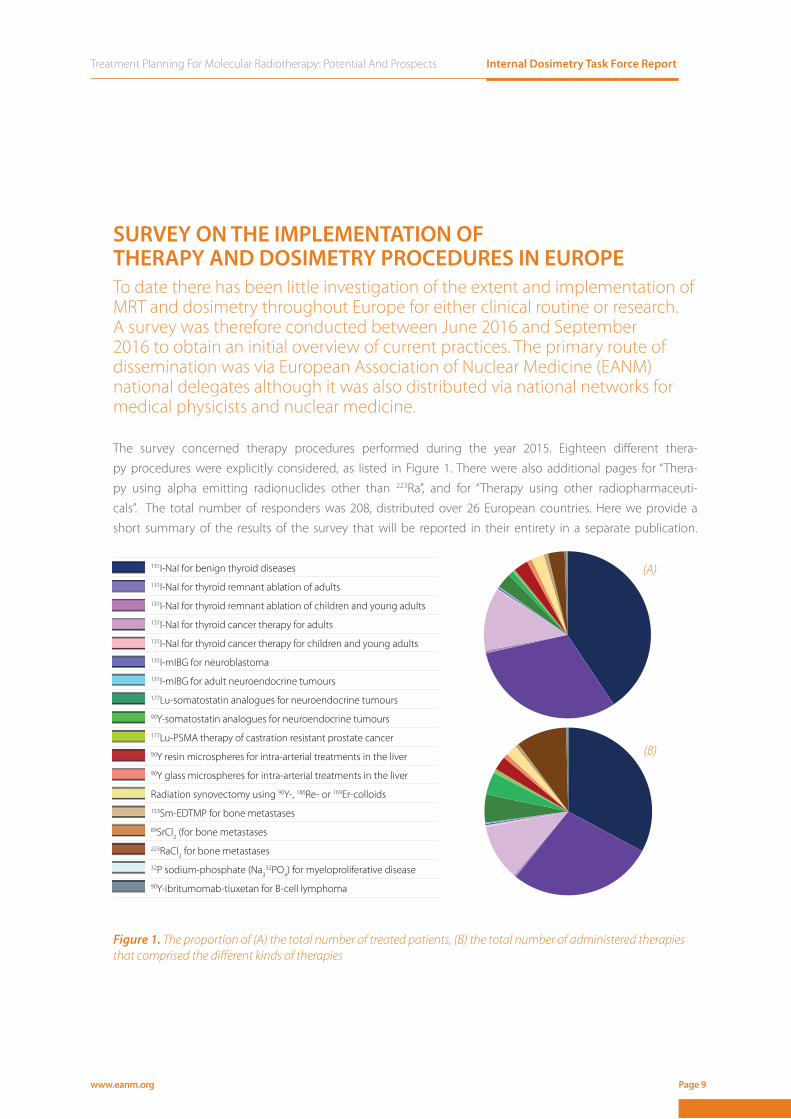
SURVEY ON THE IMPLEMENTATION OF THERAPY AND DOSIMETRY PROCEDURES IN EUROPETo date there has been little investigation of the extent and implementation of MRT and dosimetry throughout Europe for either clinical routine or research. A survey was therefore conducted between June 2016 and September 2016 to obtain an initial overview of current practices. The primary route of dissemination was via European Association of Nuclear Medicine (EANM) national delegates although it was also distributed via national networks for medical physicists and nuclear medicine.
The survey concerned therapy procedures performed during the year 2015. Eighteen diff erent thera-
py procedures were explicitly considered, as listed in Figure 1. There were also additional pages for “Thera-
py using alpha emitting radionuclides other than 223Ra”, and for “Therapy using other radiopharmaceuti-
cals”. The total number of responders was 208, distributed over 26 European countries. Here we provide a
short summary of the results of the survey that will be reported in their entirety in a separate publication.
131I-NaI for benign thyroid diseases131I-NaI for thyroid remnant ablation of adults131I-NaI for thyroid remnant ablation of children and young adults131I-NaI for thyroid cancer therapy for adults131I-NaI for thyroid cancer therapy for children and young adults131I-mIBG for neuroblastoma 131I-mIBG for adult neuroendocrine tumours177Lu-somatostatin analogues for neuroendocrine tumours90Y-somatostatin analogues for neuroendocrine tumours177Lu-PSMA therapy of castration resistant prostate cancer90Y resin microspheres for intra-arterial treatments in the liver90Y glass microspheres for intra-arterial treatments in the liver
Radiation synovectomy using 90Y-, 186Re- or 169Er-colloids153Sm-EDTMP for bone metastases89SrCl2 (for bone metastases223RaCl2 for bone metastases32P sodium-phosphate (Na3
32PO4) for myeloproliferative disease90Y-ibritumomab-tiuxetan for B-cell lymphoma
Figure 1. The proportion of (A) the total number of treated patients, (B) the total number of administered therapies that comprised the diff erent kinds of therapies
(A)
(B)

www.eanm.orgPage 10
Internal Dosimetry Task Force Report Treatment Planning For Molecular Radiotherapy: Potential And Prospects
NUMBER OF PATIENTS AND NUMBER OF TREATMENTS Figure 1 shows the proportion of the number of treated patients and the number of administered therapies
that comprised the different procedures in 2015. In total, in all countries and the 208 centres, 34,838 patients
were treated with a total number of administrations of 42,853, as a result of some procedures being repeated.
Therapies involving 131I represented 84% of the treated patients, and 71% of the number of treatments given. Of
the total treatments, 11% consisted of 177Lu/90Y somatostatin receptor PRRT or 177Lu PSMA, and 10% of 223RaCl2.
The therapies that were most disseminated among the countries were those involving 131I-NaI for the treatment
of benign thyroid diseases and for thyroid ablation of adults, which together comprised 71% of the treated pa-
tients and 60% of the given treatments.
INVOLVEMENT OF MEDICAL PHYSICIST The level of involvement of a medical physicist was asked for, and in 68% of the cases a medical physicist was
always involved or involved in the majority of treatments. In the remaining 32% of cases a medical physicist was
never involved or involved in a minority of treatments. Responses above 80% were obtained for 177Lu PSMA ther-
apy of castration resistant prostate cancer, 90Y somatostatin receptor PRRT, 32P sodium-phosphate for myelop-
roliferative diseases, 131I mIBG for neuroblastoma, and 90Y microspheres. It is worth noting that a 100% response
was not obtained for any procedure.
POST-THERAPY IMAGING Post-therapy imaging was performed always or in the majority of treatments in 69% of the cases. However, more
than 50% of the centres reported that post-therapy imaging was never performed, or performed in the minority
of cases for therapies such as 131I NaI for benign thyroid diseases, radiation synovectomy89SrCl2 or 223RaCl2 for
bone metastases, 32P phosphate for myeloproliferative diseases, and 90Y-ibritumomab-tiuxetan for B cell lym-
phoma.
Treatment Planning For Molecular Radiotherapy: Potential And Prospects Internal Dosimetry Task Force Report
www.eanm.org Page 11
ABSORBED-DOSE PLANNINGThe absorbed dose was reported to be individually planned for each patient either always or in the majority of
treatments in only 36% of cases. In 63% of cases, absorbed dose planning was never carried out, or carried out
in a minority of treatments. The highest number of responses were obtained for 90Y-labeled microspheres, 82%
(resin) and 84% (glass), and for 131I-NaI for benign thyroid diseases (54%).
POST-THERAPY DOSIMETRYPost-therapy dosimetry was performed always or in the majority of treatments in only 26% of the cases. More
than 50% of the centres indicated that post-therapy dosimetry was performed always or in the majority of cases
for 177Lu PSMA (100%) and 131I mIBG for neuroblastoma (59%). For PRRT with 90Y or 177Lu and 131I mIBG for adult
neuroendocrine tumours this percentage was approximately 40%.
SATISFACTION ON THE IMPLEMENTATION OF PATIENT-SPECIFIC DOSIMETRY Fifty-five percent of the responders indicated that they were not satisfied with the current implementation of
patient-specific dosimetry in their centre. The main limiting factors were identified as: “Shortage of knowledge
and know-how”, “Shortage of medical physicists working in nuclear medicine”, “Shortage of other staff”, “Limited
access to scanner or other equipment needed”, “Limited access to dedicated software”, with an approximately
equal distribution of responses. It is interesting to note that 12% of participating centres identified the “Lack of
legislative requirement to perform dosimetry” as the main limiting factor.
CONCLUSIONThe results of this survey indicate the need for central registries for MRT and for the implementation of dosim-
etry. Although the level of response varied between countries, the results nevertheless demonstrated a lack of
harmonisation and implementation of individual-patient based internal dosimetry.
Internal Dosimetry Task Force Report Treatment Planning For Molecular Radiotherapy: Potential And Prospects
www.eanm.orgPage 12
DOSIMETRY FOR THERAPY PROCEDURESAs also reflected in the survey, the complexities of Molecular Radiotherapy are exacerbated by the number of
different procedures, the range of radionuclides and radiopharmaceuticals and the wide variations in patient
status. This section reviews the main therapy procedures currently in use. While the goal of the survey was to
provide a report as complete as possible on the implementation of dosimetry for a wide range of therapy proce-
dures performed in Europe, this section by necessity covers in detail only a sub-set of such therapies. However,
conclusions drawn here may be readily applied to other radiopharmaceuticals using the same radionuclides.
The procedures described for imaging and dosimetry are equally applicable to verification of the absorbed
doses delivered to tumours, target volumes and healthy tissues.
For each section a brief introduction is given, followed by the current effectiveness of the treatment, the poten-
tial for quantitative imaging that underpins organ and tumour dosimetry and existing evidence for absorbed
dose-effect correlations. The potential for personalised dosimetry-based treatment planning is then considered.
Finally, issues specific to the treatment are considered along with questions that merit further investigation.

Treatment Planning For Molecular Radiotherapy: Potential And Prospects Internal Dosimetry Task Force Report
www.eanm.org Page 13
1111131I NaI for the treatment of benign thyroid disease
DOSIMETRY FOR THERAPY PROCEDURES

Internal Dosimetry Task Force Report Treatment Planning For Molecular Radiotherapy: Potential And Prospects
www.eanm.orgPage 14
INTRODUCTIONOral administration of 131I for benign thyroid disease (hyperthyroidism) has been carried out since 1941, when
the first effective therapy was performed by Saul Hertz (1). Hyperthyroidism is a consequence of an excessive
production and secretion of thyroid hormones T3 and T4. The main causes of hyperthyroidism which are a
clear indication for radioiodine treatment include autoimmune hyperthyroidism (Graves’ disease), solitary hy-
perfunctioning thyroid nodule (autonomous adenoma), and multinodular goitre. Administration of 131I NaI is
not the only treatment and other options, such as administration of anti-thyroid drugs and surgery are usually
considered (2). Procedure guidelines given by the EANM (3) and the Society of Nuclear Medicine and Molecular
Imaging (SNMIM) (4) are available to advise clinicians on how to perform the treatment of benign thyroid dis-
ease with 131I NaI.
EFFECTIVENESSThe aim of radioiodine therapy for Graves’ disease, autonomous adenoma and toxic multinodular goitre is that
patients achieve a non-hyperthyroid condition. This means that patients may become euthyroid or hypothyroid,
which is compensated with the administration of L-thyroxine. In the case of nontoxic multinodular goitre, the
main aim is the reduction of the thyroid volume. The goal of radioiodine therapy – elimination of hyperthyroid-
ism and shrinkage of thyroid volume – is achieved in 80% of patients regardless of the approach to administra-
tion used. For calculated activities, success rates for radioiodine treatment have been reported to be higher (3-5).
IMAGINGRadioiodine uptake is usually imaged with anterior gamma-camera imaging using a high-energy parallel-hole
collimator. The count rate is increased with a thick crystal (1/2 inch or 5/8 inch) (6). Corrections for scatter and
camera dead-time may be necessary (7). Quantitative SPECT/CT 131I imaging is not common practice but is
possible, and can offer accurate quantification (7). To determine the target mass, ultrasound imaging is recom-
mended (3, 5), although an anterior view after administering 99mTc pertechnetate is also used.
ORGANS AT RISK AND NORMAL TISSUE DOSIMETRY Within the range of administered activities of 131I NaI, low absorbed doses to normal tissues are delivered (4).
Thus, normal tissue dosimetry is not usually required.
TARGET DOSIMETRY In some cases fixed activities are delivered, whereas in other cases administered activities are calculated with
different methods:
(1) Measurement of thyroidal volume and/or radioiodine uptake measurement after 24 h.
(2) Measurement of thyroidal volume, radioiodine uptake, and individual radioiodine half-life by at least two
uptake measurements, for example, after 24 h and 5 days.
To determine the absorbed dose to the target, the Quimby-Marinelli method has been widely used (8). More-
over, the EANM Dosimetry Committee has released standard operational procedures for dosimetry prior to
radioiodine therapy (6).
Treatment Planning For Molecular Radiotherapy: Potential And Prospects Internal Dosimetry Task Force Report
www.eanm.org Page 15
ABSORBED DOSE-EFFECTTreatments aimed to deliver an absorbed dose prescribed to the target have shown high success rates (9).
Graves’ disease
The success rate has proved to be dependent on the absorbed dose prescribed to the target (10, 11). Moreover,
function-orientated radioiodine treatments have aimed to deliver an absorbed dose to achieve euthyroidism. A
common approach is to deliver an ablative absorbed dose to the thyroid (12).
Autonomous adenoma
Delivery of absorbed doses of 300 Gy and 400 Gy to the solitary hyperfunctioning nodule has shown similar
high success rates (> 90%) in the elimination of its functional autonomy (13). Multinodular goitre
In toxic multinodular goitre, intended absorbed doses above 150 Gy have resulted in success rates higher than
90% (14, 15). Cure rates could be maintained with absorbed doses around 120 Gy (15). In the case of nontoxic
multinodular goitre a notable volume reduction was observed (>50%) (16).
DOSIMETRY-BASED TREATMENT PLANNINGDetermination of the activity to administer in order to deliver a prescribed absorbed dose to the target is fea-
sible, and has been widely reported (3). The target mass can be determined by ultrasound. Following the ad-
ministration of a tracer of 131I NaI, thyroid uptake with time has to be assessed (6). If the uptake is determined
with a thyroid probe, 2 MBq are sufficient, and up to 10 MBq may be needed if a gamma camera is used. Higher
activities are not recommended due to the so-called ‘stunning’ effect (6). The potential for semi-individualised
treatment planning has been investigated using a mean half-life and patient specific uptake values (17). A mod-
el to calculate the optimal absorbed dose to deliver based on the normal tissue complication probability (NTCP)
was developed and verified (18).
ISSUES TO CONSIDERConventionally, 2D dosimetry has been the standard procedure. However, SPECT/CT acquisitions are widely
available nowadays, which enables 3D dosimetry. Moreover, guidelines for SPECT dosimetry with radioiodine
following the MIRD formalism are available (7).
NEED FOR INVESTIGATIONFurther evaluation of the optimal absorbed doses is warranted, including mass reduction during treatment
and possible differences in radioiodine biokinetics prior to and during therapy. The role of recombinant human
TSH (rhTSH) prior to the treatment and its potential effect on dosimetry is also in need of evaluation (19). A key
question is whether a dosimetry based approach can maximise the number of patients rendered euthyroid
rather than hypothyroid, thereby mitigating the need for lifelong medication. Multi-centre trials are necessary
to investigate this.

Internal Dosimetry Task Force Report Treatment Planning For Molecular Radiotherapy: Potential And Prospects
www.eanm.orgPage 16
Tc-99m image of thyroid prior to treatment of Graves’ disease with radioiodine

Treatment Planning For Molecular Radiotherapy: Potential And Prospects Internal Dosimetry Task Force Report
www.eanm.org Page 17
References1. Sawin CT and Becker DV, Radioiodine and the treatment of hyperthyroidism: the early history. Thyroid 1997; 7: 163-176.2. Nyström E, Berg GEB, Jansson SKG, et al., Thyroid disease in adults. 2011.3. Stokkel MP, Handkiewicz Junak D, Lassmann M, et al., EANM procedure guidelines for therapy of benign thyroid disease.
Eur J Nucl Med Mol Imaging 2010; 37: 2218-2228.4. Silberstein EB, Alavi A, Balon HR, et al., The SNMMI practice guideline for therapy of thyroid disease with 131I 3.0. J Nucl
Med 2012; 53: 1633-1651.5. Dietlein M, Grunwald F, Schmidt M, et al., [Radioiodine therapy for benign thyroid diseases (version 5). German Guide-
line]. Nuklearmedizin 2016; 55: 213-220.6. Hanscheid H, Canzi C, Eschner W, et al., EANM Dosimetry Committee series on standard operational procedures for
pre-therapeutic dosimetry II. Dosimetry prior to radioiodine therapy of benign thyroid diseases. Eur J Nucl Med Mol Imaging 2013; 40: 1126-1134.
7. Dewaraja YK, Ljungberg M, Green AJ, et al., MIRD pamphlet No. 24: Guidelines for quantitative 131I SPECT in dosimetry applications. J Nucl Med 2013; 54: 2182-2188.
8. Marinelli LD, Quimby EH and Hine GJ, Dosage determination with radioactive isotopes; practical considerations in the-rapy and protection. Am J Roentgenol Radium Ther 1948; 59: 260-281.
9. Salvatori M and Luster M, Radioiodine therapy dosimetry in benign thyroid disease and differentiated thyroid carcinoma. Eur J Nucl Med Mol Imaging 2010; 37: 821-828.
10. Dunkelmann S, Neumann V, Staub U, et al., [Results of a risk adapted and functional radioiodine therapy in Graves’ di-sease]. Nuklearmedizin 2005; 44: 238-242.
11. Reinhardt MJ, Brink I, Joe AY, et al., Radioiodine therapy in Graves’ disease based on tissue-absorbed dose calculations: effect of pre-treatment thyroid volume on clinical outcome. Eur J Nucl Med Mol Imaging 2002; 29: 1118-1124.
12. Kobe C, Eschner W, Sudbrock F, et al., Graves’ disease and radioiodine therapy. Is success of ablation dependent on the achieved dose above 200 Gy? Nuklearmedizin 2008; 47: 13-17.
13. Reinhardt MJ, Biermann K, Wissmeyer M, et al., Dose selection for radioiodine therapy of borderline hyperthyroid pa-tients according to thyroid uptake of 99mTc-pertechnetate: applicability to unifocal thyroid autonomy? Eur J Nucl Med Mol Imaging 2006; 33: 608-612.
14. Dunkelmann S, Endlicher D, Prillwitz A, et al., [Results of TcTUs-optimized radioiodine therapy in multifocal and dissemi-nated autonomy]. Nuklearmedizin 1999; 38: 131-139.
15. Kahraman D, Keller C, Schneider C, et al., Development of hypothyroidism during long-term follow-up of patients with toxic nodular goitre after radioiodine therapy. Clin Endocrinol (Oxf ) 2012; 76: 297-303.
16. Bachmann J, Kobe C, Bor S, et al., Radioiodine therapy for thyroid volume reduction of large goitres. Nucl Med Commun 2009; 30: 466-471.
17. Kobe C, Eschner W, Wild M, et al., Radioiodine therapy of benign thyroid disorders: what are the effective thyroidal half-life and uptake of 131I? Nucl Med Commun 2010; 31: 201-205.
18. Strigari L, Sciuto R, Benassi M, et al., A NTCP approach for estimating the outcome in radioiodine treatment of hyperthy-roidism. Med Phys 2008; 35: 3903-3910.
19. Cohen O, Ilany J, Hoffman C, et al., Low-dose recombinant human thyrotropin-aided radioiodine treatment of large,
multinodular goiters in elderly patients. Eur J Endocrinol 2006; 154: 243-252.

Internal Dosimetry Task Force Report Treatment Planning For Molecular Radiotherapy: Potential And Prospects
www.eanm.orgPage 18

Treatment Planning For Molecular Radiotherapy: Potential And Prospects Internal Dosimetry Task Force Report
www.eanm.org Page 19
131I NaI for the treatment of diff erentiated thyroid cancer (DTC) with ablative intent and in the case of recurrent disease 2I NaI for the treatment 2I NaI for the treatment of diff erentiated thyroid 2of diff erentiated thyroid cancer (DTC) with ablative 2cancer (DTC) with ablative intent and in the case of 2intent and in the case of 2cancer (DTC) with ablative 2cancer (DTC) with ablative
I NaI for the treatment of diff erentiated thyroid cancer (DTC) with ablative intent and in the case of recurrent disease 2I NaI for the treatment of diff erentiated thyroid cancer (DTC) with ablative intent and in the case of 22cancer (DTC) with ablative 2cancer (DTC) with ablative
DOSIMETRY FOR THERAPY PROCEDURES

Internal Dosimetry Task Force Report Treatment Planning For Molecular Radiotherapy: Potential And Prospects
www.eanm.orgPage 20
INTRODUCTION Administration of 131I NaI for DTC treatment has been in common use since the 1940s. The activity to administer
is customarily determined (1). In comparison with normal thyroid cells, thyroid cancer cells have a reduced ex-
pression of the sodium/iodide symporter (NIS), which determines iodine uptake from the blood (2). Therefore,
to improve radioiodine uptake, high thyroid-stimulating hormone (TSH) levels are achieved before 131I NaI treat-
ment by hormone withdrawal or recombinant human TSH (rhTSH) administration.
EFFECTIVENESSThe effectiveness of the treatment is usually evaluated by measuring thyroglobulin levels and sometimes also
by acquiring a whole-body scan between 6 and 12 months after the treatment. High rates of success, of about
90% or higher, can be expected for treatments with ablative intent (3). In the presence of metastases, a 10-year
overall cause-specific survival of about 85% has been reported (4).
IMAGING Radioiodine uptake in thyroid remnants and metastases can be determined with anterior gamma-camera im-
aging or SPECT/CT imaging which affords more accurate quantification (5). Corrections for scatter and camera
dead-time may be necessary (5). PET/CT imaging can also be performed using 124I NaI, which may advantageous
for determination of remnant mass and small metastases (6).
ORGANS AT RISK AND NORMAL TISSUE DOSIMETRY In the treatment of recurrent disease, when high activities are given, red-marrow absorbed dose may be of con-
cern (4, 7). EANM guidelines recommend determining red-marrow absorbed dose from whole-body and blood
dosimetry (7), and a constraint of 2 Gy is considered when using this methodology (8). Pneumonitis and pulmo-
nary fibrosis are possible concerns in the presence of diffuse lung metastases, and absorbed dose-rate methods
have been suggested based on the whole-body retention threshold of 2.96 GBq at 48 hours post-treatment (9).
Salivary glands may show side-effects such as sialadenitis and xerostomia, which are markedly less frequent for
the case of ablative treatments (10). Currently, there is no absorbed-dose constraint reported for the salivary
glands, although toxicity has been reported for notably lower absorbed doses than the constraints used in
external beam radiotherapy (11).
TUMOUR DOSIMETRY Several studies, most of them acquiring planar images, have reported absorbed doses to remnants and metas-
tases with values ranging from <10 Gy to 1000 Gy (12-15).
Treatment Planning For Molecular Radiotherapy: Potential And Prospects Internal Dosimetry Task Force Report
www.eanm.org Page 21
ABSORBED DOSE-EFFECTSeveral studies have investigated correlations between the absorbed doses delivered to remnants and metas-
tases and response, resulting in a wide range of absorbed doses to achieve a successful ablation. For instance,
absorbed doses to remnants higher than 49 Gy (12), 90 Gy (15) and 300 Gy (14) have been reported. For metas-
tases, absorbed doses of over 40 Gy (15) and 80 Gy (14) have been documented.
DOSIMETRY-BASED TREATMENT PLANNINGCurrently, in the treatment of DTC there are no well-established values for absorbed doses to remnants and
metastases which may be used as prescription values. Thus, this treatment is performed by administration of a
fixed activity of 131I NaI ranging between 1.11 GBq and 7.4 GBq, depending on the stage of the disease. In the
case of recurrent disease, the administered activity may be calculated based on the absorbed dose constraints
of the normal tissue (usually red marrow) (7). Initial pre-therapy planning would require a tracer activity of 131I
NaI or 124I NaI (7, 15).
ISSUES TO CONSIDERDetermination of radioiodine uptake and mass of remnants and of small metastases is a challenging task. Re-
covery coefficients and thresholding techniques can be used in SPECT/CT imaging (16). The superior resolution
of 124I PET/CT imaging may be an alternative way to estimate these values (15). Treatments performed after
administration of rhTSH show different radioiodine biokinetics from those performed after thyroid hormone
withdrawal (17), which may have to be considered if treatment planning is performed. For iodine-positive bone
metastases, pronounced intra-tumour non-uniformity of iodine uptake may affect dose-response relationships
if mean absorbed dose values are used (18). In the case of patients with metastatic disease, when high activities
are delivered, appropriate radiation protection measures should be undertaken if patients are moved from the
isolation rooms to the gamma camera to acquire images.
NEED FOR INVESTIGATIONAlthough an absorbed dose-effect correlation has been reported in single-centre studies, the outcome of the
treatment can be successful in patients who receive a very low remnant-absorbed dose of only a few Gy (12, 15).
This situation calls for further investigation of the influence on the treatment outcome of other factors, such as
the stage of the disease, the surgery procedure followed, and the remnant’s mass. Multi-centre clinical trials are
necessary to gather the evidence required to optimise treatments. The stunning effect has been described as a
decrease in the uptake of 131I NaI during the therapeutic course after a previous administration of a tracer of 131I
NaI (13). However, there is no completely accepted explanation for this effect (19) and further investigation is
warranted. The use of external beam radiotherapy as an adjuvant therapy of the radioiodine therapy in the case
of metastatic disease is also an issue to be investigated (20).
Internal Dosimetry Task Force Report Treatment Planning For Molecular Radiotherapy: Potential And Prospects
www.eanm.orgPage 22
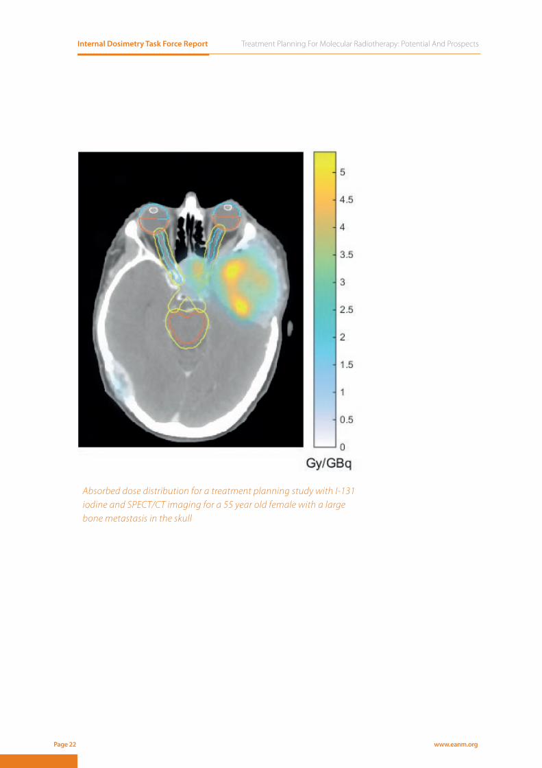
Absorbed dose distribution for a treatment planning study with I-131 iodine and SPECT/CT imaging for a 55 year old female with a large bone metastasis in the skull

Treatment Planning For Molecular Radiotherapy: Potential And Prospects Internal Dosimetry Task Force Report
www.eanm.org Page 23
References1. Silberstein EB, Alavi A, Balon HR, et al., The SNMMI practice guideline for therapy of thyroid disease with 131I 3.0. J Nucl
Med 2012; 53: 1633-1651.2. Spitzweg C, Harrington KJ, Pinke LA, et al., Clinical review 132: The sodium iodide symporter and its potential role in
cancer therapy. J Clin Endocrinol Metab 2001; 86: 3327-3335.3. Pacini F, Ladenson PW, Schlumberger M, et al., Radioiodine ablation of thyroid remnants after preparation with recombi-
nant human thyrotropin in differentiated thyroid carcinoma: results of an international, randomized, controlled study. J Clin Endocrinol Metab 2006; 91: 926-932.
4. Luster M, Clarke SE, Dietlein M, et al., Guidelines for radioiodine therapy of differentiated thyroid cancer. Eur J Nucl Med Mol Imaging 2008; 35: 1941-1959.
5. Dewaraja YK, Ljungberg M, Green AJ, et al., MIRD pamphlet No. 24: Guidelines for quantitative 131I SPECT in dosimetry applications. J Nucl Med 2013; 54: 2182-2188.
6. Nagarajah J, Jentzen W, Hartung V, et al., Diagnosis and dosimetry in differentiated thyroid carcinoma using 124I PET: comparison of PET/MRI vs PET/CT of the neck. Eur J Nucl Med Mol Imaging 2011; 38: 1862-1868.
7. Lassmann M, Hanscheid H, Chiesa C, et al., EANM Dosimetry Committee series on standard operational procedures for pre-therapeutic dosimetry I: blood and bone marrow dosimetry in differentiated thyroid cancer therapy. Eur J Nucl Med Mol Imaging 2008; 35: 1405-1412.
8. Benua RS, Cicale NR, Sonenberg M, et al., The relation of radioiodine dosimetry to results and complications in the treat-ment of metastatic thyroid cancer. Am J Roentgenol Radium Ther Nucl Med 1962; 87: 171-182.
9. Sgouros G, Song H, Ladenson PW, et al., Lung toxicity in radioiodine therapy of thyroid carcinoma: development of a dose-rate method and dosimetric implications of the 80-mCi rule. J Nucl Med 2006; 47: 1977-1984.
10. Jeong SY, Kim HW, Lee SW, et al., Salivary gland function 5 years after radioactive iodine ablation in patients with diffe-rentiated thyroid cancer: direct comparison of pre- and postablation scintigraphies and their relation to xerostomia symptoms. Thyroid 2013; 23: 609-616.
11. Liu B, Huang R, Kuang A, et al., Iodine kinetics and dosimetry in the salivary glands during repeated courses of radioio-dine therapy for thyroid cancer. Med Phys 2011; 38: 5412-5419.
12. Flux GD, Haq M, Chittenden SJ, et al., A dose-effect correlation for radioiodine ablation in differentiated thyroid cancer. Eur J Nucl Med Mol Imaging 2010; 37: 270-275.
13. Lassmann M, Luster M, Hanscheid H, et al., Impact of 131I diagnostic activities on the biokinetics of thyroid remnants. J Nucl Med 2004; 45: 619-625.
14. Maxon HR, Thomas SR, Hertzberg VS, et al., Relation between effective radiation dose and outcome of radioiodine the-rapy for thyroid cancer. N Engl J Med 1983; 309: 937-941.
15. Wierts R, Brans B, Havekes B, et al., Dose-Response Relationship in Differentiated Thyroid Cancer Patients Undergoing Radioiodine Treatment Assessed by Means of 124I PET/CT. J Nucl Med 2016; 57: 1027-1032.
16. Minguez P, Flux G, Genolla J, et al., Whole-remnant and maximum-voxel SPECT/CT dosimetry in 131 I-NaI treatments of differentiated thyroid cancer. Med Phys 2016; 43: 5279-5287.
17. Hanscheid H, Lassmann M, Luster M, et al., Iodine biokinetics and dosimetry in radioiodine therapy of thyroid cancer: procedures and results of a prospective international controlled study of ablation after rhTSH or hormone withdrawal. J Nucl Med 2006; 47: 648-654.
18. Jentzen W, Verschure F, van Zon A, et al., 124I PET Assessment of Response of Bone Metastases to Initial Radioiodine Treatment of Differentiated Thyroid Cancer. J Nucl Med 2016; 57: 1499-1504.
19. McDougall IR and Iagaru A, Thyroid stunning: fact or fiction? Semin Nucl Med 2011; 41: 105-112.20. Mikalsen LTG, Arnesen MR, Bogsrud TV, et al., Combining radioiodine and external beam radiation therapy: the potential
of integrated treatment planning for differentiated thyroid cancer. Acta Oncol 2017; 56: 894-897.

Internal Dosimetry Task Force Report Treatment Planning For Molecular Radiotherapy: Potential And Prospects
www.eanm.orgPage 24
Treatment Planning For Molecular Radiotherapy: Potential And Prospects Internal Dosimetry Task Force Report
www.eanm.org Page 25
DOSIMETRY-BASED TREATMENT PLANNINGCurrently, in the treatment of DTC there are no well-established values for absorbed doses to remnants and metastases which may be
used as prescription values. Thus, this treatment is performed by administration of a fi xed activity of 131I NaI ranging between 1.11 GBq
131I mIBG for the treatment of neuro-blastoma in children and young adults3I mIBG for the treatment of neuro-blastoma in children and young adults3I mIBG for the treatment of neuro-blastoma in children and young adults33DOSIMETRY FOR THERAPY PROCEDURES
Internal Dosimetry Task Force Report Treatment Planning For Molecular Radiotherapy: Potential And Prospects
www.eanm.orgPage 26
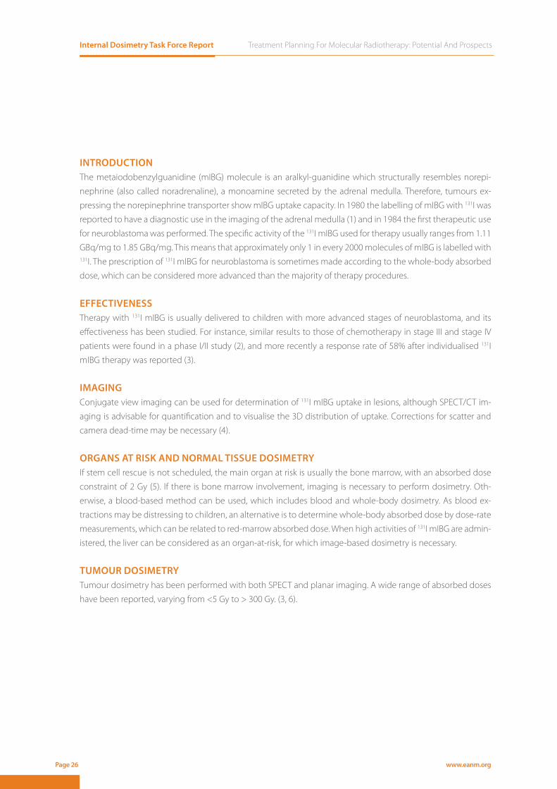
INTRODUCTION The metaiodobenzylguanidine (mIBG) molecule is an aralkyl-guanidine which structurally resembles norepi-
nephrine (also called noradrenaline), a monoamine secreted by the adrenal medulla. Therefore, tumours ex-
pressing the norepinephrine transporter show mIBG uptake capacity. In 1980 the labelling of mIBG with 131I was
reported to have a diagnostic use in the imaging of the adrenal medulla (1) and in 1984 the first therapeutic use
for neuroblastoma was performed. The specific activity of the 131I mIBG used for therapy usually ranges from 1.11
GBq/mg to 1.85 GBq/mg. This means that approximately only 1 in every 2000 molecules of mIBG is labelled with 131I. The prescription of 131I mIBG for neuroblastoma is sometimes made according to the whole-body absorbed
dose, which can be considered more advanced than the majority of therapy procedures.
EFFECTIVENESSTherapy with 131I mIBG is usually delivered to children with more advanced stages of neuroblastoma, and its
effectiveness has been studied. For instance, similar results to those of chemotherapy in stage III and stage IV
patients were found in a phase I/II study (2), and more recently a response rate of 58% after individualised 131I
mIBG therapy was reported (3).
IMAGING Conjugate view imaging can be used for determination of 131I mIBG uptake in lesions, although SPECT/CT im-
aging is advisable for quantification and to visualise the 3D distribution of uptake. Corrections for scatter and
camera dead-time may be necessary (4).
ORGANS AT RISK AND NORMAL TISSUE DOSIMETRY If stem cell rescue is not scheduled, the main organ at risk is usually the bone marrow, with an absorbed dose
constraint of 2 Gy (5). If there is bone marrow involvement, imaging is necessary to perform dosimetry. Oth-
erwise, a blood-based method can be used, which includes blood and whole-body dosimetry. As blood ex-
tractions may be distressing to children, an alternative is to determine whole-body absorbed dose by dose-rate
measurements, which can be related to red-marrow absorbed dose. When high activities of 131I mIBG are admin-
istered, the liver can be considered as an organ-at-risk, for which image-based dosimetry is necessary.
TUMOUR DOSIMETRY Tumour dosimetry has been performed with both SPECT and planar imaging. A wide range of absorbed doses
have been reported, varying from <5 Gy to > 300 Gy. (3, 6).
Treatment Planning For Molecular Radiotherapy: Potential And Prospects Internal Dosimetry Task Force Report
www.eanm.org Page 27
ABSORBED DOSE-EFFECTA correlation between the tumour absorbed dose and the response to treatment has been reported. Progres-
sive disease was seen only in those patients whose tumours received less than 17 Gy and the partial response
was much higher in those receiving more than 70 Gy (7). With regard to toxicity, a correlation between the
whole-body absorbed dose and neutropenia has been shown (8).
DOSIMETRY-BASED TREATMENT PLANNINGTreatments are often prescribed according to a whole-body absorbed dose. An increasingly common protocol is
to deliver a whole-body absorbed dose of 4 Gy in two administrations of activity separated by 2 weeks, followed by
a stem cell rescue. The first administration is delivered according to a body mass-based prescription of 444 MBq/kg
(9). If there is no stem cell rescue available, a 131I mIBG tracer study may be performed to deliver a given red-marrow
or whole-body absorbed dose. There are to date no prescription values for the tumour absorbed dose.
ISSUES TO CONSIDER123I mIBG imaging is often performed for diagnostic assessment before therapy, and using this scan could be
considered for treatment planning.’ Before performing SPECT/CT based dosimetry, it may be necessary to per-
form a whole-body scan to determine all the uptake regions during the 131I mIBG treatment. Patients are hos-
pitalised in isolation rooms, so if patients are moved to the gamma camera room during the isolation period,
appropriate radiation protection measures should be undertaken. For the elimination of the 131I mIBG from the
whole body, three to five phases can be considered (8). For tumour and liver dosimetry the uptake phase and
possibly several washout phases should be taken into account. Therefore, dose-rate measurements and gamma
camera acquisitions have to be well distributed through the whole washout of the 131I mIBG.
NEED FOR INVESTIGATIONNumerous studies have demonstrated that quantitative imaging of 131I and tumour dosimetry is feasible, which
may lead to treatment regimens based on the absorbed doses delivered to tumours and to organs-at-risk, as is
routine for external beam radiotherapy. A deeper radiobiological knowledge may also help to establish those
treatment regimens. Further possibilities for development include the use of no-carrier added mIBG with a no-
tably higher specific activity, which has been shown to improve the tumour uptake of radiolabelled molecules,
and combination treatments of 131I mIBG with chemotherapy or external beam radiotherapy.

Internal Dosimetry Task Force Report Treatment Planning For Molecular Radiotherapy: Potential And Prospects
www.eanm.orgPage 28
References1. Wieland DM, Wu J, Brown LE, et al., Radiolabeled adrenergi neuron-blocking agents: adrenomedullary imaging with
[131I]iodobenzylguanidine. J Nucl Med 1980; 21: 349-353.2. Lashford LS, Lewis IJ, Fielding SL, et al., Phase I/II study of iodine 131 metaiodobenzylguanidine in chemoresistant neu-
roblastoma: a United Kingdom Children’s Cancer Study Group investigation. J Clin Oncol 1992; 10: 1889-1896.3. George SL, Falzone N, Chittenden S, et al., Individualized 131I-mIBG therapy in the management of refractory and re-
lapsed neuroblastoma. Nucl Med Commun 2016; 37: 466-472.4. Dewaraja YK, Ljungberg M, Green AJ, et al., MIRD pamphlet No. 24: Guidelines for quantitative 131I SPECT in dosimetry
applications. J Nucl Med 2013; 54: 2182-2188.5. Chiesa C, Castellani R, Mira M, et al., Dosimetry in 131I-mIBG therapy: moving toward personalized medicine. Q J Nucl
Med Mol Imaging 2013; 57: 161-170.6. Buckley SE, Saran FH, Gaze MN, et al., Dosimetry for fractionated (131)I-mIBG therapies in patients with primary resistant
high-risk neuroblastoma: preliminary results. Cancer Biother Radiopharm 2007; 22: 105-112.7. Matthay KK, Panina C, Huberty J, et al., Correlation of tumor and whole-body dosimetry with tumor response and toxicity
in refractory neuroblastoma treated with (131)I-MIBG. J Nucl Med 2001; 42: 1713-1721.8. Buckley SE, Chittenden SJ, Saran FH, et al., Whole-body dosimetry for individualized treatment planning of 131I-MIBG
radionuclide therapy for neuroblastoma. J Nucl Med 2009; 50: 1518-1524.9. Gaze MN, Chang YC, Flux GD, et al., Feasibility of dosimetry-based high-dose 131I-meta-iodobenzylguanidine with topo-
tecan as a radiosensitizer in children with metastatic neuroblastoma. Cancer Biother Radiopharm 2005; 20: 195-199.
CT and I-123 SPECT of neuroblastoma

Treatment Planning For Molecular Radiotherapy: Potential And Prospects Internal Dosimetry Task Force Report
www.eanm.org Page 29
131I mIBG for the treatment of neuro-endocrine tumours in adults 4I mIBG for the treatment of neuro-endocrine tumours in adults 4I mIBG for the treatment of neuro-endocrine tumours 44DOSIMETRY FOR THERAPY PROCEDURES
Internal Dosimetry Task Force Report Treatment Planning For Molecular Radiotherapy: Potential And Prospects
www.eanm.orgPage 30
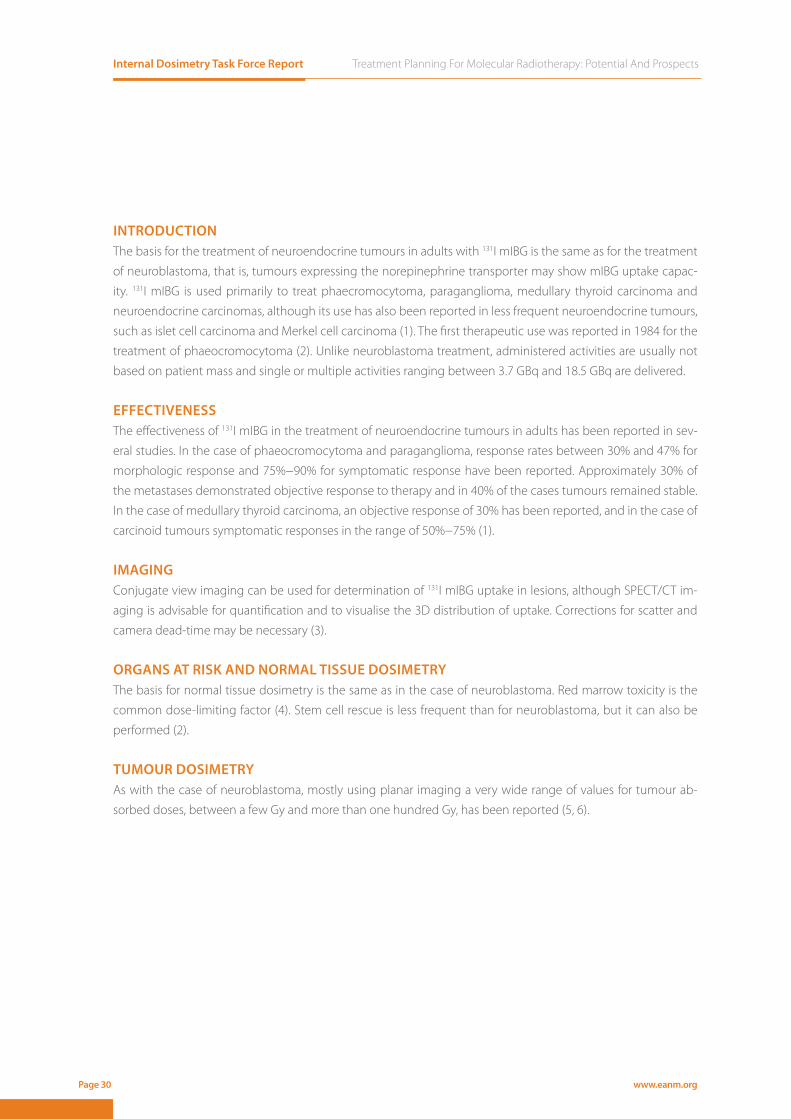
INTRODUCTION The basis for the treatment of neuroendocrine tumours in adults with 131I mIBG is the same as for the treatment
of neuroblastoma, that is, tumours expressing the norepinephrine transporter may show mIBG uptake capac-
ity. 131I mIBG is used primarily to treat phaecromocytoma, paraganglioma, medullary thyroid carcinoma and
neuroendocrine carcinomas, although its use has also been reported in less frequent neuroendocrine tumours,
such as islet cell carcinoma and Merkel cell carcinoma (1). The first therapeutic use was reported in 1984 for the
treatment of phaeocromocytoma (2). Unlike neuroblastoma treatment, administered activities are usually not
based on patient mass and single or multiple activities ranging between 3.7 GBq and 18.5 GBq are delivered.
EFFECTIVENESSThe effectiveness of 131I mIBG in the treatment of neuroendocrine tumours in adults has been reported in sev-
eral studies. In the case of phaeocromocytoma and paraganglioma, response rates between 30% and 47% for
morphologic response and 75%−90% for symptomatic response have been reported. Approximately 30% of
the metastases demonstrated objective response to therapy and in 40% of the cases tumours remained stable.
In the case of medullary thyroid carcinoma, an objective response of 30% has been reported, and in the case of
carcinoid tumours symptomatic responses in the range of 50%−75% (1).
IMAGING Conjugate view imaging can be used for determination of 131I mIBG uptake in lesions, although SPECT/CT im-
aging is advisable for quantification and to visualise the 3D distribution of uptake. Corrections for scatter and
camera dead-time may be necessary (3).
ORGANS AT RISK AND NORMAL TISSUE DOSIMETRY The basis for normal tissue dosimetry is the same as in the case of neuroblastoma. Red marrow toxicity is the
common dose-limiting factor (4). Stem cell rescue is less frequent than for neuroblastoma, but it can also be
performed (2).
TUMOUR DOSIMETRY As with the case of neuroblastoma, mostly using planar imaging a very wide range of values for tumour ab-
sorbed doses, between a few Gy and more than one hundred Gy, has been reported (5, 6).

Treatment Planning For Molecular Radiotherapy: Potential And Prospects Internal Dosimetry Task Force Report
www.eanm.org Page 31
ABSORBED DOSE-EFFECTIn a study for the treatment of the phaeocromocytoma, an absorbed dose higher than 150 Gy was considered
necessary to cause beneficial effects (5). Moreover, for the higher administered activities a better response has
been observed, which may possibly be explained under the assumption that higher tumour absorbed doses
were delivered (6).
DOSIMETRY-BASED TREATMENT PLANNINGConsidering an absorbed-dose limit of 2 Gy for the red marrow, a dosimetric study with a tracer could be per-
formed to determine the red-marrow absorbed dose per administered activity. Using this value, the activity
of 131I mIBG can be prescribed so as not to exceed the red marrow toxicity. Alternatively, the treatment can be
fractionated and the activity in subsequent activity administrations determined from the biokinetics of the first
administration (7). There are as yet no prescription values for the tumour absorbed dose.
ISSUES TO CONSIDERAs with the case of neuroblastoma, it may be useful to acquire a diagnostic or whole-body scan to determine
the uptake regions. Radiation protection measures must also be undertaken if an inpatient is moved to the
gamma camera, and it is also important to take into account all washout phases for whole-body, organs-at-risk
and tumour dosimetry.
NEED FOR INVESTIGATIONAs with neuroblastoma, it is necessary to investigate treatment regimens based on absorbed doses delivered to
tumours and to organs-at-risk, including concepts of radiobiology, as is routine for external beam radiotherapy.
The use of no-carrier added mIBG and combination of 131I mIBG treatments with chemotherapy or external
beam radiotherapy also needs to be investigated.

Internal Dosimetry Task Force Report Treatment Planning For Molecular Radiotherapy: Potential And Prospects
www.eanm.orgPage 32
References1. Voo S, Bucerius J and Mottaghy FM, I-131-MIBG therapies. Methods 2011; 55: 238-245.2. Gonias S, Goldsby R, Matthay KK, et al., Phase II study of high-dose [131I]metaiodobenzylguanidine therapy for patients
with metastatic pheochromocytoma and paraganglioma. J Clin Oncol 2009; 27: 4162-4168.3. Dewaraja YK, Ljungberg M, Green AJ, et al., MIRD pamphlet No. 24: Guidelines for quantitative 131I SPECT in dosimetry
applications. J Nucl Med 2013; 54: 2182-2188.4. Safford SD, Coleman RE, Gockerman JP, et al., Iodine-131 metaiodobenzylguanidine treatment for metastatic carcinoid.
Results in 98 patients. Cancer 2004; 101: 1987-1993.5. Sisson JC, Shapiro B, Beierwaltes WH, et al., Radiopharmaceutical treatment of malignant pheochromocytoma. J Nucl
Med 1984; 25: 197-206.6. Sudbrock F, Schmidt M, Simon T, et al., Dosimetry for 131I-MIBG therapies in metastatic neuroblastoma, phaeochromocy-
toma and paraganglioma. Eur J Nucl Med Mol Imaging 2010; 37: 1279-1290.7. Minguez P, Flux G, Genolla J, et al., Dosimetric results in treatments of neuroblastoma and neuroendocrine tumors with
(131)I-metaiodobenzylguanidine with implications for the activity to administer. Med Phys 2015; 42: 3969-3978.
Treatment Planning For Molecular Radiotherapy: Potential And Prospects Internal Dosimetry Task Force Report
www.eanm.org Page 33
177Lu-DOTATATE for the treatment of neuroendocrine tumours 5Lu-DOTATATE for the treatment of neuroendocrine tumours 5Lu-DOTATATE for the treatment of neuroendocrine 55DOSIMETRY FOR THERAPY PROCEDURES
Internal Dosimetry Task Force Report Treatment Planning For Molecular Radiotherapy: Potential And Prospects
www.eanm.orgPage 34
INTRODUCTION177Lu-DOTATATE is a radiolabelled somatostatin analogue developed for treatment of patients with somatostatin
receptor positive neuroendocrine tumours. Being a somatostatin analogue, 177Lu-DOTATATE is taken up by areas
of increased somatostatin receptor density. In 2005 results of the first large clinical study of 177Lu-DOTATATE were
reported (1). This was a study with a fractionated approach where patients in most cases were given 7.4 GBq four
times with a 6 to 10 weeks interval. No dose-limiting toxicity was observed and this schedule has been widely
adopted since it is considered both safe and effective. This is currently the most frequent treatment protocol; pa-
tients are treated with 4 therapy cycles with an activity of 7.4 GBq each time with concomitant infusion of ami-
no-acids to reduce renal uptake. However, protocols delivering cycles of 7.4 GBq, until a maximum prescribed
absorbed dose to the kidneys and the bone marrow is reached, are under investigation (2).
EFFECTIVENESSThe results of the randomised NETTER-1 phase III study of patients with somatostatin receptor positive midgut
neuroendocrine tumours showed that 177Lu-DOTATATE leads to markedly longer progression-free and overall
survival and a significantly higher response rate relative to cold somatostatin (3).
IMAGINGEven though the photon yield is relatively low, the high amount of activity administered makes quantitative
imaging of 177Lu possible. It has been demonstrated that gamma camera imaging is feasible (4) and that activity
can be quantified to within 20% accuracy, depending on the volume imaged. Medium energy general purpose
collimators and energy window centred at 208 keV is recommended (5).
ORGANS AT RISK AND NORMAL TISSUE DOSIMETRY The absorbed dose to normal tissues has been estimated in several studies based on sequential quantitative
imaging, blood sampling and in one case urine collection (4, 6, 7). Kidneys and (more seldom) bone marrow
were reported to be the dose limiting organs. In these studies the main route of excretion was found to be via
the kidneys. These studies also showed that absorbed doses delivered to kidneys vary by an order of magnitude
from 0.2 - 2.0 Gy/GBq.
TUMOUR DOSIMETRY A limited number of studies have investigated absorbed doses to measureable metastases using gamma cam-
era imaging. In one study a range of absorbed doses were calculated from SPECT/CT imaging, and reported
between 10 – 340 Gy from a standard administration (8).
ABSORBED DOSE-EFFECTA clear correlation between tumour absorbed doses and the response to the treatment was reported in pan-
creatic neuroendocrine tumours (PNETs) (8). With regard to toxicity, treatment related kidney toxicity has not
been reported, despite long follow-up for patients receiving a kidney dose over 28 Gy, indicating that this may
be a conservative limit (9).

Treatment Planning For Molecular Radiotherapy: Potential And Prospects Internal Dosimetry Task Force Report
www.eanm.org Page 35
DOSIMETRY-BASED TREATMENT PLANNINGTreatments may be performed prescribing a maximum total dose to the kidneys and the bone marrow. The
safety of 8 or more cycles has recently been described in a small patient group (10). While the maximum ab-
sorbed dose for kidneys is commonly set at limits used in EBRT, the threshold dose for late kidney toxicity for 177Lu-DOTATATE treatment is uncertain. The threshold dose for hematologic toxicity is set at 2 Gy, equivalent to
practice in 131I NaI therapy of thyroid cancer.
NEED FOR INVESTIGATIONAlthough it has been demonstrated that patient-specific absorbed doses for Lu-177 can be calculated and can
have a clinical benefit, the optimal protocol and standardised dosimetry methods are yet to be established. The
short range of the beta emissions necessitates an investigation of small scale dosimetry and microdosimetry,
both for normal and tumour tissue. The absorbed dose limits for normal tissue and the desirable absorbed dose
to the tumours also still need to be determined. The absorbed dose-based treatment with 177Lu-DOTATATE for
patients with somatostatin positive neuroendocrine tumours can increase the efficacy of the treatment to the
patient and make later external beam radiotherapy possible. Numerous studies have demonstrated that quan-
titative imaging of 177Lu and dosimetry is feasible which leads to treatment regimens based on the absorbed
doses delivered to organs-at-risk, as is routine for external beam radiotherapy. Further possibilities for develop-
ment include the use of carrier free DOTATATE and combination treatments with 131I-mIBG, chemotherapy or
external beam radiotherapy.
References1. Kwekkeboom DJ, Teunissen JJ, Bakker WH, et al., Radiolabeled somatostatin analog [177Lu-DOTA0,Tyr3]octreotate in
patients with endocrine gastroenteropancreatic tumors. J Clin Oncol 2005; 23: 2754-2762.2. Sundlov A, Sjogreen-Gleisner K, Svensson J, et al., Individualised 177Lu-DOTATATE treatment of neuroendocrine tumours
based on kidney dosimetry. Eur J Nucl Med Mol Imaging 2017.3. Strosberg J, El-Haddad G, Wolin E, et al., Phase 3 Trial of 177Lu-Dotatate for Midgut Neuroendocrine Tumors. N Engl J
Med 2017; 376: 125-135.4. Sandstrom M, Garske U, Granberg D, et al., Individualized dosimetry in patients undergoing therapy with (177)Lu-DOTA-
D-Phe (1)-Tyr (3)-octreotate. Eur J Nucl Med Mol Imaging 2010; 37: 212-225.5. Ljungberg M, Celler A, Konijnenberg MW, et al., MIRD Pamphlet No. 26: Joint EANM/MIRD Guidelines for Quantitative
177Lu SPECT Applied for Dosimetry of Radiopharmaceutical Therapy. J Nucl Med 2016; 57: 151-162.6. Garkavij M, Nickel M, Sjogreen-Gleisner K, et al., 177Lu-[DOTA0,Tyr3] octreotate therapy in patients with disseminated
neuroendocrine tumors: Analysis of dosimetry with impact on future therapeutic strategy. Cancer 2010; 116: 1084-1092.7. Sandstrom M, Garske-Roman U, Granberg D, et al., Individualized dosimetry of kidney and bone marrow in patients un-
dergoing 177Lu-DOTA-octreotate treatment. J Nucl Med 2013; 54: 33-41.8. Ilan E, Sandstrom M, Wassberg C, et al., Dose response of pancreatic neuroendocrine tumors treated with peptide recep-
tor radionuclide therapy using 177Lu-DOTATATE. J Nucl Med 2015; 56: 177-182.9. Bergsma H, Konijnenberg MW, van der Zwan WA, et al., Nephrotoxicity after PRRT with (177)Lu-DOTA-octreotate. Eur J
Nucl Med Mol Imaging 2016; 43: 1802-1811.10. Yordanova A, Mayer K, Brossart P, et al., Safety of multiple repeated cycles of 177Lu-octreotate in patients with recurrent
neuroendocrine tumour. Eur J Nucl Med Mol Imaging 2017.
Internal Dosimetry Task Force Report Treatment Planning For Molecular Radiotherapy: Potential And Prospects
www.eanm.orgPage 36
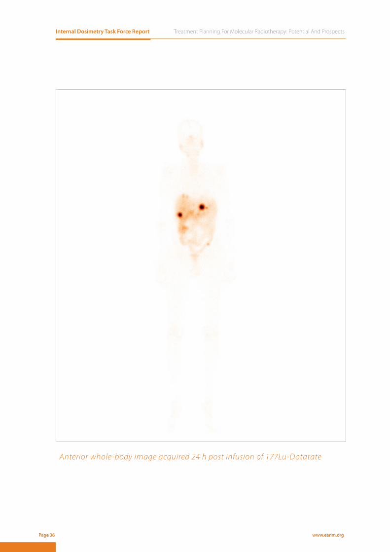
Anterior whole-body image acquired 24 h post infusion of 177Lu-Dotatate

Treatment Planning For Molecular Radiotherapy: Potential And Prospects Internal Dosimetry Task Force Report
www.eanm.org Page 37
90Y somatostatin analogues for the treatment of neuro-endocrine tumours 6Y somatostatin analogues for the treatment of neuro-endocrine tumours 6Y somatostatin analogues for the treatment of neuro-endocrine tumours 66DOSIMETRY FOR THERAPY PROCEDURES
Internal Dosimetry Task Force Report Treatment Planning For Molecular Radiotherapy: Potential And Prospects
www.eanm.orgPage 38
INTRODUCTION90Y-DOTATOC was the first somatostatin analogue developed for treatment of patients with somatostatin re-
ceptor positive neuroendocrine tumours. Phase 1 clinical trials were dosimetry guided by using prospective 86Y-DOTATOC quantitative PET imaging (1). The conclusion in this study was that individual patient dosimetry
was needed as both kidney and tumour absorbed doses showed extreme variability. In the phase 2 trial for this
compound no dosimetry was performed and patients were administered with a single or several administra-
tions of 3.7 GBq/m2 (2). Treatment protocols are mostly based on fixed activity or activity per body surface area
(typically at 1.85 - 3.7 GBq/m2) administration schemes, which are repeated with a 6-8 week interval, depending
on response and quite often adapted to (bone marrow) toxicity after previous treatment. This leads to the huge
range in reported cumulative activities of 1.1 – 26.5 GBq (3).
EFFECTIVENESS In the clinical phase 2 single-centre open-label trial overall 60% of the patients showed clinical response, bio-
chemical response, and/or morphologic disease control after a single administration of 3.7 GBq/m2 90Y-DOTA-
TOC with amino-acid infusion (2). No randomised comparative studies have been performed for 90Y DOTATOC.
Several studies have been performed to compare 90Y-labeled somatostatin receptor peptide alone with a com-
bination of 90Y and 177Lu peptides (4, 5). These combination therapies were based on equal administered activity
of both radionuclides, whereas over its cumulative decay 90Y emits 2.5 times the energy emitted by 177Lu (6).
IMAGINGAs 90Y is a pure beta-emitter, direct imaging of the therapy compound is only possible by using its induced
bremsstrahlung spectrum in planar whole body or SPECT (7). Peri-therapeutic PET imaging can also be per-
formed by using the 0.003%/decay positron emission from 90Y, which has been shown to be feasible for quan-
tifying the uptake in the renal cortex (8). Theragnostic companion compounds have been used to prospec-
tively quantify the 90Y DOTATOC biodistribution, with the gamma-emitter 111In-DOTATATE (9) or the PET emitter 86Y-DOTATATE (10). When using a surrogate peptide it is of great importance to use the same amount and type
of peptide as used in the therapeutic setting, or otherwise correct for the difference in pharmacokinetics and
binding affinity (11).
ORGANS AT RISK AND NORMAL TISSUE DOSIMETRYBoth single 90Y-DOTATOC therapy and combination treatment with 90Y and 177Lu peptides have led to perma-
nent and sometimes even fatal renal toxicity (grade 4 and 5) (2, 5). Kidneys are considered to be the critical organ
after therapy. When the peptide is cleared by the primary renal filter elements (the glomeruli), radiolabelled
peptides are reabsorbed and remain in the secondary filter elements (proximal tubuli). Dosimetry performed in
18 patients with 86Y-DOTATOC PET quantification showed interpatient variability of a factor 4, the absorbed dose
per activity ranged between 1.2 – 5.1 Gy/GBq (1), a comparable variability was observed for the 111In-DOTATOC
based dosimetry: 1.3 – 4.9 Gy/GBq (9). Bone marrow dosimetry is performed less often. Image-based methods
were used with 86Y-DOTATOC and a correlation was observed with 111In-DTPA-Octreotide thoracic spine uptake
(12). In 21 patients the bone marrow absorbed dose ranged between 0.3 and 1.7 Gy for the full therapy of 370
MBq.

Treatment Planning For Molecular Radiotherapy: Potential And Prospects Internal Dosimetry Task Force Report
www.eanm.org Page 39
TUMOUR DOSIMETRY Tumour dosimetry is seldom performed for 90Y-DOTATOC, most probably due to the highly metastasised nature
of the tumours. Nevertheless it has been performed using In-111 DOTATOC as a companion diagnostic (13) and
in the initial phase 1 clinical trial, using 86Y DOTATOC (14).
ABSORBED DOSE-EFFECTLonger follow-up in a sub-group of patients treated in Belgium revealed a dose-response relation between
renal toxicity and the Biologically Effective Dose (BED) when based on the actual kidney volume instead of the
standard size (15). It was observed that the activity and hence absorbed dose per treatment cycle significantly
influenced the incidence of renal toxicity (16). Late stage renal toxicity was shown to follow a classic sigmoidal
shaped dose-effect curve with the BED (17). The threshold for late renal toxicity was found around a BED of 40
Gy for patients without additional risk factors for renal disease, including high blood pressure, diabetes, or prior
chemotherapy. Reduction in tumour volume was shown to be significant above tumour absorbed doses of 200
Gy (14).
DOSIMETRY-BASED TREATMENT PLANNINGOne study repeated administrations according to the 1.85 GBq/m2 dosing scheme until a threshold dose of 37
Gy BED was reached, thereby preventing renal toxicity (18). The BED has been semi-empirically defined in MIRD
pamphlet 20 by using a sub-lethal damage repair half-life of 2.8 h and the radiobiology parameter α/β = 2.5 Gy
for late renal toxicity (16). A multi-factorial dose-effect model for blood platelet response was defined, using pri-
or platelet counts as additional weighting factor, leading to a correlation between the weighted bone marrow
dose and platelet count nadir after therapy (12).
ISSUES TO CONSIDER90Y is a pure high energy beta emitter (mean energy 0.93 MeV), while a minute fraction (0.0032%) leads to inter-
nal pair production photons at 511 keV. Quantitative imaging of 90Y is complex and prospective imaging with
surrogate markers may deviate from the actual biodistribution.
NEED FOR INVESTIGATIONDespite the clear relation between occurrence of late renal toxicity and absorbed dose this has not lead to
clinical protocols using this concept. The longer range of the high-energy beta-particles from 90Y results in rela-
tively homogeneous dose distributions within uptake volumes. Still inhomogeneous uptake in tumours, by e.g.
necrosis, could lead to inhomogeneity in dose distribution. This partly explains the high absorbed doses that are
needed to lead to tumour volume reduction, but this needs to be further investigated. The radiation sensitivity
of neuroendocrine tumours is not well known, but it is not considered to be extremely radio-resistant, consid-
ering the tumour dose of 50 Gy in neo-adjuvant external beam radiotherapy (19).
Internal Dosimetry Task Force Report Treatment Planning For Molecular Radiotherapy: Potential And Prospects
www.eanm.orgPage 40
References 1. Jamar F, Barone R, Mathieu I, et al., 86Y-DOTA0)-D-Phe1-Tyr3-octreotide (SMT487)--a phase 1 clinical study: pharmacoki-
netics, biodistribution and renal protective effect of different regimens of amino acid co-infusion. Eur J Nucl Med Mol Imaging 2003; 30: 510-518.
2. Imhof A, Brunner P, Marincek N, et al., Response, survival, and long-term toxicity after therapy with the radiolabeled so-matostatin analogue [90Y-DOTA]-TOC in metastasized neuroendocrine cancers. J Clin Oncol 2011; 29: 2416-2423.
3. Bodei L, Kidd M, Paganelli G, et al., Long-term tolerability of PRRT in 807 patients with neuroendocrine tumours: the value and limitations of clinical factors. European Journal of Nuclear Medicine and Molecular Imaging 2015; 42: 5-19.
4. Villard L, Romer A, Marincek N, et al., Cohort study of somatostatin-based radiopeptide therapy with [(90)Y-DOTA]-TOC versus [(90)Y-DOTA]-TOC plus [(177)Lu-DOTA]-TOC in neuroendocrine cancers. J Clin Oncol 2012; 30: 1100-1106.
5. Kunikowska J, Krolicki L, Hubalewska-Dydejczyk A, et al., Clinical results of radionuclide therapy of neuroendocrine tu-mours with 90Y-DOTATATE and tandem 90Y/177Lu-DOTATATE: which is a better therapy option? Eur J Nucl Med Mol Imaging 2011; 38: 1788-1797.
6. Eckerman KF and Menzel HG, In the book of life, the answers aren’t in the back--Charlie Brown, fictional character of the Peanuts comic strip created by Charles Schulz. Ann ICRP 2008; 38: 3-5.
7. Minarik D, Sjogreen-Gleisner K, Linden O, et al., 90Y Bremsstrahlung imaging for absorbed-dose assessment in high-dose radioimmunotherapy. J Nucl Med 2010; 51: 1974-1978.
8. Walrand S, Jamar F, van Elmbt L, et al., 4-Step Renal Dosimetry Dependent on Cortex Geometry Applied to 90Y Peptide Receptor Radiotherapy: Evaluation Using a Fillable Kidney Phantom Imaged by 90Y PET. Journal of Nuclear Medicine 2010; 51: 1969-1973.
9. Bodei L, Cremonesi M, Ferrari M, et al., Long-term evaluation of renal toxicity after peptide receptor radionuclide therapy with 90Y-DOTATOC and 177Lu-DOTATATE: the role of associated risk factors. Eur J Nucl Med Mol Imaging 2008; 35: 1847-1856.
10. Walrand S, Jamar F, Mathieu I, et al., Quantitation in PET using isotopes emitting prompt single gammas: application to yttrium-86. European Journal of Nuclear Medicine and Molecular Imaging 2003; 30: 354-361.
11. Kletting P, Muller B, Erentok B, et al., Differences in predicted and actually absorbed doses in peptide receptor radionu-clide therapy. Med Phys 2012; 39: 5708-5717.
12. Walrand S, Barone R, Pauwels S, et al., Experimental facts supporting a red marrow uptake due to radiometal transchela-tion in 90Y-DOTATOC therapy and relationship to the decrease of platelet counts. European Journal of Nuclear Medicine and Molecular Imaging 2011; 38: 1270-1280.
13. Hindorf C, Chittenden S, Causer L et al. Dosimetry for Y-90 DOTATOC therapies in patients with neuroendocrine tu-mours. Cancer Biother radiopharm 2007; 22(1) 130-135.
14. Pauwels S, Barone R, Walrand S, et al., Practical Dosimetry of Peptide Receptor Radionuclide Therapy with 90Y-Labeled Somatostatin Analogs. Journal of Nuclear Medicine 2005; 46: 92S-98S.
15. Barone R, Borson-Chazot F, Valkema R, et al., Patient-specific dosimetry in predicting renal toxicity with (90)Y-DOTATOC: relevance of kidney volume and dose rate in finding a dose-effect relationship. J Nucl Med 2005; 46 Suppl 1: 99S-106S.
16. Valkema R, Pauwels SA, Kvols LK, et al., Long-term follow-up of renal function after peptide receptor radiation therapy with (90)Y-DOTA(0),Tyr(3)-octreotide and (177)Lu-DOTA(0), Tyr(3)-octreotate. J Nucl Med 2005; 46 Suppl 1: 83S-91S.
17. Wessels BW, Konijnenberg MW, Dale RG, et al., MIRD pamphlet No. 20: the effect of model assumptions on kidney dosi-metry and response--implications for radionuclide therapy. J Nucl Med 2008; 49: 1884-1899.
18. Van Binnebeek S, Baete K, Vanbilloen B, et al., Individualized dosimetry-based activity reduction of 90Y-DOTATOC pre-vents severe and rapid kidney function deterioration from peptide receptor radionuclide therapy. European Journal of Nuclear Medicine and Molecular Imaging 2014; 41: 1141-1157.
19. Arvold ND, Willett CG, Fernandez-del Castillo C, et al., Pancreatic neuroendocrine tumors with involved surgical margins: prognostic factors and the role of adjuvant radiotherapy. Int J Radiat Oncol Biol Phys 2012; 83: e337-343.

Treatment Planning For Molecular Radiotherapy: Potential And Prospects Internal Dosimetry Task Force Report
www.eanm.org Page 41
Beta emitters for bone pain palliation 7Beta emitters for bone pain palliationfor bone pain palliationfor bone pain 7Beta emitters for bone pain 77DOSIMETRY FOR THERAPY PROCEDURES

Internal Dosimetry Task Force Report Treatment Planning For Molecular Radiotherapy: Potential And Prospects
www.eanm.orgPage 42
INTRODUCTIONSeveral beta-emitting agents exist for treatment of cancer metastatic to bone; the most commonly used are
strontium-89-chloride (89SrCl2), samarium-153-
EDTMP (153Sm-EDTMP), rhenium-186-HEDP (186Re-HEDP) and rhenium-188-HEDP (188Re-HEDP). The ligands seek
the bone matrix and accumulate in areas of increased osteoblastic activity. The radiopharmaceuticals are mainly
used for patients with castration-resistant prostate cancer, as bone metastases from prostate cancer are pre-
dominately osteoblastic. However, the applications have included treatment of skeletal metastases from pros-
tate, breast, and lung cancer. The administration is most often based on delivery of a fixed amount of radioactiv-
ity or an adjusted amount corresponding to patient body weight (1). Fractionated therapy is sometimes given.
The agents have traditionally been used for pain palliation, and there is some evidence to suggest they may
provide complete relief for months (2).
EFFECTIVENESS Response has primarily been measured as pain relief experienced. The different radiopharmaceuticals have
demonstrated similar pain reduction rates, of approx. 50-70 % (3). In addition; quality of life, analgesia consump-
tion, time to symptomatic skeletal events, and the time to increase in alkaline phosphatase or prostate-specific
antigen level can be monitored. Studies of survival have been sparse. However, the ALSYMPCA results for 223Ra
dichloride have encouraged trials investigating survival data as endpoints.
IMAGINGThe radioisotopes 153Sm and 188Re (and to a lesser degree 186Re) emit photons suitable for gamma camera
imaging (Table 1). Both low energy and high energy collimators have been utilized; the latter was previously
suggested to reduce bremsstrahlung contribution (4-6). If tumour quantitation is to be performed, 3D imaging
is recommended (7). The low yield of photons makes direct quantitative imaging of 89Sr challenging. The lack of
a carrier molecule also prevents the use of a surrogate radioisotope for imaging purposes.
Table 1. Physical characteristics
Radioisotope Half-life (d) Maximum β-energy (MeV) γ-emission (MeV)89Sr 50.5 1.46 Nil (0.01%)
153Sm 1.9 0.81 0.103 (29%)186Re 3.7 1.07 0.137 (9%)188Re 0.7 2.12 0.155 (15%)

Treatment Planning For Molecular Radiotherapy: Potential And Prospects Internal Dosimetry Task Force Report
www.eanm.org Page 43
ORGANS AT RISK AND NORMAL TISSUE DOSIMETRY The most common adverse events are myelotoxic. These can be evaluated by leukocytopenia and thrombo-
cytopenia grading. In general, bone surfaces and red bone marrow have been reported to receive the highest
absorbed doses for 153Sm-EDTMP, 186Re-HEDP and 188Re-HEDP (4, 5, 8). This is in accordance with myelosuppres-
sion being the activity-limiting side effect for bone-seeking radiopharmaceuticals. While the urine, bladder and
kidneys will also be exposed through urinary excretion, the absorbed doses are too low for these organs to be
considered dose-limiting. Normal tissue dosimetry has mainly been based on sequential planar imaging, and
the reported dose ranges have varied. For 89SrCl2 the absorbed doses to normal tissues has been estimated with
the International Commission on Radiological Protection (ICRP) model. In addition to the organs described, the
lower gastrointestinal tract will also receive an absorbed dose contribution from clearance for this treatment (9).
TUMOUR DOSIMETRY Several studies have demonstrated considerable variation in absorbed doses to metastases using gamma cam-
era imaging. For example, a range of approx. 3 Gy - 60 Gy for an administration of 222 MBq/kg 153Sm-EDTMP was
reported when state-of-the-art dosimetry was performed (6).
ABSORBED DOSE-EFFECTFor 153Sm-EDTMP, a significant dose–response relationship for transient tumour volume shrinkage has been
reported (6). The red marrow absorbed dose estimated by SPECT/CT has been shown to be predictive of my-
elotoxicity for the same treatment (10).
DOSIMETRY-BASED TREATMENT PLANNINGThe range in absorbed doses delivered by the treatments demonstrates a potential use for dosimetry to predict
toxicity or response. For 153Sm-EDTMP, 186Re-HEDP and 186Re-HEDP quantitative imaging is feasible, and a frac-
tionated regimen would allow ‘adaptive planning’ whereby the next activity given is based on the dosimetric
results from the preceding administration. Furthermore, the rate of retention of 99mTc-MDP in bone has been
demonstrated to estimate the 153Sm-EDTMP retention rate (10, 11). This can provide a simple method to predict
lesion and red marrow doses.
ISSUES TO CONSIDER A significant decrease in retained activity for normal tissue has been observed between tracer and high treat-
ment amounts of 153Sm-EDTMP (12). Saturating effects should be evaluated, and taken into account for individ-
ual treatment planning.
Care should be taken with regard to target definitions, as uptake occurs at sites of increased osteoblastic activity.
NEED FOR INVESTIGATIONFor 89SrCl2, there is a need to develop reliable dosimetric methodology for the treatment. For the rest of the
treatments, further investigations of dose-effect relationships should be conducted.

Internal Dosimetry Task Force Report Treatment Planning For Molecular Radiotherapy: Potential And Prospects
www.eanm.orgPage 44
References1. Bodei L, Lam M, Chiesa C, et al., EANM procedure guideline for treatment of refractory metastatic bone pain. Eur J Nucl
Med Mol Imaging 2008; 35: 1934-1940.2. Roque IFM, Martinez-Zapata MJ, Scott-Brown M, et al., Radioisotopes for metastatic bone pain. Cochrane Database Syst
Rev 2011: CD003347.3. Jong JM, Oprea-Lager DE, Hooft L, et al., Radiopharmaceuticals for Palliation of Bone Pain in Patients with Castration-re-
sistant Prostate Cancer Metastatic to Bone: A Systematic Review. Eur Urol 2016; 70: 416-426.4. Eary JF, Collins C, Stabin M, et al., Samarium-153-EDTMP biodistribution and dosimetry estimation. J Nucl Med 1993; 34:
1031-1036.5. Liepe K, Hliscs R, Kropp J, et al., Dosimetry of 188Re-hydroxyethylidene diphosphonate in human prostate cancer skeletal
metastases. J Nucl Med 2003; 44: 953-960.6. Senthamizhchelvan S, Hobbs RF, Song H, et al., Tumor dosimetry and response for 153Sm-ethylenediamine te-
tramethylene phosphonic acid therapy of high-risk osteosarcoma. J Nucl Med 2012; 53: 215-224.7. Plyku D, Loeb DM, Prideaux AR, et al., Strengths and Weaknesses of a Planar Whole-Body Method of (153)Sm Dosimetry
for Patients with Metastatic Osteosarcoma and Comparison with Three-Dimensional Dosimetry. Cancer Biother Radio-pharm 2015; 30: 369-379.
8. Maxon HR, 3rd, Schroder LE, Washburn LC, et al., Rhenium-188(Sn)HEDP for treatment of osseous metastases. J Nucl Med 1998; 39: 659-663.
9. Breen SL, Powe JE and Porter AT, Dose estimation in strontium-89 radiotherapy of metastatic prostatic carcinoma. J Nucl Med 1992; 33: 1316-1323.
10. Pacilio M, Ventroni G, Basile C, et al., Improving the dose-myelotoxicity correlation in radiometabolic therapy of bone metastases with 153Sm-EDTMP. Eur J Nucl Med Mol Imaging 2014; 41: 238-252.
11. Bianchi L, Baroli A, Marzoli L, et al., Prospective dosimetry with 99mTc-MDP in metabolic radiotherapy of bone metas-tases with 153Sm-EDTMP. Eur J Nucl Med Mol Imaging 2009; 36: 122-129.
12. Bartlett ML, Webb M, Durrant S, et al., Dosimetry and toxicity of Quadramet for bone marrow ablation in multiple mye-loma and other haematological malignancies. Eur J Nucl Med Mol Imaging 2002; 29: 1470-1477.
Sm-153 EDTA scan 9 hours after administration

Treatment Planning For Molecular Radiotherapy: Potential And Prospects Internal Dosimetry Task Force Report
www.eanm.org Page 45
223Ra dichloride for the treatment of bone metastases from castration resistant prostate cancer8Ra dichloride for the 8Ra dichloride for the
metastases from castration 8metastases from castration resistant prostate cancer8resistant prostate cancer8metastases from castration 8metastases from castration
Ra dichloride for the treatment of bone metastases from castration resistant prostate cancer8Ra dichloride for the treatment of bone metastases from castration resistant prostate cancer88metastases from castration 8metastases from castration
DOSIMETRY FOR THERAPY PROCEDURES

Internal Dosimetry Task Force Report Treatment Planning For Molecular Radiotherapy: Potential And Prospects
www.eanm.orgPage 46
INTRODUCTION223Ra dichloride is marketed under the tradename of Xofigo® (formerly Alpharadin®) and approved for the treat-
ment of patients with castration-resistant prostate cancer, symptomatic bone metastases and no known vis-
ceral metastatic disease. Radium is a natural bone seeker that in the form of 223RaCl2 is used for targeting of
skeletal metastases. Being an analogue to calcium, cationic radium is taken up by areas of increased osteoblastic
activity. In 2005 results from the first clinical study of 223RaCl2 were reported (1). This was a single dosage study
including patients with metastases from prostate or breast cancer. No dose-limiting toxicity was observed and
a fractionated administration regimen was then investigated. A fractionated approach is now routinely used for
the delivery of this treatment; patients are given 6 injections of 55 kBq/kg body weight with a 4 week interval (2).
EFFECTIVENESS The ALSYMPCA phase III study of patients with progressive castration-resistant metastatic prostate cancer and
pain showed that 223RaCl2 improved overall survival relative to placebo (3). In addition, the time to first symp-
tomatic skeletal event, and the time to increases in alkaline phosphatase and prostate-specific antigen levels
were prolonged.
IMAGINGThe low yield of photons, combined with the low amount of activity administered, makes quantitative imaging of 223Ra challenging. However, it has been demonstrated that prolonged gamma camera imaging is feasible (4) and
that activity can be quantified to within 20% - 50%, depending on the volume imaged. Medium energy general
purpose collimators and energy windows centred at 82 keV, and possibly 154 keV and 270 keV are recommended.
ORGANS AT RISK AND NORMAL TISSUE DOSIMETRY The absorbed dose to normal tissues has been estimated with the ICRP model for radium (5). Bone endosteum,
red bone marrow, liver, colon and intestines were reported to receive the highest absorbed doses. In an imag-
ing-based biodistribution study, 223RaCl2 was confirmed to rapidly clear from the blood and the main route of
excretion was found to be via the small bowel (6). A study of normal tissue dosimetry based on sequential quan-
titative imaging, external counting and urine and faecal collection for 6 patients treated twice with 100 kBq/
kg of 223RaCl2 showed that absorbed doses delivered to normal organs vary by an order of magnitude (7). Bone
surfaces received from 2.3 - 13.1 Gy/MBq, and the wholebody from 14 - 66 mGy/MBq. Red marrow absorbed
doses ranged from 0.2 – 1.9 Gy from a single administration of 50 kBq/kg. The short range of the alpha particles
(<100µm) emitted from 223RaCl2 distributed on trabecular bone surfaces probably contributes to a heteroge-
neous dose profile in the marrow space, explaining the low haematological toxicity.

Treatment Planning For Molecular Radiotherapy: Potential And Prospects Internal Dosimetry Task Force Report
www.eanm.org Page 47
TUMOUR DOSIMETRY To date, a single study has investigated absorbed doses to metastases using gamma camera imaging. A range of
0.2 Gy – 1.9 Gy was demonstrated. Uptake of 223RaCl2 was seen to correlate with that of 99mTc MDP (8).
ABSORBED DOSE-EFFECTThere is no evidence as yet of correlations between the absorbed doses delivered and effect.
DOSIMETRY-BASED TREATMENT PLANNINGThere is currently no indication for dosimetry in the treatment planning of 223RaCl2. The potential for treatment
planning is demonstrated by the ability to quantitatively image 223RaCl2 and the correlation of uptake with
pre-therapy 99mTc MDP. The range of delivered absorbed doses demonstrated from initial studies and the lack
of reported toxicity indicate the feasibility to administer higher activities on a personalised basis. There is a po-
tential use for dosimetry to predict toxicity or response in the treatment planning of 223RaCl2. The fractionated
regimen allows ‘adaptive planning’, whereby sequential administrations (the activity per fraction, or the number
of fractions) are based on the dosimetric results from the preceding administration.
ISSUES TO CONSIDER 223Ra is an alpha emitter with a half-life of 11.4 days which decays to stable lead via a chain of five daughters, and
most of the energy is emitted through 4 alpha particle emissions (5.6-7.4 MeV). As the third daughter (211Pb) has
a half-life of 36 minutes, there is therefore a possibility that subsequent disintegrations may occur elsewhere.
However, no redistribution of radioactive daughters has yet been reported.
NEED FOR INVESTIGATIONGiven the challenges associated with estimating patient-specific absorbed doses for 223Ra the first priority should
be to develop reliable dosimetric methodology for this treatment and to conduct clinical trials to evaluate re-
sponse in relation to the absorbed dose. The short range of the alpha emissions necessitates an investigation
of small scale dosimetry and microdosimetry, both for bone and tumour tissue. The sparse data available on
absorbed doses delivered to tumours indicates that these may be low in comparison with beta emitters. Alpha
emissions incur a relative biological effect that has been quoted as ranging from 3 – 5. Accumulation of dosim-
etry and response data will enable the Relative Biological Effect (RBE) to be determined.

Internal Dosimetry Task Force Report Treatment Planning For Molecular Radiotherapy: Potential And Prospects
www.eanm.orgPage 48
References1. Nilsson S, Larsen RH, Fossa SD, et al., First clinical experience with alpha-emitting radium-223 in the treatment of skeletal
metastases. Clin Cancer Res 2005; 11: 4451-4459.2. Xofi go® (radium Ra 223 dichloride) solution for injection Summary of Product Characteristics (SmPC). 2016, Bayer Pharma AG,
13342 Berlin, Germany.3. Parker C, Nilsson S, Heinrich D, et al., Alpha emitter radium-223 and survival in metastatic prostate cancer. N Engl J Med
2013; 369: 213-223.4. Hindorf C, Chittenden S, Aksnes AK, et al., Quantitative imaging of 223Ra-chloride (Alpharadin) for targeted alpha-emit-
ting radionuclide therapy of bone metastases. Nucl Med Commun 2012; 33: 726-732.5. Lassmann M and Nosske D, Dosimetry of 223Ra-chloride: dose to normal organs and tissues. Eur J Nucl Med Mol Imag-
ing 2013; 40: 207-212.6. Carrasquillo JA, O’Donoghue JA, Pandit-Taskar N, et al., Phase I pharmacokinetic and biodistribution study with escalating
doses of (2)(2)(3)Ra-dichloride in men with castration-resistant metastatic prostate cancer. Eur J Nucl Med Mol Imaging 2013; 40: 1384-1393.
7. Chittenden SJ, Hindorf C, Parker CC, et al., A Phase 1, Open-Label Study of the Biodistribution, Pharmacokinetics, and Dosimetry of 223Ra-Dichloride in Patients with Hormone-Refractory Prostate Cancer and Skeletal Metastases. J Nucl Med 2015; 56: 1304-1309.
8. Pacilio M, Ventroni G, De Vincentis G, et al., Dosimetry of bone metastases in targeted radionuclide therapy with al-pha-emitting (223)Ra-dichloride. Eur J Nucl Med Mol Imaging 2016; 43: 21-33.
Imaging during Ra-223 treatment. Anterior Ra-223, anterior Tc-99 MDP, F-18 Fluoride, posterior Tc-99m MDP, posterior Ra-223

Treatment Planning For Molecular Radiotherapy: Potential And Prospects Internal Dosimetry Task Force Report
www.eanm.org Page 49
177Lu-PSMA ligands for the treatment of metastatic castration-resistant prostate cancer 9metastatic castration-9metastatic castration-9Lu-PSMA ligands for the treatment of metastatic castration-resistant prostate 9Lu-PSMA ligands for the treatment of metastatic castration-resistant prostate 99DOSIMETRY FOR THERAPY PROCEDURES

Internal Dosimetry Task Force Report Treatment Planning For Molecular Radiotherapy: Potential And Prospects
www.eanm.orgPage 50
INTRODUCTIONPSMA (prostate-specific membrane antigen) is a glutamate carboxypeptidase II overexpressed in prostate can-
cer. Different radiolabelled ligands targeting PSMA have been developed for treatment of patients with ad-
vanced prostate cancer, including the small molecule inhibitors 177Lu-DKFZ-PSMA-617 and 177Lu-DOTAGA-(I-y)
fk(Sub-KuE), and the monoclonal antibody based 177Lu-DOTA-J591. Investigations of 131I or 90Y based ligands have
also been performed, however, the current focus is on 177Lu radiolabelled ligands and especially the small-mol-
ecule inhibitors. As the radiotherapeutics are still in clinical trials, no standard treatment regimens have de-
veloped. The most common way to deliver 177Lu-PSMA small-molecule inhibitors seems to be in a fractioned
approach of 2-9 GBq in each cycle (1-3). For antibodies the regimen is restricted to a single administration, and
2.59 GBq/m2 BSA was given in a phase II trial of 177Lu-DOTA-J591 (4). Commonly, the treatment is preceded by a
diagnostic assessment using an analogue ligand with a PET (or SPECT) radionuclide.
EFFECTIVENESSWhile the survival benefits of these treatments are yet to be reported in randomised trials, efficacy can be eval-
uated by biochemical response defined by decline in prostate-specific antigen (PSA), response assessed by PET/
CT imaging, radiologic response, pain relief or quality of life. In a German multi-centre study 45 % of patients had
a 50 % decline in PSA after 1-4 therapy cycles of 177Lu-PSMA-617 (5). Several other trials have also demonstrated
high efficacy for small-molecule inhibitors in patients with metastatic castration-resistant prostate cancer (2, 6, 7).
IMAGINGImaging can be performed essentially as described for 177Lu-DOTATATE, however, as lower amounts of radioac-
tivity are given, there may be a need for prolonged acquisitions. Multiple SPECT/CT scanning is recommended
using with medium energy general purpose collimators and energy window centred at 208 keV (8).
ORGANS AT RISK AND NORMAL TISSUE DOSIMETRY In studies of SPECT/CT based dosimetry for 177Lu-PSMA-617, the organs with the highest absorbed doses were
the salivary/parotid glands and kidneys, receiving mean absorbed doses of approximately 1.2-1.4 and 0.6-
1.0 Gy/GBq, respectively (9-11). The mean absorbed doses to red marrow were reported between 12 and 48
mGy/GBq. Lacrimal glands may also represent a dose-limiting organ (12). For 177Lu-DOTAGA-(I-y)fk(Sub-KuE)
similar absorbed doses are observed; of 1.3 Gy/GBq, 0.8 Gy/GBq and 14 mGy/GBq for parotid glands, kidneys
and red marrow, respectively (2). The absorbed doses to normal tissue are likely higher for the antibody-based 177Lu-DOTA-J591 (13).
TUMOUR DOSIMETRY One study has investigated absorbed doses to metastases for 177Lu-DOTAGA-(I-y)fk(Sub-KuE), demonstrating
a range from 0.03 to 78 Gy/GBq and a mean value of 3.3 Gy/GBq (2). For 177Lu-DOTA-J591, absorbed doses for
tumour lesions ranged between 1.2 and 47.5 Gy (mean 13.1 Gy/GBq) (9).

Treatment Planning For Molecular Radiotherapy: Potential And Prospects Internal Dosimetry Task Force Report
www.eanm.org Page 51
ABSORBED DOSE-EFFECTThere is as yet no study that has reported on absorbed doses compared to response or toxicity of the treatments.
DOSIMETRY-BASED TREATMENT PLANNINGFor therapy with 177Lu-PSMA small-molecule inhibitors dosimetry driven treatment planning has a potential for
both easy implementation and providing large benefits for the patients. Absorbed doses to risk organs may be
significant over the treatment cycles, and prospective absorbed dose measures may prove valuable to deter-
mine the acceptable number of cycles. Administrations of tracer amounts of 177Lu-PSMA-617 and pre-therapeu-
tic dosimetry have been investigated, and the authors concluded that the substantial individual variance seems
to make patient dosimetry mandatory (10). A more practical approach may be to perform treatment planning
based on quantitative imaging from the previous treatment cycle or the pre-therapeutic diagnostic assessment
with a surrogate ligand.
ISSUES TO CONSIDERWhile the normal tissue absorbed doses have been reported relative invariable between therapy cycles (9), it
should be investigated in a larger patient material whether Gy/GBq values from the first cycle can be used di-
rectly for treatment planning of the next cycles. Prospective imaging with positron-emitting surrogate markers
may deviate from the actual biodistribution.
NEED FOR INVESTIGATIONThe primary dose-limiting organs are thought to be salivary and lacrimal glands for the small-molecule inhib-
itors. However, this should be further investigated as clinical side effects more often include haematotoxicity
than xerostomia. The dose-effect curves, with tolerance limits for normal tissue and the desirable absorbed dose
to tumours, will likely need to be empirically determined. The experience from other molecular radiotherapies
demonstrates that the common assumption of identical tolerance limits for MRT as for EBRT can often lead to
an under-dosage of radiopharmaceuticals – as the tolerance limits are actually higher. This may be explained
by different dose-rates, energies, or the short range of the beta emissions which necessitates an investigation
of small scale dosimetry.
Further possibilities for development include combination treatments. An alpha-emitting radiopharmaceutical, 225Ac-PSMA-617, is currently under investigation, and the complementary role of the beta- and alpha-emitting
ligands should be explored.
Internal Dosimetry Task Force Report Treatment Planning For Molecular Radiotherapy: Potential And Prospects
www.eanm.orgPage 52
References1. Ahmadzadehfar H, Eppard E, Kurpig S, et al., Therapeutic response and side effects of repeated radioligand therapy with
177Lu-PSMA-DKFZ-617 of castrate-resistant metastatic prostate cancer. Oncotarget 2016; 7: 12477-12488.2. Baum RP, Kulkarni HR, Schuchardt C, et al., 177Lu-Labeled Prostate-Specific Membrane Antigen Radioligand Therapy of
Metastatic Castration-Resistant Prostate Cancer: Safety and Efficacy. J Nucl Med 2016; 57: 1006-13.3. Kratochwil C, Giesel FL, Stefanova M, et al., PSMA-Targeted Radionuclide Therapy of Metastatic Castration-Resistant Pros-
tate Cancer with 177Lu-Labeled PSMA-617. J Nucl Med 2016; 57: 1170-1176.4. Tagawa ST, Milowsky MI, Morris M, et al., Phase II study of Lutetium-177-labeled anti-prostate-specific membrane antigen
monoclonal antibody J591 for metastatic castration-resistant prostate cancer. Clin Cancer Res 2013; 19: 5182-5191.5. Rahbar K, Ahmadzadehfar H, Kratochwil C, et al., German Multicenter Study Investigating 177Lu-PSMA-617 Radioligand
Therapy in Advanced Prostate Cancer Patients. J Nucl Med 2017; 58: 85-90.6. Fendler WP, Reinhardt S, Ilhan H, et al., Preliminary experience with dosimetry, response and patient reported outcome
after 177Lu-PSMA-617 therapy for metastatic castration-resistant prostate cancer. Oncotarget 2017; 8: 3581-3590.7. Yadav MP, Ballal S, Tripathi M, et al., 177Lu-DKFZ-PSMA-617 therapy in metastatic castration resistant prostate cancer:
safety, efficacy, and quality of life assessment. Eur J Nucl Med Mol Imaging 2017; 44: 81-91.8. Ljungberg M, Celler A, Konijnenberg MW, et al., MIRD Pamphlet No. 26: Joint EANM/MIRD Guidelines for Quantitative
177Lu SPECT Applied for Dosimetry of Radiopharmaceutical Therapy. J Nucl Med 2016; 57: 151-162.9. Delker A, Fendler WP, Kratochwil C, et al., Dosimetry for (177)Lu-DKFZ-PSMA-617: a new radiopharmaceutical for the
treatment of metastatic prostate cancer. Eur J Nucl Med Mol Imaging 2016; 43: 42-51.10. Kabasakal L, AbuQbeitah M, Aygun A, et al., Pre-therapeutic dosimetry of normal organs and tissues of (177)Lu-PS-
MA-617 prostate-specific membrane antigen (PSMA) inhibitor in patients with castration-resistant prostate cancer. Eur J Nucl Med Mol Imaging 2015; 42: 1976-1983.
11. Yadav MP, Ballal S, Tripathi M, et al., Post-therapeutic dosimetry of 177Lu-DKFZ-PSMA-617 in the treatment of patients with metastatic castration-resistant prostate cancer. Nucl Med Commun 2017; 38: 91-98.
12. Hohberg M, Eschner W, Schmidt M, et al., Lacrimal Glands May Represent Organs at Risk for Radionuclide Therapy of Prostate Cancer with [(177)Lu]DKFZ-PSMA-617. Mol Imaging Biol 2016; 18: 437-445.
13. Vallabhajosula S, Goldsmith SJ, Hamacher KA, et al., Prediction of myelotoxicity based on bone marrow radiation-ab-sorbed dose: radioimmunotherapy studies using 90Y- and 177Lu-labeled J591 antibodies specific for prostate-specific membrane antigen. J Nucl Med 2005; 46: 850-858.

Treatment Planning For Molecular Radiotherapy: Potential And Prospects Internal Dosimetry Task Force Report
www.eanm.org Page 53
Radioembolisation with 90Y microspheres for the treatment of primary and metastatic liver cancer10Radioembolisation 10Radioembolisation with 90Y microspheres for 10with 90Y microspheres for the treatment of primary 10the treatment of primary and metastatic liver cancer10and metastatic liver cancer10Radioembolisation 10Radioembolisation with 90Y microspheres for 10with 90Y microspheres for the treatment of primary 10the treatment of primary and metastatic liver cancer10and metastatic liver cancer10the treatment of primary 10the treatment of primary and metastatic liver cancer10and metastatic liver cancer10with 90Y microspheres for 10with 90Y microspheres for
DOSIMETRY FOR THERAPY PROCEDURES10DOSIMETRY FOR THERAPY PROCEDURES10Radioembolisation with 90Y microspheres for the treatment of primary 10the treatment of primary 10the treatment of primary and metastatic liver cancer10and metastatic liver cancer10Radioembolisation 10Radioembolisation with 90Y microspheres for 10with 90Y microspheres for the treatment of primary 10the treatment of primary and metastatic liver cancer10and metastatic liver cancer10with 90Y microspheres for 10with 90Y microspheres for

Internal Dosimetry Task Force Report Treatment Planning For Molecular Radiotherapy: Potential And Prospects
www.eanm.orgPage 54
INTRODUCTIONIntra-arterial locoregional liver therapies have their rationale in the fact that liver lesions are fed mainly by the
arterial stream, while normal parenchyma is supplied by portal vein blood flow. As seen in the survey, at present
two products are mainly used, 90Y glass microspheres and 90Y resin microspheres, both licensed by the US Food
and Drug Administration (FDA) as medical devices to treat liver primary hepatocarcinoma (HCC) and liver me-
tastases (1). A product using 166Ho is in development (2).
EFFECTIVENESS AND TOXICITYMany studies already have demonstrated a competitive outcome of radioembolisation (TARE) with respect to
conventional treatment modalities, both in intermediate and advanced HCC stages (3) and metastases (4).
IMAGINGSimulation scanning is performed with 99mTc-albumin macro aggregate (MAA) administered under angiograph-
ic guidance for quantitative imaging and pre-treatment dosimetry (1). The permanent trapping into liver cap-
illaries of the 99mTc MAA and of therapeutic particles (90Y microspheres) allows dosimetry to be performed from
only one scan (5). This procedure can also be used to evaluate the fraction of activity that is shunted to the
lung where this occurs. Post therapy quantitative imaging can be performed by 90Y bremsstrahlung SPECT or 90Y PET with suitable corrections (6). Reproducible activity quantitation across different PET systems has been
demonstrated (7).
ORGANS AT RISK AND NORMAL TISSUE DOSIMETRYToxicity is of particular importance, as patient death from radioembolisation-induced liver disease leading to
liver failure can be a consequence of a standard treatment (5). Dosimetry for normal liver is performed from
quantitative 99mTc, 90Y bremsstrahlung or 90Y PET imaging. Lung dosimetry is also of importance as this can cause
severe toxicity and may also be performed with imaging, with suitable corrections for photon attenuation (5).
TUMOUR DOSIMETRYTumour absorbed doses can vary widely and have been reported as high as 1000 Gy (8). Dosimetry is simplified
by the assumption that the microspheres are trapped so that only physical decay occurs.
ABSORBED DOSE-EFFECTCorrelations between the absorbed doses delivered and toxicity and response have been reported both for
hepatocellular carcinoma (9-11) and colorectal metastases (12, 13).
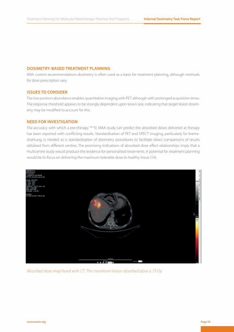
Treatment Planning For Molecular Radiotherapy: Potential And Prospects Internal Dosimetry Task Force Report
www.eanm.org Page 55
DOSIMETRY-BASED TREATMENT PLANNING With current recommendations dosimetry is often used as a basis for treatment planning, although methods
for dose prescription vary.
ISSUES TO CONSIDERThe low positron abundance enables quantitative imaging with PET although with prolonged acquisition times.
The response threshold appears to be strongly dependent upon lesion size, indicating that target lesion dosim-
etry may be modified to account for this.
NEED FOR INVESTIGATIONThe accuracy with which a pre-therapy 99mTc MAA study can predict the absorbed doses delivered at therapy
has been reported with conflicting results. Standardisation of PET and SPECT imaging, particularly for brems-
strahlung, is needed as is standardisation of dosimetry procedures to facilitate direct comparisons of results
obtained from different centres. The promising indications of absorbed dose effect relationships imply that a
multicentre study would produce the evidence for personalised treatments. A potential for treatment planning
would be to focus on delivering the maximum tolerable dose to healthy tissue (14).
Absorbed dose map fused with CT. The maximum lesion absorbed dose is 75 Gy
Internal Dosimetry Task Force Report Treatment Planning For Molecular Radiotherapy: Potential And Prospects
www.eanm.orgPage 56
References1. Giammarile F, Bodei L, Chiesa C, et al., EANM procedure guideline for the treatment of liver cancer and liver metastases
with intra-arterial radioactive compounds. Eur J Nucl Med Mol Imaging 2011; 38: 1393-1406.2. Smits MLJ, Nijsen JFW and van den Bosch MAAJ, Holmium-166 radioembolisation in patients with unresectable, che-
morefractory liver metastases (HEPAR trial): a phase 1, dose-escalation study (vol 13, pg 1028, 2012). Lancet Oncology 2012; 13: E464-E464.
3. Salem R, Mazzaferro V and Sangro B, Yttrium 90 radioembolization for the treatment of hepatocellular carcinoma: biolo-gical lessons, current challenges, and clinical perspectives. Hepatology 2013; 58: 2188-2197.
4. Rosenbaum CE, Verkooijen HM, Lam MG, et al., Radioembolization for treatment of salvage patients with colorectal can-cer liver metastases: a systematic review. J Nucl Med 2013; 54: 1890-1895.
5. Cremonesi M, Chiesa C, Strigari L, et al., Radioembolization of hepatic lesions from a radiobiology and dosimetric pers-pective. Front Oncol 2014; 4: 210.
6. Elschot M, Vermolen BJ, Lam MG, et al., Quantitative comparison of PET and Bremsstrahlung SPECT for imaging the in vivo yttrium-90 microsphere distribution after liver radioembolization. PLoS One 2013; 8: e55742..
7. Willowson KP, Tapner M, Team QI, et al., A multicentre comparison of quantitative (90)Y PET/CT for dosimetric purposes after radioembolization with resin microspheres : The QUEST Phantom Study. Eur J Nucl Med Mol Imaging 2015; 42: 1202-1222.
8. Riaz A, Gates VL, Atassi B, et al., Radiation segmentectomy: a novel approach to increase safety and efficacy of radioem-bolization. Int J Radiat Oncol Biol Phys 2011; 79: 163-171.
9. Chiesa C, Mira M, Maccauro M, et al., Radioembolization of hepatocarcinoma with (90)Y glass microspheres: develop-ment of an individualized treatment planning strategy based on dosimetry and radiobiology. Eur J Nucl Med Mol Imag-ing 2015; 42: 1718-1738.
10. Garin E, Lenoir L, Edeline J, et al., Boosted selective internal radiation therapy with 90Y-loaded glass microspheres (B-SIRT) for hepatocellular carcinoma patients: a new personalized promising concept. Eur J Nucl Med Mol Imaging 2013; 40: 1057-1068.
11. Strigari L, Sciuto R, Rea S, et al., Efficacy and toxicity related to treatment of hepatocellular carcinoma with 90Y-SIR spheres: radiobiologic considerations. J Nucl Med 2010; 51: 1377-1385.
12. Flamen P, Vanderlinden B, Delatte P, et al., CORRIGENDUM: Multimodality imaging can predict the metabolic response of unresectable colorectal liver metastases to radioembolization therapy with Yttrium-90 labeled resin microspheres. Phys Med Biol 2014; 59: 2549.
13. van den Hoven AF, Rosenbaum CE, Elias SG, et al., Insights into the Dose-Response Relationship of Radioembolization with Resin 90Y-Microspheres: A Prospective Cohort Study in Patients with Colorectal Cancer Liver Metastases. J Nucl Med 2016; 57: 1014-1019.
14. Gnesin S, Canetti L, Adib S, et al., Partition Model-Based 99mTc-MAA SPECT/CT Predictive Dosimetry Compared with 90Y TOF PET/CT Posttreatment Dosimetry in Radioembolization of Hepatocellular Carcinoma: A Quantitative Agree-
ment Comparison. J Nucl Med 2016; 57: 1672-1678.

Treatment Planning For Molecular Radiotherapy: Potential And Prospects Internal Dosimetry Task Force Report
www.eanm.org Page 57
90Y-ibritumomab tiuxetan for radioimmunotherapy of non-Hodgkin lymphoma1190Y-ibritumomab tiuxetan 1190Y-ibritumomab tiuxetan for radioimmunotherapy of 11for radioimmunotherapy of non-Hodgkin lymphoma11non-Hodgkin lymphoma
90Y-ibritumomab tiuxetan for radioimmunotherapy of 11for radioimmunotherapy of 11for radioimmunotherapy of non-Hodgkin lymphoma11non-Hodgkin lymphoma1190Y-ibritumomab tiuxetan 1190Y-ibritumomab tiuxetan for radioimmunotherapy of 11for radioimmunotherapy of non-Hodgkin lymphoma11non-Hodgkin lymphoma1190Y-ibritumomab tiuxetan 1190Y-ibritumomab tiuxetan for radioimmunotherapy of 11for radioimmunotherapy of

Internal Dosimetry Task Force Report Treatment Planning For Molecular Radiotherapy: Potential And Prospects
www.eanm.orgPage 58
INTRODUCTION.Radioimmunotherapy (RIT) with 90Y-ibritumomab tiuxetan (marketed under the tradename of Zevalin®) is ap-
proved for the treatment of non-Hodgkin, CD20-positive indolent or transformed lymphoma, in the setting of
relapse after rituximab-containing treatments or as a consolidation after first line therapy (1-3). The ibritumom-
ab antibody targets CD20 antigen found on the surface of normal and malignant B cells.
The radioactivity to be administered takes into account patient weight and platelet blood count only, and no
optimisation based on pre-therapeutic dosimetry is routinely prescribed. Usually 11 MBq or 14 MBq are admin-
istered per kg of body weight. Treatment administration is preceded by two infusions of unlabelled rituximab
(Mabthera®) 250 mg/m2, given one week apart in order to optimise RIT delivery by saturating non-tumoural
CD20-positive sites (4).
EFFECTIVENESSA randomised controlled trial of 90Y-ibritumomab tiuxetan versus rituximab in relapsed or refractory low-grade
or transformed B-cell NHL has shown an overall response rate (ORR) of 80% and complete response (CR) of 30
% for the 90Y-ibritumomab tiuxetan group (3). In follicular lymphoma, a single RIT infusion as first line treatment
shows ORR of 87% and median progression-free survival (PFS) of 26 months (5). RIT with 90Y-ibritumomab ti-
uxetan has been fractionated in two administrations showing an ORR of 94.4% with 58.3% CR. At a median
follow-up of 3.1 years, estimated 3-year PFS is 58%, and overall survival 95%. (6).
IMAGING.As 90Y is an almost pure β- emitter, the labelling of ibritumomab with a γ-emitting surrogate, such as 111In, is re-
quired for pre-therapeutic dosimetry. Both final products, namely 90Y-ibritumomab and 111In-ibritumomab, can
be assumed to have the same biodistribution in the human body (7, 8). The physical half-lives of 90Y (64 hours)
and 111In (68 hr) are closely matched. To determine the slow antibody kinetics, the biodistribution of the labelled
molecule has to be followed for several days in order to build a reliable time-activity curve. A trace amount of
185 MBq 111In-ibritumomab is usually injected one week before the planned therapeutic infusion. Three to five
imaging time points are needed for reliable curve fitting: 1-4 h, 24-48 h, 72 and 96-120 h. Suggested camera
settings (9, 10) should be validated in-house.
VERIFICATION OF PLANNED RADIATION DELIVERY.Bremsstrahlung radiation is emitted from the patient body after a treatment with 90Y-ibritumomab tiuxetan, and
gamma camera imaging of such emission is possible though of limited sensitivity and resolution (7). Although 90Y has a small branch of position emission, at the standard 90Y-ibritumomab tiuxetan administered activities this
is not sufficient and not focal enough to allow direct PET imaging as in the case of hepatic radioembolisation.

Treatment Planning For Molecular Radiotherapy: Potential And Prospects Internal Dosimetry Task Force Report
www.eanm.org Page 59
ORGANS AT RISK AND NORMAL TISSUE DOSIMETRY.Normal organ dosimetry for 90Y-ibritumomab tiuxetan has been the subject of a MIRD dose estimate report (10).
Median absorbed dose values calculated (in mGy/MBq) included Red Marrow 2.73 (± 0.90), Liver 3.64 (± 1.38),
Kidneys 2.44 (± 0.61), Spleen 4.65 (± 2.32), Lungs 0.76 (± 0.48) and Whole body 0.58 (± 0.10). This report showed
that the dosimetry approach, assumptions, and choice of parameters can substantially affect the calculated
doses (10). Using a full SPECT/CT protocol instead of planar imaging, systematically lower absorbed doses have
been found for the liver and spleen (11). A separate study determined that image-based bone marrow dosime-
try models are better predictors of myelotoxicity than methods based on blood samplings (12).
TUMOUR DOSIMETRYAccording to the experience of external beam radiotherapy, a total dose of 30-36 Gy is needed to eradicate
lymphoma lesions. However, reduced-intensity protocols of 2 x 2 Gy are successfully used in selected cases of
indolent lymphomas. When explored, the tumour absorbed dose range delivered by RIT was wide (range: 5.8
– 67 Gy) (13).
ABSORBED DOSE-EFFECTNo clear dose-response correlation has been shown (13). However, these first studies used a two-dimensional
approach, so that no refined radiobiological models could be taken into account. When this has been done in
similar types of treatments, as in the case of RIT with the anti CD20 131I-tositumomab (Bexxar®), an improved
dose-response correlation was demonstrated (14). A three dimensional, voxel-based approach to RIT with 90Y-ibritumomab tiuxetan is still to be optimised (9).
Transient and generally manageable myelotoxicity is the most common side effect of RIT. Pilot studies have
shown that, when given as standard, non myeloablative activities, only exceptionally would administration of 90Y-ibritumomab tiuxetan be contraindicated because of excessive absorbed doses to organs at risk (OARs)
(15). However, concerns regarding the absorbed dose delivered to organs at risk may arise in case of severely ill
and heavily pre-treated patients as well as if additional external beam radiotherapy is planned in the treatment
course.
DOSIMETRY-BASED TREATMENT PLANNINGThe strongest rationale for pre-therapeutic dosimetry is the prevention of unexpected toxicities in the disease
course of fragile patients or when a deviation from the standard administration protocol is foreseen, as in the
frame of myeloablative protocols. The pre-therapeutic imaging with 111In-ibritumomab tiuxetan can be used
for treatment planning, which can also take into account the planned delivered dose to lesions. Studies on
fractionated therapies have determined that median organ absorbed doses were equivalent between fractions
except for the spleen (12). Clinical studies employing myeloablative activities would always require an adequate
planning as the risk of non-haematological toxicities may not be negligible (16).

Internal Dosimetry Task Force Report Treatment Planning For Molecular Radiotherapy: Potential And Prospects
www.eanm.orgPage 60
ISSUES TO CONSIDER Radiolabelled antibodies have a slow kinetic, therefore requiring pre-therapeutic dosimetry protocols lasting for
several days. The use of a surrogate isotope (111In) might imply minimal kinetic differences with the final therapeu-
tic product. As ibritumomab is a murine antibody, there are concerns regarding the development of human anti
mouse antibodies (HAMA) for repeated administrations. Verification of the actual delivered dose is challenging.
NEED FOR INVESTIGATION » Extended indications for the use of 90Y-ibritumomab tiuxetan
» Therapy fractionation
» Re-treatment strategies
» Myeloablative strategies
» The role of patient organ and tumour dosimetry with newer camera technologies and refined radiobiolog-
ical models.
In-111 Zevalin: Anterior scan of a patient with Follicular Lymphoma, acquired 96 hours after injection. Multiple mediastinal, right axillary and bilateral inguinal lymph-nodes are seen, as well as small subcutaneous disease implants in the right arm.
Treatment Planning For Molecular Radiotherapy: Potential And Prospects Internal Dosimetry Task Force Report
www.eanm.org Page 61
References1. Morschhauser F, Radford J, Van Hoof A, et al., Phase III trial of consolidation therapy with yttrium-90-ibritumomab tiuxe-
tan compared with no additional therapy after first remission in advanced follicular lymphoma. J Clin Oncol 2008; 26: 5156-5164.
2. Witzig TE, Flinn IW, Gordon LI, et al., Treatment with ibritumomab tiuxetan radioimmunotherapy in patients with rituxi-mab-refractory follicular non-Hodgkin’s lymphoma. J Clin Oncol 2002; 20: 3262-3269.
3. Witzig TE, Gordon LI, Cabanillas F, et al., Randomized controlled trial of yttrium-90-labeled ibritumomab tiuxetan radioim-munotherapy versus rituximab immunotherapy for patients with relapsed or refractory low-grade, follicular, or transfor-med B-cell non-Hodgkin’s lymphoma. J Clin Oncol 2002; 20: 2453-2463.
4. Tennvall J, Fischer M, Bischof Delaloye A, et al., EANM procedure guideline for radio-immunotherapy for B-cell lymphoma with 90Y-radiolabelled ibritumomab tiuxetan (Zevalin). Eur J Nucl Med Mol Imaging 2007; 34: 616-622.
5. Scholz CW, Pinto A, Linkesch W, et al., (90)Yttrium-ibritumomab-tiuxetan as first-line treatment for follicular lymphoma: 30 months of follow-up data from an international multicenter phase II clinical trial. J Clin Oncol 2013; 31: 308-313.
6. Illidge TM, Mayes S, Pettengell R, et al., Fractionated (9)(0)Y-ibritumomab tiuxetan radioimmunotherapy as an initial therapy of follicular lymphoma: an international phase II study in patients requiring treatment according to GELF/BNLI criteria. J Clin Oncol 2014; 32: 212-218.
7. Minarik D, Sjogreen-Gleisner K, Linden O, et al., 90Y Bremsstrahlung imaging for absorbed-dose assessment in high-dose radioimmunotherapy. J Nucl Med 2010; 51: 1974-1978.
8. Sjogreen-Gleisner K, Dewaraja YK, Chiesa C, et al., Dosimetry in patients with B-cell lymphoma treated with [(90)Y]ibritu-momab tiuxetan or [(131)I]tositumomab. Q J Nucl Med Mol Imaging 2011; 55: 126-154.
9. Cicone F, D’Arienzo M, Carpaneto A, et al., Quantification of dose nonuniformities by voxel-based dosimetry in patients receiving 90Y-ibritumomab-tiuxetan. Cancer Biother Radiopharm 2013; 28: 98-107.
10. Fisher DR, Shen S and Meredith RF, MIRD dose estimate report No. 20: radiation absorbed-dose estimates for 111In- and 90Y-ibritumomab tiuxetan. J Nucl Med 2009; 50: 644-652.
11. Assie K, Dieudonne A, Gardin I, et al., Comparison between 2D and 3D dosimetry protocols in 90Y-ibritumomab tiuxe-tan radioimmunotherapy of patients with non-Hodgkin’s lymphoma. Cancer Biother Radiopharm 2008; 23: 53-64.
12. Ferrer L, Malek E, Bodet-Milin C, et al., Comparisons of dosimetric approaches for fractionated radioimmunotherapy of non-Hodgkin lymphoma. Q J Nucl Med Mol Imaging 2012; 56: 529-537.
13. Wiseman GA, White CA, Stabin M, et al., Phase I/II 90Y-Zevalin (yttrium-90 ibritumomab tiuxetan, IDEC-Y2B8) radioimmu-notherapy dosimetry results in relapsed or refractory non-Hodgkin’s lymphoma. Eur J Nucl Med 2000; 27: 766-777.
14. Dewaraja YK, Schipper MJ, Shen J, et al., Tumor-Absorbed Dose Predicts Progression-Free Survival Following 131I-Tositu-momab Radioimmunotherapy. Journal of Nuclear Medicine 2014; 55: 1047-1053.
15. Wiseman GA, Kornmehl E, Leigh B, et al., Radiation dosimetry results and safety correlations from 90Y-ibritumomab tiuxetan radioimmunotherapy for relapsed or refractory non-Hodgkin’s lymphoma: combined data from 4 clinical trials. J Nucl Med 2003; 44: 465-474.
16. Pacilio M, Betti M, Cicone F, et al., A theoretical dose-escalation study based on biological effective dose in radioimmu-
notherapy with (90)Y-ibritumomab tiuxetan (Zevalin). Eur J Nucl Med Mol Imaging 2010; 37: 862-873.

Internal Dosimetry Task Force Report Treatment Planning For Molecular Radiotherapy: Potential And Prospects
www.eanm.orgPage 62

Treatment Planning For Molecular Radiotherapy: Potential And Prospects Internal Dosimetry Task Force Report
www.eanm.org Page 63
Radiosynovectomy12Radiosynovectomy12RadiosynovectomyDOSIMETRY FOR THERAPY PROCEDURES12DOSIMETRY FOR THERAPY PROCEDURES1212Radiosynovectomy12Radiosynovectomy12Radiosynovectomy12Radiosynovectomy12
Internal Dosimetry Task Force Report Treatment Planning For Molecular Radiotherapy: Potential And Prospects
www.eanm.orgPage 64
INTRODUCTIONRadiosynovectomy (synoviorthesis or synoviolisis) (RSO) is an intra-articular injection of small radioactive par-
ticles (0.05 - 2 µm) to treat a synovitis. The aim of treatment is to improve mobility and to maximally decrease
soreness, discomfort and bleeding into the joint. It is most commonly used to treat rheumatoid arthritis (RA),
poly-arthritis and haemophilic arthritis (1-3). Further indications may be osteoarthritis and the articular effusion
after joint replacement (4). 90Y and 32P colloid are used for larger joints such as the knee, while 186Re colloid is fre-
quently used for smaller joints including elbows and ankle. 169Er citrate is administered for metatarsalphalangio
(4).
EFFECTIVENESSThe response to treatment differs significantly based on the grade of synovitis, the previous stage of arthritis,
and the degree of preceding cartilage destruction. For rheumatoid arthrosis response depends on systemic
inflammatory level. In general RSO is considered to be effective, tolerable and safe (5).
ADMINISTRATION AND IMAGING RSO can be preceded by two-phase bone scintigraphy (99mTc MDP/HDP/HEDP) and/or 99mTc-HIG scintigraphy of
affected joints. Puncture of joints other than knee is recommended under fluoroscopic or ultrasound guidance
(6). Intra-articular distribution may be verified by bremsstrahlung imaging to identify possible lymph nodes and
leakage for further radiation burden estimation.
DOSIMETRY Image based dosimetry is not performed due to a lack of adequate photon emissions at the activities admin-
istered. For most of the used radionuclides blood dosimetry using dicentric chromosome counting was per-
formed in a limited number of patients to prove the overall safety of the therapy. In a case of significant leakage
of 90Y colloid from a knee joint the biologically assessed dose to whole body was calculated to be 130 mGy. No
significant absorbed dose to the whole body was reported for 169Er citrate or 186Re sulphide radiosynoviorthesis
using the same methodology (7). An absorbed dose of around 130 Gy to synovium was reported, based on
commonly administered activities (5). An absorbed dose of 100 Gy is reported to achieve a similar treatment
result as a surgical synovectomy (4).
ISSUES TO CONSIDER AND INVESTIGATE RSO represents a minor low-cost intervention and the potential to simultaneously treat multiple joints in short
intervals. Nevertheless treatment prediction is challenging. Treatment can be modulated by choosing higher
energy beta-emitters for large joints and low-energy beta for small joints. The potential for individualised dosim-
etry remains an open question.
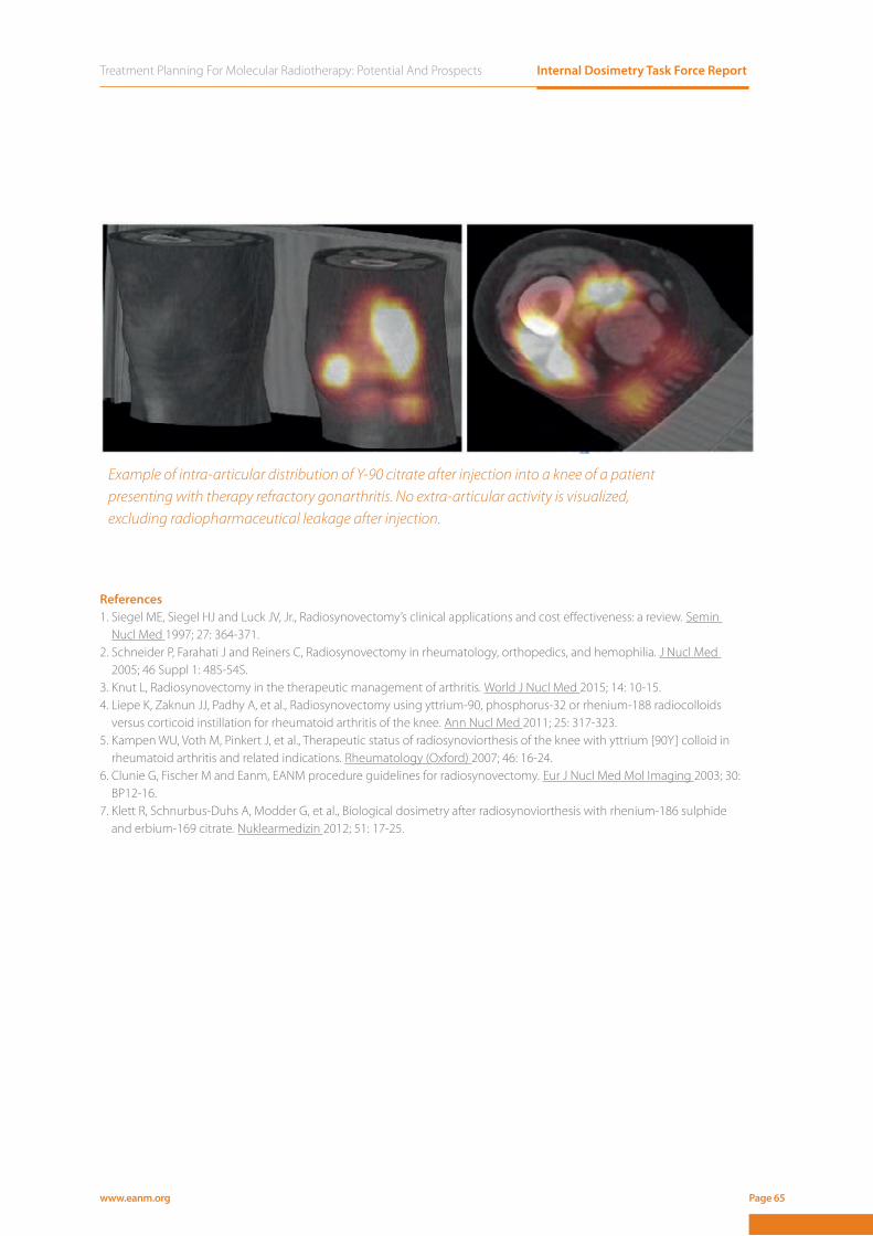
Treatment Planning For Molecular Radiotherapy: Potential And Prospects Internal Dosimetry Task Force Report
www.eanm.org Page 65
References1. Siegel ME, Siegel HJ and Luck JV, Jr., Radiosynovectomy’s clinical applications and cost eff ectiveness: a review. Semin
Nucl Med 1997; 27: 364-371.2. Schneider P, Farahati J and Reiners C, Radiosynovectomy in rheumatology, orthopedics, and hemophilia. J Nucl Med
2005; 46 Suppl 1: 48S-54S.3. Knut L, Radiosynovectomy in the therapeutic management of arthritis. World J Nucl Med 2015; 14: 10-15.4. Liepe K, Zaknun JJ, Padhy A, et al., Radiosynovectomy using yttrium-90, phosphorus-32 or rhenium-188 radiocolloids
versus corticoid instillation for rheumatoid arthritis of the knee. Ann Nucl Med 2011; 25: 317-323.5. Kampen WU, Voth M, Pinkert J, et al., Therapeutic status of radiosynoviorthesis of the knee with yttrium [90Y] colloid in
rheumatoid arthritis and related indications. Rheumatology (Oxford) 2007; 46: 16-24.6. Clunie G, Fischer M and Eanm, EANM procedure guidelines for radiosynovectomy. Eur J Nucl Med Mol Imaging 2003; 30:
BP12-16.7. Klett R, Schnurbus-Duhs A, Modder G, et al., Biological dosimetry after radiosynoviorthesis with rhenium-186 sulphide
and erbium-169 citrate. Nuklearmedizin 2012; 51: 17-25.
Example of intra-articular distribution of Y-90 citrate after injection into a knee of a patient presenting with therapy refractory gonarthritis. No extra-articular activity is visualized, excluding radiopharmaceutical leakage after injection.

Internal Dosimetry Task Force Report Treatment Planning For Molecular Radiotherapy: Potential And Prospects
www.eanm.orgPage 66
RESOURCE REQUIREMENTSImplementation of dosimetry for therapy, particularly on a routine basis, has implications for infrastructure re-
sourcing. The level of resources required will depend on the complexity of the dosimetry procedure and will
vary according to local and national protocols and guidelines. Each procedure will have resource implications
for both an initial set up of a dosimetry service and for ongoing support.
Resources fall into categories of equipment and staff. Whole-body dosimetry may be performed with an exter-
nally mounted compensated Geiger counter which enables multiple measurements to be made by either staff
or, if necessary, by comforters and carers. A portable Geiger counter may also be used and, not uncommonly, a
patient may be imaged although for high levels of 131I deadtime effects must be accounted for. Blood dosimetry
may be derived from samples measured in a well counter.
Image-based dosimetry requires a gamma camera that has been set up for the radionuclide under investigation
at activity levels relevant to the procedure. In addition to routine quality control procedures, this entails deter-
mination of calibration factors for image quantification and characterisation of camera deadtime. PET systems, if
used for dosimetry, must also be set up for quantitative imaging for each radionuclide imaged. Volume estima-
tion may be acquired from CT or, in the case of radioiodine treatment of benign thyroid disease, from ultrasound
scanning as well as from the SPECT and PET data.
As a multidisciplinary area, a range of trained staff are necessary to provide a comprehensive service. These
include medical physicists for image quantification and absorbed dose calculations, nuclear medicine technol-
ogists and radiographers with experience in high activity imaging, nurses for blood sampling, physicians and
radiologists for volume outlining and possibly paediatric specialists to help with imaging.
CONCLUSIONThe treatment of cancer with radiopharmaceuticals is undergoing a significant expansion. Many new radiother-
apeutics are being introduced into the clinic at costs in line with those for conventional chemotherapeutics
and an increasing number of patients are being treated for common as well as rare cancers. This will have a
significant effect on healthcare funding, patient management, and on the logistical and scientific challenges
faced by nuclear medicine departments. As medicine in general begins to focus on personalised cancer, and
as molecular imaging continues to attract increased attention (and funding), this growth in radiotherapeutics
offers unprecedented opportunities for recognition, support and scientific and clinical advance. As can be seen
in this report, the groundwork for dosimetry-based individualisation of treatment has been laid. Further devel-
opment will surely follow as dosimetry is introduced into the clinic.
www.eanm.org Page 67
CONTRIBUTORS
EditorsGlenn Flux, Joint Department of Physics, Royal Marsden Hospital and Institute of Cancer Research, Sutton, UK
Caroline Stokke, Department of Diagnostic Physics, Oslo University Hospital, Oslo, Norway
Responsible for the surveyKatarina Sjogreen Gleisner, Department of Medical Radiation Physics, Clinical Sciences Lund, Lund University,
Sweden
Emiliano Spezi, School of Engineering, Cardiff University, Cardiff, UK
Section authors and members of the Internal Dosimetry Task ForceMatt Aldridge, Nuclear Medicine/Radiotherapy physics, UCL Institute of Nuclear Medicine and UCL Hospitals
NHS Foundation Trust, London, UK
Klaus Bacher, Department of Basic Medical Sciences, Division of Medical Physics Ghent University, Ghent, Belgium
Boudewijn Brans, Department of Nuclear Medicine and PET Center, University Hospital, Ghent, Belgium
Carlo Chiesa, Nuclear Medicine Division, Foundation IRCCS istituto nazionale Tumori, Milan Italy
Francesco Cicone, Nuclear Medicine, Sant’Andrea Hospital, Department of Surgical and Medical Sciences
and Translational Medicine, Sapienza University of Rome, Rome, Italy
Carsten Kobe, Department for Nuclear Medicine, University Hospital of Cologne, Cologne, Germany
Mark Konijnenberg, Department of radiology & nuclear Medicine, Erasmus MC, Rotterdam, The Netherlands
Pablo Minguez Gabina, Department of Medical Physics and Radiation Protection, Gurutzeta/Cruces University
Hospital, Barakaldo, Spain
Maria Paphiti, Department of Medical Physics, Pammakaristos Hospital, Athens, Greece
Mattias Sandstrom, Section of Nuclear Medicine and PET, Department of Surgical Sciences, Uppsala
University, Uppsala, Sweden
Pavel Solny, Department of Nuclear Medicine and Endocrinology, Motol University Hospital, 2nd Faculty of
Medicine Charles University in Prague, Czech Republic
Jill Tipping, The Christie NHS Foundation Trust, Nuclear Medicine, Manchester, UK
Michael Wissmeyer, Department of Nuclear Medicine, University Hospital of Geneva, Geneva, Switzerland

www.eanm.org
Top Related