


Languages
Pages
Legal


Chapter 17

MammographyGold standard for breast cancer detection
Most common in women other than skin cancer2nd leading cause of cancer death in women1 in 8 chance of getting breast cancer1 in 35 chance of dying from it.
MammographyHighly regulated
Mammography Quality Standards Act (MQSA)
All Mammography units must meet federal standards. ACR accredited.
State of Nevada requires annual inspections
External Breast AnatomyInframammary fold
Inferior breast to the chest wall
Axillary TailSuperiolateral aspect

External Breast AnatomyAreola
Dark area surrounding nipple.Montgomery’s gland or Areolar glands (are
Sabaceous glands)
NippleProtrusion containing duct openings

Boundaries of the Breast
Superiorly: 1st or 2nd rib (clavicle)Inferiorly: 6th or 7th (IMF-Inframammary fold)Medially: SternumLaterally: Mid-axillary line at the junction of
the latissimus dorsi muscle.

Internal Breast AnatomyPectoralis Major
Chest muscle posterior to breast.
Retromammary SpaceConnective tissue attaching breast to
pectoralis major.

Parenchyma (Breast) Tissue
Glandular TissueMilk production and duct system
Adipose TissueFatty tissue surrounding glandular tissue
Fibrous (Connective) TissueSurround and support the glandular structures
(Cooper’s Ligament)
Glandular Tissue15 – 20 lobes surrounding the nipple
Alveoli (Acini)Milk producing units
LobulesGroups of alveoli
Glandular TissueDuct
Transports breast milk
AmpulaReservoir at the end of ducts
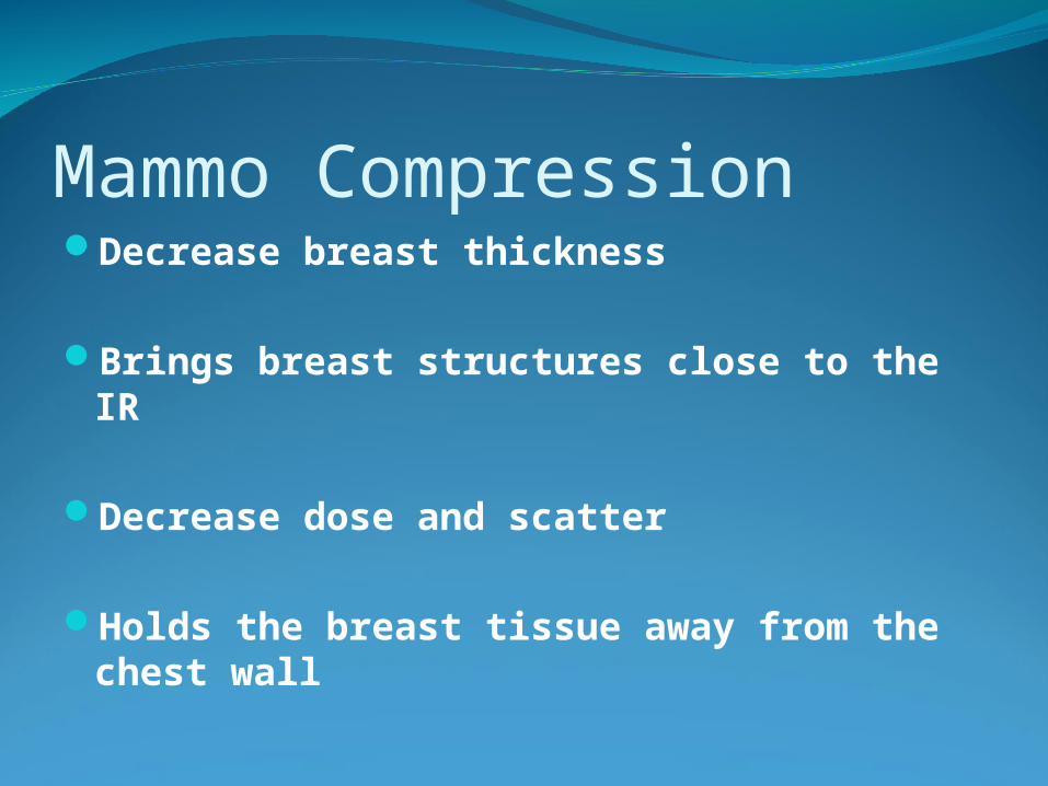
Mammo CompressionDecrease breast thickness
Brings breast structures close to the IR Decrease dose and scatter
Holds the breast tissue away from the chest wall
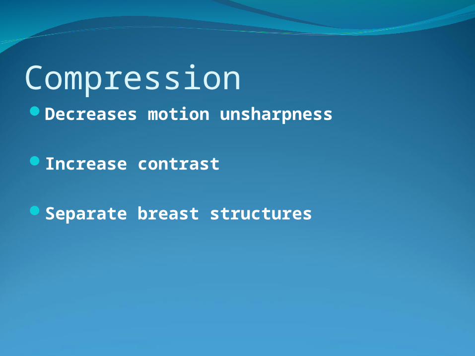
CompressionDecreases motion unsharpness
Increase contrast
Separate breast structures

PositioningCraniocaudal (CC)
Mediolateral Oblique (MLO)
Others are possible

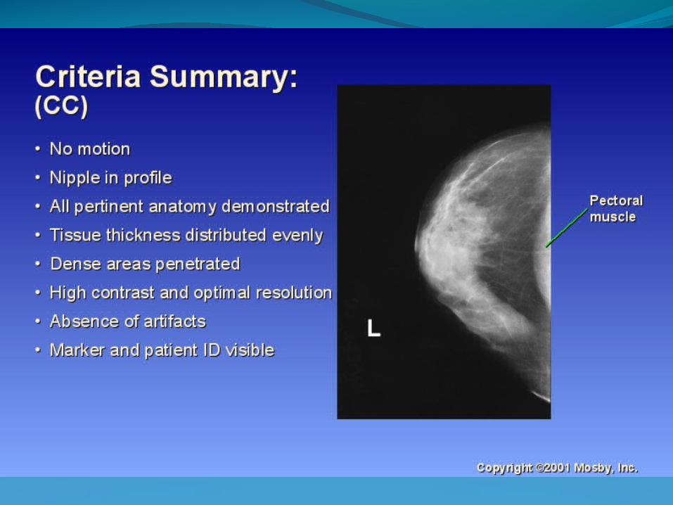
Craniocaudal (CC)IR raised for 90º chest wall angle.Compression appliedCorrect placement of the photocell in film
screen mammography is under the most glandular tissue
Typically the anterior third of the breastPectoral muscle should be seen
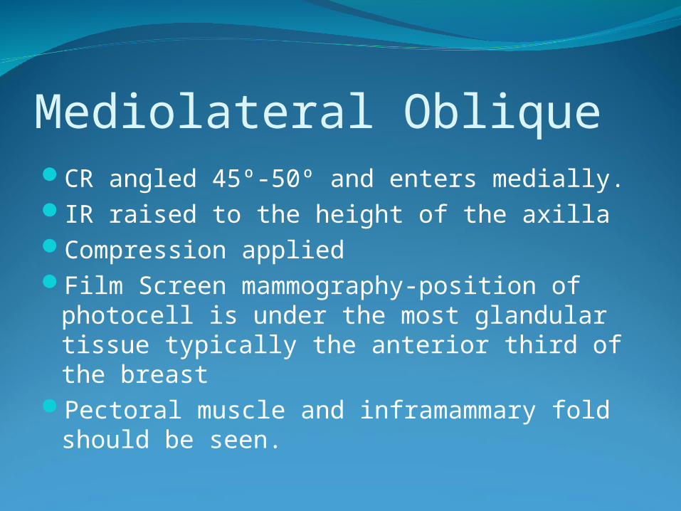
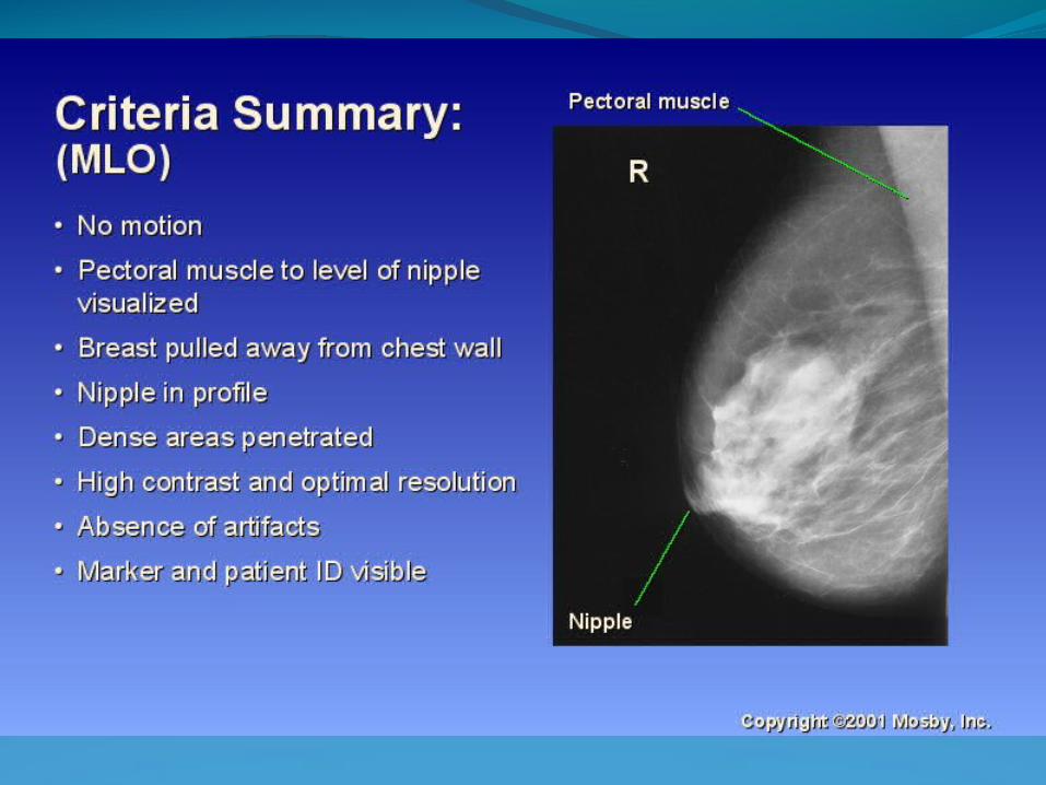
Mediolateral Oblique CR angled 45º-50º and enters medially.IR raised to the height of the axillaCompression appliedFilm Screen mammography-position of
photocell is under the most glandular tissue typically the anterior third of the breast
Pectoral muscle and inframammary fold should be seen.

Mammography AdvancementFull Field Digital MammographyComputer Aided Detection (CAD)UltrasoundMRI and MR SpectroscopyNuc MedBSGI Breast Specific Gamma Imaging (Caution-
residual radiotracers have been linked to Colon Cancer)
3 D Mammography or Breast Tomosynthesis (Standard mammographic views with approximately
50 individual images. Same compression and positioning.)
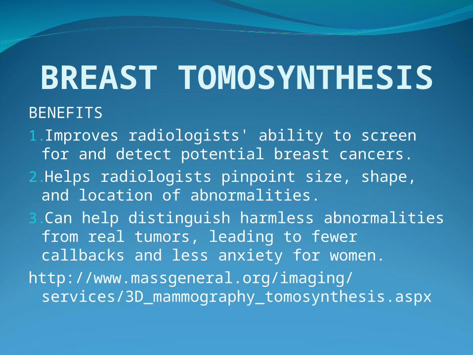
BREAST TOMOSYNTHESISBENEFITS1.Improves radiologists' ability to screen for
and detect potential breast cancers. 2.Helps radiologists pinpoint size, shape, and
location of abnormalities. 3.Can help distinguish harmless abnormalities
from real tumors, leading to fewer callbacks and less anxiety for women.
http://www.massgeneral.org/imaging/services/3D_mammography_tomosynthesis.aspx

Getting RegisteredNew Mandates from ARRT
Effective July 200940 Hours of Initial Training
Approx cost -$625 4 day programCandidates must be in compliance with MQSA
requirements for technologists, and are required to complete: (a) a specified
number of mammographicexaminations; (b) quality control procedures; (c) selected
special procedures; (d) mammographicreview and critique. All procedures must be completed
within the 24 months immediately beforeapplication for certification.
www.achievingqi.com- Las Vegas, Multiple dates 2011
Top Related