


Languages
Pages
Legal


Carpal Tunnel SyndromeCarpal Tunnel SyndromeStacey Harris-Carriman, M.D.Stacey Harris-Carriman, M.D.
Physical Medicine and RehabilitationPhysical Medicine and Rehabilitation
Noon Conference, CCRMCNoon Conference, CCRMCMay 8, 2009May 8, 2009
ObjectivesObjectives
• Be familiar with the basic neuroanatomy of the upper limb
• Understand factors involved in diagnosing CTS
• Recognize the goals and limitations of NCS
• Review treatment of CTS

OutlineOutline
• Definition
• Etiology and Risk Factors
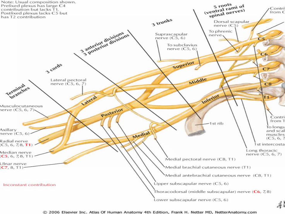
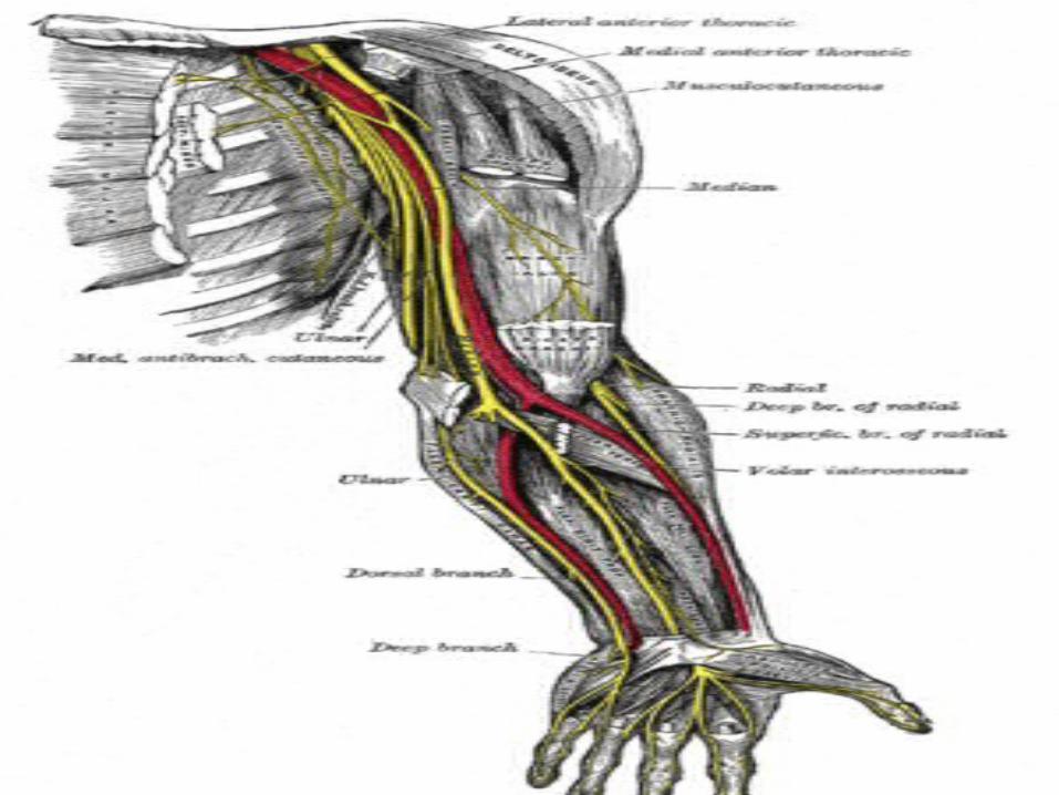
• Neuroanatomy of the Upper Limb
• Diagnosis: Symptoms and signs
• Differential diagnosis
• NCS/EMG and US
• Treatment

Definition of CTSDefinition of CTS
• Constellation of symptoms and signs secondary to a median neuropathy at the wrist

OutlineOutline
• Definition
• Etiology and Risk Factors
• Neuroanatomy of the Upper Limb
• Diagnosis: Symptoms and signs
• Differential diagnosis
• NCS/EMG and US
• Treatment

EtiologyEtiology
• Majority of CTS cases idiopathic
EtiologyEtiology
• Small percentage of CTS due to an identifiable cause, such as:– DM, RA, thyroid
disease– Conditions that
increase total body fluid (e.g. pregnancy, hemodialysis)
– Local wrist lesion (e.g. cyst, fracture, infection, tumor)
– Congenital (e.g. small carpal tunnel)
Risk FactorsRisk Factors
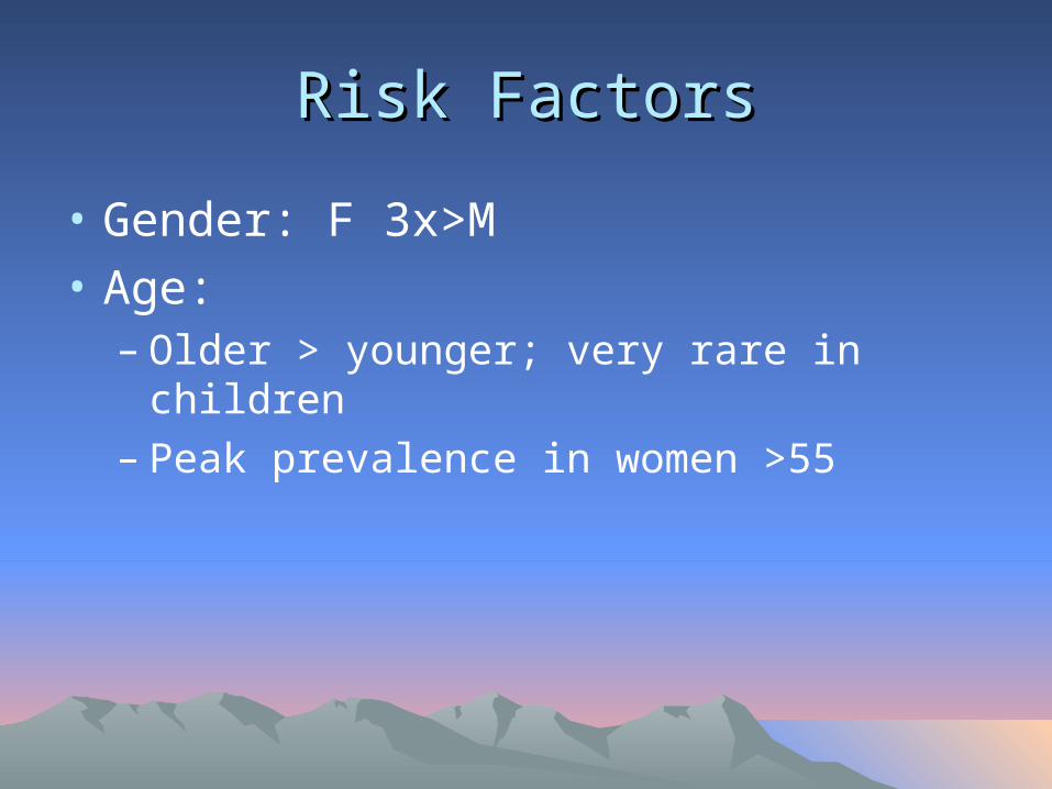
• Gender: F 3x>M
• Age: – Older > younger; very rare in children– Peak prevalence in women >55

Risk FactorsRisk Factors
• Family history• Certain medical
conditions• Workers that use
hands and wrists repetitively, especially with high force
• Musicians

Risk FactorsRisk Factors
• Other: Smoking, alcohol, poor nutrition, obesity, high cholesterol

OutlineOutline
• Definition
• Etiology and Risk Factors
• Neuroanatomy of the Upper Limb
• Diagnosis: Symptoms and signs
• Differential diagnosis
• NCS/EMG and US
• Treatment


OutlineOutline
• Definition
• Etiology and Risk Factors
• Neuroanatomy of the Upper Limb
• Diagnosis: Symptoms and signs
• Differential diagnosis
• NCS/EMG and US
• Treatment
SymptomsSymptoms
• Pattern recognition
• Wide variety of symptoms in CTS
• Some symptoms are more suggestive of CTS than other symptoms
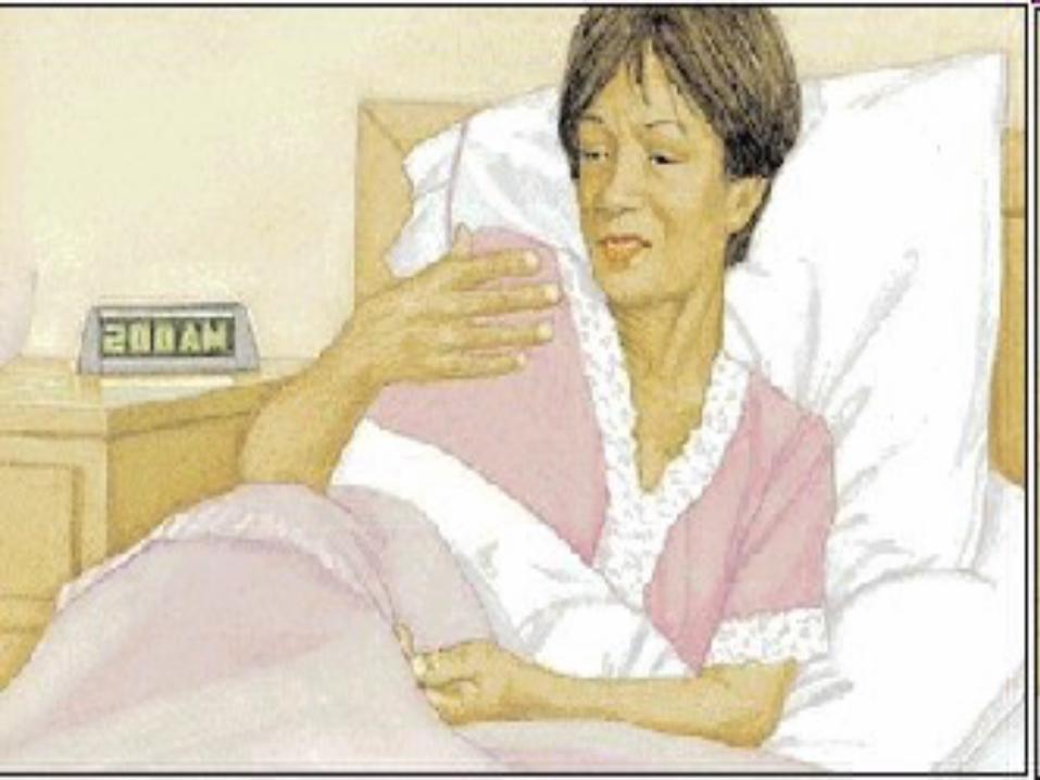

SymptomsSymptoms
• Classic symptoms in CTS:– Waking up with pain and
numbness/paresthesias of the hand – Triggered by driving, holding phone, reading
book, typing, writing– Relieving factors
• Flick sign• Changes in hand posture

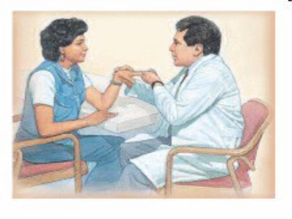
SignsSigns
• Key signs suggestive of CTS– Impaired sensation of the lateral 3-1/2 digits– Weakness of APB and other median-
innervated muscles of thenar eminence– Phalen’s, reverse Phalen’s– Tinel’s– Other: Pressure provocation test, hand
elevation test, tourniquet test


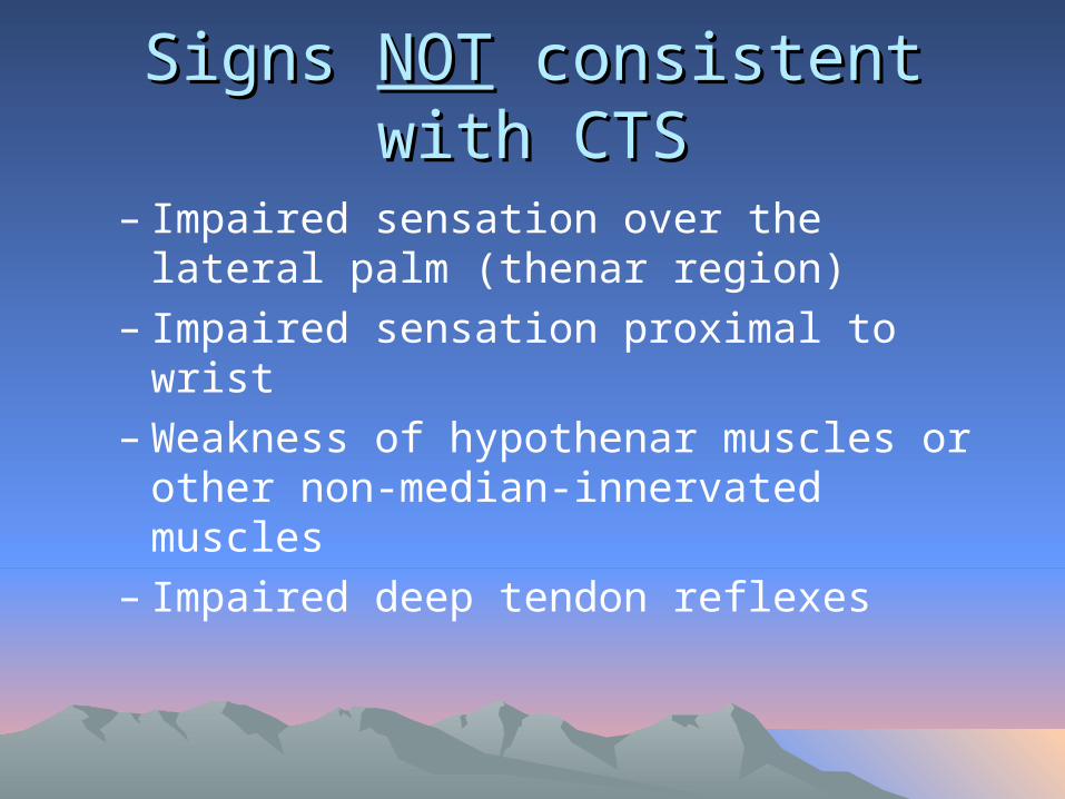
Signs Signs NOTNOT consistent with CTS consistent with CTS
– Impaired sensation over the lateral palm (thenar region)
– Impaired sensation proximal to wrist– Weakness of hypothenar muscles or other
non-median-innervated muscles– Impaired deep tendon reflexes
OutlineOutline
• Definition
• Etiology and Risk Factors
• Neuroanatomy of the Upper Limb
• Diagnosis: Symptoms and signs
• Differential diagnosis
• NCS/EMG and US
• Treatment

Differential Diagnosis of CTSDifferential Diagnosis of CTS
– Peripheral NS• Cervical radiculopathy• Brachial plexopathy• Proximal median
neuropathy (e.g. in forearm or elbow)
• Other mononeuroapthy (e.g. ulnar, radial)
• Underlying polyneuropathy
– Central NS (e.g. TIA, small lacunar infarct, myelopathy)
– Musculoskeletal • Shoulder pain with
distal paresthesias• Osteoarthritis• Cumulative trauma
disorder

Differential DiagnosisDifferential Diagnosis
• Peripheral NS: Cervical radiculopathy

DDx: Cervical RadiculopathyDDx: Cervical Radiculopathy
• Especially mild cases of cervical radiculopathy
• C6, C7
• Neck pain, radiation to shoulder, arm, +/- distally
• Worse with neck movement
• Impaired reflexes and strength
• Sensory loss beyond distribution of median nerve

Differential DiagnosisDifferential Diagnosis
• Peripheral NS: Brachial Plexopathy
DDx: Brachial PlexopathyDDx: Brachial Plexopathy
• Uncommon • Etiology: – Trauma– Tumor, Mass– Delayed radiation
injury– Plexitis– Postop (e.g. CABG)– Neurogenic TOS
DDx: Brachial PlexopathyDDx: Brachial Plexopathy
• Trauma• Most common cause of brachial
plexopathy• Mechanism:
– Traction• Car/motorcycle/bike accident, newborn • Upper trunk C5/6-Erb’s palsy• Lower trunk C8/T1-Klumpke’s palsy
– Penetrating (knife, bullet)
DDx: Brachial PlexopathyDDx: Brachial Plexopathy
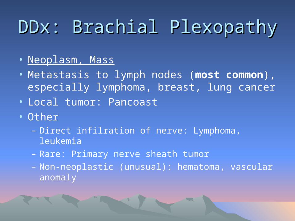
• Neoplasm, Mass• Metastasis to lymph nodes (most common),
especially lymphoma, breast, lung cancer• Local tumor: Pancoast• Other
– Direct infilration of nerve: Lymphoma, leukemia– Rare: Primary nerve sheath tumor– Non-neoplastic (unusual): hematoma, vascular
anomaly

DDx: Brachial PlexopathyDDx: Brachial Plexopathy
• Delayed Radiation VS• Onset: Progressive,
years after radiation• Risk correlated with
dose of radiation• Sensory sx
prominent (paresthesias, numbness)
• (Recurrent) Neoplasm• Onset: Slowly
progressive• Prominent pain • Horner’s syndrome

DDx: Brachial PlexopathyDDx: Brachial Plexopathy
• Brachial Plexitis• AKA Neuralgic
amyotrophy, Parsonage-Turner
• Idiopathic• Often preceded by: viral
illness or immunization; also surgery
• Long thoracic nerve, anterior interosseous nerve, other
• Shoulder pain– Onset: days to weeks after
inciting event– Severe pain, awakens from
sleep
• Weakness and atrophy– Onset: Generally after pain
subsides (1-2 weeks)
• +/- Sensory s/sx

DDx: Brachial PlexopathyDDx: Brachial Plexopathy
• Neurogenic TOS• Most cases due to
fibrous band between cervical rib and 1st thoracic rib
• Lower trunk, C8/T1
• Exam:– Muscles: hand
intrinsics, esp thenar T1; +/- FPL, FDP
– Sensory: Ulnar, MABC

Differential DiagnosisDifferential Diagnosis
• Peripheral NS: Proximal Median Neuropathy
DDx: Proximal Median NeuropathyDDx: Proximal Median Neuropathy
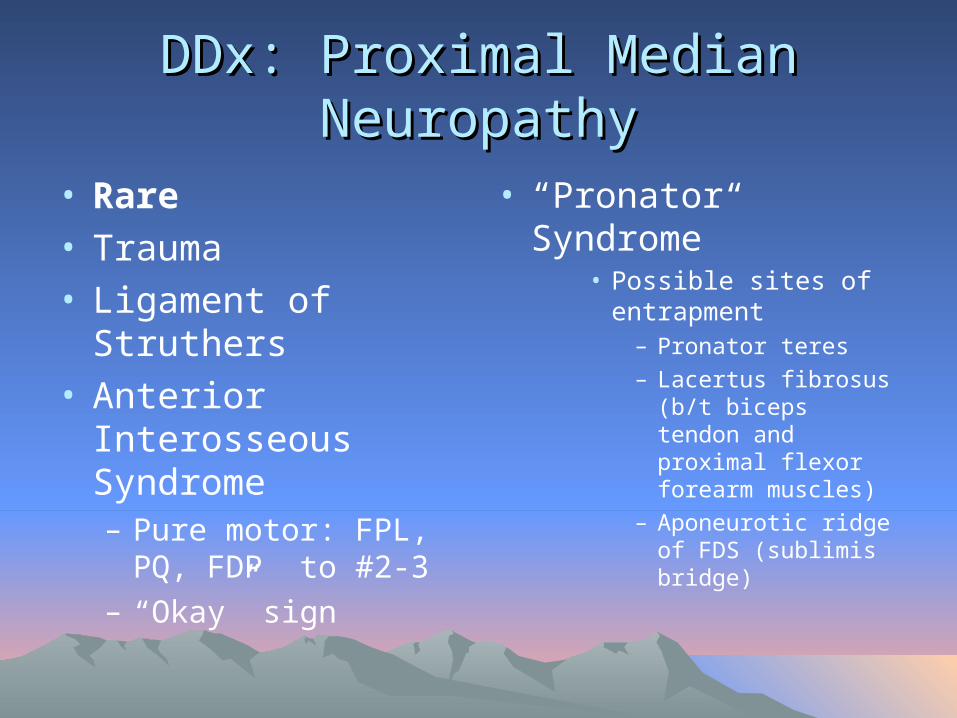
• Rare• Trauma• Ligament of Struthers• Anterior Interosseous
Syndrome– Pure motor: FPL, PQ,
FDP to #2-3– “Okay” sign
• “Pronator Syndrome”• Possible sites of
entrapment– Pronator teres
– Lacertus fibrosus (b/t biceps tendon and proximal flexor forearm muscles)
– Aponeurotic ridge of FDS (sublimis bridge)
Differential DiagnosisDifferential Diagnosis
• Peripheral NS: Other Mononeuropathy
• Ulnar, Radial

Differential DiagnosisDifferential Diagnosis
• Peripheral NS: Peripheral Polyneuropathy

Differential DiagnosisDifferential Diagnosis
• CNS: Cervical Myelopathy

Differential DiagnosisDifferential Diagnosis
• Musculoskeletal: Shoulder Pathology with Distal Paresthesias

OutlineOutline
• Definition
• Etiology and Risk Factors
• Neuroanatomy of the Upper Limb
• Diagnosis: Symptoms and signs
• Differential diagnosis
• NCS/EMG and US
• Treatment

Nerve Conduction Studies (NCS)Nerve Conduction Studies (NCS)
• [NOTE: NCS sometimes called NCV “Nerve Conduction Velocity”]

NCSNCS
• Picture here of NCS set-up
NCSNCS
• NCS can be useful in confirming CTS and assessing severity of CTS
NCSNCS
• An extension of the clinical examination
• Each NCS study must be individualized
NCSNCS
• NCS is positive in 91-98% of patients with clinically diagnosed CTS
• (Source: Keles et al, Diagnostic precision of ultrasonography in patients with CTS, Am J Phys Med Rehabil 2005)
• Risk of false negatives on NCS generally implies very mild CTS

Diagnostic Ultrasound Diagnostic Ultrasound
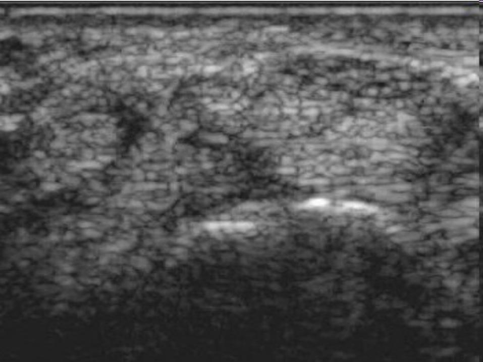
• Real-time imaging of median nerve in carpal tunnel
• Qualitative and quantitative
• Measurements can include:– Cross-sectional area (CSA) of median nerve– Bowing of flexor retinaculum– Flattening of median nerve in carpal tunnel

Diagnostic UltrasoundDiagnostic Ultrasound
• Relatively new development
• Aids in diagnosis
• Aids in treatment, ultrasound-guided injection of steroid into carpal tunnel

OutlineOutline
• Definition
• Etiology and Risk Factors
• Neuroanatomy of the Upper Limb
• Diagnosis: Symptoms and signs
• Differential diagnosis
• NCS/EMG and US
• Treatment
Treatment of CTSTreatment of CTS


Summary and ConclusionSummary and Conclusion

CTS: Summary and ConclusionCTS: Summary and Conclusion
• The diagnosis of CTS is made on clinical grounds
• Pattern recognition
• Be systematic: history, physical, differential diagnosis

Summary and ConclusionSummary and Conclusion
• NCS/EMG can be useful in confirming CTS and assessing severity of CTS
• Ultrasound can be a helpful adjunct in assessing and treating CTS

QuestionsQuestions
Top Related