
Carpal Tunnel Syndrome
63
Carpal Tunnel Syndrome Carpal Tunnel Syndrome Stacey Harris-Carriman, M.D. Stacey Harris-Carriman, M.D. Physical Medicine and Physical Medicine and Rehabilitation Rehabilitation Noon Conference, CCRMC Noon Conference, CCRMC May 8, 2009 May 8, 2009
-
Upload
walter-hyde -
Category
Documents
-
view
15 -
download
0
description
Carpal Tunnel Syndrome. Stacey Harris-Carriman, M.D. Physical Medicine and Rehabilitation Noon Conference, CCRMC May 8, 2009. Objectives. Be familiar with the basic neuroanatomy of the upper limb Understand factors involved in diagnosing CTS Recognize the goals and limitations of NCS - PowerPoint PPT Presentation
Transcript of Carpal Tunnel Syndrome

Carpal Tunnel SyndromeCarpal Tunnel SyndromeStacey Harris-Carriman, M.D.Stacey Harris-Carriman, M.D.
Physical Medicine and RehabilitationPhysical Medicine and Rehabilitation
Noon Conference, CCRMCNoon Conference, CCRMCMay 8, 2009May 8, 2009
ObjectivesObjectives
• Be familiar with the basic neuroanatomy of the upper limb
• Understand factors involved in diagnosing CTS
• Recognize the goals and limitations of NCS
• Review treatment of CTS

OutlineOutline
• Definition
• Etiology and Risk Factors
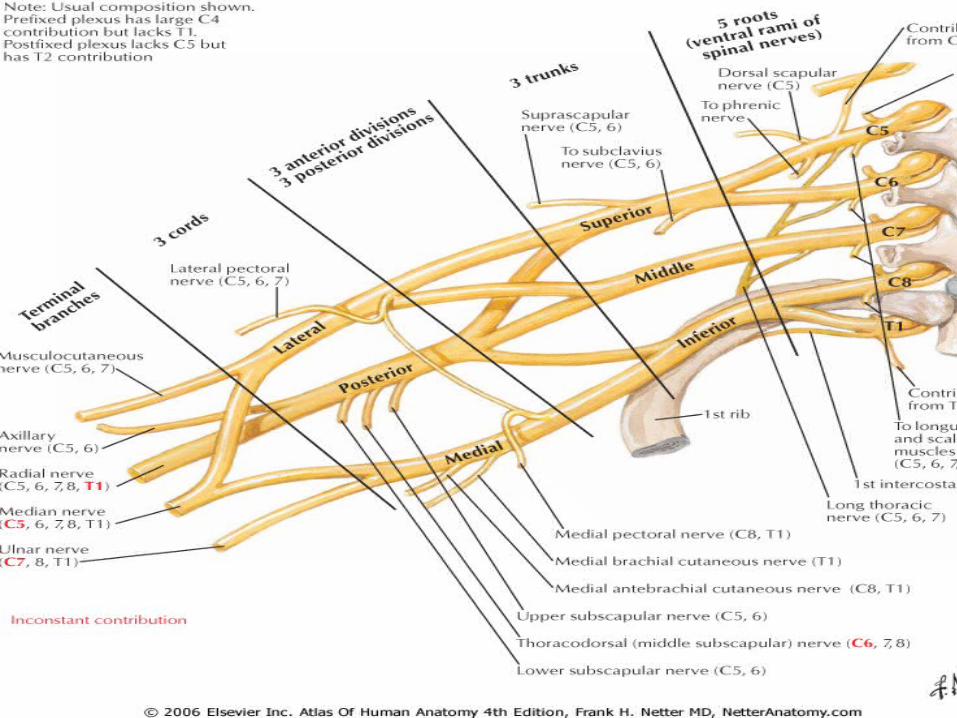
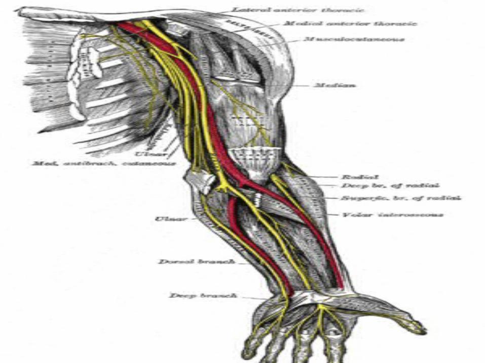
• Neuroanatomy of the Upper Limb
• Diagnosis: Symptoms and signs
• Differential diagnosis
• NCS/EMG and US
• Treatment

Definition of CTSDefinition of CTS
• Constellation of symptoms and signs secondary to a median neuropathy at the wrist

OutlineOutline
• Definition
• Etiology and Risk Factors
• Neuroanatomy of the Upper Limb
• Diagnosis: Symptoms and signs
• Differential diagnosis
• NCS/EMG and US
• Treatment

EtiologyEtiology
• Majority of CTS cases idiopathic
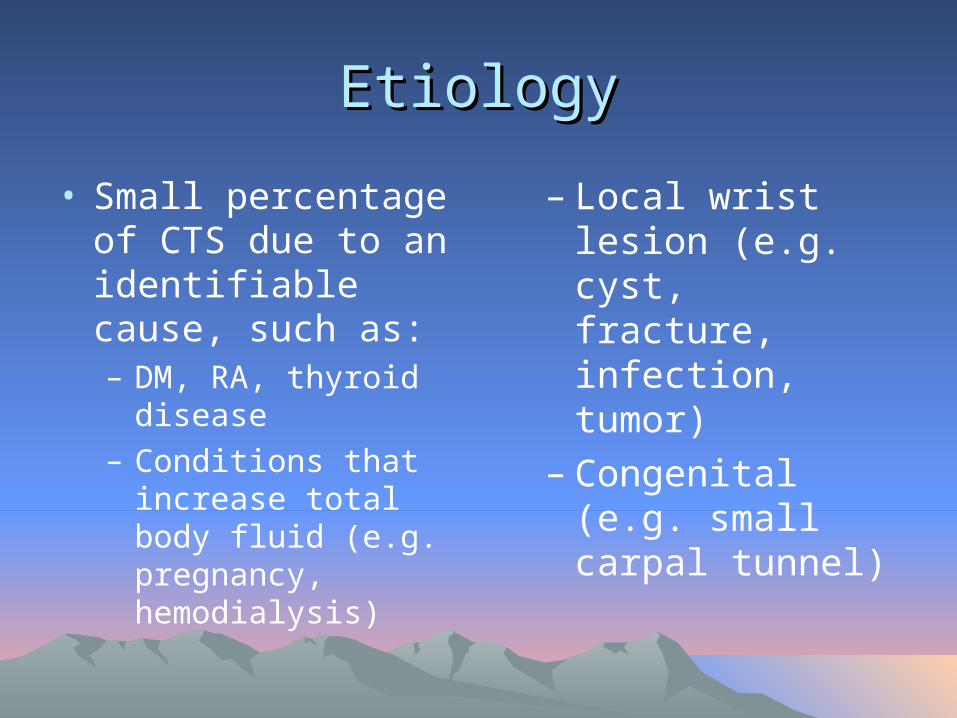
EtiologyEtiology
• Small percentage of CTS due to an identifiable cause, such as:– DM, RA, thyroid
disease– Conditions that
increase total body fluid (e.g. pregnancy, hemodialysis)
– Local wrist lesion (e.g. cyst, fracture, infection, tumor)
– Congenital (e.g. small carpal tunnel)
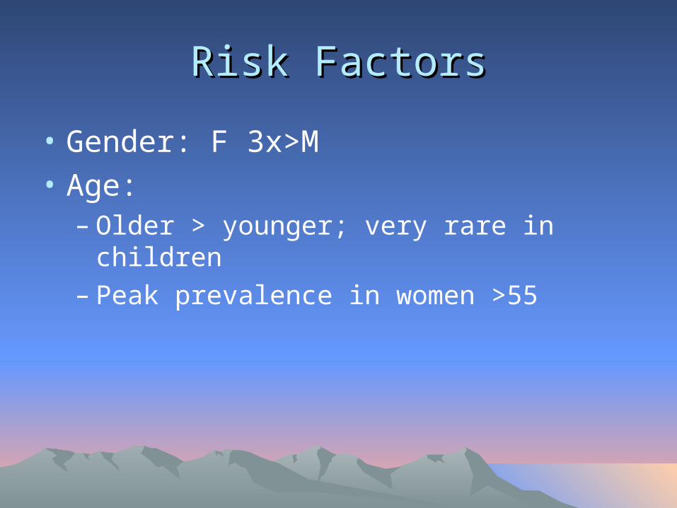
Risk FactorsRisk Factors
• Gender: F 3x>M
• Age: – Older > younger; very rare in children– Peak prevalence in women >55

Risk FactorsRisk Factors
• Family history• Certain medical
conditions• Workers that use
hands and wrists repetitively, especially with high force
• Musicians

Risk FactorsRisk Factors
• Other: Smoking, alcohol, poor nutrition, obesity, high cholesterol

OutlineOutline
• Definition
• Etiology and Risk Factors
• Neuroanatomy of the Upper Limb
• Diagnosis: Symptoms and signs
• Differential diagnosis
• NCS/EMG and US
• Treatment


OutlineOutline
• Definition
• Etiology and Risk Factors
• Neuroanatomy of the Upper Limb
• Diagnosis: Symptoms and signs
• Differential diagnosis
• NCS/EMG and US
• Treatment
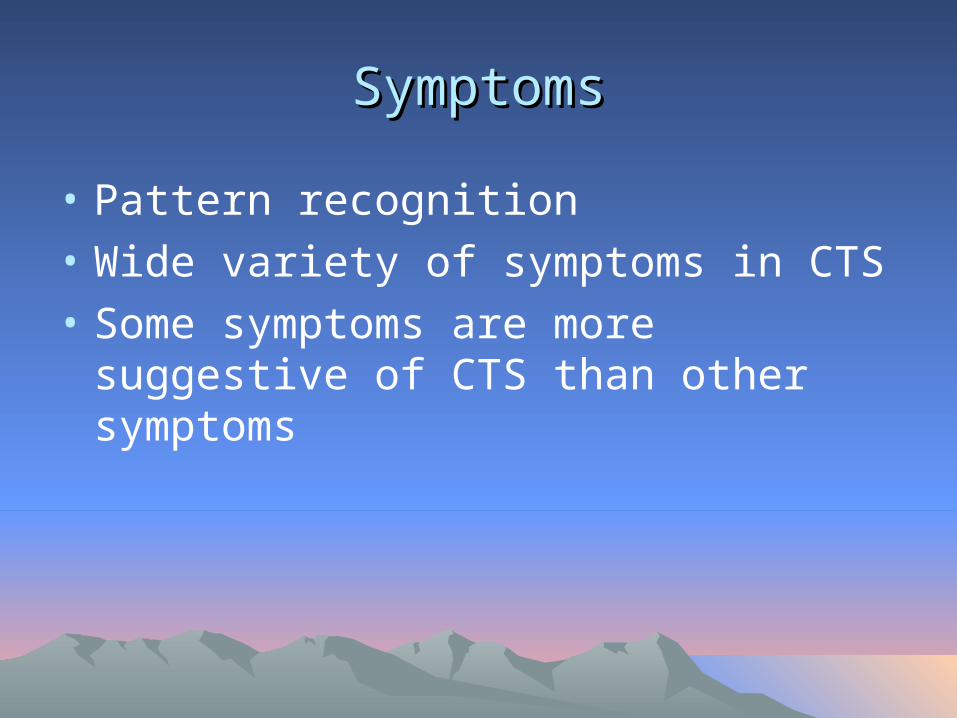
SymptomsSymptoms
• Pattern recognition
• Wide variety of symptoms in CTS
• Some symptoms are more suggestive of CTS than other symptoms

SymptomsSymptoms
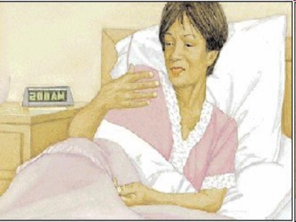
• Classic symptoms in CTS:– Waking up with pain and
numbness/paresthesias of the hand – Triggered by driving, holding phone, reading
book, typing, writing– Relieving factors
• Flick sign• Changes in hand posture

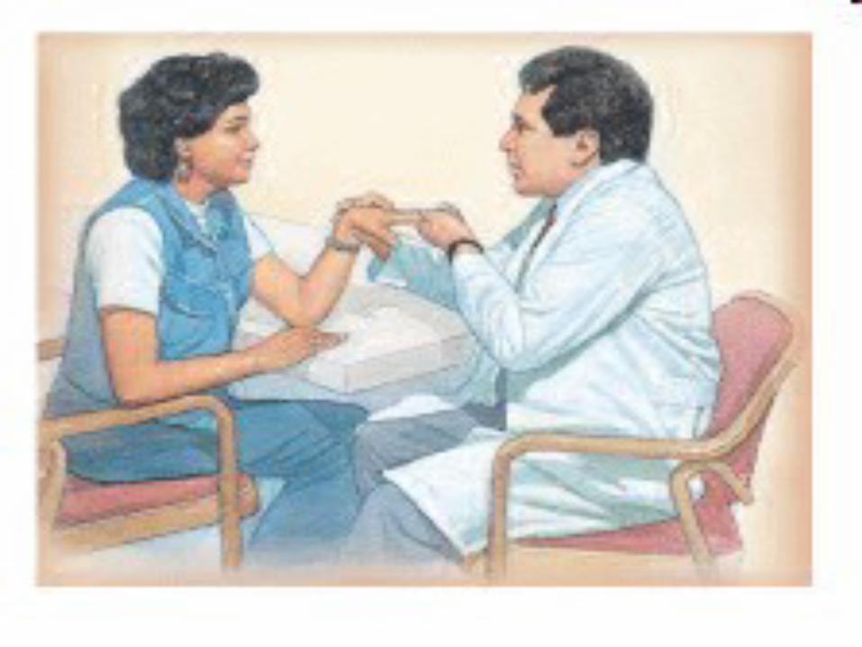
SignsSigns
• Key signs suggestive of CTS– Impaired sensation of the lateral 3-1/2 digits– Weakness of APB and other median-
innervated muscles of thenar eminence– Phalen’s, reverse Phalen’s– Tinel’s– Other: Pressure provocation test, hand
elevation test, tourniquet test


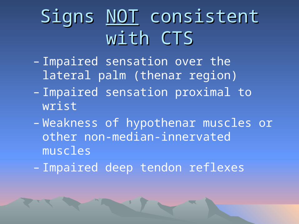
Signs Signs NOTNOT consistent with CTS consistent with CTS
– Impaired sensation over the lateral palm (thenar region)
– Impaired sensation proximal to wrist– Weakness of hypothenar muscles or other
non-median-innervated muscles– Impaired deep tendon reflexes
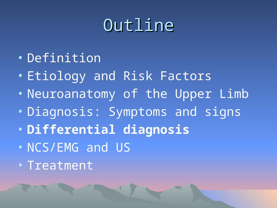
OutlineOutline
• Definition
• Etiology and Risk Factors
• Neuroanatomy of the Upper Limb
• Diagnosis: Symptoms and signs
• Differential diagnosis
• NCS/EMG and US
• Treatment

Differential Diagnosis of CTSDifferential Diagnosis of CTS
– Peripheral NS• Cervical radiculopathy• Brachial plexopathy• Proximal median
neuropathy (e.g. in forearm or elbow)
• Other mononeuroapthy (e.g. ulnar, radial)
• Underlying polyneuropathy
– Central NS (e.g. TIA, small lacunar infarct, myelopathy)
– Musculoskeletal • Shoulder pain with
distal paresthesias• Osteoarthritis• Cumulative trauma
disorder

Differential DiagnosisDifferential Diagnosis
• Peripheral NS: Cervical radiculopathy

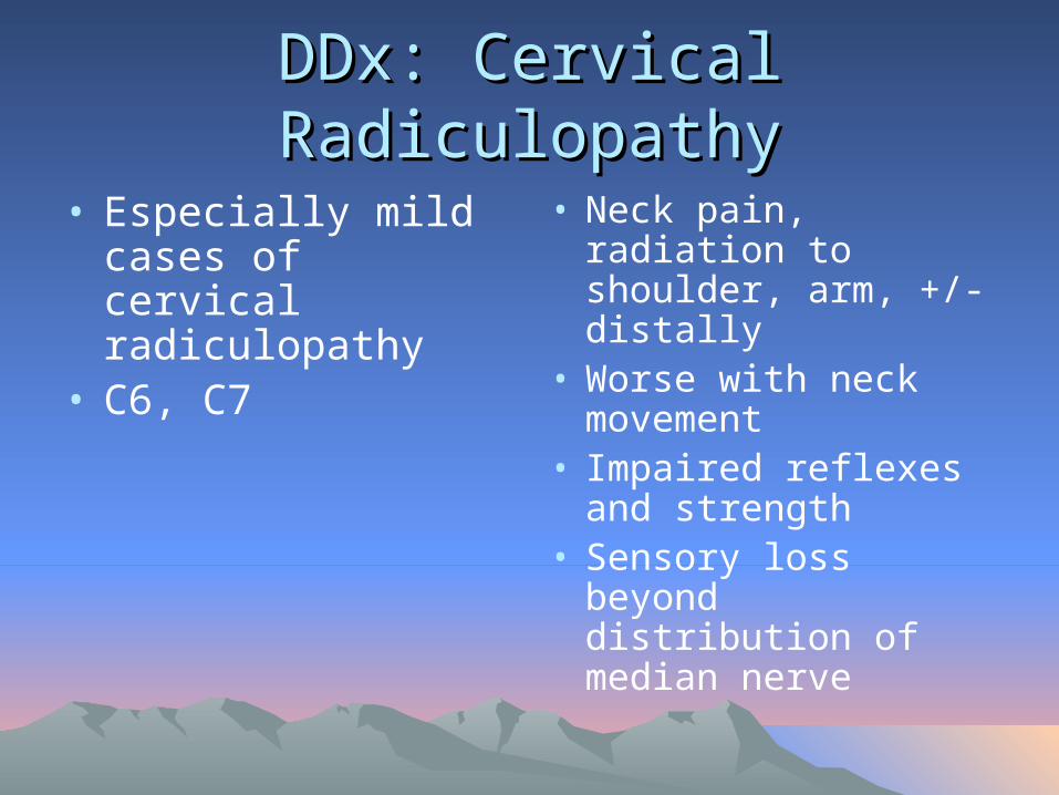
DDx: Cervical RadiculopathyDDx: Cervical Radiculopathy
• Especially mild cases of cervical radiculopathy
• C6, C7
• Neck pain, radiation to shoulder, arm, +/- distally
• Worse with neck movement
• Impaired reflexes and strength
• Sensory loss beyond distribution of median nerve

Differential DiagnosisDifferential Diagnosis
• Peripheral NS: Brachial Plexopathy
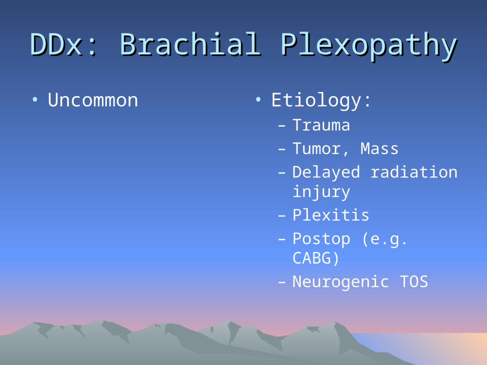
DDx: Brachial PlexopathyDDx: Brachial Plexopathy
• Uncommon • Etiology: – Trauma– Tumor, Mass– Delayed radiation
injury– Plexitis– Postop (e.g. CABG)– Neurogenic TOS
DDx: Brachial PlexopathyDDx: Brachial Plexopathy
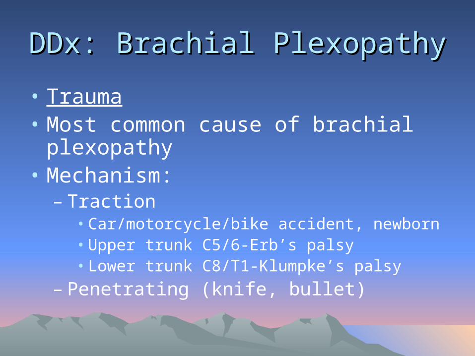
• Trauma• Most common cause of brachial
plexopathy• Mechanism:
– Traction• Car/motorcycle/bike accident, newborn • Upper trunk C5/6-Erb’s palsy• Lower trunk C8/T1-Klumpke’s palsy
– Penetrating (knife, bullet)
DDx: Brachial PlexopathyDDx: Brachial Plexopathy
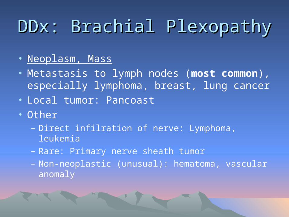
• Neoplasm, Mass• Metastasis to lymph nodes (most common),
especially lymphoma, breast, lung cancer• Local tumor: Pancoast• Other
– Direct infilration of nerve: Lymphoma, leukemia– Rare: Primary nerve sheath tumor– Non-neoplastic (unusual): hematoma, vascular
anomaly

DDx: Brachial PlexopathyDDx: Brachial Plexopathy
• Delayed Radiation VS• Onset: Progressive,
years after radiation• Risk correlated with
dose of radiation• Sensory sx
prominent (paresthesias, numbness)
• (Recurrent) Neoplasm• Onset: Slowly
progressive• Prominent pain • Horner’s syndrome

DDx: Brachial PlexopathyDDx: Brachial Plexopathy
• Brachial Plexitis• AKA Neuralgic
amyotrophy, Parsonage-Turner
• Idiopathic• Often preceded by: viral
illness or immunization; also surgery
• Long thoracic nerve, anterior interosseous nerve, other
• Shoulder pain– Onset: days to weeks after
inciting event– Severe pain, awakens from
sleep
• Weakness and atrophy– Onset: Generally after pain
subsides (1-2 weeks)
• +/- Sensory s/sx

DDx: Brachial PlexopathyDDx: Brachial Plexopathy
• Neurogenic TOS• Most cases due to
fibrous band between cervical rib and 1st thoracic rib
• Lower trunk, C8/T1
• Exam:– Muscles: hand
intrinsics, esp thenar T1; +/- FPL, FDP
– Sensory: Ulnar, MABC

Differential DiagnosisDifferential Diagnosis
• Peripheral NS: Proximal Median Neuropathy
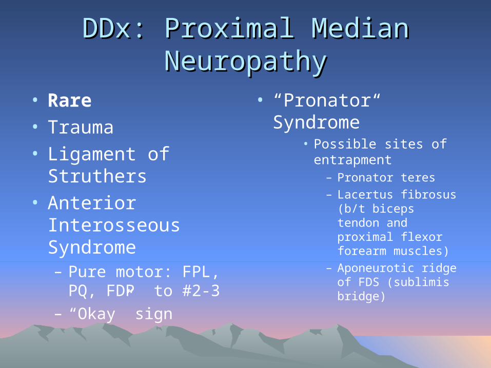
DDx: Proximal Median NeuropathyDDx: Proximal Median Neuropathy
• Rare• Trauma• Ligament of Struthers• Anterior Interosseous
Syndrome– Pure motor: FPL, PQ,
FDP to #2-3– “Okay” sign
• “Pronator Syndrome”• Possible sites of
entrapment– Pronator teres
– Lacertus fibrosus (b/t biceps tendon and proximal flexor forearm muscles)
– Aponeurotic ridge of FDS (sublimis bridge)
Differential DiagnosisDifferential Diagnosis
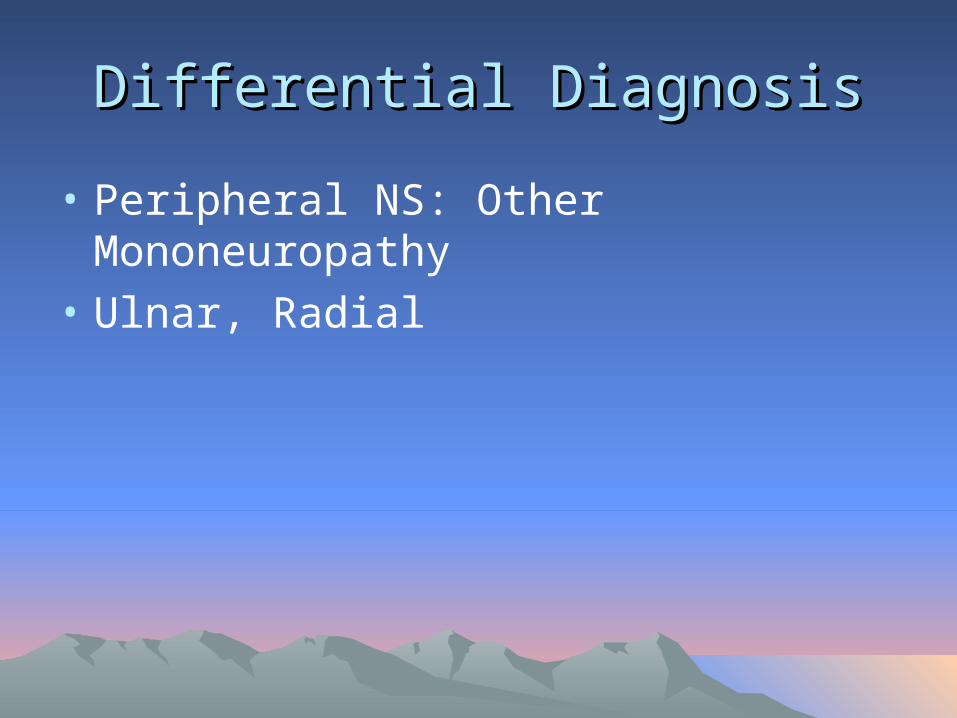
• Peripheral NS: Other Mononeuropathy
• Ulnar, Radial

Differential DiagnosisDifferential Diagnosis
• Peripheral NS: Peripheral Polyneuropathy

Differential DiagnosisDifferential Diagnosis
• CNS: Cervical Myelopathy

Differential DiagnosisDifferential Diagnosis
• Musculoskeletal: Shoulder Pathology with Distal Paresthesias

OutlineOutline
• Definition
• Etiology and Risk Factors
• Neuroanatomy of the Upper Limb
• Diagnosis: Symptoms and signs
• Differential diagnosis
• NCS/EMG and US
• Treatment

Nerve Conduction Studies (NCS)Nerve Conduction Studies (NCS)
• [NOTE: NCS sometimes called NCV “Nerve Conduction Velocity”]

NCSNCS
• Picture here of NCS set-up
NCSNCS
• NCS can be useful in confirming CTS and assessing severity of CTS
NCSNCS
• An extension of the clinical examination
• Each NCS study must be individualized
NCSNCS
• NCS is positive in 91-98% of patients with clinically diagnosed CTS
• (Source: Keles et al, Diagnostic precision of ultrasonography in patients with CTS, Am J Phys Med Rehabil 2005)
• Risk of false negatives on NCS generally implies very mild CTS

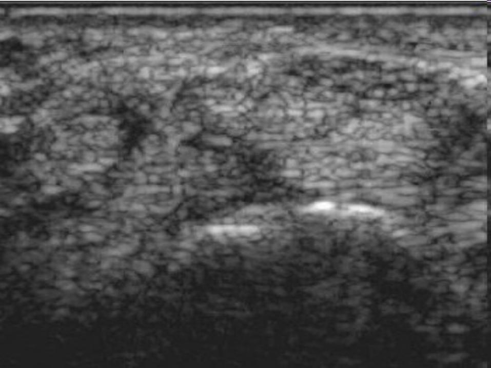
Diagnostic Ultrasound Diagnostic Ultrasound
• Real-time imaging of median nerve in carpal tunnel
• Qualitative and quantitative
• Measurements can include:– Cross-sectional area (CSA) of median nerve– Bowing of flexor retinaculum– Flattening of median nerve in carpal tunnel

Diagnostic UltrasoundDiagnostic Ultrasound
• Relatively new development
• Aids in diagnosis
• Aids in treatment, ultrasound-guided injection of steroid into carpal tunnel

OutlineOutline
• Definition
• Etiology and Risk Factors
• Neuroanatomy of the Upper Limb
• Diagnosis: Symptoms and signs
• Differential diagnosis
• NCS/EMG and US
• Treatment
Treatment of CTSTreatment of CTS


Summary and ConclusionSummary and Conclusion

CTS: Summary and ConclusionCTS: Summary and Conclusion
• The diagnosis of CTS is made on clinical grounds
• Pattern recognition
• Be systematic: history, physical, differential diagnosis

Summary and ConclusionSummary and Conclusion
• NCS/EMG can be useful in confirming CTS and assessing severity of CTS
• Ultrasound can be a helpful adjunct in assessing and treating CTS

QuestionsQuestions