

Languages
Pages
Legal

Calcium hydroxide increases
the attachment and the mineralization gene
expression of dental pulp stem cells
on the dentin treated by sodium hypochlorite
Min Jeong Park
Department of Dental Science
The Graduate School, Yonsei University
Calcium hydroxide increases
the attachment and the mineralization gene
expression of dental pulp stem cells
on the dentin treated by sodium hypochlorite
Directed by Professor Il Young Jung, D.D.S., M.S.D., Ph.D.
The Doctoral Dissertation
submitted to the Department of Dentistry,
the Graduate School of Yonsei University
in partial fulfillment of the requirements for the degree of
Doctor of Philosophy in Dental Science
Min Jeong Park
June 2014

ACKNOWLEDGEMENTS
이번 논문을 준비하면서 힘든 순간들도 있었지만, 주위의 많은
분들의 도움이 있었기 때문에 논문을 완성할 수 있었습니다.
무엇보다도 논문의 시작부터 끝까지 부족한 저에게 많은 가르침과
도움을 주신 존경하는 정일영 교수님께 진심으로 감사드립니다.
그리고 이번 논문이 완성되기까지 큰 도움을 주신 이승종 교수님,
김의성 교수님과 구강생물학 분야에서 따뜻한 조언을 아끼지
않으신 신동민 교수님과 조성원 교수님께도 깊은 감사드립니다.
실험기간 동안 물심양면으로 저에게 큰 도움을 주신 방난심
교수님과 구강종양연구소의 장향란 선생님, 그리고 박사과정을 할
수 있도록 배려해주신 일산병원 윤태철 선생님께도 감사드립니다.
또한 저에게 도움을 아끼지 않은 정유주 선생님, 박은혜 선생님,
김수연 선생님, 이상희 선생님에게 고마운 마음을 전합니다.
마지막으로 항상 저를 믿고 응원해주시는 부모님과 시부모님께
감사의 마음을 전하며 제 곁에서 큰 힘이 되어준 사랑하는 남편과
함께 이 기쁨을 나누고 싶습니다.
2014 년 6 월
박 민 정
-i-
Table of contents
List of Figures ………………………………………………………………… iii
List of Tables ………………………………………………………………….. iv
Abstract ……………………………………………………………………….. 1
I. Introduction …………………………….………………………………. 4
II. Materials and Methods …………………………………………………… 9
1. Primary human dental pulp stem cells culture …………..……..…… 9
2. Preparation of dentin slices ……………………………………….. 11
3. Cell morphology analysis by SEM ………………………………. 11
4. Adhesion analysis ………………………………………..…………. 12
A. MTT assay (Tetrazolium-based colorimetric assay) …………. 12
B. Cell attachment analysis by quantitative real-time polymerase
chain reaction ………………………………………………….. 14
5. Cell differentiation analysis by quantitative real-time polymerase chain
reaction …………………………………………………..…………. 16
6. Statistical analysis ………………………………………………….. 16
III. Results …………………………………………..………………………. 18
1. Cell morphology analysis by SEM …………………….……….…. 18
2. Adhesion analysis …………………………………………………… 18
A. MTT assay (Tetrazolium-based colorimetric assay) …………. 18
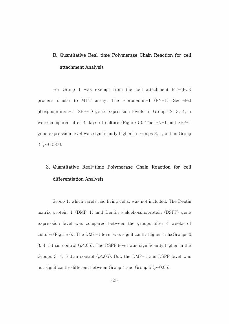
B. Quantitative real-time polymerase chain reaction for cell
attachment analysis ………………..…………………………… 21
3. Quantitative real-time polymerase chain reaction for cell differentiation
analysis ……………………………………………………………… 21
IV. Discussion ………………………………………………………………… 24
V. Conclusion …………………………………………………………..……. 32

-ii-
Raw data ………………………………………………………………………. 33
References …………………………………………………………………… 35
Abstract (In Korean) ……………………………………………..………….. 42
-iii-
List of Figures
Figure 1. Morphology of DPSCs obtained from human third molars
……………………………………………………..…….. 10
Figure 2. Experimental groups for attachment and differentiation of
DPSCs …..………………………………………………. 13
Figure 3. SEM views of DPSCs morphology in dentin surface …..19
Figure 4. Cell viability in the experimental groups ……………….. 20
Figure 5. Expression levels of adhesion molecules …….………. 22
Figure 6. Expression levels of odontogenic differentiation molecules
……………………………………….…………………... 23
-iv-
List of Tables
Table 1. PCR Primer used for cell adhesion markers ………………15
Table 2. PCR primer used for odontogenic differentiation markers ..17
-1-
Abstract
Calcium hydroxide increases the attachment and the
mineralization gene expression of dental pulp stem cells
on the dentin treated by sodium hypochlorite
Min Jeong Park
Department of Dentistry
The Graduate School, Yonsei University
(Directed by Professor Il Young Jung)
Sodium hypochlorite (NaOCl) is an excellent bactericidal agent, but it
has a profoundly detrimental effect on stem cell survival, attachment and
differentiation. Intracanal medicaments can also affect the survival and
differentiation of the dental pulp stem cells (DPSCs). Recent studies found
that Ca(OH)2 promoted greater survival and proliferation of stem cells
from apical papilla (SCAP). The purpose of this study is to evaluate the
effect of the sequential use of NaOCl and Ca(OH)2 on the attachment and
differentiation of DPSCs. Addtionally, to further investigate the optimal
protocols to reduce the cytotoxicity of NaOCl on DPSCs.
Human DPSCs were obtained from human third molars. Dentin
specimens were sterilized by ethylene oxide gas sterilization and an EDTA
-2-
treatment to produce specimens without a smear layer produced during
the preparation of the dentin specimen. Group 1 was treated by NaOCl.
Groups 2, 3, 4, 5, which underwent other treatment processes after the
NaOCl and Ca(OH)2 treatment. Group 2, for which NaOCl and Ca(OH)2
treatments were followed by PBS washing. Group 3, for which NaOCl and
Ca(OH)2 treatments were followed by EDTA. Group 4, for which NaOCl
and Ca(OH)2 treatments were followed by EDTA and culture media for 24
hours. Group 5, for which NaOCl and Ca(OH)2 treatments were followed by
instrumentation and EDTA. DPSCs morphology was observed by SEM
after 7 days of culture. A MTT assay was performed to assess the cell
survival rate by group after the treatment. After 4 days of culture, gene
expression level of cell adhesion was measured. After 4 weeks of culture,
gene expression level of odontogenic differentiation by quantitative real-
time polymerase chain reaction was investigated.
The DPSCs in the Group 1 were not attached, but the cells in the
Groups 2, 3, 4, 5 were attached to the dentin surface. The cell viability in
the Groups 2, 3, 4, 5 was lower than control group which cells were grown
in plates treated with PBS alone. The Fibronectin-1 (FN-1) and Secreted
phosphoprotein-1 (SPP-1) gene expression level was significantly higher
in Groups 3, 4, 5 than Group 2. The gene expression level of Dentin matrix
protein-1 (DMP-1) was significantly higher in the Groups 2, 3, 4, 5 than
control. The Dentin sialophosphoprotein (DSPP) level was significantly
higher in the Groups 3, 4, 5 than control. But, the DMP-1 and DSPP level
was not significantly different between Group 4 and Group 5.
-3-
In conclusion, application of Ca(OH)2 promoted the attachment and
differentiation of DPSCs. After treatment of Ca(OH)2, additional treatment
such as EDTA or instrumentation enhanced the attachment and
differentiation of DPSCs.
Keywords: regenerative endodontics, dental pulp stem cells, sodium
hypochlorite, calcium hydroxide, ethylenediaminetetraacetic
acid, cell attachment, cell differentiation
-4-
Calcium hydroxide increases
the attachment and the mineralization gene
expression of dental pulp stem cells
on the dentin treated by sodium hypochlorite
Min Jeong Park
Department of Dentistry
The Graduate School, Yonsei University
(Directed by Professor Il Young Jung)
I. Introduction
Conventional calcium hydroxide apexification has been performed for
treating immature permanent teeth with necrotic pulp. Even though
successful results have been reported, there are several drawbacks of the
calcium hydroxide (Ca(OH)2) such as incomplete root development, root
fracture, and long application period1. To overcome such shortcomings,
-5-
mineral trioxide aggregate (MTA) apexification has been introduced
recently, but this treatment also could not increase root development,
close the apex and increase thickness of dentinal walls1. Thus, the
Ca(OH)2 or MTA apexification may be less ideal treatment for many
patients, and there is a great need for alternative treatment option for
treating immature permanent necrotic teeth.
Recently, revascularization has been proposed as a new alternative
treatment option to treat immature permanent teeth with necrosis pulp.
Many clinical case reports have shown that the revascularization promotes
absence of clinical symptoms, healing of periapical lesions and increases
of dentinal wall thickness and root length as seen on radiographic
evidences2-4.
The ideal and final objective of the regenerative endodontic treatment
is to complete the dental pulp regeneration. In other words, the new vital
pulp tissue can regenerate in empty but infected root canal spaces,
reaching the coronal pulp chamber. Therefore, the pulp regeneration is
considered as an ideal treatment to keep the tooth homeostasis, protect
from reinfections and fractures, and preserve tooth longevities.
To result in the successful regenerative endodontic treatments,
disinfection of the infected root canal is essential. In order to disinfect the
-6-
entire root canal, proper irrigants should be selected on the basis of
consideration of both antimicrobial properties and proliferative capacity of
the stem cells. Sodium hypochlorite (NaOCl) is the most commonly used
irrigants in the regenerative endodontic treatments2,5,6. NaOCl is an
excellent bactericidal agent, but it has also been shown to be cytotoxic to
human periodontal ligament stem cells, cultured fibroblasts, stem cells
from human exfoliated deciduous teeth (SHEDs), stem cells from apical
papilla (SCAP), dental pulp stem cells (DPSCs) that it has a profoundly
detrimental effect on stem cell survival, attachment and differentiation7-11.
Ring et al reported that NaOCl is the most toxic treatment to DPSCs, and
Martin et al demonstrated that 6% NaOCl greatly decreased the SCAP
survival and completely abolished DSPP expression10. Also, Galler et al
found that in the dentin treated with NaOCl, resorption of lacunae was
found at the cell-dentin interface created by multinucleated cells with
clastic activity12. Both in vitro and in vivo study show that NaOCl definitely
has a negative effect on DPSCs.
Chlorhexidine (CHX) is widely used as an endodontic irrigant for a
substitute of NaOCl. CHX has shown a wide range of activity against gram
positive bacteria, gram negative bacteria, bacterial spores, lipophilic
viruses, yeasts13. CHX has demonstrated its cytotoxic potential such as
-7-
inhibition of protein synthesis, induction of apoptosis, and inhibition of
DNA synthesis14. Lessa et al reported that all concentrations of CHX had a
high direct cytotoxic effect to cultured odontoblast-like cells (MDPC-23)15.
Trevino et al also demonstrated that EDTA combination with CHX groups
showed no viable cells11. These results showed that CHX’s potential
cytotoxicity on the stem cells.
Currently, there is not any irrigant, which is as effective as NaOCl but
less cytotoxic than NaOCl at the same time. Therefore, we cannot avoid
using NaOCl. Previous studies have trials to neutralize the NaOCl
cytotoxicity used by various irrigation protocols with different
concentrations of NaOCl and neutralizing agents6,10,11.
During the regenerative endodontic treatment, after the primary
disinfection of the canals with irrigant, appropriate use of intracanal
medicaments induces removal of any residual bacteria in the canals16.
Common intracanal medicaments used in the regenerative endodontic
treatments are triple antibiotic paste (TAP), double antibiotic paste (DAP),
Ca(OH)2, formocresol, and Augmentin17-19. Other than the irrigants such as
NaOCl, these intracanal medicaments can also affect the survival and
differentiation of the DPSCs. Recent studies investigated the effect of the
intracanal medicaments on stem cells. They found that Ca(OH)2 promoted
-8-
greater SCAP survival and proliferation than TAP and DAP20,21.
Until now, there is not a known way to decrease the cytotoxicity of
NaOCl. Moreover, the effect of sequential application of NaOCl and
Ca(OH)2 on DPSCs survival has never been investigated previously.
Therefore, the purpose of this study is to evaluate the effect of the
sequential use of NaOCl and Ca(OH)2 on the attachment and differentiation
of DPSCs. Addtionally, to further investigate the optimal protocols to
reduce the cytotoxicity of NaOCl on DPSCs.
-9-
II. Materials and Methods
1. Primary human dental pulp stem cells culture
Protocol of primary human DPSCs culture was directly imported from
an earlier study22. Human third molars were collected from young adults
(16-22 years of age) at the Department of Advanced General Dentistry,
Yonsei University Dental Hospital. The pulp tissue was separated from the
apex of the extracted third molars with a barbed broach and was then cut
into 1㎣ blocks and placed in 60㎜ culture dishes (BD Falcon, Franklin
Lakes, NJ, USA) with a counting chamber cover glass (Marienfeld-
Superior, Lauda-Königshofen, Germany) to allow for the outgrowth of
cells. The tissues were cultured in α-modified Eagle medium (Invitrogen,
Carlsbad, CA, USA) supplemented with 10% fetal bovine serum (FBS;
Invitrogen), 2 mM/L glutamine, 100 μM/L ascorbic acid-2-phosphate
(WAKO, Tokyo, Japan), 100 U/㎖ penicillin, and 100 ㎍/㎖ streptomycin
(Biofluids, Rockville, MD, USA) at 37℃ in 5% CO2. The outgrowth cells
were transferred to 5 X 10㎝ culture flasks (passage 1) and grown to
confluence. The obtained cells were harvested and kept frozen in liquid N2.
Cells from the passage 3 were used in this experiment.
-10-
A. B.
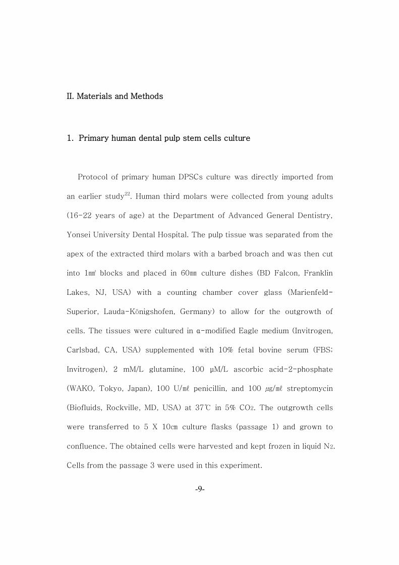
Figure 1. Morphology of DPSCs obtained from human third molars. The
DPSCs showed elongated, spindle shaped and had typical
fibroblast-like cell morphology. (A) magnification x100, (B)
magnification x200
-11-
2. Preparation of dentin slices
The dentin slices were prepared from the human third molars. The
coronal dentin was cut into a disc shape approximately 1㎜ thick by low
speed diamond cutter RB205 Metsaw-LS™ (R&B Inc., Daejeon, Korea)
under sterile phosphate buffered saline irrigation (Mediatech Inc.,
Manassas, VA, USA). The dentin slices were sterilized by ethylene oxide
gas sterilization.
3. Cell morphology analysis by Scanning Electron Microscope
After 7 days of culture, one dentin slice from each group was selected
randomly. And then they were washed 3 times with PBS followed by fixed
in 2% glutaraldehyde for 5 minutes. The slices were dehydrated in a
graded series of ethanol and dried with hexamethyldisilane. After gold
coating, cell morphology was assessed in each group with a scanning
electron microscope (JEOL JSM-820; JEOL, Tokyo, Japan).
-12-
4. Adhesion analysis
A. MTT assay (Tetrazolium-based colorimetric assay)
The dentin slices were randomly assigned to the following groups
(Figure 2). All dentin slices were 5 times wash-out with PBS and placed in
the 100㎜ x 15㎜ sterile Nunclon® cell culture dishes (NUNC™, Roskilde,
Denmark) in single layer. Harvested cells were seeded onto the dentin
specimens (1x10⁶ cells/plate) and cultured in normal growth medium for 7
days. At the end of the culture period, the dentin slices were removed and
placed in new Nunclon cell culture dishes. Cells were detached with 0.25%
trypsin/EDTA, and 2 minutes incubation in Thermo® Steri-Cycle CO2
Incubators (Forma Scientific Inc., Mariotla, Ohio, USA) at 37℃ in 5% CO2
for trypsin activation. After 5 minutes centrifugation was performed, the
cells were placed in new plates and then MTT [3-(4,5-dimethylthiazol-
2yl)-2,5-diphenyl-2H-tetrazolium bromide] (MTT, Sigma Chemical Co.,
St. Louis, MO, USA) assay was performed for assessing cell viability. The
absorbance at 570nm was measured using a spectrophotometer (Bio-Rad
Laboratories, Hercules, CA, USA).
-13-
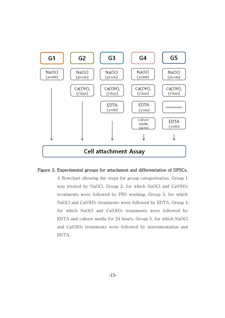
Figure 2. Experimental groups for attachment and differentiation of DPSCs.
A flowchart showing the steps for group categorization. Group 1
was treated by NaOCl. Group 2, for which NaOCl and Ca(OH)2
treatments were followed by PBS washing. Group 3, for which
NaOCl and Ca(OH)2 treatments were followed by EDTA. Group 4,
for which NaOCl and Ca(OH)2 treatments were followed by
EDTA and culture media for 24 hours. Group 5, for which NaOCl
and Ca(OH)2 treatments were followed by instrumentation and
EDTA.
-14-
B. Cell attachment analysis by Quantitative Real-time
Polymerase Chain Reaction
Cell attachment was evaluated based on the expression level of
Fibronectin-1 (FN-1) and Secreted phosphoprotein-1 (SPP-1). Total
cellular RNA was extracted from DPSCs in each group using the RNeasy
Mini Kit (Qiagen Inc., Valencia, CA, USA) according to the manufacturer’s
instructions. The RNA was treated with the RNase-free DNase-set
(Qiagen) during the RNA extraction. The Complementary DNA samples
were prepared from the isolated RNA using the RT First Strand Kit
(Qiagen) according to the manufacturer’s instructions. Quantitative real-
time polymerase chain reaction (RT-qPCR) analysis was performed using
ABI 7500 software (Applied Biosystems, Foster City, CA, USA) according
to the standard protocol. The real-time polymerase chain reaction cycles
included 40 cycles of general denaturation at 94℃ (30 seconds), annealing,
and elongation at 60℃ (45 seconds), except for the first cycle with a 15-
minute denaturation and the last cycle with a 7-minute elongation at 72℃.
The primers used for assessing the FN-1, SPP-1 expression level are
shown in Table 1.
-15-
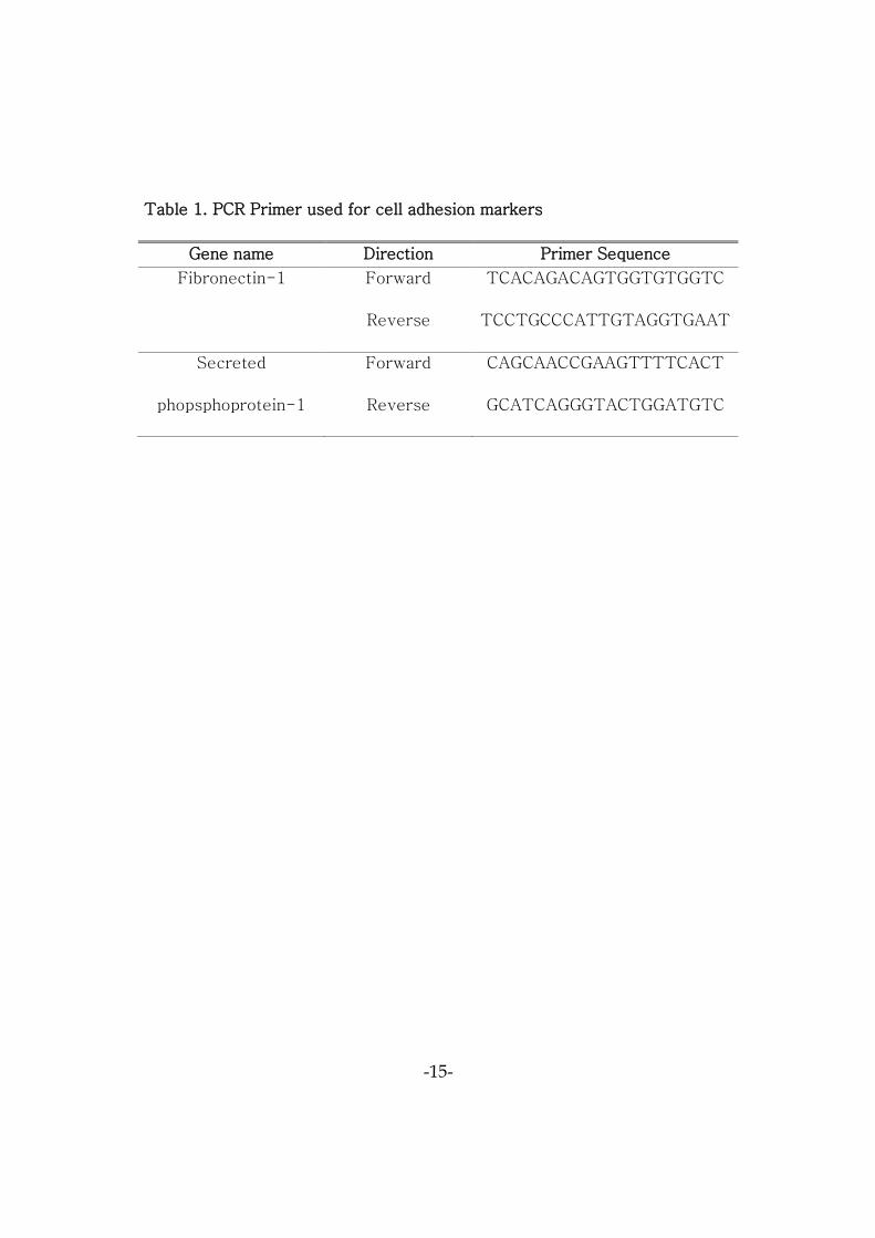
Table 1. PCR Primer used for cell adhesion markers
Gene name Direction Primer Sequence
Fibronectin-1 Forward
Reverse
TCACAGACAGTGGTGTGGTC
TCCTGCCCATTGTAGGTGAAT
Secreted
phopsphoprotein-1
Forward
Reverse
CAGCAACCGAAGTTTTCACT
GCATCAGGGTACTGGATGTC
-16-
5. Cell differentiation analysis by Quantitative Real-time Polymerase
Chain Reaction
To compare differential gene expressions under various conditions,
RT-qPCR analysis of the mineralization-related genes was performed.
The primers for the differentiation markers of mineralization are given in
Table 2. All DPSCs were cultured in vitro at 37℃ in a humidified
atmosphere of 5% CO2 for 4 weeks. The culture medium in all groups was
replenished every 3 days. The procedures were the same as those for the
cell attachment assay.
6. Statistical Analysis
Mann-Whitney U tests was used to determine the statistical
differences between the experimental groups by using SPSS software
version 21.0 (SPSS, Chicago, IL, USA). Adjusted p value less than 0.05
was considered to be statistically significant.
-17-
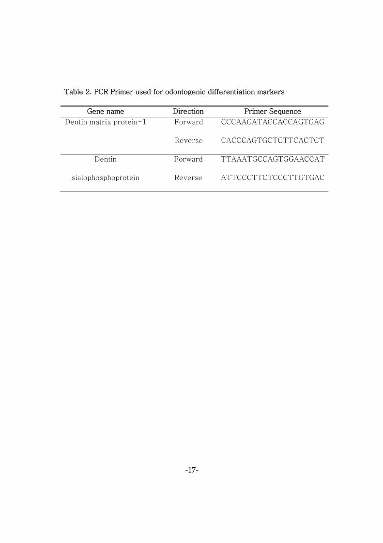
Table 2. PCR Primer used for odontogenic differentiation markers
Gene name Direction Primer Sequence
Dentin matrix protein-1 Forward
Reverse
CCCAAGATACCACCAGTGAG
CACCCAGTGCTCTTCACTCT
Dentin
sialophosphoprotein
Forward
Reverse
TTAAATGCCAGTGGAACCAT
ATTCCCTTCTCCCTTGTGAC
-18-
III. Results
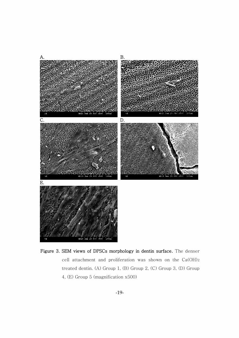
1. Cell morphology analysis by Scanning Electron Microscope
After 7 days of culture, the DPSCs in the Group 1 were not attached
the dentin surfaces (Figure 3A). This result was consistent with
preliminary studies. However, the cells in the Groups 2, 3, 4, 5 were
attached to the dentin surface. Especially, Dentin surface was overlapped
by proliferating cell layers in Group 5 (Figure 3E).
2. Adhesion analysis
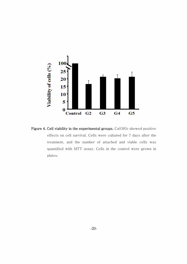
A. MTT assay (Tetrazolium-based colorimetric assay)
For Group 1, the SEM images confirmed that there were few if any
living cells. Thus, this group was exempt from the MTT assay (Figure 4).
The cell viability of Groups 2, 3, 4, 5 was significantly lower than control
group (p=0.037). The cell viability between the Groups 2, 3, 4, 5 was not
significantly different (p>.05).
-19-
A. B.
C. D.
E.
Figure 3. SEM views of DPSCs morphology in dentin surface. The denser
cell attachment and proliferation was shown on the Ca(OH)2
treated dentin. (A) Group 1, (B) Group 2, (C) Group 3, (D) Group
4, (E) Group 5 (magnification x500)
-20-
Figure 4. Cell viability in the experimental groups. Ca(OH)2 showed positive
effects on cell survival. Cells were cultured for 7 days after the
treatment, and the number of attached and viable cells was
quantified with MTT assay. Cells in the control were grown in
plates.
-21-
B. Quantitative Real-time Polymerase Chain Reaction for cell
attachment Analysis
For Group 1 was exempt from the cell attachment RT-qPCR
process similar to MTT assay. The Fibronectin-1 (FN-1), Secreted
phosphoprotein-1 (SPP-1) gene expression levels of Groups 2, 3, 4, 5
were compared after 4 days of culture (Figure 5). The FN-1 and SPP-1
gene expression level was significantly higher in Groups 3, 4, 5 than Group
2 (p=0.037).
3. Quantitative Real-time Polymerase Chain Reaction for cell
differentiation Analysis
Group 1, which rarely had living cells, was not included. The Dentin
matrix protein-1 (DMP-1) and Dentin sialophosphoprotein (DSPP) gene
expression level was compared between the groups after 4 weeks of
culture (Figure 6). The DMP-1 level was significantly higher in the Groups 2,
3, 4, 5 than control (p<.05). The DSPP level was significantly higher in the
Groups 3, 4, 5 than control (p<.05). But, the DMP-1 and DSPP level was
not significantly different between Group 4 and Group 5 (p=0.05)
-22-
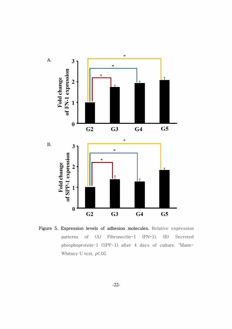
Figure 5. Expression levels of adhesion molecules. Relative expression
patterns of (A) Fibronectin-1 (FN-1), (B) Secreted
phosphoprotein-1 (SPP-1) after 4 days of culture. *Mann-
Whitney U test, p<.05
A.
B.
-23-
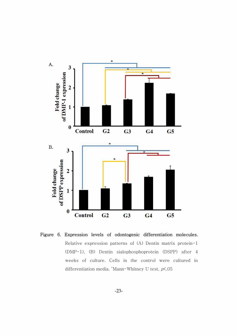
Figure 6. Expression levels of odontogenic differentiation molecules.
Relative expression patterns of (A) Dentin matrix protein-1
(DMP-1), (B) Dentin sialophosphoprotein (DSPP) after 4
weeks of culture. Cells in the control were cultured in
differentiation media. *Mann-Whitney U test, p<.05
A.
B.
-24-
IV. Discussion
This present study aimed to evaluate the effect of the sequential
application of NaOCl and Ca(OH)2 on the attachment and differentiation of
DPSCs. For the purpose of this study, an experiment setting similar to a
clinical trial was planned. Regenerative endodontics in clinical practice
does not produce a smear layer, as it minimizes instrumentation within the
root canal to protect the thin root dentin wall of an immature permanent
tooth. The smear layer is 1-5㎛-thick denatured cutting debris produced
on instrumented dentin surfaces. It is composed of dentin, odontoblastic
processes, nonspecific inorganic contaminants and microorganisms23-25.
The existence of a smear layer in regenerative endodontics is a critical
factor which may be responsible for the failure of the surgery, as it
interrupts the adhesion to the DPSCs26. Therefore, unlike previous
research, this study involved the sterilization of dentin specimens with
ethylene oxide gas and an EDTA treatment to produce specimens without
a smear layer produced during the preparation of the dentin specimen22.
The Ca(OH)2 concentration was 1mg/㎖, a level at which the survival rate
of SCAP was significantly higher according to Ruparel et al20.
-25-
The first step in regeneration is to obtain proper stem cells. There
were many previous studies to describe the characteristics of the DPSCs.
It has been shown that DPSCs have a self-renewal potential and
multilineage differentiation ability into odontoblasts, osteoblasts,
adipocytes, chodrocytes, neurons27-29. Canine dental pulp stem cells from
dog teeth (cDPSCs) showed superiority in the proliferation rate and
multilineage differentiation over human bone marrow mesenchymal stem
cells (hBMMSCs), but neurogenic differentiation ability was inferior than
human dental pulp stem cells (hDPSCs). Also there is no evidence that
cDPSCs could produce the uniform dentin30. The difference between
SHEDs and adult DPSCs was that SHEDs were able to induce bone-like
structure when implanted into immunocompromised mice subcutaneously,
whereas DPSCs generated a dentin-like, pulp-like tissue31. Therefore, the
cell sources used in this study were young adults’ DPSCs that isolated
from extracted pulps of human third molars.
First, The SEM images showed cells in Groups 2, 3, 4, 5 successfully
adhering, as opposed to those in Group 1, which failed. In 7 days, it was
confirmed that the cells had stretched and had long cytoplastic processes.
Primarily, the Ca(OH)2 treatment enhanced cell adhesion and proliferation.
Secondarily, culture media, EDTA and instrumentation used during the
-26-
removal of Ca(OH)2 changed the properties of the dentin surface, thereby
creating an environment which facilitates better DPSCs adhesion.
A MTT assay was performed to assess the cell survival rate by group
after the treatment. In the MTT assay, cell viability does not simply
account for living cells in a plate. Rather, it evaluates living cells that are
attached to the dentin specimens. Considering Groups 2, 3, 4, 5, which
underwent other treatment processes after the Ca(OH)2 treatment, which
is believed to show positive effects of Ca(OH)2 on cell survival. The
interpretation here matched that of the SEM images. A high alkaline pH
level of Ca(OH)2 encourages the survival, migration and proliferation of
pulp stem cells, helping the formation of reparative dentin20,21,32. Therefore,
it is understood that Ca(OH)2 plays a decisive role in boosting cell viability.
Next, cell attachment was assessed by means of the adhesion
molecule RT-qPCR in response to Fibronectin-1 (FN-1) and Secreted
phosphoprotein-1 (SPP-1). FN-1 is an extracellular matrix molecule
important in cell adhesion33. SPP-1 is stored in bone matrix as one of the
abundant noncollageous bone matrix proteins and plays a role in bone-cell
attachment34. Group 2, for which NaOCl and Ca(OH)2 treatments were
followed by PBS washing, was chosen as a control for Groups 3, 4, 5. The
results demonstrated a significant difference between the Group 2 and
-27-
Groups 3, 4, 5. The high expression of FN-1 and SPP-1 means that cell
attachment has been also increased. The significant difference between
Group 2 and 3 indicates that EDTA additionally decalcifies the dentin
surface and increases the wettability. EDTA plays a role in cell adhesion
and odontoblast differentiation by causing changes in the dentin surfaces
of the specimens22. This is similar to a previous research which showed
changes in the dentin surfaces caused by the chelating effect of EDTA35.
The results for Groups 2 and 4 illustrate that EDTA initially created an
environment for cells to adhere successfully, after which culture media
including FBS neutralized the cell toxicity of NaOCl, thus increasing cell
adhesion. Fetal bovine serum (Gibco, Tulsa, OK, USA) is a serum media
rich in protein, electrolytes, inorganic substances, and nutrients. It dilutes
or neutralizes the toxicity irrigants and helps protect living cells36. The
use of instrumentation, as shown in Groups 2 and 5, can effectively
remove any remaining Ca(OH)2. Although Ca(OH)2 is commonly used in
various clinical situations as an antiseptic for root canal treatment,
imperfectly removed Ca(OH)2 can block the penetration of the sealer into
dentinal tubules and increase apical leakage, resulting in a failed root canal
treatment37. Therefore, Ca(OH)2 applied to the root canal should be
thoroughly removed to completely seal it off. An experiment by Kenee et
-28-
al. in which rotary and ultrasonic instrumentation techniques were more
effective in removing Ca(OH)2 than an instrument with an irrigant was
considered. Thus, the instrumentation was standardized at the #40/.04
taper profile at 300rpm and scraping of the entire dentin surfaces was
performed once38. The removal of Ca(OH)2 by instrumentation was
followed by the decalcification of the dentin surfaces using EDTA, which
caused the exposure of the dentinal tubule and collagen fiber, creating an
environment in which stem cells could easily adhere.
For the cell differentiation RT-qPCR analysis, the control group
underwent a cell culture assay in differentiation media without dentin
samples. Dentin matrix protein-1 (DMP-1), an essential noncollagenous
and acidic phosphorylated extracellular matrix protein, is highly expressed
in odontoblast. DMP-1 has been shown to play a prime role in dentin
mineralization39. Dentin sialophosphoprotein (DSPP) is the only protein
produced uniquely by odontoblasts. DSPP plays an important role in the
regulation of mineral deposition40. As a result, DMP-1 was expressed at
higher levels in Groups 2, 3, 4, 5 than the control group, while DSPP
showed higher levels in Groups 3, 4, 5. The significantly higher DMP-1
and DSPP levels expressed as compared to the control group can be
interpreted as showing that after the NaOCl treatment, the Ca(OH)2
-29-
treatment triggered odontogenesis, leading to odontoblast differentiation.
In particular, the lack of a significant difference between Group 4 and
Group 5 demonstrates that there is no difference between the technique in
which culture media is used for 24 hours as a neutralizing treatment agent
for NaOCl and the technique in which instrumentation is performed
followed by a NaOCl and Ca(OH)2 treatment. In other words, creating an
environment to facilitate better stem cell attachment and differentiation in
the root canal through instrumentation and an EDTA treatment is more
effective than the cumbersome process of using culture media for 24
hours as a neutralizing treatment agent for NaOCl.
In this study, a Ca(OH)2 treatment was shown to improve the reduced
survival and attachment of cells after a NaOCl treatment. The Ca(OH)2
treatment is believed to have caused a favorable change in cell attachment
and differentiation on the dentin surfaces. Possible mechanisms include
that Ca(OH)2 exposes the collagen fiber on the dentin surfaces, allowing
better cell attachment41, and Ca(OH)2 releases a growth factor from dentin
and enhances cell attachment and differentiation42. If this could lead to the
improvement of biomechanical and biochemical environments, the survival
and differentiation of stem cells are expected to increase.
-30-
Numerous studies of revascularization were clinical case reports, with
animal studies showing that newly formed tissues into the root canal space
have little similarity to normal pulp tissue but with more cementum-like,
periodontal ligament-like, and bone-like tissue43,44. Recently, some
interesting studies have tried to use tissue engineering technique in pulp
regeneration. Huang et al reported that vascularized pulp-like tissues and
dentin-like mineral structures were formed in the root canal space by
containing synthetic scaffolds seeded with stem/progenitor cells from
apical papilla and dental pulp45. Iohara et al demonstrated complete pulp
regeneration in the canine model after pulpectomy by transplantation of
CD105+ with stromal cell-derived factor-146. Wang et al achieved that
autologous cDPSCs combined with Gelfoam transplanted into pulpless root
canals led to generate pulp-like tissues containing blood vessels and
dentin-like tissues and thickening of the root canal wall was also
observed47.
This present research is an in vitro study which takes place
immediately before a transplantation experiment, a practice currently in
full swing targeting large animals for the complete regeneration of pulp. It
can be used as the basis for in vivo experiments.
-31-
Regenerative endodontics in clinical practice involves intracanal
medication after gradually using an irrigant in an infected root canal.
Previous studies have performed NaOCl treatments and Ca(OH)2
treatments independently. This study is significant in that it integrated
both treatments for use, creating an environment closer to that of actual
clinical practice. However, due to the in vitro limitations, additional in vivo
studies are necessary down the road. Also, a long-term study is required,
as the RT-qPCR results for the attachment factor and for the
differentiation factor were measured at specific points and therefore have
limitations. In addition, studies of a counteragent to neutralize the toxicity
of NaOCl and research on scaffolding and growth factors for enhanced
tissue engineering are needed. If the technique elaborated in this study
can be successfully performed in regenerative endodontics for immature
permanent teeth in clinical practice, it will be proven as applicable to
endodontics for mature permanent teeth as well. Ultimately, the goal of
regenerative endodontics will be met through complete pulp-dentin
complex regeneration.
-32-
V. Conclusion
Based on the results of this study, the following can be concluded.
1. Application of Ca(OH)2 promotes dental pulp stem cells
attachment and differentiation.
2. After treatment of Ca(OH)2, additional treatment such as EDTA
or instrumentation enhanced the attachment and differentiation
of dental pulp stem cells.
-33-
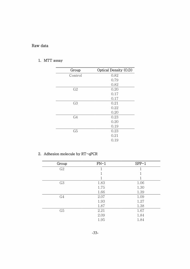
Raw data
1. MTT assay
Group Optical Density (O.D)
Control 0.82
0.79
0.82
G2 0.20
0.17
0.17
G3 0.21
0.22
0.20
G4 0.23
0.20
0.19
G5 0.23
0.21
0.19
2. Adhesion molecule by RT-qPCR
Group FN-1 SPP-1
G2 1
1
1
1
1
1
G3 1.83
1.75
1.66
1.06
1.30
1.39
G4 2.07
1.93
1.87
1.09
1.27
1.38
G5 2.21
2.09
1.95
1.67
1.84
1.84
-34-
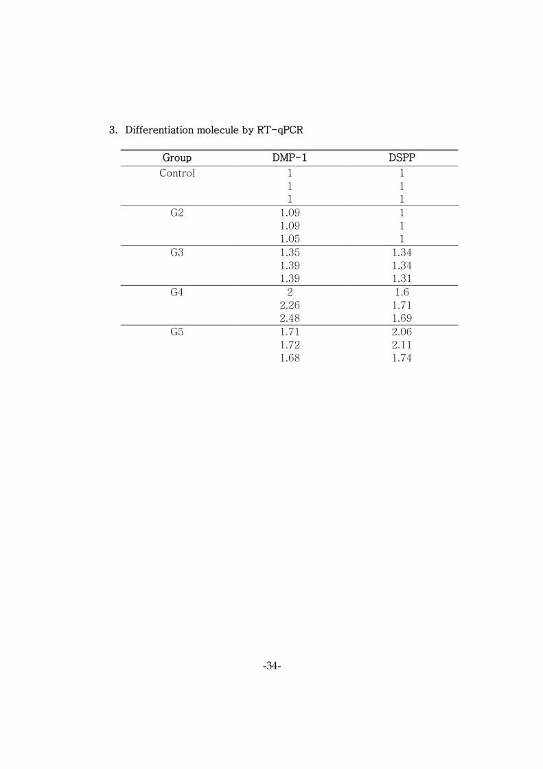
3. Differentiation molecule by RT-qPCR
Group DMP-1 DSPP
Control 1
1
1
1
1
1
G2 1.09
1.09
1.05
1
1
1
G3 1.35
1.39
1.39
1.34
1.34
1.31
G4 2
2.26
2.48
1.6
1.71
1.69
G5 1.71
1.72
1.68
2.06
2.11
1.74
-35-
References
1. Jeeruphan T, Jantarat J, Hargreaves KM. Mahidol study 1:
comparison of radiographic and survival outcomes of immature
teeth treated with either regenerative endodontic or apexification
methods: a retrospective study. Journal of Endodontics
2012;38(10):1330-6.
2. Jung IY, Lee SJ, Hargreaves KM. Biologically based treatment of
immature permanent teeth with pulpal necrosis: a case series.
Journal of Endodontics 2008;34(7):876-87.
3. Ding RY, Cheung GS, Chen J, Yin XZ, Wang QQ, Zhang CF. Pulp
revascularization of immature teeth with apical periodontitis: a
clinical study. Journal of Endodontics 2009;35(5):745-9.
4. Bose R, Nummikoski P, Hargreaves K. A retrospective evaluation of
radiographic outcomes in immature teeth with necrotic root canal
systems treated with regenerative endodontic procedures. Journal
of Endodontics 2009;35(10):1343-9.
5. Banchs F, Trope M. Revascularization of immature permanent teeth
with apical periodontitis: new treatment protocol? Journal of
Endodontics 2004;30(4):196-200.
6. Martin G, Ricucci D, Gibbs JL, Lin LM. Histological findings of
revascularized/revitalized immature permanent molar with apical
periodontitis using platelet-rich plasma. Journal of Endodontics
2013;39(1):138-44.
7. Chang YC, Huang FM, Tai KW, Chou MY. The effect of sodium
hypochlorite and chlorhexidine on cultured human periodontal
-36-
ligament cells. Oral Surg Oral Med Oral Pathol Oral Radiol Endod
2001;92(4):446-50.
8. Heling I, Rotstein I, Dinur T, Szwec-Levine Y, Steinberg D.
Bactericidal and cytotoxic effects of sodium hypochlorite and
sodium dichloroisocyanurate solutions in vitro. Journal of
Endodontics 2001;27(4):278-80.
9. Wennberg A. Biological evaluation of root canal antiseptics using in
vitro and in vivo methods. Scandinavian journal of dental research
1980;88(1):46-52.
10. Ring KC, Murray PE, Namerow KN, Kuttler S, Garcia-Godoy F. The
comparison of the effect of endodontic irrigation on cell adherence
to root canal dentin. Journal of Endodontics 2008;34(12):1474-9.
11. Trevino EG, Hargreaves KM, Diogenes A, et al. Effect of irrigants
on the survival of human stem cells of the apical papilla in a
platelet-rich plasma scaffold in human root tips. Journal of
Endodontics 2011;37(8):1109-15.
12. Galler KM, Schmalz G, et al. Dentin conditioning codetermines cell
fate in regenerative endodontics. Journal of Endodontics
2011;37(11): 1536-41.
13. Davies A. The mode of action of chlorhexidine. Journal of
Periodontal Research. Supplement 1973;12:68-75.
14. Emilson CG. Susceptibility of various microorganisms to
chlorhexidine. Scandinavian journal of dental research
1977;85(4):255-65.
-37-
15. Lessa FC, Aranha AM, Nogueira I, Giro EM, Hebling J, Costa CA.
Toxicity of chlorhexidine on odontoblast-like cells. Journal of
Applied Oral Science 2010;18(1):50-8.
16. Chong BS, Pitt Ford TR. The role of intracanal medication in root
canal treatment. International Endodontic Journal 1992;25(2):97-
106.
17. Hoshino E, Kurihara-Ando N, Sato I, Uematsu H, Sato M, Kota K,
Iwaku M. In-vitro antibacterial susceptibility of bacteria taken from
infected root dentine to a mixture of ciprofloxacin, metronidazole
and minocycline. International Endodontic Journal 1996;29(2):125-
30.
18. Chueh LH, Ho YC, Kuo TC, Lai WH, Chen YH, Chiang CP.
Regenerative endodontic treatment for necrotic immature
permanent teeth. Journal of Endodontics 2009;35(2):160-4.
19. Shah N, Logani A, Bhaskar U, Aggarwal V. Efficacy of
revascularization to induce apexification/apexogensis in infected,
nonvital, immature teeth: a pilot clinical study. Journal of
Endodontics 2008;34(8):919-25; Discussion 1157.
20. Ruparel NB, Teixeira FB, Ferraz CC, Diogenes A. Direct effect of
intracanal medicaments on survival of stem cells of the apical
papilla. Journal of Endodontics 2012;38(10):1372-5.
21. Althumairy RI, Teixeira FB, Diogenes A. Effect of dentin
conditioning with intracanal medicaments on survival of stem cells
of apical papilla. Journal of Endodontics 2014;40(4):521-5.
-38-
22. Pang NS, Lee SJ, Kim E, Shin DM, Cho SW, Park W, Zhang X, Jung
IY. Effect of EDTA on attachment and differentiation of dental pulp
stem cells. Journal of Endodontics 2014;40(6):811-7.
23. Brännström M. Smear layer: pathological and treatment
considerations. Operative Dentistry. Supplement 1984;3:35-42.
24. Czonstkowsky M, Wilson EG, Holstein FA. The smear layer in
endodontics. Dental clinics of North America 1990;34(1):13-25.
25. Takeda FH, Harashima T, Kimura Y, Matsumoto K. A comparative
study of the removal of smear layer by three endodontic irrigants
and two types of laser. International Endodontic Journal 1999;
32(1):32-9.
26. Torabinejad M, Handysides R, Khademi AA, Bakland LK. Clinical
implications of the smear layer in endodontics: a review. Oral Surg
Oral Med Oral Pathol Oral Radiol Endod 2002;94(6):658-66.
27. Zhang W, Walboomers XF, Shi S, Fan M, Jansen JA. Multilineage
differentiation potential of stem cells derived from human dental
pulp after cryopreservation. Tissue Engineering 2006;12(10):2813-
23.
28. Koyama N, Okubo Y, Nakao K, Bessho K. Evaluation of pluripotency
in human dental pulp cells. Journal of oral and maxillofacial surgery
2009;67(3):501-6.
29. Gronthos S, Brahim J, Li W, Fisher LW, Cherman N, Boyde A,
DenBesten P, Robey PG, Shi S. Stem cell properties of human
dental pulp stem cells. Journal of dental research 2002;81(8):531-5.
-39-
30. Dissanayaka WL, Zhu X, Zhang C, Jin L. Characterization of dental
pulp stem cells isolated from canine premolars. Journal of
Endodontics 2011;37(8):1074-80.
31. Miura M, Gronthos S, Zhao M, Lu B, Fisher LW, Robey PG, Shi S.
SHED: stem cells from human exfoliated deciduous teeth. Proc Natl
Acad Sci U S A 2003;100(10):5807-12.
32. Guven EP, Yalvac ME, Sahin F, Yizici MM, Rizvanov AA, Bayirli G.
Effect of dental materials calcium hydroxide-containing cement,
mineral trioxide aggregate, and enamel matrix derivative on
proliferation and differentiation of human tooth germ stem cells.
Journal of Endodontics 2011;37(5):650-6.
33. Kapila YL, Lancero H, Johnson PW. The response of periodontal
ligament cells to fibronectin. Journal of periodontology
1998;69(9):1008-19.
34. Noda M, Vogel RL, Craig AM, Prahl J, DeLuca HF, Denhardt DT.
Identification of a DNA sequence responsible for binding of the
1,25-dihydroxyvitamin D3 receptor and 1,25-dihydroxyvitamin D3
enhancement of mouse secreted phosphoprotein 1 (SPP-1 or
osteopontin) gene expression. Proc Natl Acad Sci U S A
1990;87(24):9995-9.
35. Dalby MJ, Gadegaard N, Tare R, Andar A, Riehle MO, Herzyk P,
Wilkinson CD, Oreffo RO. The control of human mesenchymal cell
differentiation using nanoscale symmetry and disorder. Nature
Materials 2007;6(12):997-1003.

-40-
36. Hayman EG, Pierschbacher MD, Suzuki S, Ruoslahti E. Vitronectin-
-a major cell attachment-promoting protein in fetal bovine serum.
Experimental cell research 1985;160(2):245-58.
37. Calt S, Serper A. Dentinal tubule penetration of root canal sealers
after root canal dressing with calcium hydroxide. Journal of
Endodontics 1999;25(6):431-3.
38. Kenee DM, Allemang D, Johnson JD, Hellstein J, Nichol BK. A
quantitative assessment of efficacy of various calcium hydroxide
removal techniques. Journal of Endodontics 2006;32(6):563-5.
39. Yue J, Wu B, Gao J, Huang X, Li C, Ma D, Fang F. DMP1 is a target
of let-7 in dental pulp cells. International Journal of Molecular
Medical Science 2012;30(2): 295-301.
40. Martini D, Breschi L, Mazzoni A, Teti G, Falconi M, Ruggeri A Jr.
Dentin matrix protein 1 and dentin sialophosphoprotein in human
sound and carious teeth: an immunohistochemical and colorimetric
assay. European journal of histochemistry 2013;57(4):e32.
41. Yassen GH, Chu TM, Eckert G, Platt JA. Effect of medicaments
used in endodontic regeneration technique on the chemical
structure of human immature radicular dentin: an in vitro study.
Journal of Endodontics 2013;39(2):269-73.
42. Graham L, Cooper PR, Cassidy N, Nor JE, Sloan AJ, Smith AJ. The
effect of calcium hydroxide on solubilisation of bio-active dentine
matrix components. Biomaterials 2006; 27(14):2865-73.
43. Zhu X, Zhang C, Huang GT, Cheung GS, Dissanayaka WL, Zhu W.
Transplantation of dental pulp stem cells and platelet-rich plasma
for pulp regeneration. Journal of Endodontics 2012;38(12):1604-9.
-41-
44. Wang X, Thibodeau B, Trope M, Lin LM, Huang GT. Histologic
characterization of regenerated tissues in canal space after the
revitalization/revascularization procedure of immature dog teeth
with apical periodontitis. Journal of Endodontics 2010;36(1):56-63.
45. Huang GT, Yamaza T, Shea LD, Djouad F, Kuhn NZ, Tuan RS, Shi S.
Stem/progenitor cell-mediated de novo regeneration of dental pulp
with newly deposited continuous layer of dentin in an in vivo model.
Tissue Engineering. Part A 2012;16(2):605-15.
46. Iohara K, Imabayashi K, Ishizaka R, Watanabe A, Nabekura J, Ito M,
Matsushita K, Nakamura H, Nakashima M. Complete pulp
regeneration after pulpectomy by transplantation of CD105+ stem
cells with stromal cell-derived factor-1. Tissue Engineering. Part A
2011;17(15-16):1911-20.
47. Wang Y, Zhao Y, Jia W, Yang J, Ge L. Preliminary study on dental
pulp stem cell-mediated pulp regeneration in canine immature
permanent teeth. Journal of Endodontics 2013;39(2):195-201.

-42-
Abstract (In Korean)
NaOCl 로 처리한 상아질에서 수산화칼슘 처치로 인한
치수줄기세포의 부착과 경조직 형성 유전자의 발현증가
<지도교수 정일영>
연세대학교 대학원 치의학과
박 민 정
Sodium hypocholorite (NaOCl)는 재생근관치료에서 근관 내 소독을 위해
사용하는 세척용액으로 우수한 항균 효과를 갖고 있지만 줄기세포의 생존을
방해한다. 세척제와 더불어 근관 내 첩약제도 재생근관치료에 사용되는데,
특히 Calcium hydroxide (Ca(OH)2)는 줄기세포의 생존과 증식에 도움이 되는
것으로 알려져 있다. 아직까지 NaOCl 의 부정적인 효과를 줄이는 방법은
아직 알려지지 않았고 NaOCl 과 Ca(OH)2 를 연이어 사용한 후 평가한
연구는 없다. 따라서 본 연구에서는 임상환경과 유사하게 상아질 시편에
NaOCl 과 Ca(OH)2 를 순차적으로 사용한 후 NaOCl 의 치수줄기세포에 대한
부정적인 효과를 줄이는 방법을 찾아보고자 한다.
발치된 제 3 대구치에서 치수조직을 채취해 일차배양을 시행하여
줄기세포를 준비하였다. 발치된 대구치의 치관부를 1mm 두께로 절삭하여
상아질 시편을 만들고, 임의로 5 개 군으로 나눈 후 다른 처치를 하였다. 이전
연구와는 달리 Ethylene oxide gas 로 상아질 시편을 멸균하고 EDTA 3 분
처리를 하여 시편제작과정 중에 발생한 도말층을 제거하였다. 먼저 모든
-43-
그룹은 NaOCl 30 분 처리하였다. 그룹 1 은 NaOCl 처리만 하였고 그룹 2, 3,
4, 5 는 추가로 Ca(OH)2 를 일주일 동안 처치하였다. 그룹 2 는 Ca(OH)2
처치까지 한 군이고 그룹 3 은 추가로 EDTA 처리한 군, 그룹 4 는 EDTA
처리하고 culture media 24 시간 처리한 군, 그룹 5 는 기구조작하고 EDTA
처리한 군이다. DPSCs 의 생존과 부착은 MTT assay 로 정량화하였고 상아질
시편에 부착되어 있는 세포의 모습은 전자현미경을 통해 관찰하였다.
그리고 4 일, 4 주간 배양하여 세포부착과 경조직 형성 세포로의 분화와 관련된
유전자의 발현양상을 정량적 실시간 중합효소 연쇄반응으로 분석하였다.
전자현미경 사진에서는 cell 이 부착하지 못한 그룹 1 에 비해 그룹 2, 3, 4,
5 는 7 일 뒤 cell 이 붙었고 성장해 나간 것을 확인할 수 있었다. MTT 실험
결과 그룹 2, 3, 4, 5 는 대조군에 비해 세포생존력이 낮았으나, 그룹 간의
유의차는 없었다. 세포부착 유전자의 발현결과 Fibronectin-1 (FN-1) 과
Secreted phosphoprotein-1 (SPP-1)은 그룹 2 에 비해 그룹 3, 4, 5 가 모두
유의하게 증가하였다. 세포분화 유전자의 발현결과 Dentin matrix protein-1
(DMP-1)은 대조군보다 그룹 2, 3, 4, 5 가 높게 발현되었고 Dentin
sialophosphoprotein (DSPP)은 그룹 3, 4, 5 가 높게 발현되었다. DMP-1 과
DSPP 는 그룹 4, 5 간의 유의차는 없었다.
결론적으로 Ca(OH)2 처치과정은 NaOCl 로 처리한 상아질 시편에서
치수줄기세포의 부착과 분화가 일어날 수 있도록 도움을 준다. 또한 Ca(OH)2
처치 후 추가적으로 EDTA 처리나 기구조작을 통해 상아질 시편을 처리하면
치수줄기세포의 부착과 분화가 증진된다.
핵심되는 말: 재생근관치료, 치수줄기세포, sodium hypochlorite, calcium
hydroxide, 세포부착, 세포분화
Top Related