The Intracanal Diffusion of Camphorated Para-mono ... · One of the most commonly used intracanal...
44
Loyola University Chicago Loyola eCommons Master's eses eses and Dissertations 1971 e Intracanal Diffusion of Camphorated Para- mono-chlorophenol in Endodontics: An Autoradiographic Study Gerald R. Heiman Loyola University Chicago is esis is brought to you for free and open access by the eses and Dissertations at Loyola eCommons. It has been accepted for inclusion in Master's eses by an authorized administrator of Loyola eCommons. For more information, please contact [email protected]. is work is licensed under a Creative Commons Aribution-Noncommercial-No Derivative Works 3.0 License. Copyright © 1971 Gerald R. Heiman Recommended Citation Heiman, Gerald R., "e Intracanal Diffusion of Camphorated Para-mono-chlorophenol in Endodontics: An Autoradiographic Study" (1971). Master's eses. Paper 2543. hp://ecommons.luc.edu/luc_theses/2543
Transcript of The Intracanal Diffusion of Camphorated Para-mono ... · One of the most commonly used intracanal...
Loyola University ChicagoLoyola eCommons
Master's Theses Theses and Dissertations
1971
The Intracanal Diffusion of Camphorated Para-mono-chlorophenol in Endodontics: AnAutoradiographic StudyGerald R. HeimanLoyola University Chicago
This Thesis is brought to you for free and open access by the Theses and Dissertations at Loyola eCommons. It has been accepted for inclusion inMaster's Theses by an authorized administrator of Loyola eCommons. For more information, please contact [email protected].
This work is licensed under a Creative Commons Attribution-Noncommercial-No Derivative Works 3.0 License.Copyright © 1971 Gerald R. Heiman
Recommended CitationHeiman, Gerald R., "The Intracanal Diffusion of Camphorated Para-mono-chlorophenol in Endodontics: An Autoradiographic Study"(1971). Master's Theses. Paper 2543.http://ecommons.luc.edu/luc_theses/2543
THE INTRACANAL DIFFUSION OF CAMPHORATED
PARA-MONO-CHLOROPHENOL IN ENDODONTICS:
AN AUTORADIOGRAPHIC STUDY
BY
GERALD R. HEIMAN, D.D.S.
A Thesis Submitted to the Faculty of the Graduate School
of Loyola University in .Partial Fulfillment
of the Requirements for the Degree of
Master of Science
JUNE
1971
library --Loyola University Medical Center
DEDICATION
To my loving wife Catherine, whose devotio~, loyalty and patience are unending, I dedicate this thesis.
ACKNOWLEDGMENTS
My sincere gratitude and appreciation to the following:
To John V. Madonia, D.D.S., Ph.D., Chairman, Department of Micro
biology, my advisor, for his guidance in preparing this thesis.
To Dale C. Birdsell, M.S., Ph.D., for his advice and encourage
ment.
To Michael A. Heuer, D.D.S., M.S., for his helpful suggestions
and honest criticism.
To Norman K. Wood, D.D.S., M.S., Ph.D., Chairman, Department of
Oral Diagnosis, for his untiring and invaluable assistance.
To Marshall H. Smulson, D.D.S., Chairman, Department of Endo
dontia, for whom I have the deepest admiration. He is truly a
teacher and a friend.
To my parents for their love, understanding, encouragement and
faith throughout the years.
To Mrs. Mickevicius and Mrs. Tumosa for their help in the
laboratory.
AUTOBIOGRAPHY
Gera1d R. Heiman was born in Chicago, Illinois, on October 5,
1942. There he attended St. Priscilla Elementary School and
St. Patrick High School.
In September, 1960, he entered St. Mary's College in Winona,
Minnesota, and in 1962 he transferred to Loyola University of Chicago.
He was accepted to Loyola University School of Dentistry in 1963 and
graduated with the degree of Doctor of Dental Surgery in June of 1967.
After graduation from dental school he entered the United States
Navy Dental Corps as a commissioned officer and was stationed in
San Francisco California, for two years. He then returned to Loyola
University School of Dentistry, Chicago, Illinois, in 1969 to pursue
a two-year graduate program in the Department of Oral Biology leading
to the degree of Master of Science and a postgraduate certificate in
Endodontics.
The author is married and has two daughters.
CHAPTER
1.
2.
3.
4.
s.
6.
1.
8.
I
TABLE OF CONTENTS
INTRODUCTION ...••.••••.••••.••...••.•.•••••.•••
LI TE RA TURE REV I EW .........••..•.....•••........
PAGE
1
2
A. DYE PENETRATION STUDIES.................... 2
B. RADIOACTIVE ISOTOPE STUDIES................ 3
C. MEDICAMENT PENETRATION STUDIES............. 6
1. NON-RADIOLABELED MEDICAMENT STUDIES.... 6
2. RADIOLABELED MEDICAMENT STUDIES........ 7
D. TR I Tl UM LABELED STUD I ES........ • . . . . . . • . . . • 9
MATERIALS AND METHODS ••.••••••••.••••••.••.••.•
RES UL TS • •••••••••••••••••••••••••••••••••••••••
DISCUSSION •••••••••••••••••••••••••••••••••••••
SUMMARY ••••••••••••••••••••••••••••••••••••••••
BIBLIOGRAPHY • •••.••..••••••••.••••••••.••.•••••
APPENDIX •.•...•.•••.•.•••••••.••.••••••.•.•.•••
11
16
18
22
23
27
CHAPTER
INTRODUCTION
One of the most commonly used intracanal medicaments in endo
dontics has been camphorated para-mono-chlorophenol. This medicament
was introduced by Dr. Otto Wolkoff in 1891 as a eutectic solution of
35% para-mono-chlorophenol in a camphor vehicle. As with many other
drugs used in endodontics, investigations have been confined to its
antimicrobial spectrum and toxicity.
The dentinal tubules of teeth which are undergoing endodontic
treatment can and do harbor many different micro-organisms. As
endodontists we should, therefore, be concerned not only with the
spectrum and toxicity but also with the ability of our intracanal
medicaments to penetrate into these infected dentinal tubules in order
for it to be effective against the micro-organisms.
It is the purpose of this research to determine, by means of
tritium labeling, the ability of camphorated para-mono-chlofophenol
to penetrate into the dentinal tubules of endodontically-treated teeth.
4
CHAPTER 2
LITERATURE REVIEW
A. Dye Penetration Studies
Harlan (1891) recommended root canal medicaments which would
penetrate into the dentin. Compounds such as phenol, zinc chloride,
sulfuric acid and creosote were felt to be self-limiting due to
their protein coagulation.
Coolidge (1929) agreed with Harlan (1891) in reporting that
drugs causing protein coagulation have poor dentin penetration and
that low surface tension is of value only if the compound does not
precipitate protein.
Buest (1931) found that dye penetration was greater in the
dentin of young teeth than old teeth. He used fuchsin stain and
.examined ground sections under transmitted light.
In 1933, Fish completed one of the classic dentin penetration
studies using methylene blue. Fish packed solid methylene blue
dye into the pulp chamber of extracted teeth, moistened the dye
with saliva and closed the chamber with wax. The teeth were
incubated at 37°c. in a moist chamber for 24 hours. He then pre
pared ground sections and found that the stain penetrated the
dentin to varying degrees. In older teeth the peripheral dentin
in the root was at times hypercalcified. A translucent dentin
seemed to occlude the ends of the tubules.
Bodecker and Lefkowitz (1937) reported that dentin perme-
ability was reduced by maturation, aging, abrasion and caries.
Permeability, however, was temporarily increased after pulp
removal.
B. Radioactive Isotope Studies
One of the first papers published on the use of radioactive
isotopes in teeth was that of Hevesy, Holst and Krogh in 1937.
They showed that the highest degree of P32 absorption from the
blood was in the circumpulpal area. A similar experiment in
1939, by Manly and Bale also used P32 • They fed rats 120 mg. of
P32 in a single dose daily by a stomach tube. At various intervals
after feedings the animals were sacrificed. The rat incisors were
.divided into tips, middles and roots. The samples were dissolved
in 2 cc. of dilute hydrochloric acid and their radioactivity II
measured with a Geiger-Muller counter. They showed that' teeth
without pulps take up P32 faster than intact teeth and that tips
of roots show high activity measurements.
In 1941, Sognnaes and Volker fed animals with p32 by way of
a stomach tube. The animals were sacrificed and the teeth cleaned,
dried and separated. Under a microscope the enamel was ground off
II
and the specimens examined with a Geiger-Muller counter. The
dentin was also separated into apical and coronal sections and
the dentin permeability of P32 varied with different morphological
and pathological varieties of dentin.
Gilda and McCauley (1943) found that devitalization of dogs'
teeth lowered the uptake of systemic P32 . The pulps were extri-
pated and the canals filled with zinc oxide and eugenol. The
animals were sacrificed 36 hours after intravenous injection of
P32• The sectioned teeth were then autoradiographed.
Amler and Bevelander (1945) agreed with Bodecker (.1943) that
the absorption of P32 is dependent upon the structure of dentin •
. In comparing their results with those of Van Huysen, Hodge and
Warren (1937) who measured the density of dentin, Amler and
Bevelander found that P32 absorption decreases as .density increases.
They also showed that dentin absorbs P32 in the same order under
in vivo and in vitro conditions.
Amler (1948} and Amler and Bevelander (1951) continued with
their studies of P32 penetration into dentin in the presence or
absence of various medicaments. They prepared cavities in canine
and molar teeth of dogs. Before applying P32 (Na2 (HP0} 4] the
cavities were treated with either phenol; phenol followed by
alcohol; fluorine; silver nitrate followed by eugenol; cavity
varnish; or zinc oxyphosphate cement and sealed with silver
amalgam. The amalgam fillings were removed at 7 days, 14 days
or 70 days and the P32 was placed in the cavity and sealed again
with silver amalgam. The animals were sacrificed and thin ground
sections were prepared and autoradiographed. The investigators
reported that all of the medicaments except zinc oxyphosphate
cement increased the permeability of the dentin. The zinc oxyphos
phate cement served as an impervious barrier to P32 . These results
were also confirmed by Martin (1951) who used a direct tissue
autoradiography technique on human teeth. He sectioned the teeth
and covered the sections with a very fine grain film emulsion
which remained permanently in contact with the tooth exposing the
emulsion to the isotope. The emulsion was then developed while
still in contact with the tooth.
In 1960, Marshall, Hassler and Dute concentrated for the first
time on the effects of medicaments used in endodontic procedures
on the permeability of dentin. The root canals of 253 freshly
extracted teeth were reamed and filed and the apices sealed with
sticky wax. Various solutions were placed in the canal either
alone or in combination for a specific time period. The canal
was then dried and one of the isotope solutions (p32, Na22 , I l3l,
or s35) was placed in the canal. The teeth were stored for 24
hours at 37°c. and 100% humidity. Ground sections were prepared
I
and autoradiographed. They concluded that silver nitrate alone or
hydrogen peroxide and sodium hypochlorite solutions used alternately
produced significant increased dentin permeability, whereas EDTA
(ethylene-diamine tetra acetic acid), hydrogen peroxide alone,
sodium hypochlorite alone, silver nitrate and formalin, formalin
alone, eugenol alone and sodium bicarbonate alone showed no signi
ficant changes in dentin permeability . They further concluded
s35 was the superior radioactive label.
C. Medicament Penetration Studies
1. Non-Radiolabeled Medicament Studies
It was shown by Shuttleworth in 1950 that penicillin
diffused from the root canal through both the dentin and
cementum and beyond into the surrounding media. He divided
freshly extracted teeth into four groups: (1) whole teeth;
(2) teeth with their crowns removed; (3) those cut at mid
root; (4) teeth that had the apices cut off. Each of the
teeth had the pulps removed and paper points with penicillin
placed in the canals. The teeth were sealed at both ends and
placed on blood agar plates inoculated with Staphylococcus
aureus. A significant zone of inhibition around the teeth was
reported.
Stamps (1953) stated that phenol could penetrate to a
depth of 2 mm. in dentin. In 1956, Coolidge and Kesel reported
that parachlorophenol as described by Wolkoff in 1891 would
penetrate deeper than phenol because it does not coagulate
protein or cauterize tissue.
The research of Stewart, Kapsimalas and Rappaport in 1969
showed that root canals prepared with a combination of
ethylene-diamine tetra acetic acid (EDTA) and urea peroxide
were more penetrable by a 2% aniline dye than root canals
prepared with EDTA alone.
2. Radiolabeled Medicament Studies
Wainwright and Lemoine (1950) employed radioactive urea
labeled with c14 in their study of urea penetration in human
teeth. Freshly extracted teeth were washed, pumiced, washed
again, dried, washed with benzene, dried and mounted in wax.
After 10 minutes the radioactive urea was applied to the
surface of the teeth and allowed to dry. The teeth were then
embedded in clear plastic and ground sections were prepared.
They used a Tracerlab mica window tube and laboratory monitor
with a counting rate meter to measure radioactivity. Auto-
radiographs were also prepared which showed the greatest
penetration of urea near the gingiv~l line and near occlusal
fissures of the teeth.
Penicillin labeled with 535 was used in 1955 by Wach,
et al, to study dentinal penetration of the penicillin.
Fourteen freshly extracted human teeth were each placed in
an individual bottle in a moist atmosphere and stored in a
refrigerator for not more than 24 hours. The teeth were
divided into four groups. Group one contained teeth with
carious pulp exposures. These teeth had 535 labeled penicillin
applied by placing a saturated cotton pellet in the carious
lesion. The other three groups had the pulps removed from the
teeth and minimal enlargements of the canals. Group two had
paper points containing 5,000 units of penicillin inserted in
the canal without sealing it. The third group was handled the
same as the second but was sealed. The fourth group had the
isotope incorporated into the powder portion of a root canal
filling material mixed to a paste, and the canal filled with
the paste. All the teeth were stored in a moist atmosphere
for an arbitrary period of time and then subjected to auto
radiographic and chromatographic evaluations. They found that
the uptake of 535 labeled penicillin in dentin was variable;
little or no penicillin penetrated the dentin in the apical
third; there was no penetration through intact enamel or
cementum.
In 1964, Hampson and Atkinson studied the effects of
centrimides, chlorhexidine solution and hypochlorite on dentin
permeability. The pulps of freshly extracted teeth were
extripated and the canals enlarged. Paper points containing
the medicaments were placed in the canals and radioactive
solutions of either iodine or sulphur were added using an
Agla micrometer syringe. The teeth were sectioned longi
tudinally through the root canals and the sections placed on
fast dental X-ray film for autoradiographs. The results were
analyzed by placing the films in a photographic enlarger and
projecting an image of the tooth on squared graph paper.
they found the apical dentin to be impermeable while the
cervical and mid-root dentin showed variable degrees of
permeability. Permeability was increased by chloramine
cetrimide and chlorhexidene.
Nicholson, Stark, Nguyen and Scott (1968) utilized ca45
labeled EDTA in their penetration study of pulpless monkey
and human teeth. After inserting the labeled EDTA, ground
sections were prepared and autoradiographed. They concluded
that the self-limiting properties of the compound appeared
questionable.
D. Tritium Labeled Studies
In recent years tritium labeling services have been made
readily available to researchers through commercial suppliers
10
(Evans, 1968). Feinendfgen (1967) noted that tritium was the
isotope of his choice because of its versatility and the relative
ease of labeling compounds with it. Because tritium is a low
energy beta emitter, the label is localized and prevents excessive
scatter to and false labeling of tissue components distant from
the actual labeled area (Zach, 1969). According to Hughes (1958)
a beta-ray will travel a maximum distance of six microns in tissue
and half of the particles will travel less than one micron. Con
sequently, the majority of the activated silver grains in an
autoradiogram will lie within one micron of their source.
Tritiated thymidine has been used for the evaluation of
cellular synthesis in studies of general wound healing (Montagna
and Billingham, 1964), skeletal repair (Tonna, 1966), gingival
healing (Engler et al, 1966; Ramfjord et al, 1966; and Stahl et al,
1968); and more recently pulp and periapical responses to injury
.(Zach et al, 1969; Stahl et al, 1969).
In 1970 for the first time Avny used a tritiated intracanal
medicament to study its penetration into dentin. He used tritium
labeled parachlorophenol in a 2% aqueous solution and sealed it
in freshly extracted human teeth for 48 hours. Serial sections
were prepared and autoradiographed. The results indicated that
the medicament penetrated to the cemento-dentinal junction.
II
• CHAPTER 3
MATERIALS AND METHODS
1 2 For this study camphor and para-mono-chlorophenol were supplied
to a conmercial laboratory3 to be radiolabeled and compounded. The
catalytic ion exchange method was used to prepare the tritium labeled
para-mono-chlorophenol with a specific activity of 5 mCi/millimole.
A eutectic solution of 35% tritium labeled para-mono-chlorophenol and
65% camphor was then prepared.
Sixteen freshly extracted human intact maxillary teeth were
obtained from the Oral Surgery Department of Loyola University School
of Dentistry and placed in sterile saline. These teeth were inmediately
randomly divided into four equal groups and prepared as follows:
Group I: Control Group
Access cavity preparations were made with a high speed tapered
fissure #701 carbide bur and a #4 slow speed long shank round carbide
bur. 4 A fine-fine barbed broach was then used to remove the pulp
tissue from the root canal. The root canal was completely cleansed
1. Eastman Organic Chemical; Rochester, New York. 2. Eastman Organic Chemical; Rochester, New York. 3. Amersharn/Searle Corporation; DesPlaines, Illinois. 4. Union Broach; Long Island, New York.
It--
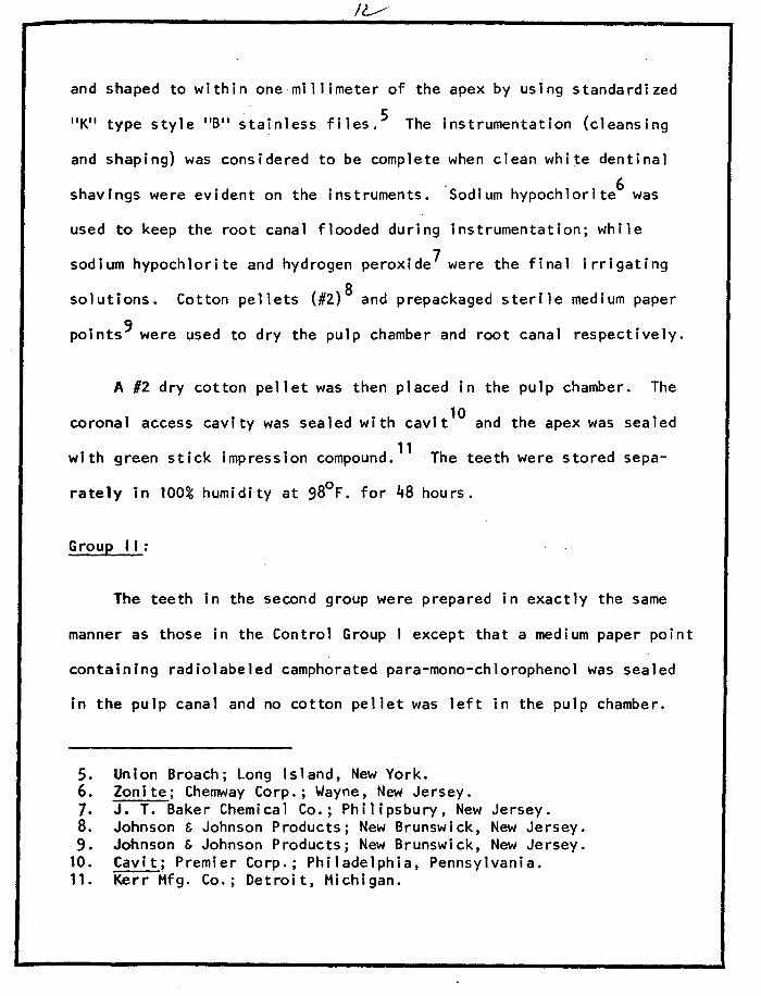
and shaped to within one millimeter of the apex by using standardized
11 K11 type style 11611 stainless files. 5 The instrumentation (cleansing
and shaping) was considered to be complete when clean white dentinal
shavings were evident on the instruments. 'sodium hypochlorite6 was
used to keep the root canal flooded during instrumentation; while
sodium hypochlorite and hydrogen peroxide7 were the final irrigating
solutions. Cotton pellets (#2) 8 and prepackaged sterile medium paper
points9 were used to dry the pulp chamber and root canal respectively.
A #2 dry cotton pellet was then placed in the pulp chamber. The
coronal access cavity was sealed with cavit 10 and the apex was sealed
11 with green stick impression compound. The teeth were stored sepa-
rately in 100% humidity at 98°F. for 48 hours.
Group ti:
The teeth in the second group were prepared in exactly the same
manner as those in the Control Group I except that a medium paper point
containing radiolabeled camphorated para-mono-chlorophenol was sealed
in the pulp canal and no cotton pellet was left in the pulp chamber.
5. Union Broach; Long Island, New York. 6. Zonite; Chemway Corp.; Wayne, New Jersey. 7. J. T. Baker Chemical Co.; Philipsbury, New Jersey. 8. Johnson & Johnson Products; New Brunswick, New Jersey. 9. Johnson & Johnson Products; New Brunswick, New Jersey.
10. Cavit; Premier Corp.; Philadelphia, Pennsylvania. 11. Kerr Mfg. Co.; Detroit, Michigan.
Group I II:
In Group I II the preparation of the teeth differed in that the
cleansing and shaping was confined to the buccal surfaces in order to
leave some pulpal tissue on the lingual wall. A #2 cotton pellet with
radiolabeled camphorated para-mono-chlorophenol was sealed in the
tooth.
Group IV:
The teeth in Group IV were prepared in exactly the same manner
as those in Control Group I except that the #2 cotton pellet sealed
in the pulp chamber contained radiolabeled camphorated para-mono
chlorophenol.
All of the teeth were handled individually with rubber gloves to
prevent cross-contamination. Care was taken to prevent excess cam
phorated para-mono-chlorophenol from flowing on to the external surface
of the teeth and all instruments were thoroughly washed and scrubbed
before entering each tooth.
After 48 hours in 100% humidity all of the teeth were placed in
separate decalcifying solutions containing 50% of 88% formic acid and
50% of 20% sodium citrate (Preece, 1965). The teeth were kept in this
solution for twelve days. All four groups of teeth were bisected
bucco-lingually using razor blades. A one-blade per tooth procedure
was followed to prevent contamination.
!~
The teeth were then sealed in paraffin and central sections with
a thickness of 4 microns were made. Autoradiographs were produced by
coating the sections with radiographic emulsion and placing them in a
light tight box for four days. After developing and fixing, the
sections were stained with nuclear fast red Indigo-Carmine dye and
picric acid. The specimens were then examined microscopically and the
silver grains produced by the radiolabeled camphora·ted para-mono
chlorophenol were counted with the aid of an automatic hand counter.
A 50 micron square grid was superimposed over the grains to facilitate
counting. All counting was done under oil emersion (lOOOX). Ten
random background counts were made in areas free of specimen on each
slide examined. This was necessary to establish counts above back
ground on each tooth section.
Each section in all groups was divided into apical 1/3, middle
1/3, and coronal 1/3 areas. In Groups II, I II, and IV the grid was
superimposed over the dentin directly against the pulpal wall. The
grains within the grid were counted and recorded and the grid was
moved one grid width at a time away from the pulp wall and perpen
dicular to it until no significant counts above background were
recorded. This procedure was repeated ten times at random points
along the pulp wall in each 1/3 of each section.
In Group I, grain counts were started at the pulpal wall and
moved one grid width at a time until ten counts were made. This was
/,J
done at six random points along the pulpal wall in each 1/3 of each
section.
The mean counts above background were determined for the various
fields in each group. Statistical analysis of the results was carried
out using the t-test for comparing two means.
IG
CHAPTER 4
RESULTS
Statistical analysis using the t-test to compare mean grain
counts above background of Groups I and II are shown in Table 1. In
the coronal 1/3 of the teeth in Group II (teeth with paper points
containing the tritium labeled medicament sealed in) there was a
significant (P = 0.01) penetration of labeled camphorated para-mono-
chlorophenol 0.20 mm. from the pulpal wall. In the middle 1/3,
significant penetration was noted 0.25 mm. from the pulpal wall.
Significant penetration was minimal in the apical 1/3 where the radio-
labeled medicament traveled only 0.05 mm. from the pulpal wall. The
greatest penetration then in Group I I was in the middle 1/3.
Turning to Group I II (teeth in which the cleansing and shaping
was confined to the buccal surface of the canal and medicament sealed
on a cotton pellet) and its statistical comparison with the Control
Group I (Table 2), it can be seen that the greatest penetration was in
• the coronal 1/3. No significant penetration was seen in the apical
1/3 while penetration to 0.05 mm. was seen in the middle 1/3 and to
0.40 mm. in the coronal 1/3. All counting was confined to the
partially cleansed lingual surface.
Table 3 shows a significant penetration in the coronal 1/3 of
teeth in Group IV (those cleansed and shaped with placement of
r7
radiolabeled camphorated para-mono-chlorophenol on a cotton pellet)
to a depth of 0.25 mm. from the pulpal wall. In the middle 1/3
penetration was only to 0.15 mm. Again, in the apical 1/3 penetration
was limited to 0.05 mm. from the pulpal wall.
It was also observed in all sections in the experimental groups
that there_ was an extremely high concentration of activated grains in
the pulp chamber or canal immediately adjacent to the cotton pellet or
paper point.
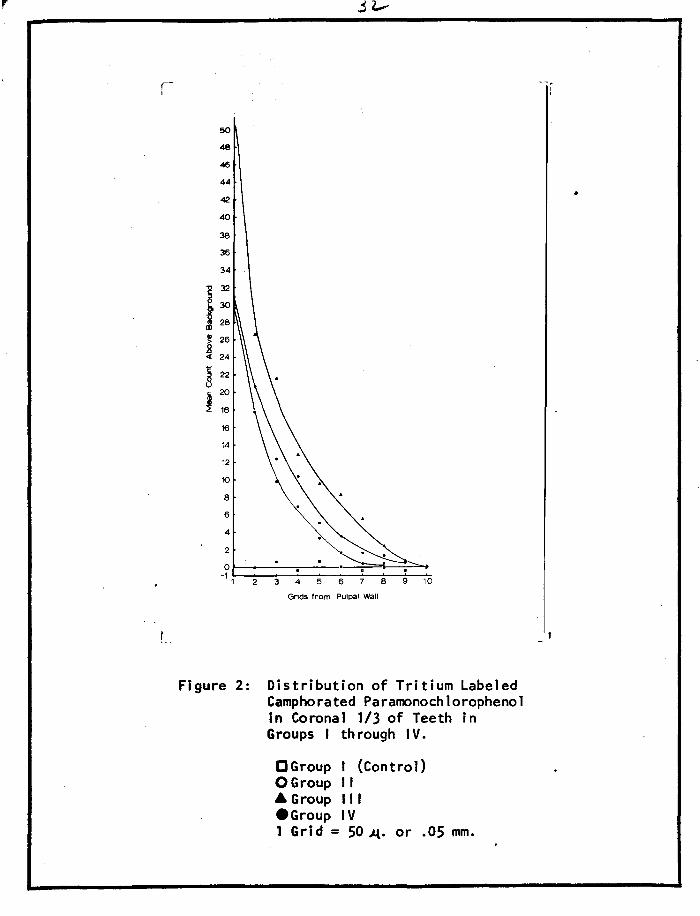
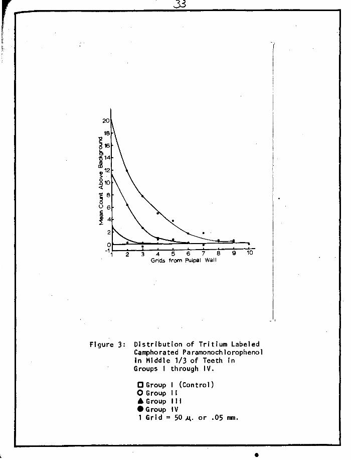
Figures 2 and 3 show the distribution of tritium labeled cam
phorated para-mono-chlorophenol in the various areas of the teeth
in Groups I through IV.
CHAPTER 5
DISCUSSION
It is well established that bacteria in a tooth are rarely con
fined to one area of a tooth such as the pulp chamber or the pulp
canal. Instead they are found throughout the whole tooth and
especially in the dentinal tubules (Gurney, 1963).
Since the natural defense mechanisms of the body have been
destroyed inside a tooth undergoing endo~ontic treatment, micro
organisms in these teeth must be brought under control by some other
means. Large numbers of microorganisms can be removed through bio
mechanical preparation. Chemotherapeutic agents, however, are required
to eliminate the remaining microorganisms.
The minimum requirements of an ideal root canal medication are:
(1) that it be germicidal to most organisms; (2) that it has deep
penetration; (3) that it exhibits rapid effectiveness; and (4) that
it is effective in the presence of organic matter (Sommer, Ostrander
and Crowley, 1961).
Camphorated para-mono-chlorophenol has been considered clinically
as a highly effective antimicrobial agent (Ostrander, 1958). Little
experimental evidence, however, has been presented to date to classify
camphorated para-mono-chlorophenol as an ideal root canal medicament.
If
This study was concerned only with the second minimum requirement
of an ideal intracanal medicament, the ability to penetrate into
dentin.
Tritium was used in this study because of the ease with which
compounds can be labeled with it. It has a convenient half life and
is a low energy beta emitter which gives an accurate relation of
silver grains to labeled material.
Thin sections were essential in order to accurately measure the
exact depth of penetration into the dentinal tubules. Decalcified
sections were used rather than ground sections because of the problems
encountered in a similar study by Avny in 1970. He found that in
preparing thin ground sections there was a scattering of radioactive
material over the surface of the tooth which gave inaccurate and
false results.
It is apparent from Tables 1 through 3 that the greatest penetra
tion of camphorated para-mono-chlorophenol in the coronal 1/3 occurs
in Group I II where the medicament was placed on a cotton pellet in the
chamber and sealed in. In the middle 1/3 the greatest penetration was
in Group I I where the medicament was placed on a paper point in the
canal. Penetration in the middle 1/3 appeared to be greatly reduced
by the pulpal tissue along the lingual wall in Group II I as compared
to Group IV, both of which had radiolabeled CPC on a cotton pellet in
the chamber. The poorest penetration was in the apical 1/3 in all
groups. The teeth in the Control Group showed grain counts very
similar to background (insignificant difference at P = 0.50) and
equally dispersed throughout the dentin thus allowing the comparison
of mean counts above background of the Control Group and each of the
other groups.
From the results it would seem that camphorated para-mono-
chlorophenol stays predominantly in the area where it is placed and
if used, would be most effectively placed on a paper point or cotton
pellet in intimate contact with the pulpal wall along its entire
length. The idea held by many practitioners, that because this
medicament is highly volatile, it penetrates rapidly and deeply into
all the dentinal tubules, appears to be in great error,
The antimicrobial activity of camphorated para-mono-chlorophenol
has been reported to be due solely to the para-mono-chlorophenol while
the camphor serves merely as a vehicle. While camphor possesses no
antiseptic value, it does have a slight anodyne effect (Sommer,
Ostrander and Crowley, 1961). From this study it would appear that
camphor also acts as a reservoir releasing the para-mono-chlorophenol
very s 1ow1 y.
The results of Harrison's 1969 experiments indicated:
11 1) CPC (camphorated para-mono-chlorophenol) is highly toxic, 2) a reduced parachlorophenol concentration in a vehicle of water is far less toxic than
CPC and 3) parachlorophenol is an effective antimicrobial agent in extremely small concentrations in aqueous solution against a variety of microorganisms commonly found in the root canal."
Avny's study in 1970 indicated that parachlorophenol in a vehicle
of water has deep penetration capability and that the use of either a
paper point or a cotton pellet to carry the medicament into the tooth
resulted in penetration throughout the dentin in the apical, middle
and coronal 1/3's to the cemento-dentinal junction, a distance at
least 7 to 10 times greater than the present study.
Considering the advantages of 2% aqueous parachlorophenol as
pointed out by Harrison and Avny in their studies and the failure of
camphorated para-mono-chlorophenol to penetrate beyond a maximum of
0.40 mm. into the dentin in the present study, perhaps the choice of
one of these two intracanal medicaments in endodontic therapy should
be re-evaluated. Since camphor serves only as a vehicle for the
para-mono-chlorophenol and CPC is highly toxic to the tissues, it
would seem that para-mono-chlorophenol in an aqueous vehicle would
be preferred.
CHAPTER 6
SUMMARY
A study of the ability of camphorated para-mono-chlorophenol
to penetrate into the dentinal tubules of endodontically-treated
teeth was conducted using tritium labeling. Autoradiographic evi
dence indicated that this intracanal medicament placed on cotton
pellets and paper points penetrated a maximum of 0.40 mm. into the
dentin of the coronal 1/3, 0.25 mm. into the dentin of the middle
1/3, and 0.05 mm. into the dentin of the apical 1/3.
CHAPTER 7
BIBLIOGRAPHY
Amler, M. H.: Radioactive Phosphate Permeability in Dentin Following the Use of Medicaments, J. Dent. Res. 27: 635-639, 1948.
Amler, M. H., and Bevelander, G.: Radioactive Phosphate Absorption by Dentin and Enamel, J. Dent. Res. 24: 45-51, 1945.
Amler, H. H., and Bevelander, G.: Dentin Permeability to Radioactive Phosphorus After Specific Time Intervals Following the Application of Various Drugs, New York J. Dent. 21: 295-300, 1951.
Avny, W. Y.: The lntracanal Diffusion of 2% Aqueous Parachlorophenol in Endodontics, an Autoradiographic Study, Masters Thesis, Loyola University Medical Center, June, 1970.
Bartels tone, H. J.: Survey of the Use of Radioactive Isotopes in Dentistry, New York J. Dent. 20: 320-336, 1950.
Bodecker, C. W.: Tooth Condition, a Factor in Experimental Isotope Absorption, J. Dent. Res. 22: 281-285, 1943.
Bodecker, C. W., and Lefkowitz, W.: Concerning the Vitality of the Calcified Dental Tissues, J. Dent. Res. 16: 463-470, 1937.
Buest, T. B.: Posteruptive Changes in the Maturation of Teeth, J. Amer. Dent. Ass. 18: 2186-2192, 1931.
Coolidge, Edgar D.: Studies of Germicides for the Treatment of Root Canals, J. Amer. Dent. Ass. 16: 698-712, 1929.
Coolidge, E. D., and Kesel, R. G.: A Textbook of Endodontology, ed. 2, Philadelphia, 1956, Lea and Febiger, p. 201 and p. 229.
Engler, W. D., Ramfjord, S. P., and Hinicker, J. J.: Healing Following Simple Gingivectomy, a Tritiated Thymidine Autoradiographic Study I. Epithelization, J. Periodont. 37: 298, 1966.
Evans, E. Anthony: A Guide to Tritium Labeling Services, England, 1968, Radiochemical Centre, Amersham Buck~.
Feinendfgen, L. E.: Tritium-Labeled Molecules in Biology and Medicine, New York and London, 1967, Academic Press.
Fish, E. W.: An Experimental Investigation of Enamel, Dentin, and the Dental Pulp, London, 1933, John Bale, Sons and Danielson, Ltd., p. 118.
Fitzgerald, P., Eidenoff, M., Knoll, J., and Simmel, E.: Tritium in Autoradiographs, Science 114: 494-498, 1951.
Fitzgerald, P., Semmel, E., Weinstein, J., and Martin, C.: Radioautography: Theory, Technique, and Applications, Lab. Invest. 2: 181-182, 1963.
Gurney, B. F.: Modern Methods in Bacteriologic Control, Dent. Cl in. N. Amer. pp. 321-339, July, 1963.
Hampson E. L., and Atkinson, Anne M.: The Relation Between Drugs Used in Root Canal Therapy and the Permeability of the Dentin, Brit. Dent. J. 116: 546-550, June 16, 1964.
Harlan, A. W.: The Use of Diffusable Medicaments in an Around the Roots of Teeth, D. Review 4: 466-475, July, 1891.
Harrison, J. W.: Aqueous Parachlorophenol: Its Toxicity and Antimicrobial Effectiveness, Masters Thesis, Loyola University, June 1969.
Hevesy, G. C., Holst, J. J., Krogh, A.: Investigation on Exchange of Phosphorus in Teeth Using Radioactive Phosphorus as Indicator, Kgl. Danske Viedensk. Biol. Medd. 13: Nov. 13, 1937,
Manly, M. L., and Bale, W. F.: The Metabolism of Inorganic Phosphorus of Rat Bones and Teeth as Indicated by the Radioactive Isotope, J. Biol. Chem. 129: 125-134, 1939.
Marshall, F. J., Hassler, M., and Dute, A. L.: Effects of Endodontic Treatments on Permeability of Root Dentine, Oral Surg., Oral Med., and Oral Path. 13: 208-223, Feb., 1960.
Martin, N. 0.: The Permeability of the Dentin to P32 Using the Direct Tissue Radioautography Technique, Oral Surg., Oral Med., and Oral Path. 4: 1461-1464, 1951.
Montagna, W., and Billingham, R. E.: Wound Healing, New York, 1964, The Macmillan Co.
McCauley, H. B., a~2 Gilda, J. E.: In Vivo Distribution of Radiophosphorus (P ) In Vital and Pulpless Teeth of a Dog as Indicated by Radioautographs, J. Dent. Res. 22: 200-203, 1943.
··,
National Formulary, 12th Ed., Washington, D.C., 1965, American Pharmaceutical Association, p. 76.
Nicholson, R., Stark, M., ij§uyen, N., and Scott, H.: Autoradiographic Tracings Utilizing Ca Labeled E.D.T.A.C., Oral Surg., Oral Med., and Oral Path. 26: 563-566, Oct., 1968.
Ostrander, F. D.: The Development of Antiseptics and Antibiotics for Use in Endodontics, In: Transaction of the Second International Conference on Endodontics, L. I. Grossman, Editor, pp. 81-95, 1958.
Preece, Ann: A Manual for Histologic Technicians, Ed. 2, Boston, 1965, Little Brown and Co.
Ramfjord, S. P., Engler, W. D., and Hiniker, J. J.: An Autoradiographic Study of Healing Following Simple Gingivectomy. II. The Connective Tissue, J. Periodont. 37: 179, 1966.
Shuttelworth, C. W.: The Diffusion of Penicillin from the Dental Root Canal, Brit. Dent. J. 89: 127-130, 1950.
Sognnaes, R. F., and Volker, J. F.: Studies on the Distribution of Radioactive Phosphorus in the Tooth Enamel of Experimental Animals, Am. J. Physiol. 133: 112-120, 1941.
Sommer, R. F., Ostrander, F., Darl, and Crowley, M. C.: Clinical Endodontics, Ed. 2, W. B. Saunders, Co., p. 194, 1961.
Stahl, S.S., Tonna, E. A., and Weiss, R.: Autoradiographic Evaluation of Gingival Response to Injury. I. Surgical Trauma in Young Adult Rats, Ach. Oral Biol. 13: 71, 1968.
Stahl, S. S., Weiss, R., and Tonna, E. A.: Autoradiographic Evaluation of Periapical Responses to Pulpal Injury. I. Young Rats, Oral Surg., Oral Med., and Oral Path. 28: 249-258, Aug., 1962.
Stamps, Herman F.: The Testing of Dentine Sterilizing Agents, University of Michigan Alumni Bulletin, p. 16, Oct., 1953.
Stewart, G. G., Kapsimalas, P., Rappaport, H.: E.D.T.A. and Ur841 Peroxide for Root Canal Preparation, J. Amer. Dent. Ass. 78: 335-338, March, 1969.
Tonn a, E. A. : Response of the Ce 11u1 ar Phase of the S ke 1 eton >.IO Trauma, Periodontics 4: 105, 1966.
Van Huysen, G., Hodge, H. C., and Warren, S. L.: A Quantitative Roentgenodensitometric Study of the Changes in Teeth Due to Attrition, J. Dent. Res. 16: 243-265, 1937.
Wach, E. C., Hauptfuehrer, J. D., and Kes35, R. G.: Endodontic Significance of the Penetration of S Labeled Penicillin in Extracted Human Teeth, Oral Surg~, Oral Surg., Oral Med., and Oral Path. 8: 639-643, 1955·. -----------
Wainwright, W.W., and Lemoine, F. +4: Rapid Diffuse Penetration of Intact Enamel and Dentine by C Labeled Urea, J. Amer. Dent. Ass. 41: 135-145, 1950.
Zach, Leo, Topal, R., Cohen, G.: Pulpal Repair Following Operative Procedures, Oral Surg., Oral Med., and Oral Path. 28: 587-597, Oct., 1969.
27
CHAPTER 8
.APPENDIX
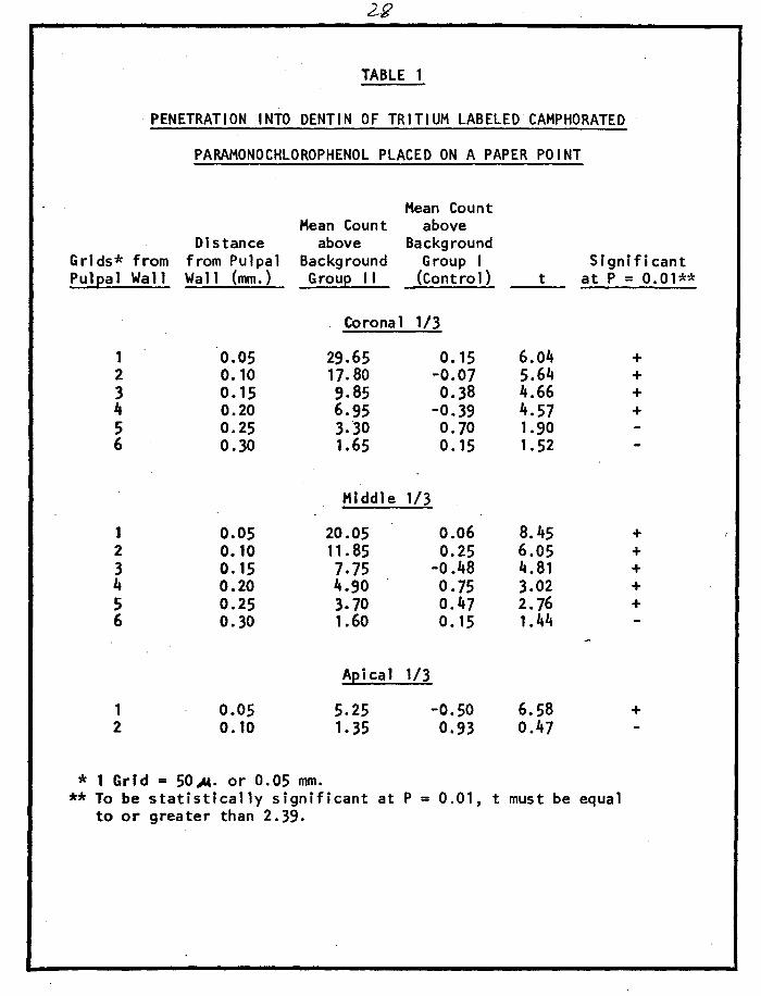
TABLE 1
PENETRATION INTO DENTIN OF TRITIUM LABELED CAMPHORATED
PARAMONOCHLOROPHENOL PLACED ON A PAPER POINT
Mean Count Hean Count above
Di stance above Background Grids* from from Pulpal Background Group I Significant Pulpal Wall Wall (mm.) Group 11 (Contro 1) t at P = 0.01**
Coronal 1/3
1 0.05 29.65 0. 15 6.04 + 2 o. 10 17.80 -0.07 5.64 + 3 o. 15 9.85 0.38 4.66 + 4 0.20 6.95 -0.39 4.57 + 5 0.25 3. ·30 0.10 1.90 6 0.30 1.65 0. 15 1.52
Middle 1/3
1 0.05 20.05 0.06 8.45 + 2 0.10 11. 85 0.25 6.05 + 3 o. 15 7.75 -o.48 4.81 + 4 0.20 4.90 o. 75 3.02 + 5 0.25 3.70 0.47 2. 76 + 6 o. 30 1.60 o. 15 1.44
Apical 1/3
1 0.05 5.25 -0.50 6.58 + 2 0. 10 1. 35 0.93 0.47
* 1 Grid = 50-"'. or 0.05 mm. **To be statistically significant at P = 0.01, t must be equal
to or greater than 2.39.
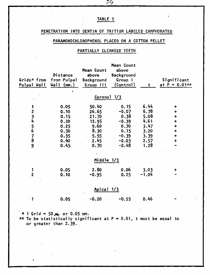
TABLE 2
PENETRATION INTO DENTIN OF TRITIUM LABELED CAHPHORATED
PARAMONOCHLOROPHENOL PLACED ON A COTTON PELLET
PARTIALLY CLEANSED TEETH
Hean Count Mean Count above
Distance above Background Grids* from from Pulpal Background Group I Significant Pulpal Wall Wall (mm.) Group 111 (Control) t at P = 0.01**
Coronal 1/3
1 0.05 50.40 o. 15 6.44 + 2 0. 10 26.65 -0.07 6.78 + 3 o. 15 21. 70 0.38 5.08 + 4 0.20 12.95 -0.39 4.61 +
·5 0.25 9.60 0. 70 3.47 + 6 0.30 8.30 0. 15 3.20 + 7 0.35 5.55 -0.39 3. 39 + 8 o.4o 2.45 -0.03 2.57 + 9 o.45 0.10 -0.48 1.28
Middle 113
1 0.05 2.80 0.06 3.03 + 2 o. 10 -0.95 0.25 -1.04
Apical 1/3
1 0.05 -0.20 -0.53 0.46
* 1 Grid = 50 .M.- or 0.05 mm. **To be statistically significant at P = 0.01, t must be equal to
or greater than 2.39.
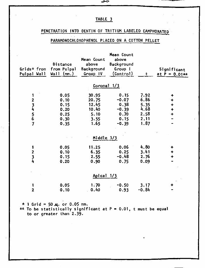
TABLE 3
PENETRA Tl ON INTO DENTIN OF TRITIUM LABELED CAMPHORATED
PARAMONOCHLOROPHENOL PLACED ON A COTTON PELLET
Hean Count Hean Count above
Distance above Background Grids* from from Pulpal Background Group I S i g n i fi cant Pulpal Wall Wa 11 (nvn.) Group IV (Centro 1) t at P = 0.01**
Coronal 1/3
1 0.05 30.95 0. 15 7.92 + 2 o. 10 20. 75 -0.07 6.86 + 3 0. 15 12.45 0.38 5.35 + 4 0.20 10.40 -0.39 4.68 + 5 0.25 5. 10 0.10 2.58 + 6 0.30 3.55 0. 15 2. 11 7 0.35 1.65 -0. 39 1.87
Middle 1/3
1 0.05 11.25 0.06 4.80 + 2 o. 10 6.35 0.25 3.41 + 3 o. 15 2.55 -o.48 2.76 + 4 0.20 0.90 0.75 0.09
Apical 1/3
1 0.05 1. 70 -0.50 3. 17 + 2 0. 10 0.40 0.93 -0.84
* 1 Grid = 50 ~· or 0.05 mm. **To be statistically significant at P = 0.01, t must be equal
to or greater than 2.39.
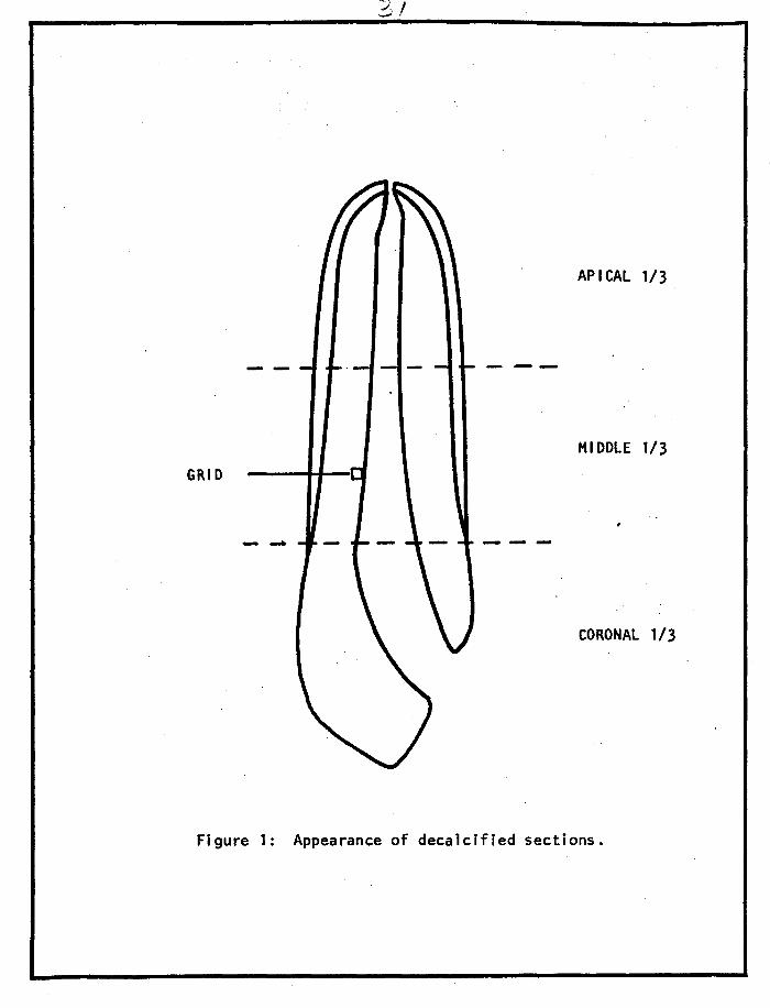
APICAL 1/3
---
MIDDLE 1/3
GRID
- -
CORONAL 1/3
Figure 1: Appearance of decalcified sections.
t __
50
48
46
44
40
38
36
34
"B 32
i3 30
J 28
~ 26 .8 <I: 24
"§ 22 u c; 20
' 18 16
14
12
10
8
6
4
2 o,__ ____ _
-1
1 2 3 4 5 6 7 8 9 10
Grids from Pulpal Wall
Figure 2: Distribution of Tritium Labeled Camphorated Paramonochlorophenol in Coronal 1/3 of Teeth in Groups I through IV.
C Group I (Contro 1) OGroup 11 •Group 111 eGroup IV 1 Grid = 50 ..A.l· or .OS mm.
20
1: 8 :I 0 u 6 c
i 4
0 -1
1 2 3 4 5 6 7 8 9 10 Grids from Pulpal Wall
Figure 3: Distribution of Tritium Labeled Camphorated Paramonochlorophenol in Middle 1/3 of Teeth in Groups I through IV.
CGroup I {Control) 0 Group 11 6 Group 111 •Group IV 1 Grid = 50 ,t.t. or .05 mm.
•
- I
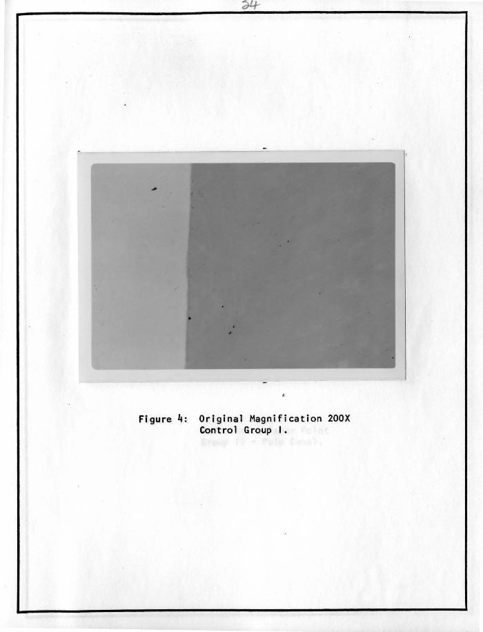
Figure 4: Original Magnification 200X Control Group I.
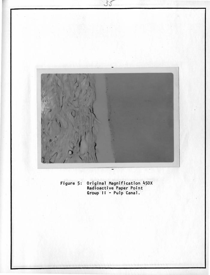
Figure 5: Original Magnification 450X Radioactive Paper Point Group I I - Pulp Canal.
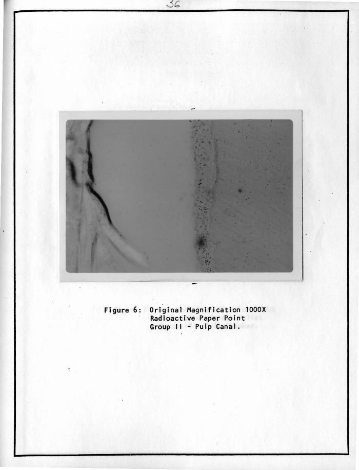
Figure 6: Original Magnification lOOOX Radioactive Paper Point Group II - Pulp Canal.
7
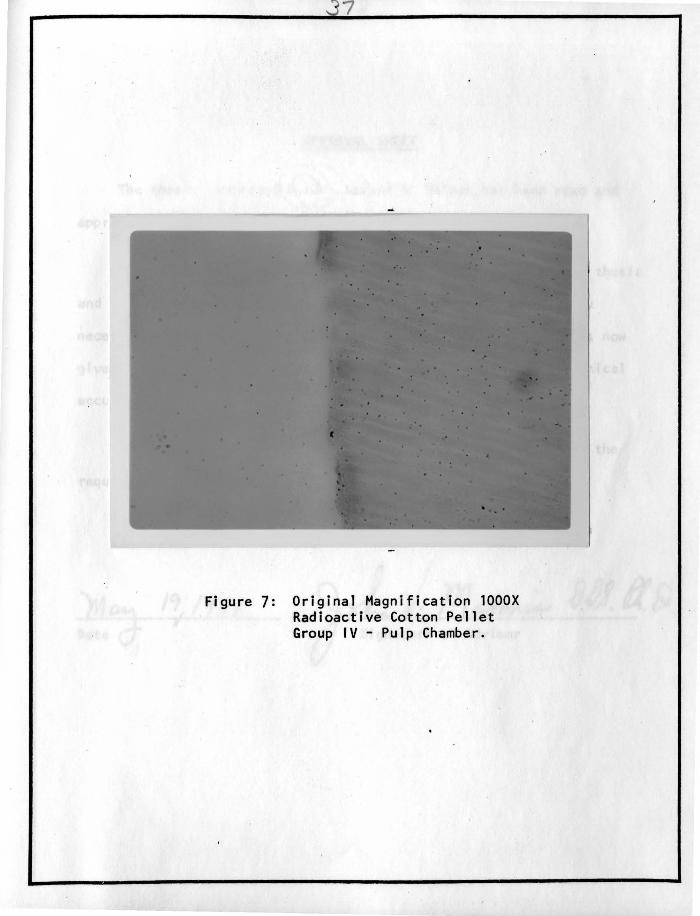
Figure 7: Original Magnification lOOOX Radioactive Cotton Pellet Group IV - Pulp Chamber.
APPROVAL SHEET
The thesis submitted by Dr. Gerald R. Heiman has been read and
approved by three members of the Graduate School faculty.
The final copies have been examined by the director of the thesis
and the signature which appears below verifies the fact that any
necessary changes have been incorporated, and that the thesis is now
given final approval with references to content, form and mechanical
accuracy.
The thesis is therefore accepted in partial fulfillment of the
requirements for the degree of Master of Science.
Im J
Signature of Advisor