


Languages
Pages
Legal


9/9/15
1
Evaluation and Management of Acute Abdominal Pain in Primary
Care Joanna Guenther, PhD, RN, FNP-BC, CNE
Objectives
! Discuss the symptoms of acute abdominal pain in relationship to the patient’s history and clinical presentation.
! Review the etiology of acute abdominal pain in relationship to anatomic location and the patient’s age.
! Review diagnostic testing to evaluate acute abdominal pain and discuss appropriate treatment plans.
Epidemiology ! Abdominal Pain (AP) is a common reason for
patients to seek primary care. ! Acute AP is defined in terms of symptoms lasting
<1 wk. ! Admission rates for patients with acute AP range
from 20-40% (and even higher in the elderly population).
! The diagnosis is undetermined in at least 50% of patients at the time of discharge.
! Initial diagnosis is accurate in only 50-65% of cases.

9/9/15
2
Abdominal Pain
! Differential very broad – Work-up based on symptoms, medical history,
physical exam, lab, and location (RUQ, RLQ, LUQ, LLQ, Epigastric, Flank)
– Determine which patients can be safely observed or treated symptomatically and which require further investigation and/or referral
HISTORY Onset Location
Duration Intensity
Relieving & aggravating
factors
Med Hx Surg Hx Family Hx
Travel Hx Social Hx Rx
Triggers & associated symptoms
Mechanisms of Pain Transmission
Visceral
Parietal
Referred
9/9/15
3
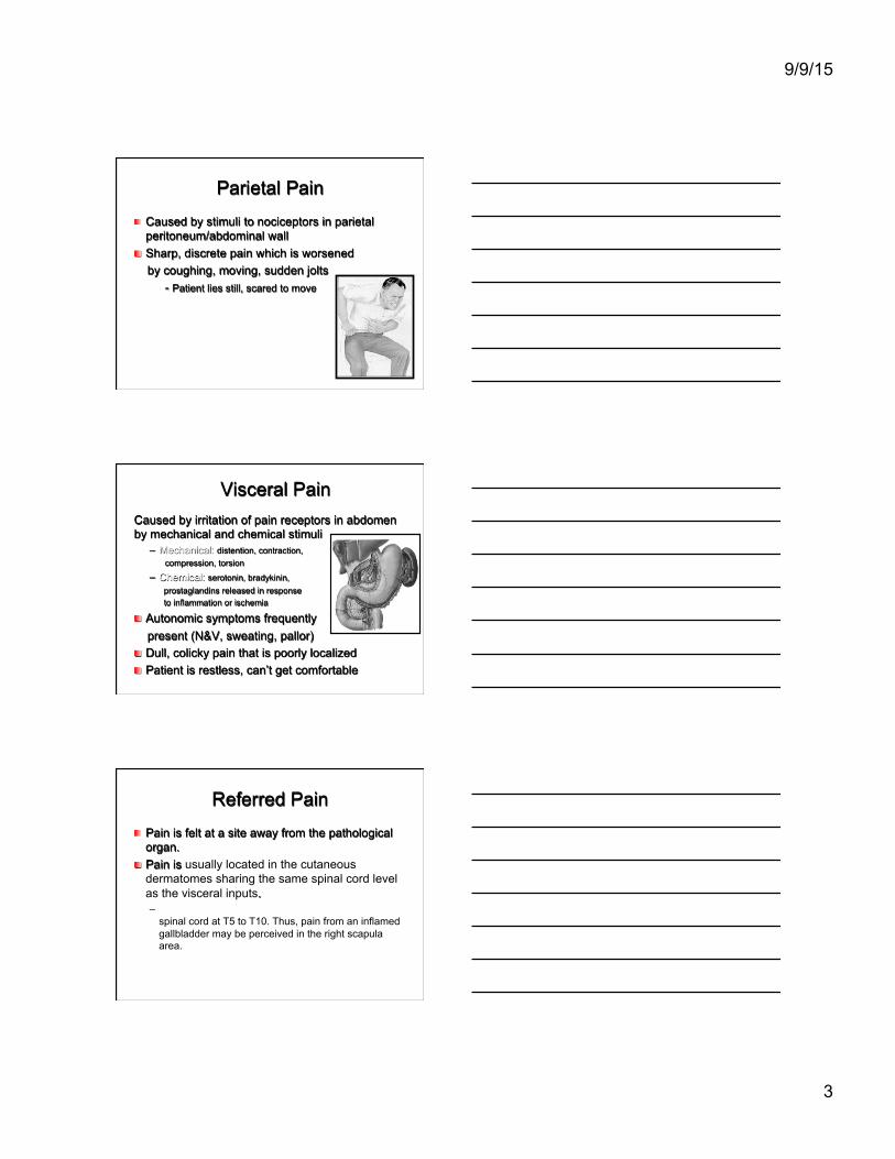
Parietal Pain ! Caused by stimuli to nociceptors in parietal
peritoneum/abdominal wall ! Sharp, discrete pain which is worsened by coughing, moving, sudden jolts
- Patient lies still, scared to move
Visceral Pain Caused by irritation of pain receptors in abdomen by mechanical and chemical stimuli
– Mechanical: distention, contraction, compression, torsion
– Chemical: serotonin, bradykinin, prostaglandins released in response to inflammation or ischemia
! Autonomic symptoms frequently present (N&V, sweating, pallor) ! Dull, colicky pain that is poorly localized ! Patient is restless, can’t get comfortable
Referred Pain ! Pain is felt at a site away from the pathological
organ. ! Pain is usually located in the cutaneous
dermatomes sharing the same spinal cord level as the visceral inputs. – Nociceptive inputs from the gallbladder enter the
spinal cord at T5 to T10. Thus, pain from an inflamed gallbladder may be perceived in the right scapula area.

9/9/15
4
FIRST - ASSESS q Patient’s overall
demeanor q Skin/Eyes for jaundice q Vital Signs q Expose the entire
abdomen ! Xiphoid to pubis ! Observe for pulsations
Description of abdomen – Scaphoid – Flat – Rounded
SECOND - AUSCULTATE ! Auscultate in all
quadrants ! Quantitative
– Absent – Decreased – Hyperactive
! Qualitative – Normal – High-pitched
! Additional – Arterial bruits
9/9/15
5
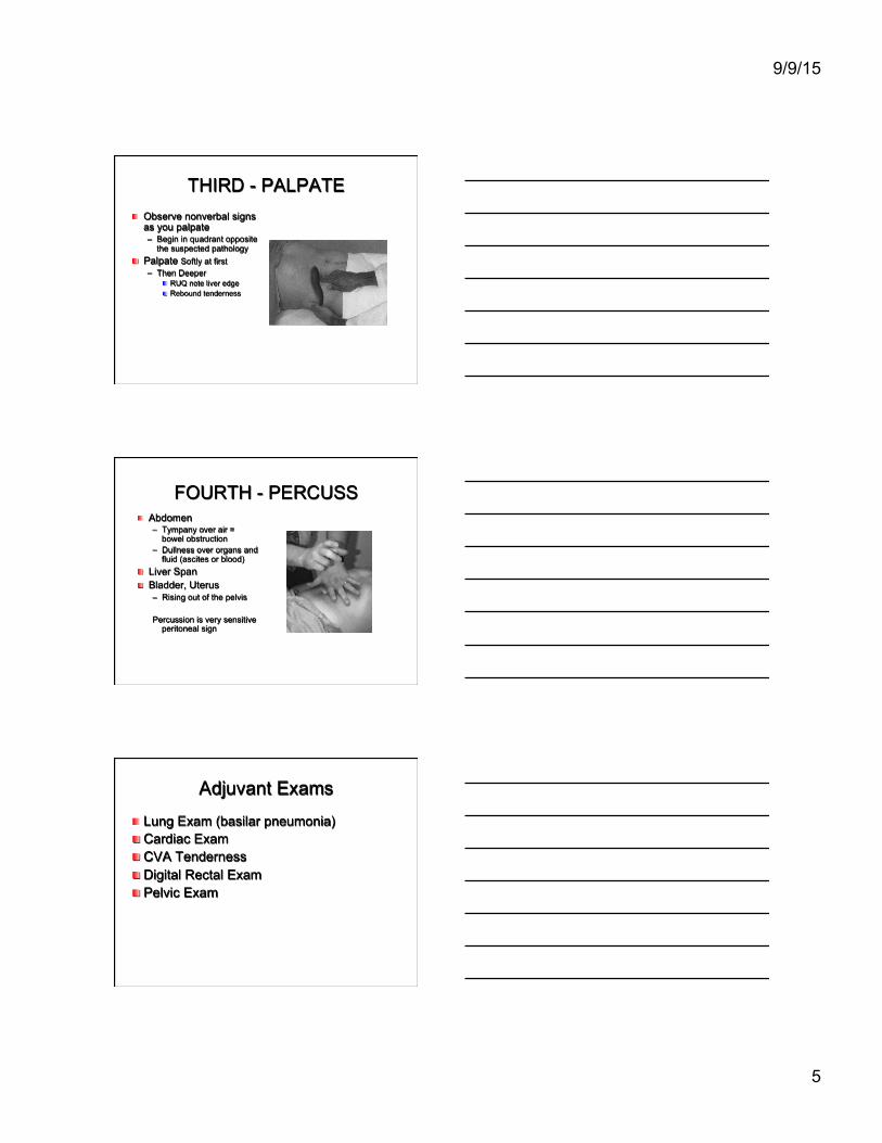
THIRD - PALPATE ! Observe nonverbal signs
as you palpate – Begin in quadrant opposite
the suspected pathology ! Palpate Softly at first
– Then Deeper ! RUQ note liver edge ! Rebound tenderness
FOURTH - PERCUSS ! Abdomen
– Tympany over air = bowel obstruction
– Dullness over organs and fluid (ascites or blood)
! Liver Span ! Bladder, Uterus
– Rising out of the pelvis Percussion is very sensitive
peritoneal sign
Adjuvant Exams
! Lung Exam (basilar pneumonia) ! Cardiac Exam ! CVA Tenderness ! Digital Rectal Exam ! Pelvic Exam

9/9/15
6
Peritoneal Signs of the Acute Abdomen
! Constant tenderness and guarding ! Involuntary rigidity of abdominal muscles ! Reduced or absent bowel sounds ! Positive Heel-Jar test - stand on toes/fall on heels;
hop (Markle sign) ! Rebound tenderness -worsens on release of hand
after deep palpation (McBurney sign) ! Referred pain - deep palpation of LLQ causes pain
in RLQ (Rovsing sign)
Classification of Acute Abdominal Pain
! Three main categories of acute abdominal pain: 1. Intra-abdominal (arising from within the
abdominal cavity/retroperitoneum): ! GI (Biliary Tract Disease, Appendicitis, Diverticulitis, Pancreatitis,
SBO, IBS, Constipation, Intussusception)
! GU (Renal Colic, Acute urinary retention, UTI)
! Gyn (Acute PID, Ectopic pregnancy)
! Vascular systems (AAA, Mesenteric Ischemia, Ischemic Colitis)
Classification of Abdominal Pain 2. Extra-abdominal (less common):
! Cardiopulmonary (AMI, etc)
! Abdominal wall (Hernia, Zoster etc)
! Toxic-metabolic (DKA, OD, lead, etc)
! Neurogenic pain (Zoster, etc)
! Psychic (Anxiety, Depression, etc)
3. Nonspecific abdominal pain (NSAP) – not well explained or described.

9/9/15
7
Acute Abdominal Pain: Common Causes by Age
Infants:
• Constipation • Colic • Intussusception
Childhood:
• Appendicitis • UTI
Adolescence:
• Appendicitis, PID, IBS
Adults/Elders:
• IBS, cholecystitis, pancreatitis, appendicitis, dyspepsia, diverticulitis, biliary tract disease, bowel obstruction
Diagnostic Approach ! Essential Questions:
– Stable or Unstable? ! Vital Signs
– Is surgical referral needed? ! Obstruction ! Peritonitis ! Vascular
– Or is watchful waiting okay?

9/9/15
8
Diagnostic Approach ! What are the differential diagnoses?
– Based on history and physical exam, and possibly labs ! Location of pain has strong predictive value
! Work up in stable, less obvious cases may include: – CBC with differential – Electrolytes, BUN, creatinine, and glucose – Amylase, lipase – Bilirubin – LFTs – UA – Pregnancy test in women of childbearing potential – Imaging
Diagnostic Approach
! Primary Imaging for Acute Abdominal Pain:
– Radiographs Abdomen (and possibly chest) ! Free air (perforation), bowel obstruction, masses
– US Abdomen (and possibly pelvis) ! Stones (GB, CBD), masses, enlarged organs
– CT Abdomen (and possibly pelvis) ! Better at identifying site and cause of obstruction, can identify
complications such as ischemia, perforation
Algorithm RUQ Pain
Reference: Cartwright, S.L., & Knudson, M.P. (2008). Evaluation of acute abdominal pain in adults. American Family Physician, (77)7, 971-978.

9/9/15
9
Algorithm RLQ Pain
Algorithm LLQ Pain
Acute Appendicitis
! Clinical features with some predictive value include:
! Pain located in the RLQ (74%) ! Pain migration from the periumbilical area to the
RLQ (54%) ! Positive psoas or obturator sign (52%) ! Fever (51%) ! Rebound tenderness (40%) ! Rigidity (38%) ! Anorexia (26%)

9/9/15
10
Appendicitis: Psoas Sign
Appendicitis: Obturator Sign
Passively flex right hip and knee then internally rotate the hip
ACR Rating Scale: 1,2,3 = Usually not appropriate 4,5,6 = May be appropriate 7,8,9 = Usually appropriate

9/9/15
11
Acute Appendicitis
! Ultrasound abdomen is usually used for detection (ACR Rating 9)
! CT abdomen may be ordered in adults
and non-pregnant women (ACR Rating 6).
! Ultrasound preferred in children.
Biliary Tract Disease ! Most common diagnosis of pts > 50 years. ! Generally associated with RUQ pain
– Composed of: ! Acute Cholecystitis (acalculus/calculus) ! Biliary Colic ! Common Bile Duct Obstruction
! Of those patients found to have acute cholecystitis, the majority lack fever and 40% lack leukocytosis.
! Abdominal US is the test of choice for patients with suspected biliary tract disease (ACR Rating 9), as compared to CT scan with contrast (ACR Rating 6).

9/9/15
12
Bowel Obstruction Ÿ Can be either large or small bowel
Ÿ Most common causes : Ÿ Adhesions from prior surgery, incarcerated
hernia, cancer, volvulus, mass of parasites, inflammatory bowel disease
Ÿ CT abdomen and pelvis with IV contrast (ACR Rating 9); x-ray abdomen and pelvis (ACR Rating 5)
Acute Pancreatitis ! 80% of cases are due to ETOH abuse or gallstones in CBD.
! Other common causes: – Drugs (Valproic acid, Tetracycline, Hydrochlorothiazide, Furosemide) – Pancreatic cancer – Abdominal trauma/surgery – Ulcer with pancreatic involvement – Familial pancreatitis (Hypertriglycerides / Hypercalcemia)
! Definition : – Inflammation of the pancreas associated with edema, pancreatic
autodigestion, necrosis and possible hemorrhage
Acute Pancreatitis ! Only a minority number of pts present with pain
and tenderness limited to the anatomic area of the pancreas in the upper half of the abdomen.
! 50% of pts present with c/o pain extending well beyond the upper abdomen to cause generalized tenderness.

9/9/15
13
Acute Pancreatitis
! The inflammatory process around the pancreas may cause other signs and symptoms such as: – Pleural effusion – Grey Turner's sign (flank discoloration) – Cullen's sign ( discoloration around the
umbilicus) – Ascites – Jaundice
Acute Pancreatitis
Radiological study: Ultrasound abdomen – Indicated if increased amylase and lipase with high
clinical certainty of diagnosis; with < 48-72 hours after onset of symptoms; Commonly ordered to evaluate for CBD obstruction. (ACR Rating 9)
CT abdomen with contrast – Indicated with high index of severity score and
> 48-72 hours after onset of symptoms
(ACR Appropriateness Criteria)

9/9/15
14
Acute Diverticulitis ! 1/3 of pts present with pain to the lower half of the
abdomen.
! 20% of elderly pts with operatively confirmed diverticulitis lacked abdominal tenderness.
! Elderly pts are at risk for a severe and often fatal complication of diverticulitis (perforation of the colon)
Diverticulitis ! CT abdomen and pelvis with contrast to
confirm diagnosis, determine extent of disease and evaluate for complications: perforation, abscess, fistulas, or bowel obstruction – Perforated colon CA can mimic acute
diverticulitis

9/9/15
15
Renal Colic ! Pts may present with abrupt, colicky, unilateral flank pain
that radiates to the groin, testicle, or labia.
! Hematuria and plain abd films can be helpful however do not provide a strong support in the diagnostic evaluation of suspected renal colic.
! CT (with and without contrast): masses and stones well
seen (ACR Rating 9) ! US: Pregnancy, pediatric age group
Hematuria
Acute Pelvic Inflammatory Disease
! Patient may complain of pain/tenderness in lower abdomen, adnexa or cervix.
! Most importantly patient may complain of abnormal vaginal discharge (most common finding).
! Fever, palpable mass, ↑WBC have been inconsistently associated with PID.
! The best noninvasive test is transvaginal ultrasound.

9/9/15
16
Ectopic Pregnancy ! Symptoms include lower abdominal pain (most common)
and vaginal bleeding (maybe the only complaint). ! Female pts (child bearing age) that present with these
symptoms automatically get a urine pregnancy test and HCG quantitative level.
! If the pt is pregnant, then order a transvaginal US to evaluate for ectopic pregnancy (ACR Rating 9).
! Clear view of an IUP in 2 perpendicular views essentially excludes an ectopic pregnancy.
Abdominal Aortic Aneurysm ! Dissections produce chest or upper back pain that
often migrates to abdomen as the dissection extends distally.
! AAA rather than dissect, it often enlarges, leaks, and then ruptures.
! Approx. 50% of pts with AAA present with hypotension, abdominal/back pain, and/or pulsatile abd mass. Sxs often similar to renal colic.
! Neither the presence or the absence of femoral pulse or an abdominal bruit are helpful clinically.
Abdominal Aortic Aneurysm ! Palpation is an important part of physical exam. May
be able to detect an enlarged aorta.
! Pt > 50 yrs old presenting with recent onset of abd/flank/low back pain should have either US abdomen (ACR Rating 9) or CT scan abdomen without contrast (ACR Rating 8) to confirm/exclude AAA from the differential diagnosis. – Smoking history significantly increases risk

9/9/15
17
Extrabdominal Diagnoses of Acute Abdominal Pain: Cardiopulmonary ! Pain is usually in upper half of abdomen.
! A chest film should be done to look for pneumonia, pulmonary infarction, pleura effusion, and/or pneumothorax.
! A neg. film plus pleuritic pain could mean PE.
! If epigastric pain is present one should inquire about cardiac history, get an ECG, and consider further cardiac evaluation.
Extrabdominal Diagnoses of Acute Abdominal Pain: Hernias
! Characterized by a defect through which intraabdominal contents protrude during increases in the intraabdominal pressure
! Several types exist: inguinal, incisional, periumbilical, and femoral (common in female).
! Uncomplicated hernias can be asymptomatic, aching/uncomfortable, and reducible on exam.
! Significant pain could mean strangulation (blood supply is compromised)/incarceration (not reducible).
Nonspecific Abdominal Pain (NSAP)
! A good portion of patients will have nonspecific abdominal pain.
! Patients may have nausea, midepigastric pain, or RLQ tenderness.
! The lab workup is usually normal. ! WBC may be elevated. ! If NSAP is the working diagnosis, patients
must be re-examined in 24 hours.

9/9/15
18
Abdominal Pain Clinical Pearls ! Do not restrict the diagnosis solely by the location of the pain. ! Consider appendicitis in all patients with abdominal pain who have
an appendix, especially in patients with the presumed diagnosis of gastroenteritis, PID or UTI.
! Any woman with childbearing potential and abdominal pain has an ectopic pregnancy until her pregnancy test comes back negative.
! Obtain an ECG in patients with cardiac risk factors presenting with abdominal pain.
! Pain almost always precedes vomiting in surgical causes; converse is true for most gastroenteritis and NSAP.
! Acute cholecystitis is the most common surgical emergency in the elderly.
! If the pain of biliary colic lasts more than 6 hours, suspect early cholecystitis.
Medical-Legal Risk Successful Lawsuits -Fractures and dislocations -Foreign bodies in wound -Tendon and nerve damage
associated with limb lacerations -Myocardial Infarction -Appendicitis -Meningitis -Subarachnoid hemorrhage -Spinal Cord injury -Ectopic pregnancy
Largest Damage Awards -Myocardial Infarction
-Meningitis
-Fractures
-Ectopic Pregnancy
-Tendon and nerve damage associated with limb lacerations
-Foreign bodies in wounds
Top Related