Acute Abdominal Pain Handout - c.ymcdn.com · Acute Abdominal Pain: Common Causes by Age ... Acute...
18
9/9/15 1 Evaluation and Management of Acute Abdominal Pain in Primary Care Joanna Guenther, PhD, RN, FNP-BC, CNE Objectives Discuss the symptoms of acute abdominal pain in relationship to the patient’s history and clinical presentation. Review the etiology of acute abdominal pain in relationship to anatomic location and the patient’s age. Review diagnostic testing to evaluate acute abdominal pain and discuss appropriate treatment plans. Epidemiology Abdominal Pain (AP) is a common reason for patients to seek primary care. Acute AP is defined in terms of symptoms lasting <1 wk. Admission rates for patients with acute AP range from 20-40% (and even higher in the elderly population). The diagnosis is undetermined in at least 50% of patients at the time of discharge. Initial diagnosis is accurate in only 50-65% of cases.
Transcript of Acute Abdominal Pain Handout - c.ymcdn.com · Acute Abdominal Pain: Common Causes by Age ... Acute...

9/9/15
1
Evaluation and Management of Acute Abdominal Pain in Primary
Care Joanna Guenther, PhD, RN, FNP-BC, CNE
Objectives
! Discuss the symptoms of acute abdominal pain in relationship to the patient’s history and clinical presentation.
! Review the etiology of acute abdominal pain in relationship to anatomic location and the patient’s age.
! Review diagnostic testing to evaluate acute abdominal pain and discuss appropriate treatment plans.
Epidemiology ! Abdominal Pain (AP) is a common reason for
patients to seek primary care. ! Acute AP is defined in terms of symptoms lasting
<1 wk. ! Admission rates for patients with acute AP range
from 20-40% (and even higher in the elderly population).
! The diagnosis is undetermined in at least 50% of patients at the time of discharge.
! Initial diagnosis is accurate in only 50-65% of cases.

9/9/15
2
Abdominal Pain
! Differential very broad – Work-up based on symptoms, medical history,
physical exam, lab, and location (RUQ, RLQ, LUQ, LLQ, Epigastric, Flank)
– Determine which patients can be safely observed or treated symptomatically and which require further investigation and/or referral
HISTORY Onset Location
Duration Intensity
Relieving & aggravating
factors
Med Hx Surg Hx Family Hx
Travel Hx Social Hx Rx
Triggers & associated symptoms
Mechanisms of Pain Transmission
Visceral
Parietal
Referred
9/9/15
3
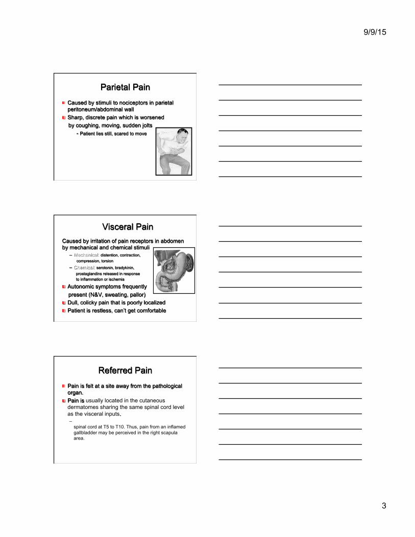
Parietal Pain ! Caused by stimuli to nociceptors in parietal
peritoneum/abdominal wall ! Sharp, discrete pain which is worsened by coughing, moving, sudden jolts
- Patient lies still, scared to move
Visceral Pain Caused by irritation of pain receptors in abdomen by mechanical and chemical stimuli
– Mechanical: distention, contraction, compression, torsion
– Chemical: serotonin, bradykinin, prostaglandins released in response to inflammation or ischemia
! Autonomic symptoms frequently present (N&V, sweating, pallor) ! Dull, colicky pain that is poorly localized ! Patient is restless, can’t get comfortable
Referred Pain ! Pain is felt at a site away from the pathological
organ. ! Pain is usually located in the cutaneous
dermatomes sharing the same spinal cord level as the visceral inputs. – Nociceptive inputs from the gallbladder enter the
spinal cord at T5 to T10. Thus, pain from an inflamed gallbladder may be perceived in the right scapula area.

9/9/15
4
FIRST - ASSESS q Patient’s overall
demeanor q Skin/Eyes for jaundice q Vital Signs q Expose the entire
abdomen ! Xiphoid to pubis ! Observe for pulsations
Description of abdomen – Scaphoid – Flat – Rounded
SECOND - AUSCULTATE ! Auscultate in all
quadrants ! Quantitative
– Absent – Decreased – Hyperactive
! Qualitative – Normal – High-pitched
! Additional – Arterial bruits
9/9/15
5
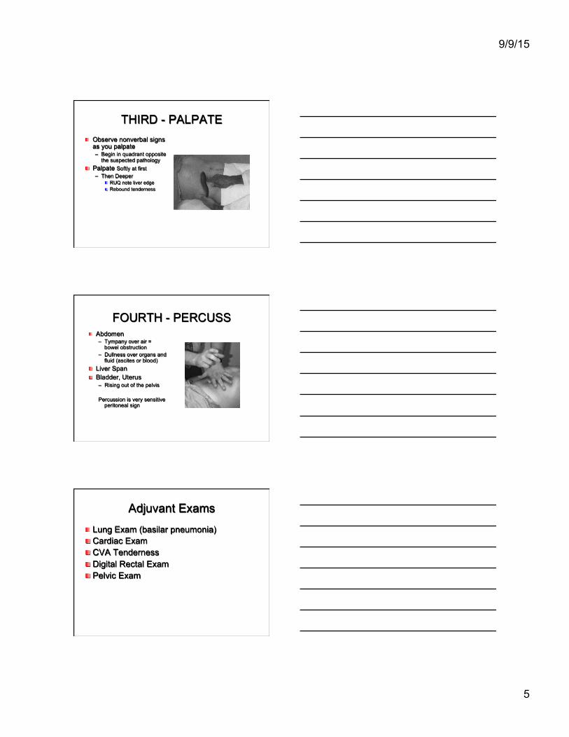
THIRD - PALPATE ! Observe nonverbal signs
as you palpate – Begin in quadrant opposite
the suspected pathology ! Palpate Softly at first
– Then Deeper ! RUQ note liver edge ! Rebound tenderness
FOURTH - PERCUSS ! Abdomen
– Tympany over air = bowel obstruction
– Dullness over organs and fluid (ascites or blood)
! Liver Span ! Bladder, Uterus
– Rising out of the pelvis Percussion is very sensitive
peritoneal sign
Adjuvant Exams
! Lung Exam (basilar pneumonia) ! Cardiac Exam ! CVA Tenderness ! Digital Rectal Exam ! Pelvic Exam

9/9/15
6
Peritoneal Signs of the Acute Abdomen
! Constant tenderness and guarding ! Involuntary rigidity of abdominal muscles ! Reduced or absent bowel sounds ! Positive Heel-Jar test - stand on toes/fall on heels;
hop (Markle sign) ! Rebound tenderness -worsens on release of hand
after deep palpation (McBurney sign) ! Referred pain - deep palpation of LLQ causes pain
in RLQ (Rovsing sign)
Classification of Acute Abdominal Pain
! Three main categories of acute abdominal pain: 1. Intra-abdominal (arising from within the
abdominal cavity/retroperitoneum): ! GI (Biliary Tract Disease, Appendicitis, Diverticulitis, Pancreatitis,
SBO, IBS, Constipation, Intussusception)
! GU (Renal Colic, Acute urinary retention, UTI)
! Gyn (Acute PID, Ectopic pregnancy)
! Vascular systems (AAA, Mesenteric Ischemia, Ischemic Colitis)
Classification of Abdominal Pain 2. Extra-abdominal (less common):
! Cardiopulmonary (AMI, etc)
! Abdominal wall (Hernia, Zoster etc)
! Toxic-metabolic (DKA, OD, lead, etc)
! Neurogenic pain (Zoster, etc)
! Psychic (Anxiety, Depression, etc)
3. Nonspecific abdominal pain (NSAP) – not well explained or described.

9/9/15
7
Acute Abdominal Pain: Common Causes by Age
Infants:
• Constipation • Colic • Intussusception
Childhood:
• Appendicitis • UTI
Adolescence:
• Appendicitis, PID, IBS
Adults/Elders:
• IBS, cholecystitis, pancreatitis, appendicitis, dyspepsia, diverticulitis, biliary tract disease, bowel obstruction
Diagnostic Approach ! Essential Questions:
– Stable or Unstable? ! Vital Signs
– Is surgical referral needed? ! Obstruction ! Peritonitis ! Vascular
– Or is watchful waiting okay?

9/9/15
8
Diagnostic Approach ! What are the differential diagnoses?
– Based on history and physical exam, and possibly labs ! Location of pain has strong predictive value
! Work up in stable, less obvious cases may include: – CBC with differential – Electrolytes, BUN, creatinine, and glucose – Amylase, lipase – Bilirubin – LFTs – UA – Pregnancy test in women of childbearing potential – Imaging
Diagnostic Approach
! Primary Imaging for Acute Abdominal Pain:
– Radiographs Abdomen (and possibly chest) ! Free air (perforation), bowel obstruction, masses
– US Abdomen (and possibly pelvis) ! Stones (GB, CBD), masses, enlarged organs
– CT Abdomen (and possibly pelvis) ! Better at identifying site and cause of obstruction, can identify
complications such as ischemia, perforation
Algorithm RUQ Pain
Reference: Cartwright, S.L., & Knudson, M.P. (2008). Evaluation of acute abdominal pain in adults. American Family Physician, (77)7, 971-978.

9/9/15
9
Algorithm RLQ Pain
Algorithm LLQ Pain
Acute Appendicitis
! Clinical features with some predictive value include:
! Pain located in the RLQ (74%) ! Pain migration from the periumbilical area to the
RLQ (54%) ! Positive psoas or obturator sign (52%) ! Fever (51%) ! Rebound tenderness (40%) ! Rigidity (38%) ! Anorexia (26%)

9/9/15
10
Appendicitis: Psoas Sign
Appendicitis: Obturator Sign
Passively flex right hip and knee then internally rotate the hip
ACR Rating Scale: 1,2,3 = Usually not appropriate 4,5,6 = May be appropriate 7,8,9 = Usually appropriate

9/9/15
11
Acute Appendicitis
! Ultrasound abdomen is usually used for detection (ACR Rating 9)
! CT abdomen may be ordered in adults
and non-pregnant women (ACR Rating 6).
! Ultrasound preferred in children.
Biliary Tract Disease ! Most common diagnosis of pts > 50 years. ! Generally associated with RUQ pain
– Composed of: ! Acute Cholecystitis (acalculus/calculus) ! Biliary Colic ! Common Bile Duct Obstruction
! Of those patients found to have acute cholecystitis, the majority lack fever and 40% lack leukocytosis.
! Abdominal US is the test of choice for patients with suspected biliary tract disease (ACR Rating 9), as compared to CT scan with contrast (ACR Rating 6).

9/9/15
12
Bowel Obstruction Ÿ Can be either large or small bowel
Ÿ Most common causes : Ÿ Adhesions from prior surgery, incarcerated
hernia, cancer, volvulus, mass of parasites, inflammatory bowel disease
Ÿ CT abdomen and pelvis with IV contrast (ACR Rating 9); x-ray abdomen and pelvis (ACR Rating 5)
Acute Pancreatitis ! 80% of cases are due to ETOH abuse or gallstones in CBD.
! Other common causes: – Drugs (Valproic acid, Tetracycline, Hydrochlorothiazide, Furosemide) – Pancreatic cancer – Abdominal trauma/surgery – Ulcer with pancreatic involvement – Familial pancreatitis (Hypertriglycerides / Hypercalcemia)
! Definition : – Inflammation of the pancreas associated with edema, pancreatic
autodigestion, necrosis and possible hemorrhage
Acute Pancreatitis ! Only a minority number of pts present with pain
and tenderness limited to the anatomic area of the pancreas in the upper half of the abdomen.
! 50% of pts present with c/o pain extending well beyond the upper abdomen to cause generalized tenderness.

9/9/15
13
Acute Pancreatitis
! The inflammatory process around the pancreas may cause other signs and symptoms such as: – Pleural effusion – Grey Turner's sign (flank discoloration) – Cullen's sign ( discoloration around the
umbilicus) – Ascites – Jaundice
Acute Pancreatitis
Radiological study: Ultrasound abdomen – Indicated if increased amylase and lipase with high
clinical certainty of diagnosis; with < 48-72 hours after onset of symptoms; Commonly ordered to evaluate for CBD obstruction. (ACR Rating 9)
CT abdomen with contrast – Indicated with high index of severity score and
> 48-72 hours after onset of symptoms
(ACR Appropriateness Criteria)

9/9/15
14
Acute Diverticulitis ! 1/3 of pts present with pain to the lower half of the
abdomen.
! 20% of elderly pts with operatively confirmed diverticulitis lacked abdominal tenderness.
! Elderly pts are at risk for a severe and often fatal complication of diverticulitis (perforation of the colon)
Diverticulitis ! CT abdomen and pelvis with contrast to
confirm diagnosis, determine extent of disease and evaluate for complications: perforation, abscess, fistulas, or bowel obstruction – Perforated colon CA can mimic acute
diverticulitis

9/9/15
15
Renal Colic ! Pts may present with abrupt, colicky, unilateral flank pain
that radiates to the groin, testicle, or labia.
! Hematuria and plain abd films can be helpful however do not provide a strong support in the diagnostic evaluation of suspected renal colic.
! CT (with and without contrast): masses and stones well
seen (ACR Rating 9) ! US: Pregnancy, pediatric age group
Hematuria
Acute Pelvic Inflammatory Disease
! Patient may complain of pain/tenderness in lower abdomen, adnexa or cervix.
! Most importantly patient may complain of abnormal vaginal discharge (most common finding).
! Fever, palpable mass, ↑WBC have been inconsistently associated with PID.
! The best noninvasive test is transvaginal ultrasound.

9/9/15
16
Ectopic Pregnancy ! Symptoms include lower abdominal pain (most common)
and vaginal bleeding (maybe the only complaint). ! Female pts (child bearing age) that present with these
symptoms automatically get a urine pregnancy test and HCG quantitative level.
! If the pt is pregnant, then order a transvaginal US to evaluate for ectopic pregnancy (ACR Rating 9).
! Clear view of an IUP in 2 perpendicular views essentially excludes an ectopic pregnancy.
Abdominal Aortic Aneurysm ! Dissections produce chest or upper back pain that
often migrates to abdomen as the dissection extends distally.
! AAA rather than dissect, it often enlarges, leaks, and then ruptures.
! Approx. 50% of pts with AAA present with hypotension, abdominal/back pain, and/or pulsatile abd mass. Sxs often similar to renal colic.
! Neither the presence or the absence of femoral pulse or an abdominal bruit are helpful clinically.
Abdominal Aortic Aneurysm ! Palpation is an important part of physical exam. May
be able to detect an enlarged aorta.
! Pt > 50 yrs old presenting with recent onset of abd/flank/low back pain should have either US abdomen (ACR Rating 9) or CT scan abdomen without contrast (ACR Rating 8) to confirm/exclude AAA from the differential diagnosis. – Smoking history significantly increases risk

9/9/15
17
Extrabdominal Diagnoses of Acute Abdominal Pain: Cardiopulmonary ! Pain is usually in upper half of abdomen.
! A chest film should be done to look for pneumonia, pulmonary infarction, pleura effusion, and/or pneumothorax.
! A neg. film plus pleuritic pain could mean PE.
! If epigastric pain is present one should inquire about cardiac history, get an ECG, and consider further cardiac evaluation.
Extrabdominal Diagnoses of Acute Abdominal Pain: Hernias
! Characterized by a defect through which intraabdominal contents protrude during increases in the intraabdominal pressure
! Several types exist: inguinal, incisional, periumbilical, and femoral (common in female).
! Uncomplicated hernias can be asymptomatic, aching/uncomfortable, and reducible on exam.
! Significant pain could mean strangulation (blood supply is compromised)/incarceration (not reducible).
Nonspecific Abdominal Pain (NSAP)
! A good portion of patients will have nonspecific abdominal pain.
! Patients may have nausea, midepigastric pain, or RLQ tenderness.
! The lab workup is usually normal. ! WBC may be elevated. ! If NSAP is the working diagnosis, patients
must be re-examined in 24 hours.

9/9/15
18
Abdominal Pain Clinical Pearls ! Do not restrict the diagnosis solely by the location of the pain. ! Consider appendicitis in all patients with abdominal pain who have
an appendix, especially in patients with the presumed diagnosis of gastroenteritis, PID or UTI.
! Any woman with childbearing potential and abdominal pain has an ectopic pregnancy until her pregnancy test comes back negative.
! Obtain an ECG in patients with cardiac risk factors presenting with abdominal pain.
! Pain almost always precedes vomiting in surgical causes; converse is true for most gastroenteritis and NSAP.
! Acute cholecystitis is the most common surgical emergency in the elderly.
! If the pain of biliary colic lasts more than 6 hours, suspect early cholecystitis.
Medical-Legal Risk Successful Lawsuits -Fractures and dislocations -Foreign bodies in wound -Tendon and nerve damage
associated with limb lacerations -Myocardial Infarction -Appendicitis -Meningitis -Subarachnoid hemorrhage -Spinal Cord injury -Ectopic pregnancy
Largest Damage Awards -Myocardial Infarction
-Meningitis
-Fractures
-Ectopic Pregnancy
-Tendon and nerve damage associated with limb lacerations
-Foreign bodies in wounds