Zuur-Base Stoornissen De Anesthesist als Peri-Operatief ... · PDF file• Definition...
110
Zuur-Base Stoornissen De Anesthesist als Peri-Operatief Geneesheer Dr. M. Verhaegen Co-assistenten 3-10-2012
Transcript of Zuur-Base Stoornissen De Anesthesist als Peri-Operatief ... · PDF file• Definition...

Zuur-Base StoornissenDe Anesthesist als Peri-Operatief Geneesheer
Dr. M. VerhaegenCo-assistenten 3-10-2012
Section III:Preoperative Preparation and Intraoperative Managem ent
Chapter 21: Acid-Base Balance and Blood Gas Analysis (p. 334 - 343)Linda L. Liu
Basics of Anesthesia, 6 th editionRonald D. Miller and Manuel C. Pardo, Jr.
Acid - Base Balance
• Maintaining a physiologic pH ([H +]) is important– Oxygen transport– Enzyme activity– Biochemical reactions– Cellular function– Organ function
• Anesthesia and surgery affect acid-base balance– Changes in ventilation– Hemodynamic changes– Intravenous fluid therapy

Topics
• Definitions• Regulation of [H +]
– Buffer systems– Ventilatory response– Renal response
• Common acid-base disturbances– Respiratory acidosis– Respiratory alkalosis– Metabolic acidosis– Metabolic alkalosis
• Interpretation of acid-base disturbances: practical approach

DEFINITIONS
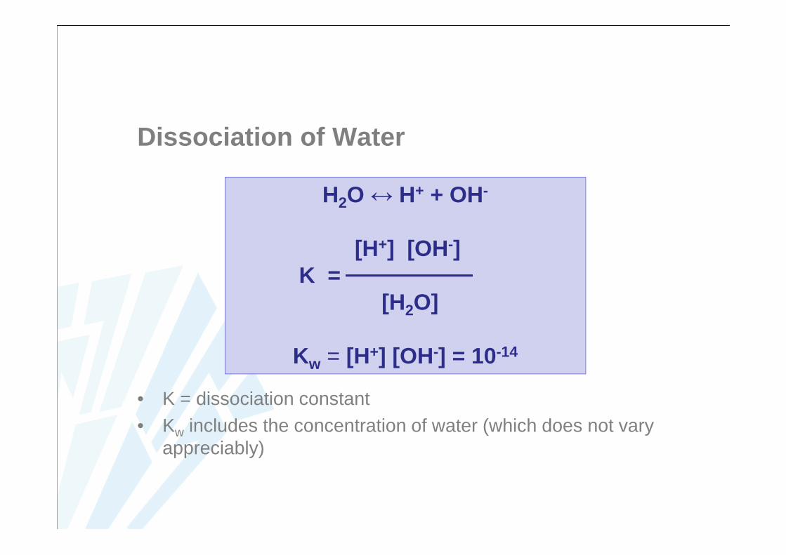
Dissociation of Water
• K = dissociation constant • Kw includes the concentration of water (which does not vary
appreciably)
H2O ↔ H+ + OH-
[H+] [OH -]K =
[H2O]
Kw = [H+] [OH -] = 10-14

Acid
• Definition– Traditionally: a substance that can act as a proton (H+) donor– A substance that increases [H+] when added to a solution
• Strong acid– Readily and irreversibly donates H+
� Increases [H+]
• Weak acid– Reversibly donates H+
� Less effect on [H+]

Base
• Definition– Traditionally: a substance that can act as a proton (H+) acceptor– A substance that decreases [H+] when added to a solution
• Strong base– Avidly binds H+
� Decreases [H+]
• Weak base– Reversibly binds H+
� Less effect on [H+]

Acidosis and Alkalosis
The terms acidosis and alkalosis refer to a primary pro cessand its compensatory respons altering arterial pH• Acidosis: a process that reduces blood pH• Alkalosis: a process that increases blood pH� Acidosis and alkalosis can occur concomitantly

Acidemia and Alkalemia
The terms acidemia and alkalemia denote the net effect o n arterial pH of primary processes and their compensatoryphysiologic responses • Acidemia: blood pH < 7.35• Alkalemia: blood pH > 7.45� Acidemia and alkalemia are mutually exclusive terms

Base Excess
Base excess (BE) is the amount of strong acid (BE > 0 ) or strong base (BE < 0) required to return 1 L of fully oxygenatedblood to a pH of 7.4 (at 37°C and a PaCO 2 of 40 mmHg)• Refers to the nonrespiratory (= metabolic) component o f an
acid-base disturbance– Normal value: -2 - 3 mmol/L– A positive value indicates metabolic alkalosis– A negative value indicates metabolic acidosis
• Graphically or electronically derived from a nomogr am (Siggaard-Andersen) or algorithm– Utilizes plasma pH, blood PCO2 and hemoglobin concentration
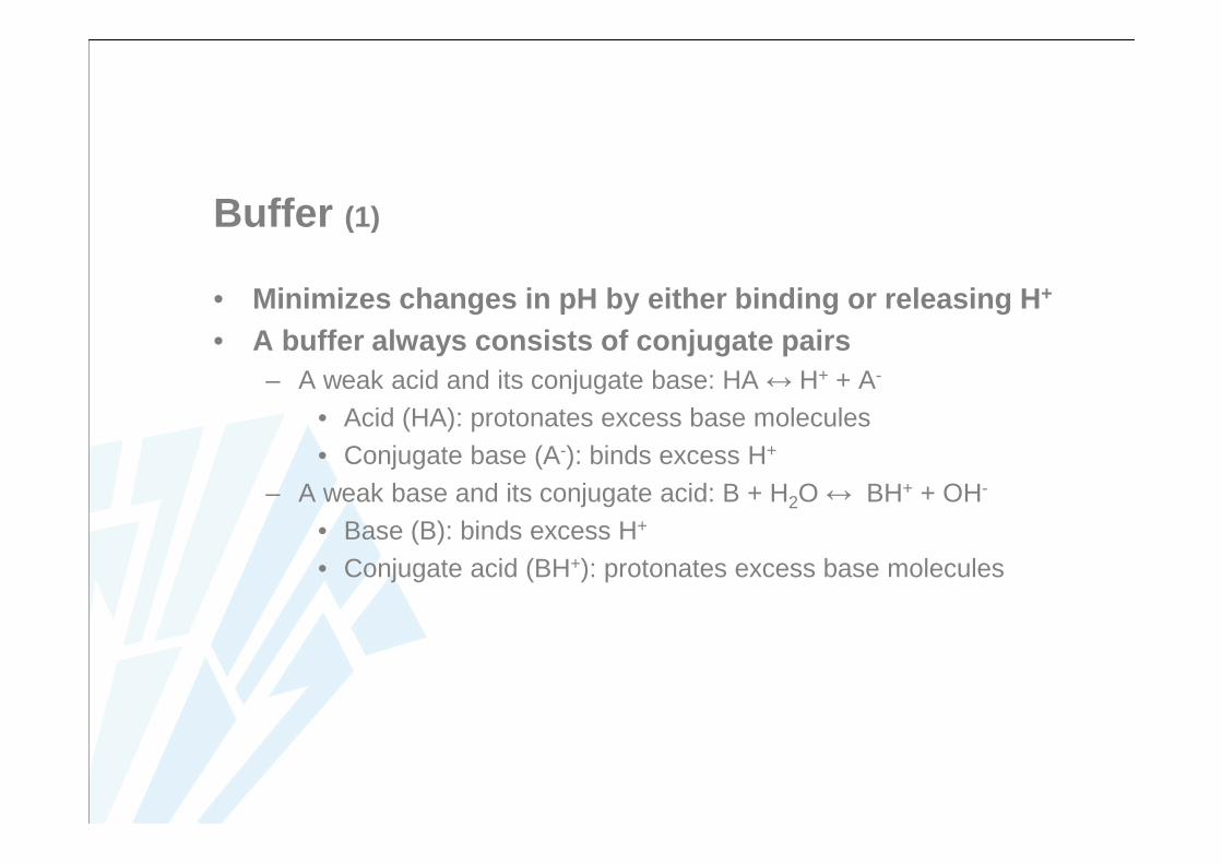
Buffer (1)
• Minimizes changes in pH by either binding or releasing H+
• A buffer always consists of conjugate pairs– A weak acid and its conjugate base: HA ↔ H+ + A-
• Acid (HA): protonates excess base molecules• Conjugate base (A-): binds excess H+
– A weak base and its conjugate acid: B + H2O ↔ BH+ + OH-
• Base (B): binds excess H+
• Conjugate acid (BH+): protonates excess base molecules
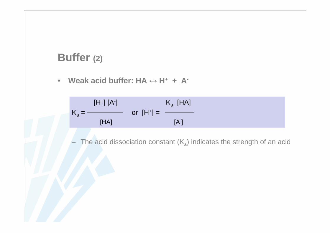
Buffer (2)
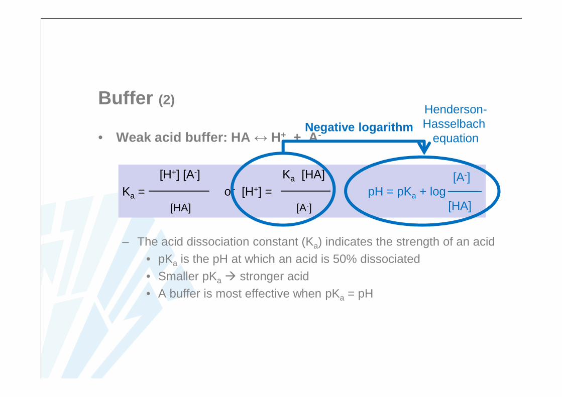
• Weak acid buffer: HA ↔ H+ + A-
[H+] [A-] Ka [HA]Ka = or [H+] =
[HA] [A-]
– The acid dissociation constant (Ka) indicates the strength of an acid
Buffer (2)
• Weak acid buffer: HA ↔ H+ + A-
[H+] [A-] Ka [HA]Ka = or [H+] =
[HA] [A-]
– The acid dissociation constant (Ka) indicates the strength of an acid• pKa is the pH at which an acid is 50% dissociated• Smaller pKa � stronger acid• A buffer is most effective when pKa = pH
Negative logarithmHenderson-Hasselbach
equation
[A-]pH = pKa + log
[HA]

Henderson -Hasselbach Equation
• The Henderson-Hasselbalch equation describes the relationship between the pH of a solution and the rati o of the concentration of a weak acid or weak base and its salt.
[Base] pH = pKa + log
[Weak acid]
• Bicarbonate buffer (H 2CO3 / HCO3-)
[HCO3-]
pH = pKa + log[H2CO3]
– pH is related to the ratio between [H2CO3] and [HCO3-]
– CO2 tension (PaCO2) may be substituted for H2CO3
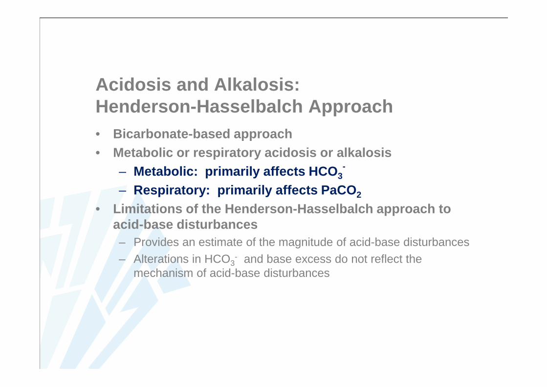
Acidosis and Alkalosis:Henderson -Hasselbalch Approach• Bicarbonate-based approach• Metabolic or respiratory acidosis or alkalosis
– Metabolic: primarily affects HCO 3-
– Respiratory: primarily affects PaCO 2
• Limitations of the Henderson-Hasselbalch approach toacid-base disturbances– Provides an estimate of the magnitude of acid-base disturbances– Alterations in HCO3
- and base excess do not reflect the mechanism of acid-base disturbances
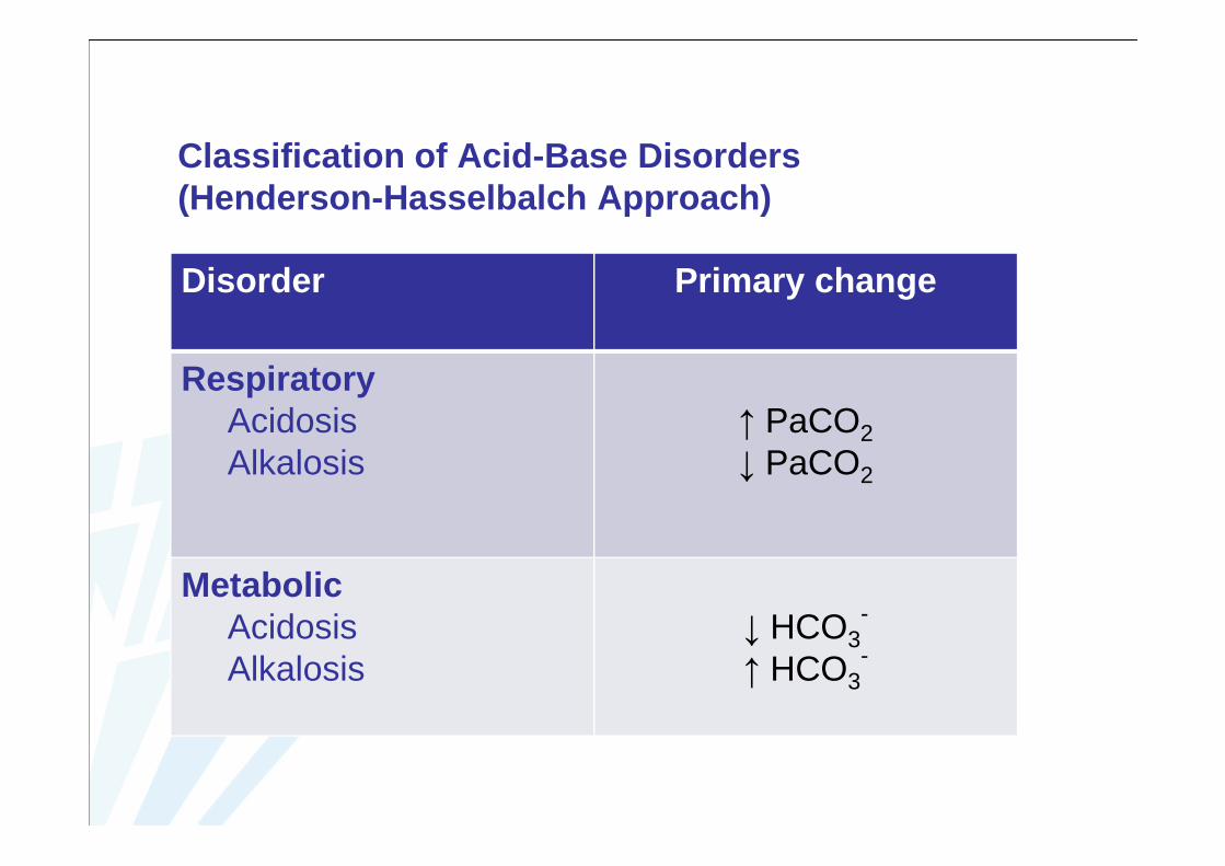
Classification of Acid-Base Disorders (Henderson-Hasselbalch Approach)
Disorder Primary change
RespiratoryAcidosisAlkalosis
↑ PaCO2↓ PaCO2
MetabolicAcidosisAlkalosis
↓ HCO3-
↑ HCO3-
Stewart Approach
• Physicochemical approach• Plasma [HCO 3
-] is a dependent variable and therefore does not alter blood pH
• 3 independent variables influence the dissociation of water and the [H +] ���� responsible for acid-base disturbances– Strong ion difference (SID)– Plasma concentration of nonvolatile weak acids (ATot)– Arterial carbon dioxide tension (PaCO2)

Strong Ion Difference
SID = (Strong cations ) – (Strong anions )= ([Na+]+[K +]+[Ca 2+]+[Mg 2+]) - ([Cl -]+[other strong anions] )= ([Na+]+[K +]) - [Cl -]
• Normal SID = 40 - 44 mEq/L– Balanced by an equal amount of buffer base (phosphate, albumin,
bicarbonate)
• Decreased SID ���� decreased blood pH – E.g. massive volumes of NaCl 0.9%: major ions are Na+ and Cl-
and SID = 0 � reduction of SID
• Increased SID ���� increased blood pH– E.g. gastric fluid loss: major ion is Cl- (> strong cations)

Nonvolatile Weak Acids
• Inorganic phosphates, albumin, and other plasma protei ns– The major practical difference with the Henderson-Hasselbalch
approach is the inclusion of the serum albumin concentration– However, in the Henderson-Hasselbalch approach the anion gap
can be corrected for albumin (hypoalbuminemia)
• Weak acids have not a great influence on the acid-base balance in healthy persons
• Hypoalbuminemia and hypophosphatemia���� Metabolic alkalosis
• Hyperalbuminemia and hyperphosphatemia���� Metabolic acidosis

Classification of Acid-Base Disorders (Stewart Approach)
Acidosis
Respiratory Increased PaCO 2
Metabolic
Decreased SID Increased Cl-
Decreased Na+/increased free water
Increased A Tot HyperphosphatemiaHyperproteinemia
Alkalosis
Respiratory Decreased PaCO 2
Metabolic
Increased SID Decreased Cl-
Increased Na+/decreased free water
Decreased A Tot HypophosphatemiaHypoalbuminemia

REGULATION OF HYDROGEN ION CONCENTRATION
Buffers
Hydrogen Ion Concentration
• Arterial blood and extracellular fluid: [H +] = 35 - 40 nmol/L– Normal arterial pH = 7.35 - 7.45– Blood pH between 6.8 – 7.8 ([H+] between 16 and 160 nmol/L) is
compatible with life
• The body has a large buffering capacity to maintain a normal [H +] despite a large amount of daily acid production• Daily approximately 15 000 mmol of CO2 (� carbonic acid)• Daily 50 – 100 mEq of nonvolatile acid
Regulation of [H +]
• Buffer systems: immediate chemical response– Bicarbonate buffer system– Hemoglobin buffer system
• Ventilatory response: fast response• Renal Response: slow response, but nearly complete
restoration of pH– Reabsoption of HCO3
-
– Excretion of titratable acid– Production of ammonia
Bicarbonate Buffer System (1)
Carbonic anhydrase
CO2 + H2O ↔ H2CO3 ↔ H+ + HCO3-
• Bicarbonate buffer: base = HCO 3-
weak acid = H 2CO3
• Carbonic anhydrase (in endothelium , erythrocytes andkidneys) greatly accelerates this reaction
• Most important buffering system when combined withrenal control of HCO 3
- and pulmonary control of CO 2
• Bicarbonate buffer is effective against metabolic, but notagainst respiratory acid-base disturbances

Bicarbonate Buffer System (2)
H+ + HCO3- ↔ H2CO3 ↔ H2O + CO2
• Henderson-Hasselbach equation for bicarbonate buffer
[HCO3-]
pH = pK + log [H2CO3]
• CO2 tension (PaCO 2) may be substituted for H 2CO3 [HCO3
-]pH = pK’ + log
0.3 PaCO2
– Solubility coefficient for CO2 = 0.03 mmol/L– Adjusted dissociation constant pK’ = 6.1
Bicarbonate Buffer System (3)
• pK’ = 6.1– Not close to normal arterial pH of 7.4– Nevertheless: bicarbonate buffer is important
• Relatively high concentrations in extracellular fluid• Kidneys and lungs have important influences on arterial pH by
altering the [HCO3-] /PaCO2 ratio
– Lungs regulate PaCO2
– Kidneys regulate plasma [HCO3-]
→System adapts to changing acid-base conditions

Ext
race
llula
rflu
idIn
trac
ellu
larf
luid
H+ + HCO3- ↔ H2CO3 ↔ H2O + CO2
H+ + HCO3- ↔ H2CO3 ↔ H2O + CO2
H+B-
B-
LungsKidneys
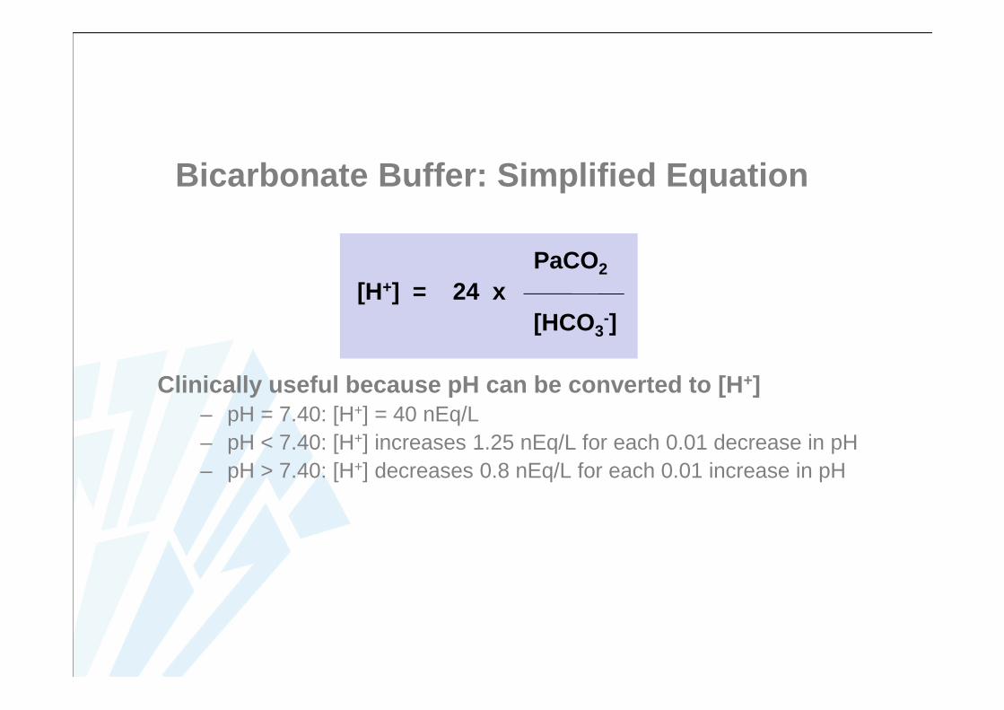
Bicarbonate Buffer: Simplified Equation
PaCO2
[H+] = 24 x [HCO3
-]
Clinically useful because pH can be converted to [H +]– pH = 7.40: [H+] = 40 nEq/L– pH < 7.40: [H+] increases 1.25 nEq/L for each 0.01 decrease in pH– pH > 7.40: [H+] decreases 0.8 nEq/L for each 0.01 increase in pH
Hemoglobin Buffer System (1)
• Histidine buffer– Multiple buffering sites on imidazole side chains– Histidine is an effective buffer from pH 5.7 – 7.7 (pKa 6.8)
• Dependent on the HCO 3- system: a large fraction of extra-
pulmonary CO 2 is transported to the lungs as plasma HCO 3-
– Tissues: CO2 diffuses into RBC where it is eliminated as HCO3-
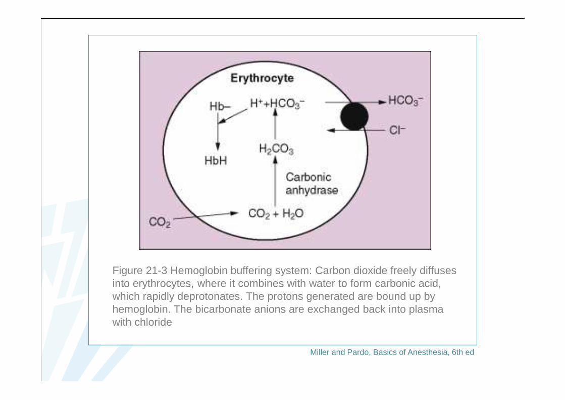
Figure 21-3 Hemoglobin buffering system: Carbon dioxide freely diffuses into erythrocytes, where it combines with water to form carbonic acid, which rapidly deprotonates. The protons generated are bound up by hemoglobin. The bicarbonate anions are exchanged back into plasma with chloride
Miller and Pardo, Basics of Anesthesia, 6th ed
Hemoglobin Buffer System (2)
• Hb buffer depends on the HCO 3- system
– Tissues: CO2 diffuses into RBC where it is eliminated as HCO3-
– Lungs: HCO3- enters the RBC (in exchange for Cl-) where it is
converted to CO2 which is released into the plasma and eliminatedby the lungs.
• Oxygenated and deoxygenated hemoglobin have different affinities for H + and CO2
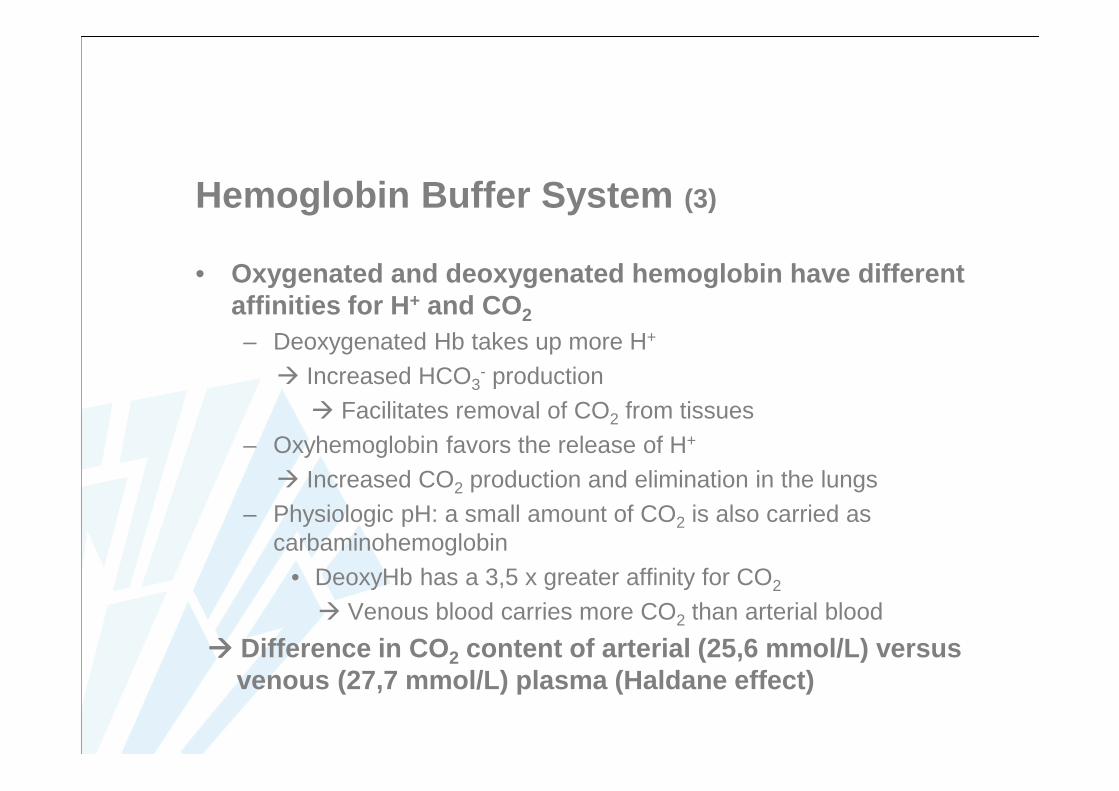
Hemoglobin Buffer System (3)
• Oxygenated and deoxygenated hemoglobin have different affinities for H + and CO2
– Deoxygenated Hb takes up more H+
� Increased HCO3- production
� Facilitates removal of CO2 from tissues – Oxyhemoglobin favors the release of H+
� Increased CO2 production and elimination in the lungs– Physiologic pH: a small amount of CO2 is also carried as
carbaminohemoglobin• DeoxyHb has a 3,5 x greater affinity for CO2
� Venous blood carries more CO2 than arterial blood
���� Difference in CO 2 content of arterial (25,6 mmol /L) versus venous (27,7 mmol/L) plasma (Haldane effect)

Ventilatory Response (1)
• Central chemoreceptors (medular) respond to changes in pH of CSF– CO2 diffuses across the blood-brain barrier
�[H+] in CSF increases � pH in CSF decreases�Activation of chemoreceptors
� Increased alveolar ventilation– Relationship between PaCO2 and minute ventilation: almost linear
• Exceptions: very high PaCO2 (carbonarcosis) and very low PaCO2 (apneic threshold)
• PaCO2/ventilation response curve: wide interindividual variation• Generally: minute volume increases 1 - 4 L/min for every
1 mmHg increase in PaCO2 (for PaCO2 40-50 mmHg)

Ventilatory Response (2)
• Peripheral chemoreceptors (bifurcation of common carot idarteries, aortic arch) are sensitive to changes in PaO 2, PaCO2, pH, and arterial perfusion pressure– Most sensitive to PaO2
– Glossopharyngeal nerves: communication with central respiratorycenters
• There is no complete ventilatory correction– Pulmonary response diminishes when pH approaches 7.4
• Pulmonary response to metabolic alkalosis is generallyless than pulmonary response to metabolic acidosis

Ventilatory Response (3)
• Pulmonary response to metabolic acidosis– Rapid response, but steady state after 12 - 24 h– Appropriate compensation?
• PaCO2 decreases 1 - 1.5 mmHg for each 1 mEq/L decrease in plasma [HCO3
-]• Winter’s formula: PaCO2 = (1.5 x [HCO3
-]) + 8
Ventilatory Response (4)
• Pulmonary response to metabolic alkalosis– Less predictable response than to metabolic acidosis
• Progressive hypoventilation results in hypoxemia• Hypoxemia activates O2-sensitive chemoreceptors stimulating
ventilation• PaCO2 usually does not rise above 55 mmHg in response to metabolic
alkalosis
– Appropriate compensation?• Generalization: PaCO2 increases 0.25 - 1 mmHg for each 1
mEq/L increase in plasma [HCO3-]
• PaCO2 = (0.7 x [HCO3-]) + 21
Renal Response and Metabolic Acidosis
• Slow onset (12 - 24 h)• Maximal effect after 5 d• Mechanisms compensating for metabolic acidosis
1. Reabsorption of filtered HCO3-
2. Excretion of titratable acids3. Production of ammonia� Generation and return of HCO3
- into the blood stream

Figure 21-4 Three mechanisms of renal compensation during acidosis to sequester hydrogen ions and reabsorb bicarbonate: (1) reabsorption of the filtered HCO3-, (2) excretion of titratable acids, and (3) production of ammonia
Miller and Pardo, Basics of Anesthesia, 6th ed
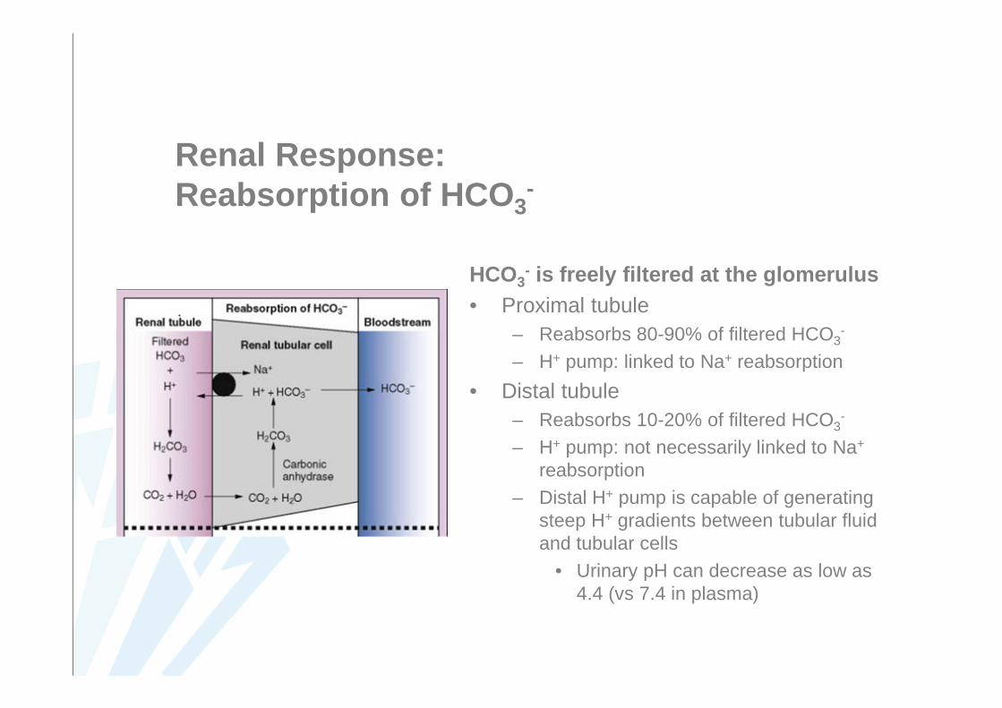
Renal Response: Reabsorption of HCO3
-
HCO3- is freely filtered at the glomerulus
• Proximal tubule– Reabsorbs 80-90% of filtered HCO3
-
– H+ pump: linked to Na+ reabsorption
• Distal tubule– Reabsorbs 10-20% of filtered HCO3
-
– H+ pump: not necessarily linked to Na+
reabsorption– Distal H+ pump is capable of generating
steep H+ gradients between tubular fluidand tubular cells
• Urinary pH can decrease as low as 4.4 (vs 7.4 in plasma)

Renal Response: Excretion of Titratable Acids
Phosphate (H 2PO4- / HPO4
2-) buffer: increased H + excretion• Starts after all the HCO3
- in the tubularfluid is reclaimed
• H+ combines with HPO42- and is
excreted as H2PO4-
• H2PO4- /HPO4
2- : pK = 6.8– Normally an ideal urinary buffer– pH 4.4: H2PO4
- ions are no longeravailable for H+ elimination

Renal Response: Production of Ammonia
Ammonia (NH 3/NH4+) buffer:
increased H + elimination• NH3/NH4
+ buffer becomes important after complete reabsorption of HCO3
-
and after consumption of the phosphatebuffer
– More ammonia is produced duringacidosis
• Excretion of NH4+ in urine
– NH3 is produced from deamination of glutamine
– NH3 combines in the tubular fluid with H+
to form NH4+
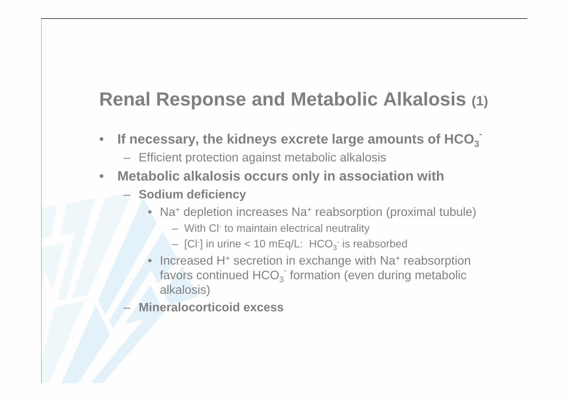
Renal Response and Metabolic Alkalosis (1)
• If necessary, the kidneys excrete large amounts of HCO 3-
– Efficient protection against metabolic alkalosis
• Metabolic alkalosis occurs only in association with– Sodium deficiency
• Na+ depletion increases Na+ reabsorption (proximal tubule)– With Cl- to maintain electrical neutrality– [Cl-] in urine < 10 mEq/L: HCO3
- is reabsorbed
• Increased H+ secretion in exchange with Na+ reabsorptionfavors continued HCO3
- formation (even during metabolicalkalosis)
– Mineralocorticoid excess

Renal Response and Metabolic Alkalosis (2)
• Metabolic alkalosis occurs only in association with– Sodium deficiency– Mineralocorticoid excess
• Increased aldosterone-mediated Na+ reabsorption in exchange for H+ secretion in the distal tubules� Increased HCO3
- formation
ACID-BASE DISTURBANCES
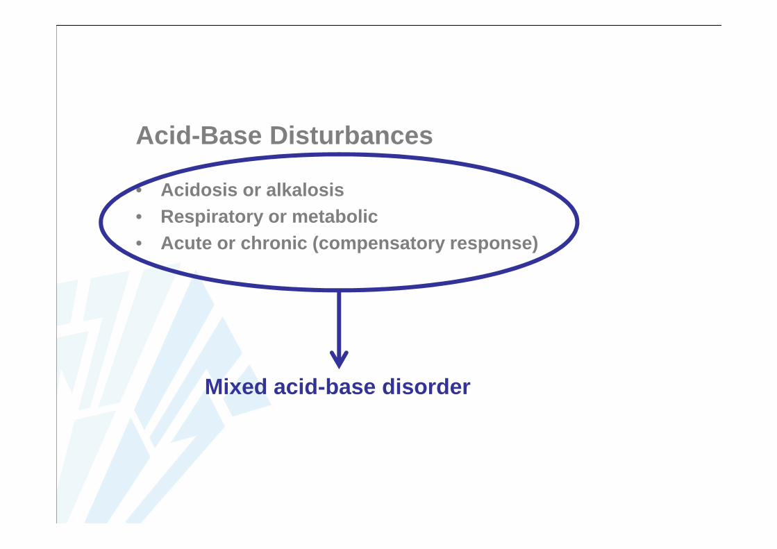
Acid -Base Disturbances
• Acidosis or alkalosis• Respiratory or metabolic• Acute or chronic (compensatory response)
Mixed acid-base disorder
Acidemia: Adverse Effects (1)
• Result of balance between direct depressant effects andsympathoadrenal activation
• Mild acidemia– Release of catecholamines mitigate myocardial depression– No significant effect on systemic vascular resistance, pulmonary
vascular resistance, cardiac output, or systemic O2 delivery
• Severe acidemia– pH < 7.20: direct depressant effects predominate
Acidemia: Adverse Effects (2)
• Severe acidemia– pH < 7.20: direct depressant effects predominate– Decreased myocardial responsiveness to catecholamines with
myocardial depression and hypotension– Progressive hyperkalemia
• Movement of K+ out of cells in exchange for extracellular H+
• Plasma [K+] increase of 0.6 mEq/L for each 0.10 decrease in pH
– Central nervous system effects (respiratory > metabolic acidosis)• Severe acute intracellular acidosis � confusion, loss of
consciousness, seizures• Increased cerebral blood flow � increased intracranial pressure
Acidemia and Anesthesia: Risks
• Hemodynamic instability– Arteriolar vasodilatation and decreased cardiac output– Exaggerated cardiovascular depressant effects of anesthetics– Decreased response to vasopressors and inotropes
• Arrhythmias• Hyperkalemia (avoid succinylcholine)• Insulin resistance and hyperglycemia• Acidemia may augment nondepolarizing neuromuscular blockade• Postoperative mechanical ventilation may be necessaryIn severe acidemia, consider postponing surgery until the underlyingcause is treated or the patient is preoperatively optimized. However: surgery may be the treatment that is required
Alkalemia: Adverse Effects
• Severe alkalemia (pH > 7.6)– Arteriolar vasoconstriction
� Decreased cerebral and coronary blood flow– Consequences are more severe with respiratory alkalosis
• Rapid movement of CO2 across cell membranes• Acute hyperventilation � confusion, myoclonus, flapping
tremor, depressed consciousness, seizures– Risk of hypokalemia
• Movement of H+ out of cells in exchange for extracellular K+

Alkalemia and Anesthesia: Risks
• Coronary vasospasm and risk of myocardial ischemia• Hypokalemia and reduced threshold for arrhythmias• Hypocalcemia• Reduction in CBF and risk of cerebral ischemia
– Particularly during hypotension
• Potentiation of nondepolarizing neuromuscular blockade• Respiratory alkalosis prolongs the duration of opioid-
induced respiratory depression– Increased protein binding of opioids?

Acid-Base Disorders
Disorder Primary change Compensatoryresponse
RespiratoryAcidosisAlkalosis
↑ PaCO2↓ PaCO2
↑ HCO3-
↓ HCO3-
MetabolicAcidosisAlkalosis
↓ HCO3-
↑ HCO3-
↓ PaCO2↑ PaCO2
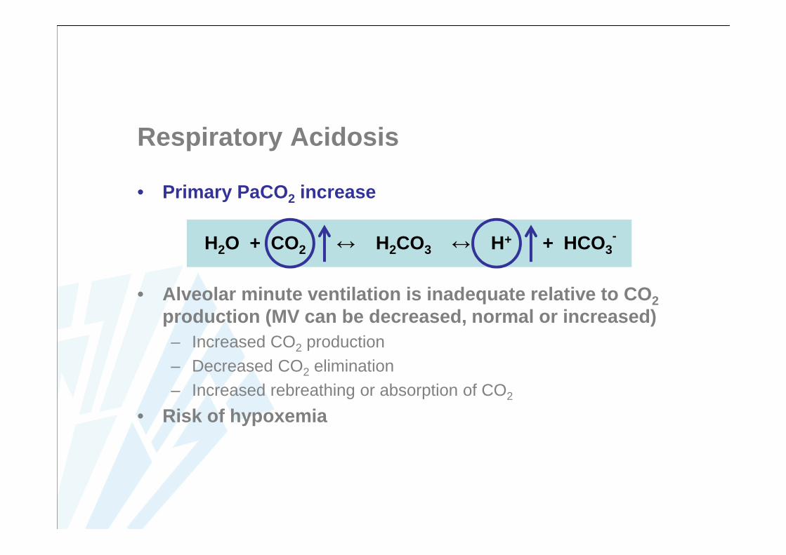
Respiratory Acidosis
• Primary PaCO 2 increase
H2O + CO2 ↔ H2CO3 ↔ H+ + HCO3-
• Alveolar minute ventilation is inadequate relative to C O2production (MV can be decreased, normal or increased)– Increased CO2 production– Decreased CO2 elimination– Increased rebreathing or absorption of CO2
• Risk of hypoxemia

Causes of Respiratory Acidosis
Increased CO 2 productionMalignant hyperthermiaHyperthyroidismSepsisOverfeeding
Decreased CO 2 eliminationIntrinsic pulmonary disease (pneumonia, ARDS, fibrosis, edema)Upper airway obstruction (laryngospasm, foreign body, OSA)Lower airway obstruction (asthma, COPD)Chest wall restriction (obesity, scoliosis, burns)CNS depression (anesthetics, opioids, CNS lesions)Decreased skeletal muscle strength (residual effects of neuromuscular blockingdrugs, myopathy, neuropathy)
Increased CO 2 rebreathing or absorptionExhausted soda limeIncompetent one-way valveLaparoscopic surgery (CO2-pneumoperitoneum)
ARDS = acute respiratory distress syndrome; CNS = central nervous sytem; COPD = chronic obstructive pulmonary disease; OSA = obstructive sleep apnea)

Respiratory Acidosis: Compensatory Response (1)
• [HCO3-] increase
– Increased H+ secretion and HCO3- reabsorption
– Initially [HCO3-] is minimally affected
• Limited acute (6 - 12 h) compensatory response– Hb buffer– Exchange of extracellular H+ for Na+ and K+ from bone and
intracellular fluid– Renal response to retain HCO3
- is very limited acutely• Plasma [HCO3
- ] increases only about 1 mEq/L for each10 mmHg increase in PaCO2 > 40 mmHg

Respiratory Acidosis: Compensatory Response (2)
• Renal compensation– Appreciable only after 12 – 24 hours– Maximal after 3 – 5 days
• Chronic respiratory acidosis: nearly normal pH (increasedPaCO2)
– Plasma [HCO3- ] increases approximately 4 mEq/L for each
10 mmHg increase in PaCO2 > 40 mmHg
Respiratory Acidosis: Treatment (1)
• Causal treatment– Reduction of CO2 production (in specific situations)
• Status epilepticus: muscle paralysis• TPN: reduced carbohydrate intake• Thyroid storm: antithyroid medication• Malignant hyperthermia: dantrolene
– Improving alveolar ventilation (temporary measures)• Bronchodilatation• Reversal of narcosis• Administration of a respiratory stimulant (doxapram)• Improving lung compliance (diuretics)

Respiratory Acidosis: Treatment (2)
• Mechanical ventilation may be indicated– Severe acidosis (pH < 7.2)– CO2 narcosis– Respiratory muscle fatigue
• Intravenous buffers are only rarely indicated– Very severe acidosis (pH < 7.10 and HCO3
- < 15 mEq/L)– NaHCO3: transient PaCO2 increase: H+ + HCO3
- ↔ CO2 + H2O– Buffers that do not increase PaCO2: no proven benefit
Respiratory Acidosis: Treatment (3)
• Avoid overventilation in patients with chronic respirato ryacidosis– Acute respiratory failure superimposed on chronic respiratory
acidosis– Goal: return PaCO2 to the patient’s baseline PaCO2
• PaCO2 = 40 mmHg in chronic respiratory acidosis will result in metabolic alkalosis
�Renal loss of HCO3-
� Increased work of breathing
– In some patients, respiratory drive is dependent on PaO2

Respiratory Alkalosis
• Primary PaCO 2 decrease– PaCO2 is decreased relative to [HCO3
-] � pH increases– Prolonged respiratory alkalosis: central chemoreceptors reset to a
lower CO2 level• Active transport of HCO3
- out of the CSF
• Generally, there is an inappropriate increase in alveol arventilation relative to CO 2 production

Causes of Respiratory Alkalosis
Increased minute ventilationHypoxia (high altitude, low FiO2, severe anemia)Iatrogenic (mechanical ventilation)Anxiety and painCNS disease (tumor, infection, trauma)Fever, sepsisDrugs (salicylates, progesterone, doxapram)Liver diseasePregnancyRestrictive lung diseasePulmonary embolism
Decreased CO 2 productionHypothermiaSkeletal muscle paralysis
CNS = central nervous sytem

Respiratory Alkalosis: Compensatory Response• Decreased reabsorption of HCO 3
- from the renal tubulesand increased urinary excretion
• Acute respiratory alkalosis– Plasma [HCO3
-] usually decreases 2 mEq/L for each 10 mmHg acute decrease in PaCO2 below 40 mmHg
• Chronic respiratory alkalosis: variable compensatoryresponse– Plasma [HCO3
-] decreases 2-5 mEq/L for each 10 mmHg decrease in PaCO2 below 40 mmHg

Respiratory Alkalosis: Treatment
• Correction of the underlying disorder• Mild alkalemia usually does not require treatment• Severe acute respiratory alkalosis (pH > 7.60): int ravenous
drug therapy may be indicated– Hydrochloric acid– Arginine chloride– Ammonium chloride
Metabolic Acidosis
• Primary decrease in [HCO 3-]
• Mechanisms– Consumption of HCO3
- by a strong nonvolatile acid– Renal or gastrointestinal wasting of HCO3
-
– Rapid dilution of extracellular fluid with a HCO3- free fluid
• Traditionally, the differential diagnosis is based on the anion gap calculation– More recent: Stewart method based on the strong ion difference
• Frequently accompanied by hyperkalemia andhyperphosphatemia
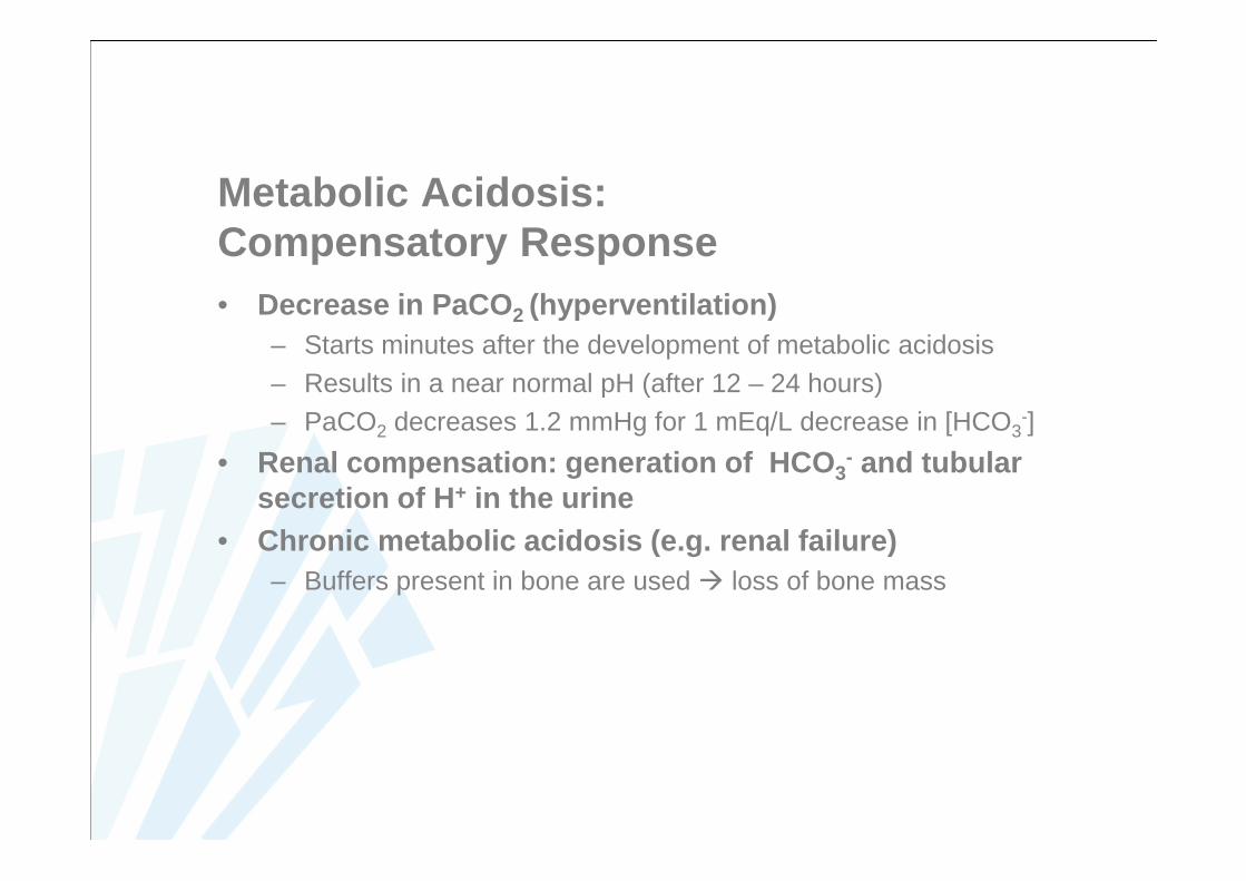
Metabolic Acidosis:Compensatory Response• Decrease in PaCO 2 (hyperventilation)
– Starts minutes after the development of metabolic acidosis– Results in a near normal pH (after 12 – 24 hours)– PaCO2 decreases 1.2 mmHg for 1 mEq/L decrease in [HCO3
-]
• Renal compensation: generation of HCO 3- and tubular
secretion of H + in the urine• Chronic metabolic acidosis (e.g. renal failure)
– Buffers present in bone are used � loss of bone mass
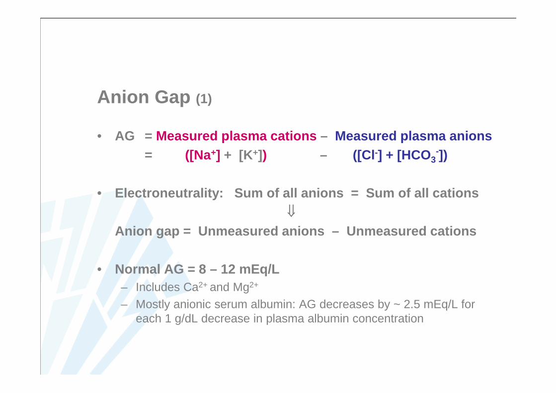
Anion Gap (1)
• AG = Measured plasma cations – Measured plasma anions= ([Na+] + [K+]) – ([Cl -] + [HCO 3
-])
• Electroneutrality: Sum of all anions = Sum of all cations⇓⇓⇓⇓
Anion gap = Unmeasured anions – Unmeasured cations
• Normal AG = 8 – 12 mEq/L– Includes Ca2+ and Mg2+
– Mostly anionic serum albumin: AG decreases by ~ 2.5 mEq/L for each 1 g/dL decrease in plasma albumin concentration

UMAs = strong unmeasured anions (e.g. lactate)A- = weak acids (phosphate, albumin)

Anion Gap (2)
• AG increases when the ion replacing HCO 3- is not routinely
measured– Most common unmeasured anions: lactic acid, keto acids
• Weakness: AG may be normal in the presence of unmeasured anions– E.g. hypoalbuminemia and hypophosphatemia
→ Use corrected anion gap to compensate for hypoalbuminemia:Measured AG + 2.5 (normal – measured albumin g/dL)
– Use of HCO3- in the equation
� [HCO3-] can change independently of metabolic disturbances
(e.g. hyperventilation)
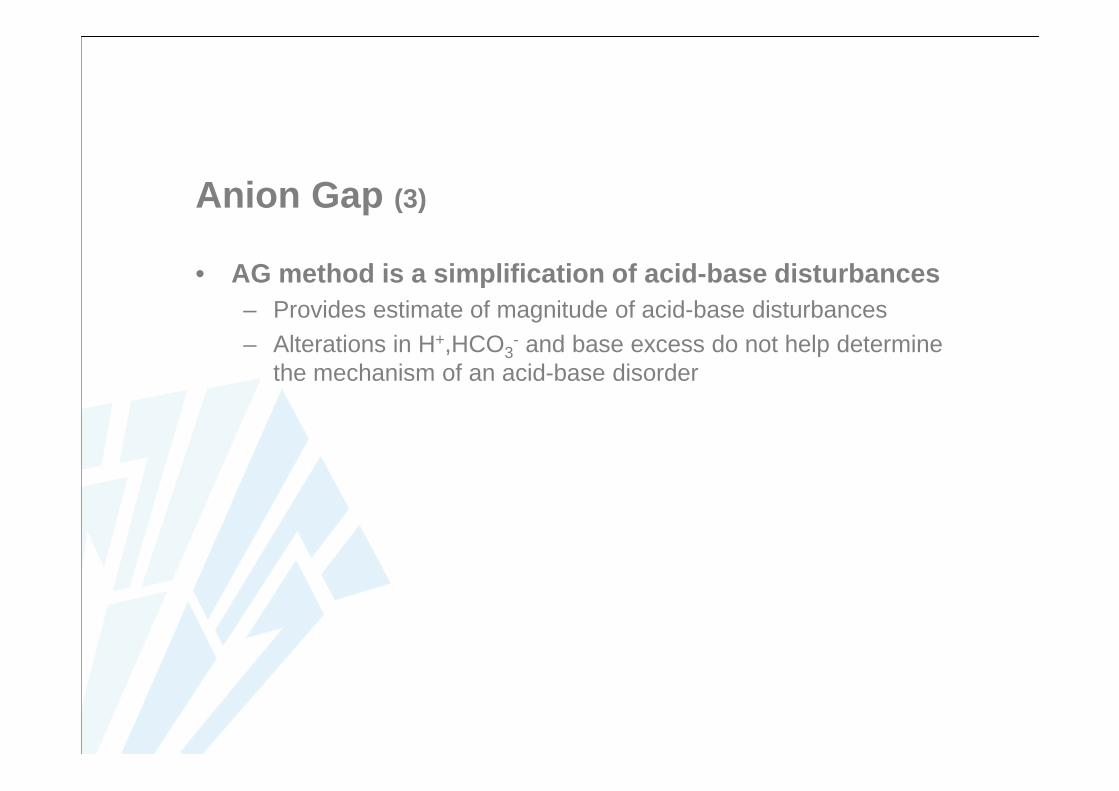
Anion Gap (3)
• AG method is a simplification of acid-base disturbanc es– Provides estimate of magnitude of acid-base disturbances– Alterations in H+,HCO3
- and base excess do not help determinethe mechanism of an acid-base disorder

Anion Gap: Classification of Metabolic Acidosis• High anion gap metabolic acidosis
– Acidosis is due to acid accumulation, with HCO3- being replaced
by an unmeasured anion
• Normal anion gap metabolic acidosis– Acidosis is due to gastrointestinal or renal HCO3
- losses (HCO3- is
lost with Na+: no change in AG)– Administration of large volumes of NaCl 0.9% (> 30 mL/kg/hour)
• Excessive Cl- administration
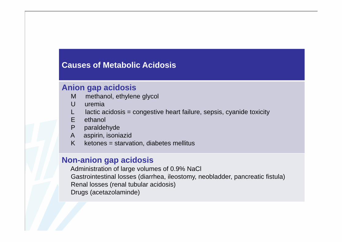
Causes of Metabolic Acidosis
Anion gap acidosisM methanol, ethylene glycolU uremiaL lactic acidosis = congestive heart failure, sepsis, cyanide toxicityE ethanolP paraldehydeA aspirin, isoniazidK ketones = starvation, diabetes mellitus
Non-anion gap acidosisAdministration of large volumes of 0.9% NaClGastrointestinal losses (diarrhea, ileostomy, neobladder, pancreatic fistula)Renal losses (renal tubular acidosis)Drugs (acetazolaminde)
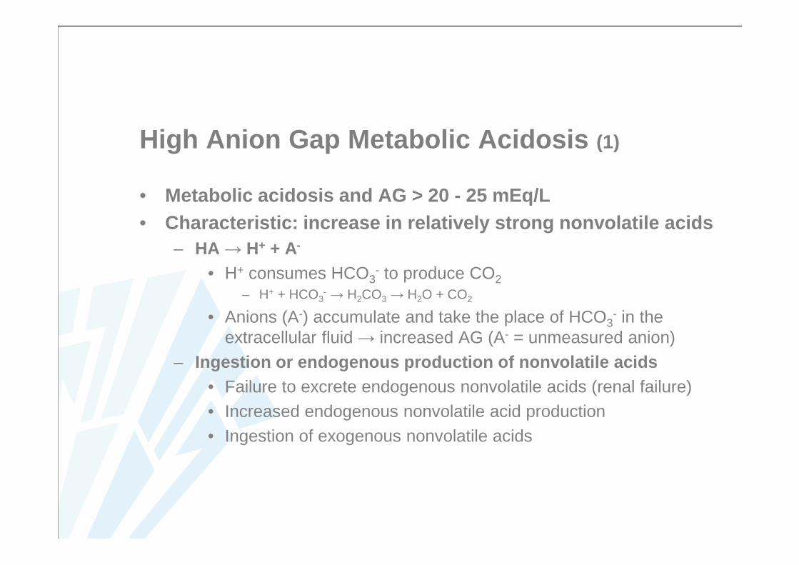
High Anion Gap Metabolic Acidosis (1)
• Metabolic acidosis and AG > 20 - 25 mEq/L• Characteristic: increase in relatively strong nonvola tile acids
– HA → H+ + A-
• H+ consumes HCO3- to produce CO2
– H+ + HCO3- → H2CO3 → H2O + CO2
• Anions (A-) accumulate and take the place of HCO3- in the
extracellular fluid → increased AG (A- = unmeasured anion)– Ingestion or endogenous production of nonvolatile acids
• Failure to excrete endogenous nonvolatile acids (renal failure)• Increased endogenous nonvolatile acid production• Ingestion of exogenous nonvolatile acids
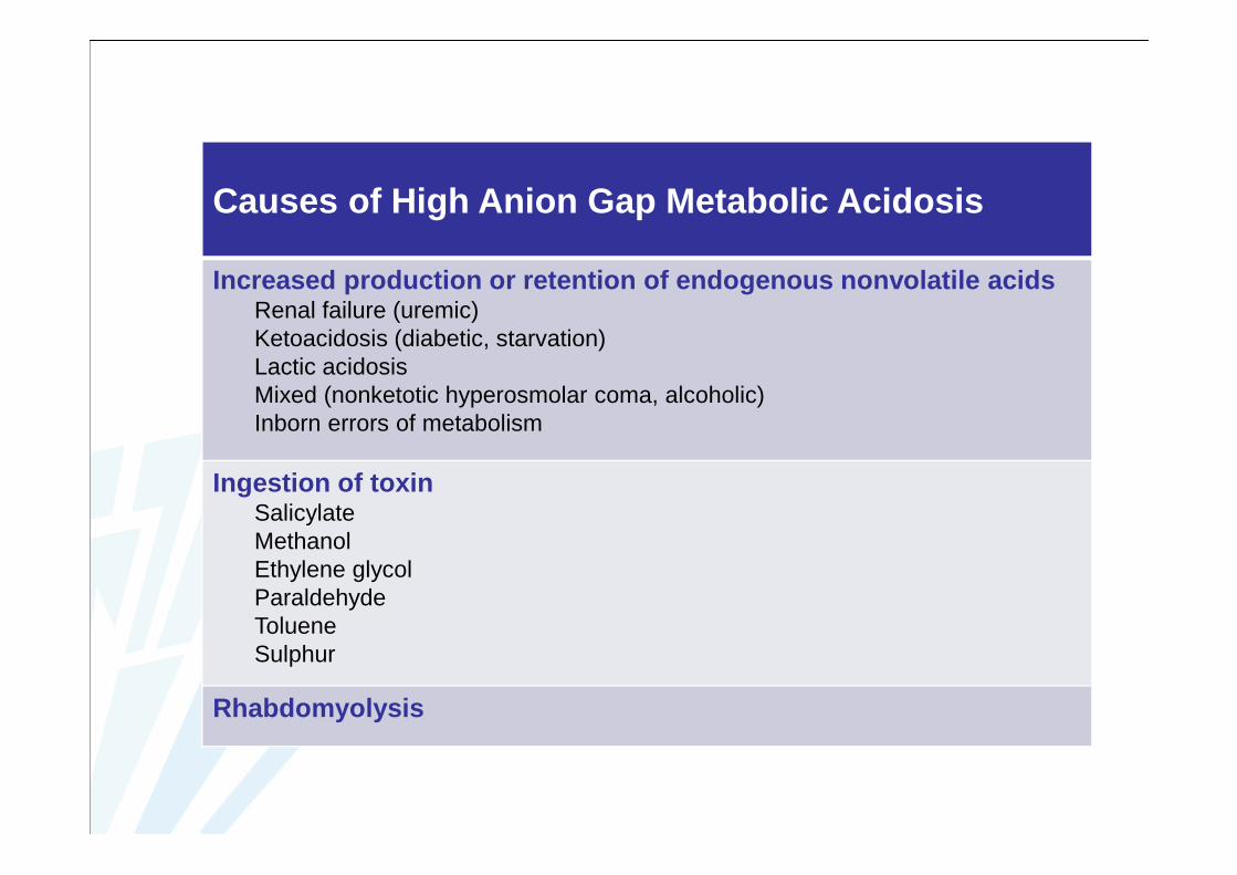
Causes of High Anion Gap Metabolic Acidosis
Increased production or retention of endogenous nonvola tile acidsRenal failure (uremic)Ketoacidosis (diabetic, starvation)Lactic acidosisMixed (nonketotic hyperosmolar coma, alcoholic)Inborn errors of metabolism
Ingestion of toxinSalicylateMethanolEthylene glycolParaldehydeTolueneSulphur
Rhabdomyolysis
High Anion Gap Metabolic Acidosis (2)
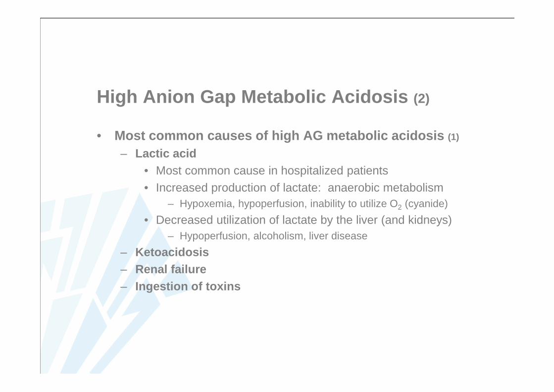
• Most common causes of high AG metabolic acidosis (1)
– Lactic acid• Most common cause in hospitalized patients• Increased production of lactate: anaerobic metabolism
– Hypoxemia, hypoperfusion, inability to utilize O2 (cyanide)
• Decreased utilization of lactate by the liver (and kidneys)– Hypoperfusion, alcoholism, liver disease
– Ketoacidosis– Renal failure– Ingestion of toxins
High Anion Gap Metabolic Acidosis (3)
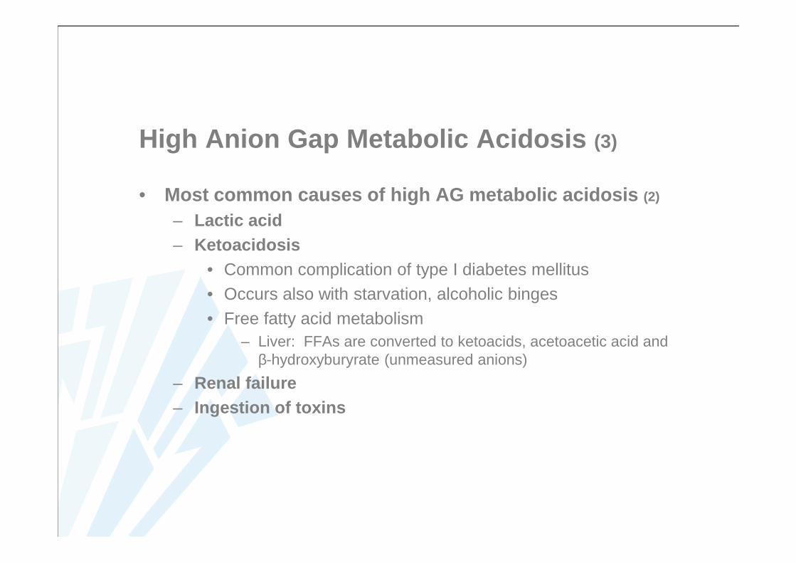
• Most common causes of high AG metabolic acidosis (2)
– Lactic acid– Ketoacidosis
• Common complication of type I diabetes mellitus• Occurs also with starvation, alcoholic binges• Free fatty acid metabolism
– Liver: FFAs are converted to ketoacids, acetoacetic acid and β-hydroxyburyrate (unmeasured anions)
– Renal failure– Ingestion of toxins

High Anion Gap Metabolic Acidosis (4)
• Most common causes of high AG metabolic acidosis (3)
– Lactic acid– Ketoacidosis– Renal failure
• Decreased acid excretion• Decreased HCO3
- reabsorption• High AG due to accumulation of suphates, phosphates, urate,
and hippurate– Ingestion of toxins
• Acidic metabolites or triggering of lactic acidosis• Salicylate, methanol, ethylene glycol, paraldehyde…

Delta Ratio
Assessment of high anion gap metabolic acidosis• Delta ratio = ∆∆∆∆ Anion gap / ∆∆∆∆ [HCO3
-]= (Anion gap – 12) / (24 - [HCO 3
-])= Increase in AG / [HCO 3
-] deficit• A delta ratio < 1 indicates a greater [HCO3
-] deficit than wouldbe expected by the increase in AG
– Hyperchloremic metabolic acidosis– Mixed high AG and normal AG acidosis
• A delta ratio > 2 indicates a lesser [HCO3-] deficit than would be
expected by the increase in AG– Metabolic acidosis with concurrent metabolic alkalosis– Metabolic acidosis with pre-existing compensated respiratory acidosis
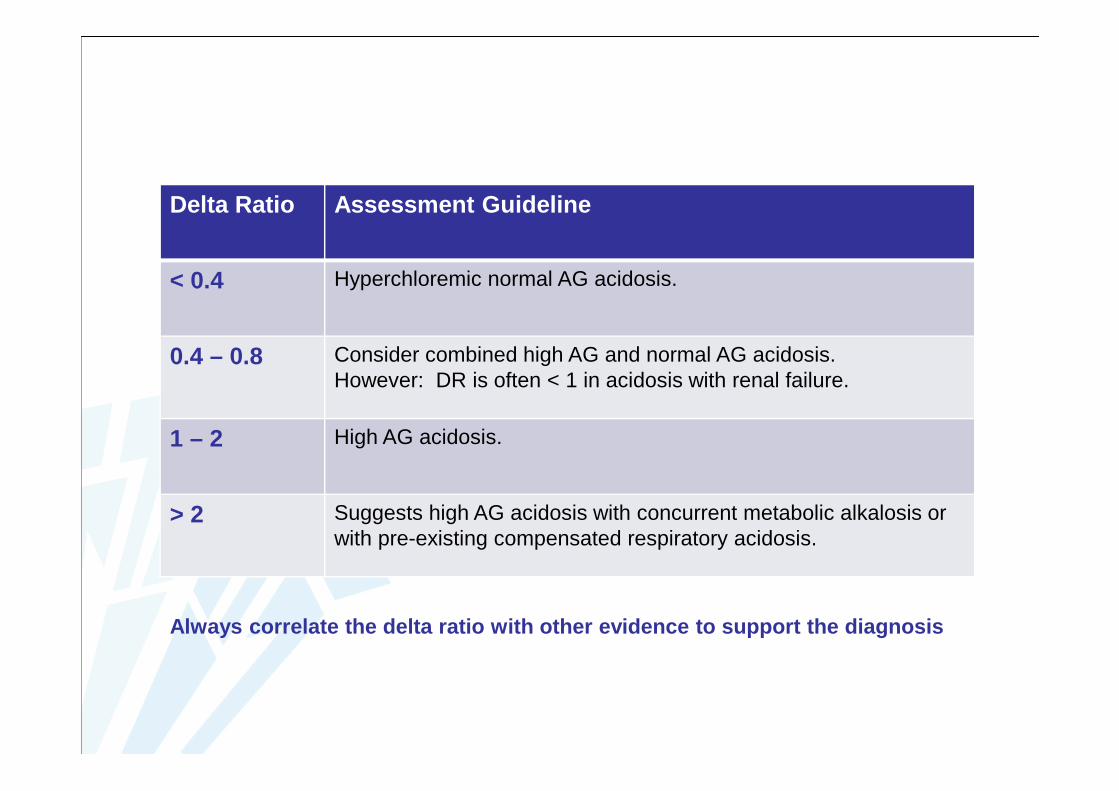
Delta Ratio Assessment Guideline
< 0.4 Hyperchloremic normal AG acidosis.
0.4 – 0.8 Consider combined high AG and normal AG acidosis.However: DR is often < 1 in acidosis with renal failure.
1 – 2 High AG acidosis.
> 2 Suggests high AG acidosis with concurrent metabolic alkalosis orwith pre-existing compensated respiratory acidosis.
Always correlate the delta ratio with other evidence to support the diagnosis
Normal Anion Gap Metabolic Acidosis (1)
• Normal anion gap and metabolic acidosis• Characteristic: hyperchloremic metabolic acidosis
– Plasma [Cl-] increase to replace lost HCO3-
• Causes of normal AG metabolic acidosis– Abnormal losses of HCO3
-
– Other causes

Causes of Normal Anion Gap Metabolic Acidosis
Increased gastrointestinal losses of HCO 3-
DiarrheaAnion exchange resinsIngestion of CaCl2, MgCl2Fistulas (pancreatic, biliary, or small bowel)Ureterosigmoidostomy or obstructed ileal loop
Increased renal losses of HCO 3-
Renal tubular acidosisCarbonic anhydrase inhibitorsHypoaldosteronism
Rapid infusion of a large amount of bicarbonate-free f luids
Total parenteral nutrition (Cl - salts of amino acids)
Increased intake of Cl - containing acidsAmmonium chlorideLysine hydrochlorideArginine hydrochloride

Normal Anion Gap Metabolic Acidosis (2)
• Abnormal losses of HCO 3-: gastrointestinal or renal
– Diarrhea– Pancreatic or biliary fistulae– Ureterosigmoidostomy
• Colon secretes HCO3- in exchange for Cl-
• Colon absorbes urinary ammonium (dissociates in NH3 and H+)– Renal tubular acidosis
• Impaired H+ secretion• Impaired HCO3
- absorption– Acetazolamide (Diamox®)

Normal Anion Gap Metabolic Acidosis (3)
• Other causes of normal AG metabolic acidosis– Rapid infusion of a bicarbonate-free fluid: e.g. normal saline
• Fast infusion of large volumes – Parenteral nutrition: organic cations > organic anions
• Cl- is commonly used as anion for the cationic amino acids– Excessive administration of chloride-containing acids– Adrenal insufficiency

Metabolic Acidosis: Treatment (1)
• Causal treatment if possible– Lactic acidosis
• Restoration of adequate oxygenation and tissue perfusion withoxygen, fluid resuscitation and circulatory support
– Diabetic ketoacidosis• Correction of fluid deficit• Insulin• Potassium, phosphate, magnesium
– Methanol or ethylene glycol intoxication• Ethanol
Metabolic Acidosis: Treatment (2)
• NaHCO3 therapy if pH < 7.1 - 7.15 (no respiratory component)– Therapy should always be guided using arterial blood gas
measurements (goal: pH 7.2)– Other indications
• pH 7.2 in case of ARDS with permissive hypercapnia• Acidosis and chronic renal insufficiency or renal tubular acidosis• Acidosis with life threatening hyperkalemia• Salicylate intoxication (alkalinization of urine)

Metabolic Acidosis: Treatment (3)
• Amount of NaHCO 3
– Calculated• NaHCO3 (mEq) = (24 - [HCO3
-]s) x 0.5 x body weight (kg)• Generally 50% of the calculated dose is given initially
– Empirically• A fixed dose of ≈ 1 mEq/kg • 50 – 100 mEq
• NaCO3 8.4 %: 1000 mEq Na +/L, 2000 mOsm /L� Side effects
Metabolic Acidosis: Treatment (4)
• Side effects of treatment with NaHCO 3 8.4%– Increased production of CO2
• Intracellular acidosis and possibly electromechanicaldissociation
– Hypernatremia and hyperosmolarity• Hypervolemia, pulmonary edema
– Inhibition of peripheral chemoreceptors• Decrease of hyperventilation, hypercapnia
– Negative inotropic effect• Lactate has also a negative inotropic effect
– Hemoglobin: increased affinity for oxygen
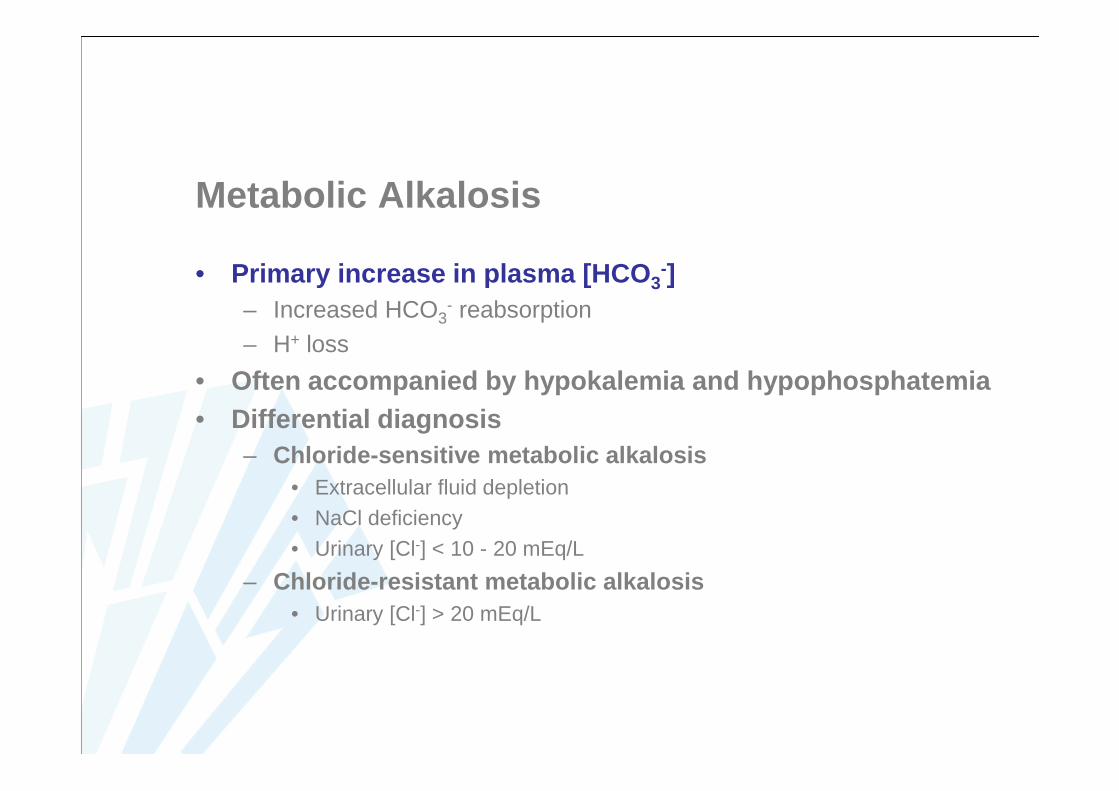
Metabolic Alkalosis
• Primary increase in plasma [HCO 3-]
– Increased HCO3- reabsorption
– H+ loss
• Often accompanied by hypokalemia and hypophosphatemia• Differential diagnosis
– Chloride-sensitive metabolic alkalosis• Extracellular fluid depletion• NaCl deficiency• Urinary [Cl-] < 10 - 20 mEq/L
– Chloride-resistant metabolic alkalosis• Urinary [Cl-] > 20 mEq/L

Metabolic Alkalosis:Compensatory Response• Decrease in PaCO 2 (hypoventilation)
– PaCO2 increases 0.7 mmHg for every 1 mEq/L increase in [HCO3-]
• Renal compensation by HCO 3- excretion is less efficient

Chloride-Sensitive Metabolic Alkalosis (1)
• Depletion of extracellular fluid�Na+ reabsorption in the renal tubules�There is not enough Cl- available to accompany all reabsorbed Na+
� Increased H+ secretion to maintain electroneutrality� HCO3
- ions that might otherwise have been excreted are reabsorbed
� Enhanced K+ secretion
� Urinary [Cl -] < 10 - 20 mEq/L

Chloride-Sensitive Metabolic Alkalosis
Depletion of extracellular fluid
Na+ reabsorption in the renal tubules
Cl- absorption: insufficient to maintain electroneutrality
� H+ secretion HCO3- absorption � K+ secretion
++
Hypokalemia
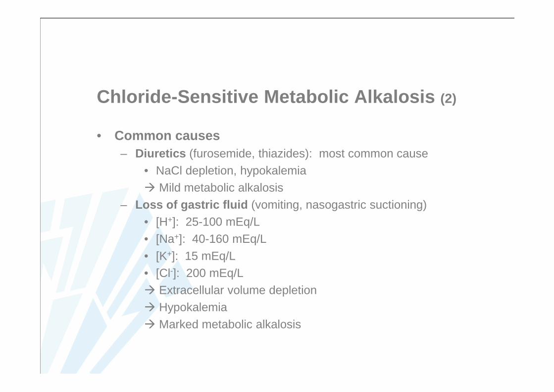
Chloride-Sensitive Metabolic Alkalosis (2)
• Common causes– Diuretics (furosemide, thiazides): most common cause
• NaCl depletion, hypokalemia� Mild metabolic alkalosis
– Loss of gastric fluid (vomiting, nasogastric suctioning)• [H+]: 25-100 mEq/L• [Na+]: 40-160 mEq/L• [K+]: 15 mEq/L• [Cl-]: 200 mEq/L� Extracellular volume depletion� Hypokalemia� Marked metabolic alkalosis
Chloride-Sensitive Metabolic Alkalosis (3)
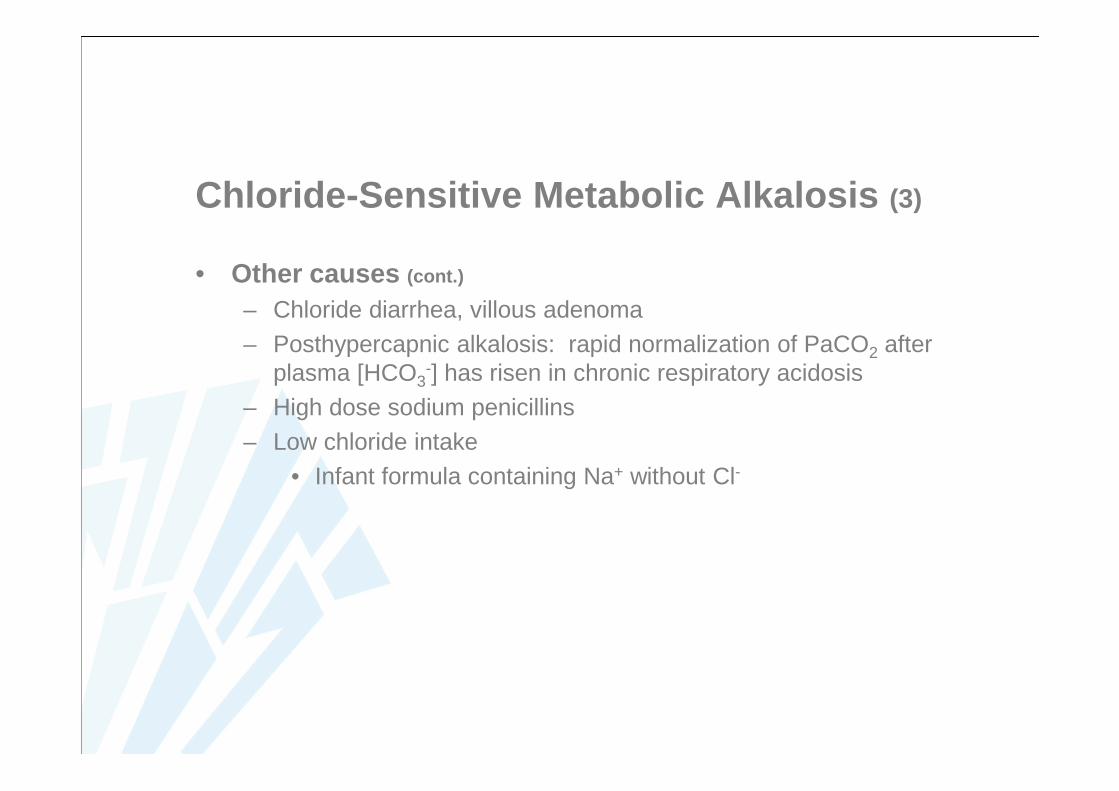
• Other causes (cont.)
– Chloride diarrhea, villous adenoma– Posthypercapnic alkalosis: rapid normalization of PaCO2 after
plasma [HCO3-] has risen in chronic respiratory acidosis
– High dose sodium penicillins– Low chloride intake
• Infant formula containing Na+ without Cl-
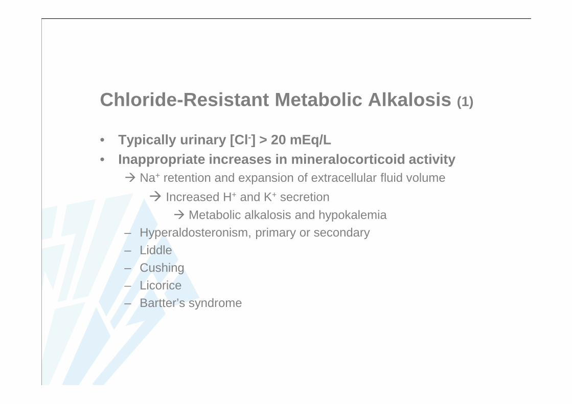
Chloride-Resistant Metabolic Alkalosis (1)
• Typically urinary [Cl -] > 20 mEq/L• Inappropriate increases in mineralocorticoid activity
� Na+ retention and expansion of extracellular fluid volume
� Increased H+ and K+ secretion
� Metabolic alkalosis and hypokalemia– Hyperaldosteronism, primary or secondary– Liddle– Cushing– Licorice– Bartter’s syndrome
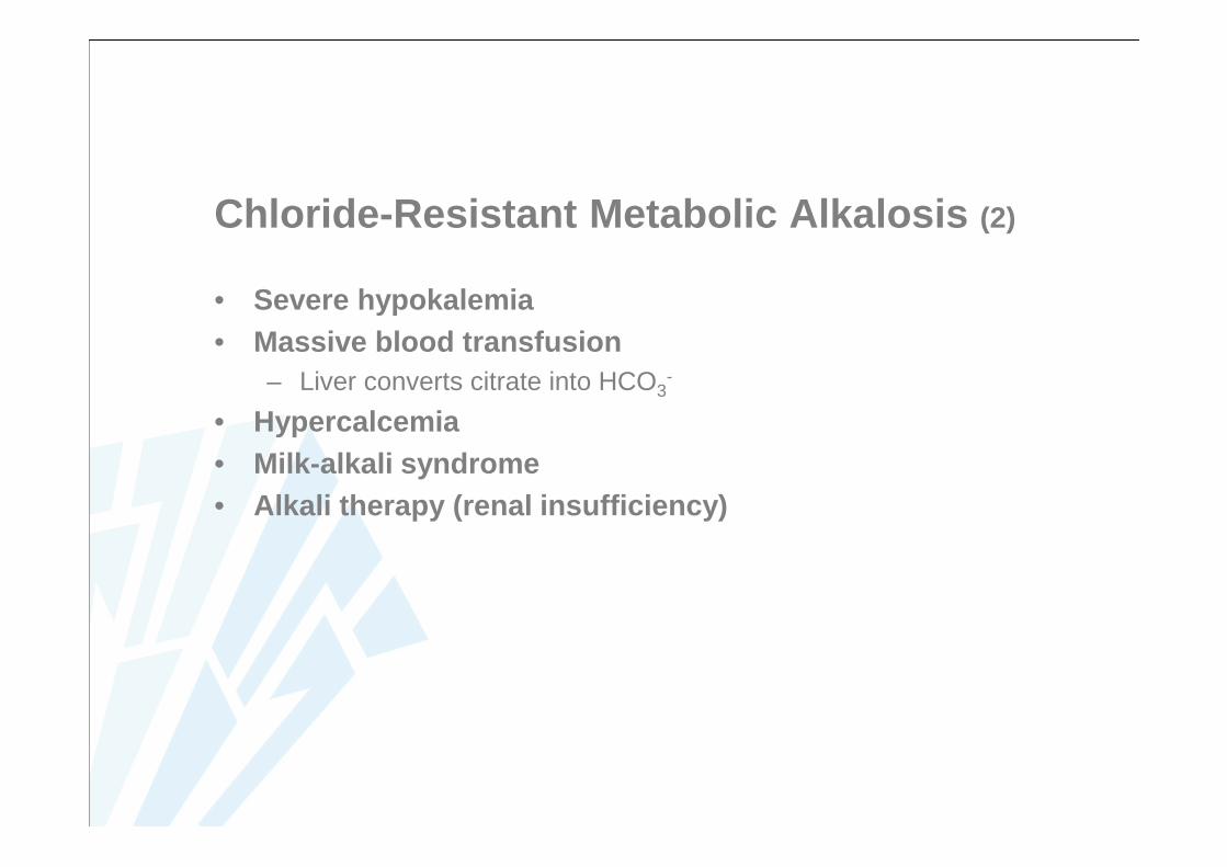
Chloride-Resistant Metabolic Alkalosis (2)
• Severe hypokalemia• Massive blood transfusion
– Liver converts citrate into HCO3-
• Hypercalcemia• Milk-alkali syndrome• Alkali therapy (renal insufficiency)

Metabolic Alkalosis: Treatment (1)
• Treatment of underlying disorder• Chloride-sensitive metabolic alkalosis
– Intravenous saline (NaCl)• Intravenous potassium (KCl) may be indicated
– Depending on the cause• H2-blocker therapy (in case of excessive loss of gastric fluid)• Acetazolamide (edematous patients)
• Primary increase in mineralocorticoid activity– Aldosterone antagonist (spironolactone)
Metabolic Alkalosis: Treatment (2)
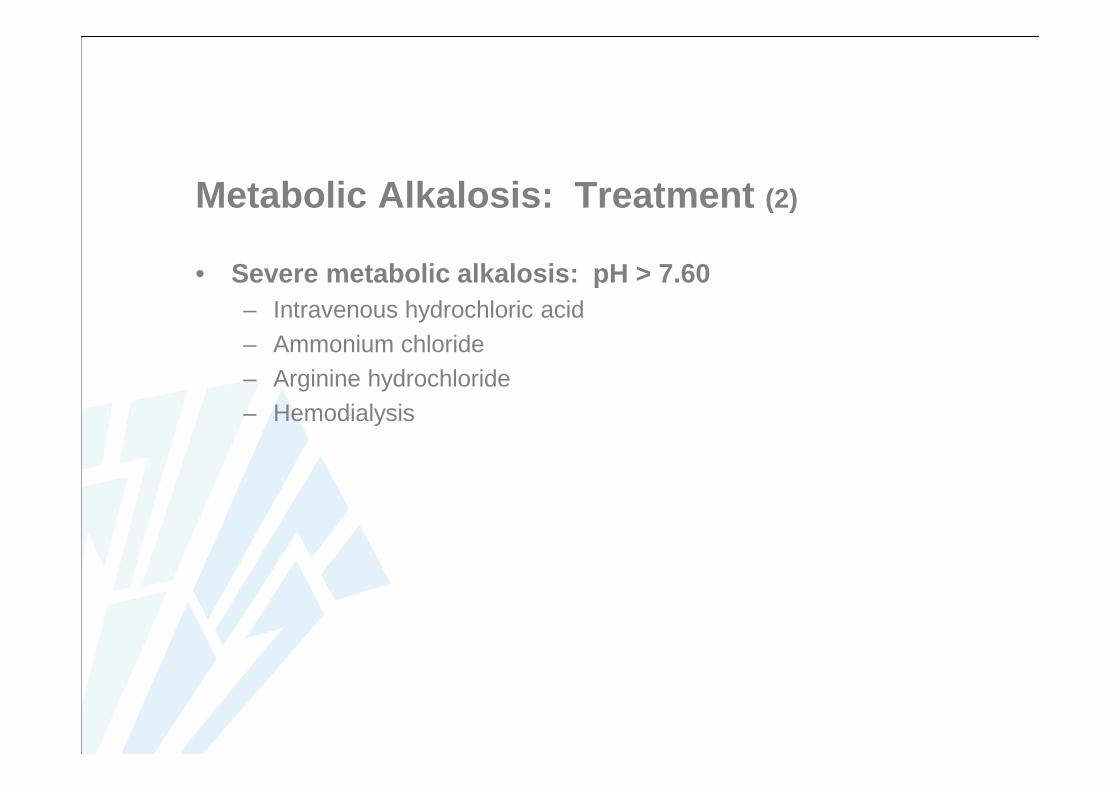
• Severe metabolic alkalosis: pH > 7.60– Intravenous hydrochloric acid– Ammonium chloride– Arginine hydrochloride– Hemodialysis

ASSESSMENT OF ACID-BASE DISORDERSPractical Approach

Miller and Pardo, Basics of Anesthesia, 6th ed

Assessment of Acid -Base Disorders: Practical Approach (1)
1. Evaluate the clinical context2. Determine pH: acidemia or alkalemia present?
– pH < 7.35: acidemia– pH > 7.45: alkalemia– pH 7.35 – 7.45: normal or more than one acid-base disturbance
Assessment of Acid -Base Disorders: Practical Approach (2)
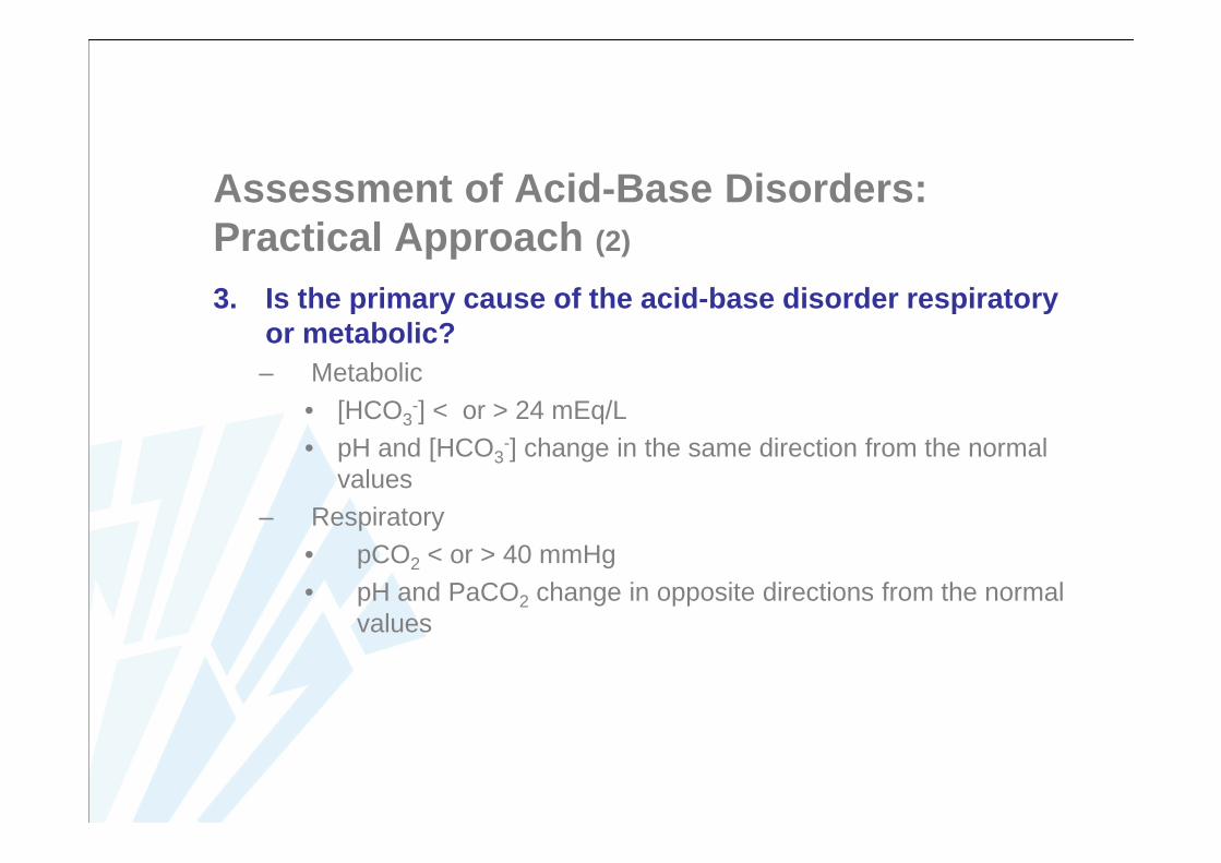
3. Is the primary cause of the acid-base disorder resp iratoryor metabolic?– Metabolic
• [HCO3-] < or > 24 mEq/L
• pH and [HCO3-] change in the same direction from the normal
values– Respiratory
• pCO2 < or > 40 mmHg• pH and PaCO2 change in opposite directions from the normal
values
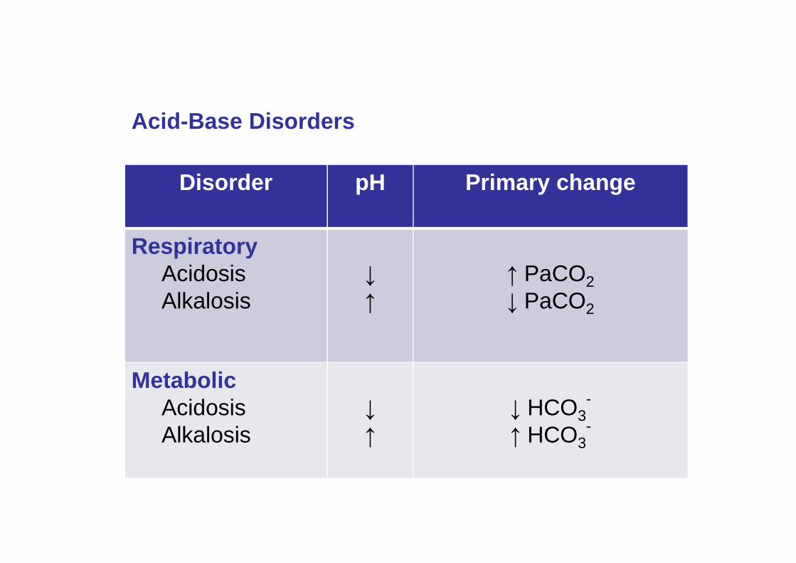
Acid-Base Disorders
Disorder pH Primary change
RespiratoryAcidosisAlkalosis
↓
↑
↑ PaCO2↓ PaCO2
MetabolicAcidosisAlkalosis
↓
↑
↓ HCO3-
↑ HCO3-
Assessment of Acid -Base Disorders: Practical Approach (3)
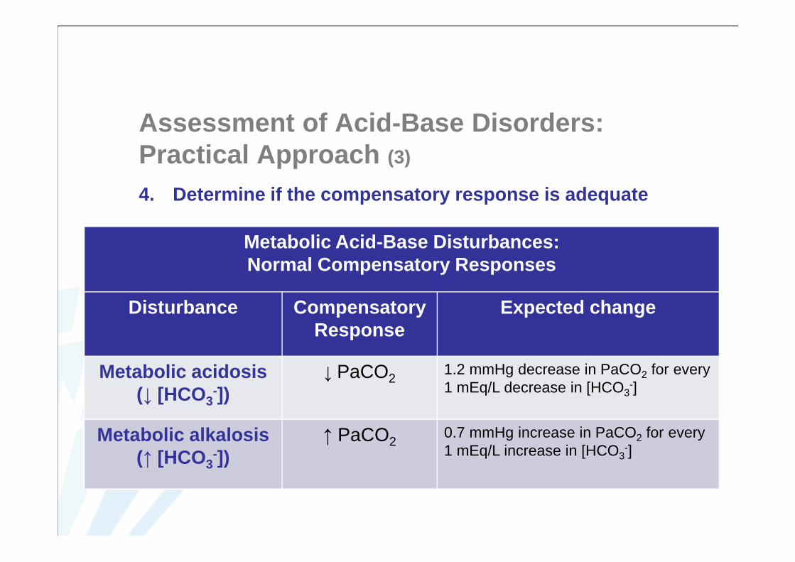
4. Determine if the compensatory response is adequate
Metabolic Acid-Base Disturbances: Normal Compensatory Responses
Disturbance CompensatoryResponse
Expected change
Metabolic acidosis(↓ [HCO3
-])↓ PaCO2
1.2 mmHg decrease in PaCO2 for every1 mEq/L decrease in [HCO3
-]
Metabolic alkalosis(↑ [HCO3
-])↑ PaCO2
0.7 mmHg increase in PaCO2 for every1 mEq/L increase in [HCO3
-]
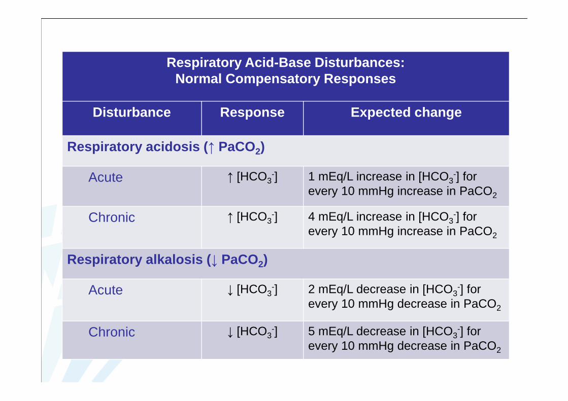
Respiratory Acid-Base Disturbances: Normal Compensatory Responses
Disturbance Response Expected change
Respiratory acidosis ( ↑ PaCO2)
Acute ↑ [HCO3-] 1 mEq/L increase in [HCO3
-] forevery 10 mmHg increase in PaCO2
Chronic ↑ [HCO3-] 4 mEq/L increase in [HCO3
-] forevery 10 mmHg increase in PaCO2
Respiratory alkalosis ( ↓ PaCO2)
Acute ↓ [HCO3-] 2 mEq/L decrease in [HCO3
-] forevery 10 mmHg decrease in PaCO2
Chronic ↓ [HCO3-] 5 mEq/L decrease in [HCO3
-] forevery 10 mmHg decrease in PaCO2

Assessment of Acid -Base Disorders: Practical Approach (4)
5. Is it a simple or mixed acid-base disorder?1. In a simple acid-base disorder only one primary acid-base
disturbance is present. A simple acid-base disorder is suspected if• Primary and compensatory changes of PaCO2 and [HCO3
-] occurin the same direction (↑ or ↓)
• The measured compensatory response equals the calculatedcompensatory response
2. In a mixed acid-base disorder more than one acid-basedisturbance is present.
Assessment of Acid -Base Disorders: Practical Approach (5)
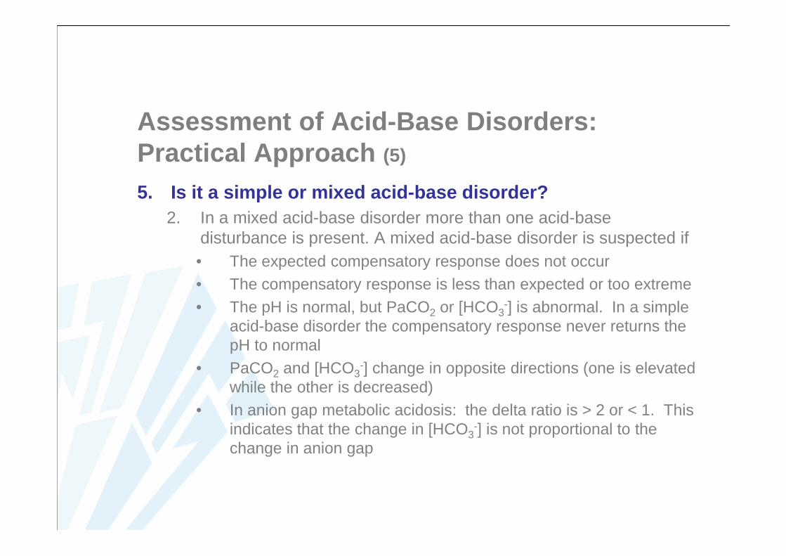
5. Is it a simple or mixed acid-base disorder?2. In a mixed acid-base disorder more than one acid-base
disturbance is present. A mixed acid-base disorder is suspected if• The expected compensatory response does not occur• The compensatory response is less than expected or too extreme• The pH is normal, but PaCO2 or [HCO3
-] is abnormal. In a simpleacid-base disorder the compensatory response never returns the pH to normal
• PaCO2 and [HCO3-] change in opposite directions (one is elevated
while the other is decreased)• In anion gap metabolic acidosis: the delta ratio is > 2 or < 1. This
indicates that the change in [HCO3-] is not proportional to the
change in anion gap

Mixed metabolic disorders
• High anion gap and normal anion gap metabolic acidosis
• High anion gap metabolic acidosis and metabolic alkalosis
• Normal anion gap metabolic acidosis and metabolic alkalosis
Mixed respiratory disorders
• Chronic respiratory acidosis with superimposed acute respiratory acidosis
• It is impossible to have concurrent respiratory acidosis and respiratoryalkalosis
Mixed respiratory – metabolic disorders
• Chronic respiratory acidosis and high anion gap metabolic acidosis
• Chronic respiratory acidosis and metabolic alkalosis
• Respiratory alkalosis and metabolic acidosis
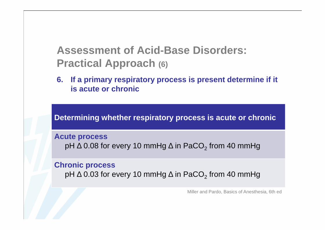
Assessment of Acid -Base Disorders: Practical Approach (6)
6. If a primary respiratory process is present determine if itis acute or chronic
Determining whether respiratory process is acute or chr onic
Acute processpH ∆ 0.08 for every 10 mmHg ∆ in PaCO2 from 40 mmHg
Chronic processpH ∆ 0.03 for every 10 mmHg ∆ in PaCO2 from 40 mmHg
Miller and Pardo, Basics of Anesthesia, 6th ed

Disturbance Response Expected change
Respiratory acidosis ( ↑ PaCO2)
Acute ↑ [HCO3-] 1 mEq/L increase in [HCO3
-] for every10 mmHg increase in PaCO2
Chronic ↑ [HCO3-] 4 mEq/L increase in [HCO3
-] for every10 mmHg increase in PaCO2
Respiratory alkalosis ( ↓ PaCO2)
Acute ↓ [HCO3-] 2 mEq/L decrease in [HCO3
-] for every10 mmHg decrease in PaCO2
Chronic ↓ [HCO3-] 5 mEq/L decrease in [HCO3
-] for every10 mmHg decrease in PaCO2
Respiratory Acid-Base Disturbances: Normal Compensatory Responses
Assessment of Acid -Base Disorders: Practical Approach (7)
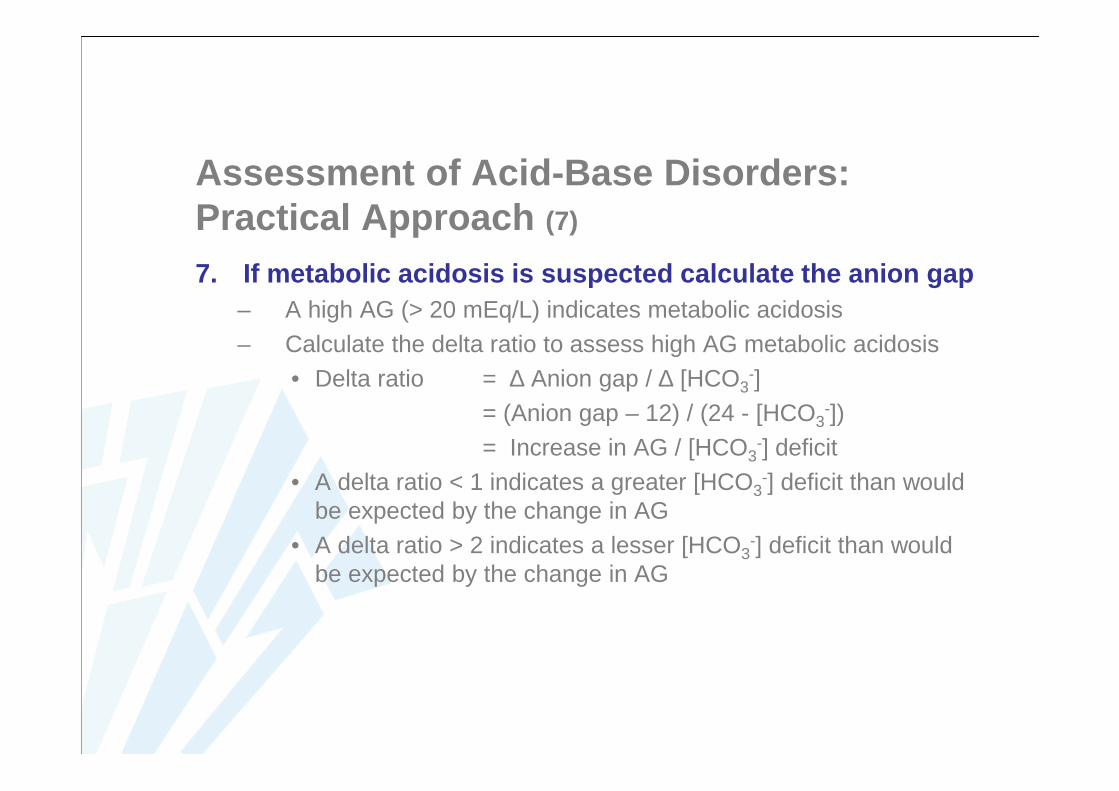
7. If metabolic acidosis is suspected calculate the anio n gap – A high AG (> 20 mEq/L) indicates metabolic acidosis– Calculate the delta ratio to assess high AG metabolic acidosis
• Delta ratio = ∆ Anion gap / ∆ [HCO3-]
= (Anion gap – 12) / (24 - [HCO3-])
= Increase in AG / [HCO3-] deficit
• A delta ratio < 1 indicates a greater [HCO3-] deficit than would
be expected by the change in AG• A delta ratio > 2 indicates a lesser [HCO3
-] deficit than wouldbe expected by the change in AG

Delta Ratio Assessment Guideline
< 0.4 Hyperchloremic normal AG acidosis.
0.4 – 0.8 Consider combined high AG and normal AG acidosis.However: DR is often < 1 in acidosis with renal failure.
1 – 2 High AG acidosis.
> 2 Suggests high AG acidosis with concurrent metabolic alkalosis orwith pre-existing compensated respiratory acidosis.
Always correlate the delta ratio with other evidence to support the diagnosis