Roland N. Dickerson, Pharm.D., BCNSP, FCCP, FCCM, · PDF fileDifferentiate causes for...
112
Updates in Therapeutics ® 2015: Critical Care Pharmacy Preparatory Review Course Fluids, Electrolytes, Acid-Base Disorders, and Nutrition Support Roland N. Dickerson, Pharm.D., BCNSP, FCCP, FCCM, FASHP, FASPEN University of Tennessee College of Pharmacy Memphis, TN
Transcript of Roland N. Dickerson, Pharm.D., BCNSP, FCCP, FCCM, · PDF fileDifferentiate causes for...

Updates in Therapeutics® 2015: Critical Care Pharmacy Preparatory Review Course
Fluids, Electrolytes, Acid-Base Disorders, and Nutrition Support
Roland N. Dickerson, Pharm.D., BCNSP, FCCP, FCCM, FASHP, FASPEN
University of Tennessee College of PharmacyMemphis, TN

Conflict of Interest Disclosures
Nestlé Nutrition (consultant; speaker) NPS Pharmaceuticals (consultant)

Learning Objectives1. Assess hypo and hypernatremia and develop an
appropriate treatment plan.2. Discuss causes and treatment of common
intracellular electrolyte disorders.3. Differentiate causes for metabolic acidosis and
alkalosis and construct a therapeutic treatment.4. Specify appropriate route for nutrition therapy,
amounts, and component formulation.5. Identify markers for assessing tolerance, safety,
and efficacy of enteral or parenteral nutrition.

Learning Objectives (continued)5. Describe methods for ensuring glycemic control.6. Identify pertinent drug-nutrient interactions, and
provide recommendations for the safe and effective delivery of medications to patients receiving enteral or parenteral nutrition therapy.
7. Discuss current controversies regarding nutrition therapy for critically ill patients.

Lecture Outline1. Fluid and Electrolyte Overview (pg 54-56)2. Water and Sodium Aberrations (pg 56-57)3. Intracellular Electrolytes (pg 57-68)4. Acid-Base Disorders (pg 68-74)5. Nutritional Assessment, Energy/Protein
Requirements (pg 74-80)6. Enteral Nutrition (pg 80-82)7. Parenteral Nutrition (pg 82-85)8. Glycemic Control (pg 86-88)9. Controversies in Nutrition Support for Critically Ill
Patients (pg 88-94)

Body Water and Fluids Total body water: ~60% of weight for males;
55% for females – lower % for obese/elderly Daily fluid requirements: 30 to 35 mL/kg/d
(over-estimates obese, under-estimates small person). Alternative: ~1.4 to 1.5 L/m2/d BSA
100 mL/kg for first 10 kg, 50 mL/kg for next 10 kg, and then 20 mL/kg for each kg thereafter
Insensible losses ~550 mL/m2/d BSA –increased ~10% to 15% per degree > 37oC

Fluid Balance Assessment Recorded volume intake – volume output “Normal” fluid balance ~ 600 to 800 mL/d “Normal” fluid balance decreased with
ventilator dependency (less humidified air lost)
“Normal” fluid balance increased with fever Physical exam (mucous membranes,
decreased skin turgor, edema)

Fluid Balance Assessment Hemodynamic markers
(Swan-Ganz catheter measurements, CVP, tachycardia?)
Urine output; I/Os, fever BUN, BUN/creatinine
ratio Serum sodium change Alkalemia (“contraction
alkalosis”) Glycemic control

Electrolyte Content of GI Fluids (Table 4)Fluid Daily
Volume(mL)
Na(mEq/L)
K(mEq/L)
CL(mEq/L)
HCO3(mEq/L)
Mag (mEq/L)
Stomach 1000 –2000
60 –90 10 –15 100 –130 0.9
Duodenum 400 – 600 140 5 –10 90 –120 80
Small intestine
2000 –2500
140 5 –10 90 –120 30 – 40 6 –12
Colon < 300 60 20 –30 50 6 –12
Pancreas 600 – 800 140 5 –10 75 115 0.4
Bile 300 – 600 140 5 –10 100 30 1.1

Self Assessment Question #2 (pg 52)(from Tables 4 and 5):
Which of the following would be the best replacement fluid for nasogastric losses?A. 0.9% sodium chloride and KCL 20 mEq/L
B. 0.45% sodium chloride and KCL 20 mEq/L
C. 5% dextrose in 0.225% sodium chloride and KCL 20 mEq/L
D. Lactated ringers solution

Evaluation of Hyponatremia Rule out pseudo or factitious hyponatremia
(hyperproteinemia, hyperlipidemia, hypoglycemia)
Exclude mannitol, glycine for unmeasured osmoles
Evaluate ECF volume (American method) or urine sodium/osmolality (European method)
Evaluate urine sodium/osmolality (American method) or ECF volume (European method)
Consider patient conditions/diagnoses

Correcting Serum Sodium Concentration for Hyperglycemia
For every 100 g/dL increase in BG > 100 mg/dL; serum Na will decrease by ~ 1.6 to 2.4 mEq/L
Example: A patient with a serum glucose of 300 mg/dL and a Na of 130 mEq/L is given insulin therapy. What is the corrected serum Na once his BG is treated to normal?
300 – 100 = 200; 200/100 = 2 2 X (1.6 to 2.4 mEq/L) = 3 to 5
mEq/L His “corrected” serum Na would be =
130 + (3 to 5) = 133 to 135 mEq/L
Hillier, TA. Am J Med.1999;106:399-403.

Hyponatremia Defined as a serum Na < 135
mEq/L (some clinicians are unconcerned unless serum Na < 130 mEq/L; severe: Na < 120 mEq/L)
Sodium content within a defined volume of water
MUST be interpreted with assessment of ECF status
Patients can be hypovolemic, euvolemic, or hypervolemic (and have a low serum Na)
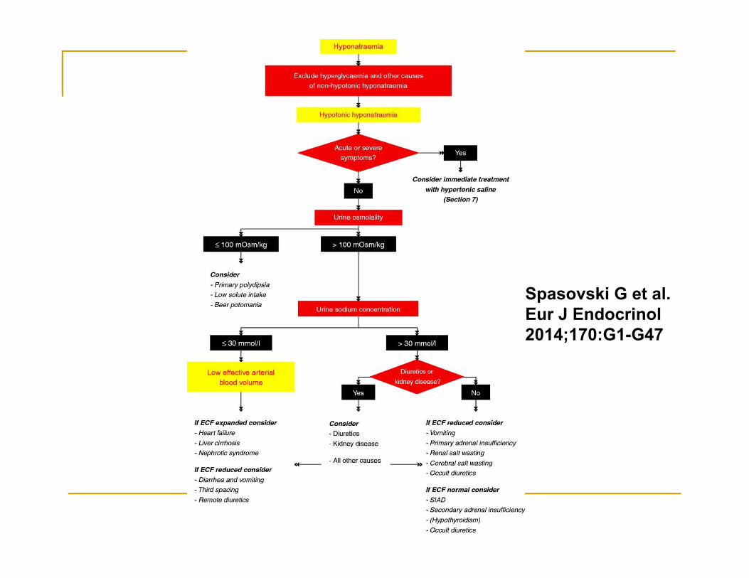
Spasovski G et al. Eur J Endocrinol2014;170:G1-G47

Dickerson RN. Hospital Pharmacy. 2002;37:1336-1339.

Treatment of Hyponatremia Acute vs severe symptoms – hypertonic
saline vs. conivaptan Do not increase serum Na > 10-12 mEq/L/d
(increased risk for central pontinemyelinolysis); most clinicians prefer 6-8mEq/L/d change in absence of seizures, etc.
ECF expanded – fluid and Na restriction ECF depleted – 0.9% NaCL

Treatment of Hyponatremia ECF normal – SIADH or
adrenal insufficiency Fluid restriction first Consider conivaptan or
tolvaptan if available Fluid restriction with use of
0.9% NaCL solution (make PN or EN solutions isonatremic); diuretic therapy may be needed
Marik PE et al. Pharmacotherapy. 2013;33:51-55.
4.3 + 2.6 mEq/L increase in serum Na 24 hrs after a single20 mg bolus of conivaptan

Patient Case 55 yo 70 kg woman with pneumonia. Serum Na
of 125 mEq/L on day 5 of hospitalization. BG is 167 mg/dL, BUN 20 mg/dL, creatinine 1.1 mg/dL
Receiving EN (1kcal/mL, 62 g protein/L) @ 60 mL/hr + D5 ½ NS @ 25 mL/hr
Fluid balance: + 300 to 600 mL/d for past 3 days. No edema.
Serum osmolality 265 mOsm/kg, urine osmolality 490 mOsm/kg, urine Na 67 mEq/L
Chapter Case, Pg 57

What is the most likely etiology for the patient’s hyponatremia?
A. Factitious hyponatremia
B. Adrenal insufficiency
C. Cerebral salt wasting
D. SIADH
Chapter Case, Pg 57

What would be the most appropriate treatment for this woman?
A. Sodium chloride tablets 1 g TID
B. Limit fluids (fluid restriction)
C. Change the intravenous fluid to 0.9% NaCL
D. Provide a short-term infusion of 3% NaCL
Chapter Case, Pg 57

What change in the enteral feeding formula would be best?
A. Add NaCL 100 mEq/L to the EN formula
B. Change the formula to a fish oil enriched formula
C. Change the formula to a low carbohydrate, high fat formula
D. Change the formula to a 2 kcal/mL formula and decrease the rate
Chapter Case, Pg 57

Hypernatremia Hypervolemic: restrict water and
Na, diuretics Hypovolemic: dehydration (water
loss in excess of sodium) Euvolemic: primarily free water
loss (fever, thermal injury, diabetes insipidous)
Water deficit (L): body weight(kg) X 0.6 X [(serum Na/140)-1]*
*commonly reported but ? accuracy – usually underestimates total body water and free water losses. Cheuvront SN et al. Am J Clin Nutr. 2013;97:79-85

Hypernatremia Assess ECF volume status Do not decrease > 10-12 mEq/L/d (cerebral
edema) Hypervolemic: reduce Na and fluid intake Hypovolemic: hypotonic fluids (e.g., ½ NS,
D5 ¼ NS, D5W, water boluses per tube/po; delete Na in PN temporarily)
Euvolemic: hypotonic fluids, (central DI: nasal desmopressin 10 mcg BID, IV desmopressin1- 2 mcg BID), (nephrogenic DI: Na restriction, thiazides –paradoxical Tx)

Potassium Homestasis Total body stores 35 - 50 mEq/kg in normal
healthy adults (25 – 30 mEq/kg if undernourished)
Serum K influenced by changes in pH (for every 0.1 increase in pH, serum K decreases by ~ 0.6 mEq/L and vice-versa
Average requirement ~ 0.5 to 1.2 mEq/Kg/d Kidney is primary route of elimination Magnesium is required for normal potassium
homeostasis

Etiologies for Hypokalemia Increased requirements due to
building of new muscle/tissue Increased gastrointestinal fluid
losses Hypomagnesemia Medications –Beta agonists,
catecholamines, insulin, amphotericin B, diuretics, bicarbonate
Increased urinary losses (DI, DKA)

Estimating Potassium Deficit Based on estimate of total body potassium Serum K = 3 mEq/L ~ 10% total body deficit Serum K = 2.5 mEq/L ~20% total body deficit
Sterns RH et al. Medicine.1981;60:339.

Treatment of HypokalemiaEnteral vs Parenteral
Enteral K is safer due to slower/controlled absorption rate
Enteral K is safer due to feedforward regulation of K homeostasis
Enteral K may be more difficult for some patients
Greenlee ML al. Ann Intern Med 2009;150:619-625

Intravenous Potassium Therapy When po potassium not possible or if patient has
severe hypokalemia Maximum concentration of 60 mEq/L of KCL for
a peripheral IV. “Boluses” (e.g., 40 mEq) cannot be given via
peripheral IV: burning, phlebitis, pain. Must be given via central vein.
Maximum infusion rate of 10 mEq/hr (when pt is not on an ECG monitor); 20 mEq/hr if pt is being monitored.
40 mEq/hr only if patient has paralysis or life-threatening arrhythmia.

Intravenous Potassium Dosage For every 40 mEq
IV KCL given, serum K increases by 0.5 to 0.6 mEq/L?
495 infusion sets in 190 patients
KCL 20 mEq IV over 1 hour
0.25 mEq/L increased per 20 mEq dose
Kruse et al. Arch Int Med. 1990;150:613-617

Potassium Dosing:Effect of Body Size, Renal Function, Nutrition Therapy?

Empiric Dosing Guidelines for Potassium Chloride
Serum K (mEq/L)
Potassium Dosage (mEq)*
3.5 – 3.9 40 mEq X 1 dose
3.0 – 3.4 40 mEq X 2 doses
< 3 40 mEq X 3 doses
*Use half dose for renal impairment; may need to be adjusted based on body size, nutrition therapy, and ongoing losses. Given at 10 mEq/hr.
Johnston C et al. ASPEN meeting. February 2015.
Hyperkalemia Rule out factitious hyperkalemia Etiologies: excessive intake,
acidemia, renal impairment, tissue catabolism (chemotherapy, rhabdomyolysis), elderly are susceptible, hyporeninemichypoaldosteronism, blood transfusion
Etiologies – Drugs (K sparing diuretics, ACE and ARBs, NSAIDs, Heparin, Trimethoprim, Octreotide, Pen G (1.6 mEq K/million units).

Hyperkalemia

Treatment of Hyperkalemia
Dextrose/Insulin 50 g / 15 units IV Sodium bicarbonate 50 – 100 mEq IV Calcium gluconate 2 g IV Potent beta-2 agonist (albuterol) Sodium polystyrene sulfonate 15 to 60 g up to
every 6 hrs (in-vivo exchange capacity of ~ 1 mEq K per g).
Discontinue all sources of K intake ? Loop diuretic therapy Dialysis

Magnesium Homeostasis
50% of magnesium is in bone; 1% of total body magnesium is in the ECF
Normal serum magnesium: 1.8 to 2.4 mg/dL Serum: 60% ionized; 15% complexed; 25%
protein bound. Kidney is primary route of elimination Influences potassium and calcium metabolism

Hypomagnesemia Definition: < 1.8 mg/dL Symptomatic: < 1.5 mg/dL Causes hypokalemia Causes hypocalcemia Chvostek’s/Trousseau’s/QT
prolongation, weakness, tetany Etiologies: diarrhea, alcohol,
sepsis/critical illness, pancreatitis, burns, brain injury, drugs (see next slide)

Drug-Induced Hypomagnesemia Diuretics Amphotericin B Cyclosporin/tacrolimus Foscarnet Pentamidine Cisplatin/Carboplatin/ifosfamide/Cetuximab Lactulose/orlistat

Treatment of Hypomagnesemia
32 to 48 mEq/d (4-6 g/d) sufficient to maintain normal serum Mg concentrations
Estimated magnesium deficit: for a serum Mg concentration < 1.5 mg/dL, a 1 to 2 mEq/kg deficit can be expected
Should be replaced over 4-5 days Treat the etiology (ies) Takes 48 hrs for the serum concentration to
equilibrate following a short term infusion

Empiric Magnesium Dosing Guidelines
Serum magnesium
(mg/dL)
IntravenousMagnesium Sulfate
dosage (g/kg)*1.6 to 1.8 0.05 g/kg1.0 to 1.5 0.1 g/kg
< 1.0 0.15 g/kg
*Use half dose for renal impairment; may need to be adjusted based on body size, nutrition therapy, and ongoing losses. Given at 1 g/hr.
Sacks et al. Nutrition.1997;13:303-8.

Common Oral Magnesium Products and Dosing
Salt Form Strength (mg) Elemental Mg (mEq)
Usual Dosing
Oxide 400100
19.86.9
1-2 tablets BID-TID
Gluconate 500 2.2 1-2 tablets BID-TID
Chloride 100 2.6 1-2 tablets BID-TID

Calcium Homeostasis
Ionized calcium (iCa): physiologically active fraction of serum total calcium
“Normal” serum calcium concentration is 8.5 to 10.5 mg/dL
“Normal” serum ionized calcium concentration is 1.12 – 1.32 mmol/L

Calcium Homeostasis Ionized calcium (iCa):
physiologically active fraction of serum total calcium
“Normal” serum calcium concentration is 8.5 to 10.5 mg/dL
“Normal” serum ionized calcium concentration is 1.12 – 1.32 mmol/L
50%
40%
10%
Free Protein-Bound Complexed

Calcium Homeostasis
Average daily requirement with PN: 15 mEq/d calcium gluconate
Kidney is primary route of elimination Magnesium can influence calcium
homeostasis (end organ resistance to PTH +/- impaired secretion of PTH)
Mild hypocalcemia will correct within ~ 48 hrs after correction of hypomagnesemia

Correcting Serum Calcium Conc for a Low Serum Albumin Conc
For every 1 g/dL decrease in serum albumin concentration, serum calcium concentration will decrease by 0.8 mg/dL- Endres, 1999; Orrell, 1971;35:483-489
Only use in NON-ICU patients! Use of the modified Orrell equation (above)
correctly identified 1 out of 21 hypocalcemicpatients (from a total of 100 NSS trauma patients) - Dickerson RN. JPEN. 2004;28:133-41.

Correcting Serum Calcium Conc for a Low Serum Albumin Conc
Example: A patient with a serum albumin 2 g/dL, serum calcium 7 mg/dL
Normal serum albumin = 4 g/dL 4 – 2 = 2 2 X 0.8 = 1.6 “Corrected” serum calcium = 7 + 1.6 = 8.6
mg/dL

Hypocalcemia Prolonged QTc, parasthesias,
Chvostek’s and Trousseau’s signs, PVCs, seizures, tetany, torsadesdes pointes
Mild: iCa 1 – 1.12 mmol/L Moderate to severe: iCa < 1
mmol/L Multiple causes: critical illness,
CRRT, massive blood transfusion, alkalemia, hypomagnesemia, hyperphosphatemia, pancreatitis, drugs (see hypoMg drugs)

When Should Hypocalcemia be Treated?
Significant or symptomatic hypocalcemia Massive blood transfusion with pre-existing
myocardial disease Calcium channel blocker overdose Receiving inotropic agents and/or
vasopressors Adjunct to emergent management of severe
hyperkalemia Coagulopathy? Prevention of worsening hypocalcemia?

Treatment of Hypocalcemia
iCa(mmol/L)
IV calcium gluconate dose
1 – 1.12 2 g in 100 mL over 2 hrs< 1 4 g in 100 mL over 4 hrs
Treat the cause if possible!
Dickerson. JPEN.2005;29:436-431.Dickerson. JPEN.2007;31:228-233. Dickerson. Nutrition.2007;23:9-15.

Etiologies for Hypercalcemia Immobilization Metabolic bone disease Immobility Excessive intake Malignancy Drugs (vitamin D) Dehydration Granulomatous diseases
(TB, sarcoidosis)

Treatment of Hypercalcemia
IV fluids/Rehydrate! Add furosemide if necessary.
Mobilize the patient Calcitonin Pamidronate

Phosphorus Homeostasis Regulated by vitamin D and PTH Normal serum concentration: 2.5 – 4.5 mg/dL During critical illness, goal is to achieve ~ 4
mg/dL (based on Zazzo and Aubier studies) Kidney is primary route of elimination Mean Renal Phosphate Threshold Conc
(TmP/GFR) is ~ 3 – 3.3 mg/dL (trauma and thermally injured patients – Dickerson, 2001)

Etiologies of Hypophosphatemia Malnourishment Alcoholism Refeeding syndrome Drugs (insulin,
catecholamines) Critical Illness/TBI/Thermal
injury/DKA/alkalemia Hepatic Resection Hyperparathyroidism Cancer (fibroblast growth
factors)

Hollywood and Refeeding Syndrome
HBO’s Band of Brothers. Easy Company, 101st Airborne, US Army during WWII.
Liberation of LandsbergConcentration Camp.

Loven L. J Trauma.1986;26:348-52. Sheldon GF. J Trauma.1973;15:971-9.

Effect of Phosphorus Supplementation on Organ Function
Variable Before After P <
Phos 1.0 + 0.4 3.8 + 1.4 0.01
HR 102 + 17 105 + 13 NS
CI 3.8 + 1.9 4.5 + 1.8 0.01
Zazzo. Inten Care Med.1995;21:826-31.
Aubier. NEJM.1995;313:420-4.

IV Phosphorus Dosing for Hospitalized Patients
Serum Phosphorus Conc (mg/dL)
2.3 to 3 mg/dL
1.6 to 2.2 mg/dL
< 1.5 mg/dL
Dosage (mmol/kg) Trauma/Burn 0.32
0.64
1
Dosage (mmol/kg)General/ICU
0.16
0.32
0.64
Infuse intravenous phosphorus at 7.5 mmol/hr

Hyperphosphatemia
Renal failure Immobility/chronic critical
illness-associated metabolic bone disease
Vitamin D toxicity Excessive phosphorus
intake

Hyperphosphatemia
Velentzas. Adv Exper Med Biol. 1978;103:195-201.

Treatment of HyperphosphatemiaPhosphate Binders
Drug P-binding capacity Initial DoseAluminum hydroxide 22.3 mg/ 5 ml 30 ml Q6hr
(not recommended in renal failure)
Calcium carbonate 43 mg/ g calcium 1 g QID(less effective at higher gastric pH; higher Ca content)
Calcium acetate 106 mg/ g calcium 1334 to 2001 mg TID(lower in calcium content than Ca carbonate)
Sevelamer 800 mg/cap or packet 800 mg TID(Maximal binding at pH 7; powder or capsule)
Lanthanum data not available 500 mg TID
Schucker et al. Am J Health-Syst Pharm.2005;62:2355-61.

Patient Case 55 yo 70 kg man, s/p total colectomy and
hepatic resection for stage IV colon Ca. Twenty pound unintentional weight loss. Frequent ETOH. NG output > 2 L/d. PN initiated.
Na 140 mEq/L, K 3.2 mEq/L, CL 102 mEq/L, tot CO2 25 mEq/L, BUN 14 mg/dL, creat 0.9 mg/dL, Ca 8.1 mg/dL, phos 2 mg/dL, mag 1.4 mg/dL, albumin 2.5 g/dL.
Patient Case, Pg 63

Which potassium-phosphorus dosing regimen is best for this patient?
A. KCL liquid 40 mEq per NGT X 2 doses, 2 Neutra-Phos capsules in water per NGT
B. Kphos 30 mmol IV X 1 dose, 2 doses of KCL liquid 40 mEq per NGT
C. KCL 40 mEq IV X 1, Kphos 45 mmol IV X 1D. KCL 40 mEq IV X 1, Kphos 30 mmol IV X 1
Patient Case, Pg 63, Question #4

The patient is also given Mag Sulfate 6 g IV over 6 hrs. His repeat serum Mag the next day is 1.9 mg/dL. Which next therapeutic option is best?
A. Magnesium oxide 500 mg BID X 4-5days
B. Magnesium sulfate 2-4 g IV daily X 4-5 days
C. Give a second dose of 6 g of Mag Sulfate IV
D. No additional treatment is necessary
Patient Case, Pg 63, Question #5

Patient Case
24 yo 70 kg man s/p GSW abdomen with multiple abdominal injuries. He received 10 units of PRBC.
Serum iCa is 0.86 mmol/L, K 4.6 mEq/L, Mag 1.8 mEq/L.
Good renal function (sCr 0.8 mg/dL and UOP > 0.5 mL/kg/hr).
Patient Case, Pg 65

What is the most likely etiology for his hypocalcemia?
A. Hypomagnesemia
B. Excessive urinary diuresis
C. Blood transfusion
D. Critical illness
Patient Case, Pg 65, Question #6

Which therapeutic regimen would be best for this patient?
A. Calcium gluconate 2 g IV over 2 hrs
B. Calcium gluconate 4 g IV over 4 hrs
C. Calcium chloride 1 g IV push over 5 to 10 min
D. No calcium therapy necessary
Patient Case, Pg 65, Question #7

Lecture Outline1. Fluid and Electrolyte Overview (pg 54-56)2. Water and Sodium Aberrations (pg 56-57)3. Intracellular Electrolytes (pg 57-68)4. Acid-Base Disorders (pg 68-74)5. Nutritional Assessment, Energy/Protein
Requirements (pg 74-80)6. Enteral Nutrition (pg 80-82)7. Parenteral Nutrition (pg 82-85)8. Glycemic Control (pg 86-88)9. Controversies in Nutrition Support for Critically Ill
Patients (pg 88-94)

Acid-Base Disorders
Severe Acidemia (pH < 7.25) Severe Alkalemia (pH > 7.55) Metabolic acidosis Metabolic alkalosis Compensatory response to A-B disorders Base Excess – freedom from memorizing
acid-base correction formulas?


Metabolic Acidosis: Anion GapAnion Gap = Na – CL – HCO3
Normal range: ~3 – 11 or 12 mEq/L
Every 1 g/dL decrease in serum Alb < 4 g/dLcontributes to an unmeasured ~2.5 mEq/L in gap
Example: Na 145 mEq/L, Cl 110 mEq/L, HCO3 20 mEq/L, albumin 2 g/dL
Anion Gap = 145 - 110 - 20 = 15 Correction Factor = 4-2 = 2 X 2.5 = 5 Corrected Anion Gap = 15 + 5 = 20

Causes of an Anion Gap AcidosisA MUD PIE
Aspirin (salicylates) Methanol Uremia (renal failure) including
rhabdomyolysis Diabetes (Diabetic Ketoacidosis) Paraldehyde Infection or Ischemia (Lactic acidosis) Ethylene Glycol or Ethanol toxicity

Causes of a Non-Anion Gap AcidosisACCRUED
Ammonium Chloride or Acetozolamide (urine bicarbonate loss)
Chloride intake (sources: PN, IVs, etc.) Cholestyramine (GI bicarbonate loss) Renal Tubular Acidosis- Type I, II, IV Urine diverted into the bowel Endocrine disorders (e.g., aldosterone
deficiency) Diarrhea (also enterocutaneous fistulas)

Anion Gap Metabolic AcidosisUse of the Delta Ratio
sometimes used in the assessment of elevated anion gap metabolic acidosis to determine if a mixed acid base disorder is present.
Delta ratio = ∆ Anion gap/∆ [HCO3-] or ↑anion gap/ ↓ [HCO3-]
DR = Measured anion gap – Normal anion gap Normal [HCO3-] – Measured [HCO3-]
DR = (AG – 14)(24 - [HCO3-])

Evaluating Metabolic Acidosis: the Delta Ratio
Delta Ratio
Assessment Guidelines
< 0.4 Hyperchloremic normal anion gap acidosis
< 1 High anion gap acidosis and normal anion gap acidosis
1 to 2 Anion gap acidosis
> 2 High anion gap acidosis and concurrent metabolic alkalosis OR a pre-existing compensated respiratory alkalosis

Determining Extent of Compensation and Mixed Acid-Base Disorders (see Table 17)
Chronic Respiratory Acidosis: For every 10 mm Hg increase in pC02, serum HCO3 should increase by ~ 4 mEq/L
Chronic Respiratory Alkalosis: For every 10 mm Hg decrease in pCO2, serum HCO3 should decrease by ~ 5 mEq/L
Metabolic Acidosis: pCO2 = ~ (1.5 X HCO3) + 8 Metabolic Alkalosis: widely variable increase
in pCO2 pCO2 = ~(0.7 X HCO3) + 21

Evaluating Acid-Base Disorders at the Bedside
Know the clinical details of the patient Is the “snap-shot” reflective of the current
clinical situation Find the cause for the acid-base disorder Is compensation appropriate? Is the pH severe enough to warrant therapy? Plan a therapeutic treatment regimen

Treatment of Metabolic AcidosisIntravenous sources of alkali
Sodium bicarbonate Sodium acetate Sodium citrate THAM (0.3 N Tromethamine) Lactate

Treatment of Metabolic AcidosisUse of bicarbonate
Total dose of bicarbonate = 0.5 X Wt (kg) X (24 – [HCO3])
Give one half to one third (OR 1 to 2 mEq/kg) over several hours (avoid boluses) to get pH > 7.25
Once pH > 7.25, slower correction without increasing HCO3 > 4 to 6 mEq/L to avoid overalkalinization
Serial ABGs (e.g., Q6h)

Treatment options for Metabolic Alkalosis depends on:
Sodium Chloride Responsive (urinary Cl < 25 mEq/L)
Sodium Chloride Resistant (urinary Cl > 40 mEq/L)

Causes of Sodium Chloride Responsive Metabolic Alkalosis
Excessive gastric fluid losses Diuretic therapy (especially loop) Dehydration (contraction alkalosis) Hypokalemia

Causes of Sodium Chloride Resistant Metabolic Alkalosis
Excessive mineralocorticoid activity (hyperaldosteronism, glucocorticoid excess/Cushing’s disease, glycyrrhizicacid)
Massive blood transfusion Milk alkali syndrome Large doses of large penicillins

Treatment of Saline Responsive Alkalemia
Treat the etiology 0.9% NaCl + KCL to replenish deficits Acetozolamide 250 – 375 mg QD-TID HCL therapy

Hydrochloric Acid Therapy
Only if NaCl and KCl not possible Central venous administration only Glass bottle 0.1 or 0.2 N HCL (0.2 N for fluid restricted
patients)

Hydrochloric Acid TherapyCalculating the Dose
Chloride deficit methodmEq HCL= 0.2 X WT (kg) X (103 – serum Cl) Bicarbonate excess methodmEq HCL= 0.5 X WT (kg) X (serum HCO3 - 24) Give ~ ½ of the above calculated dose over 12
hrs and reassess. ABGs Q6h. Stop infusion at ~ pH 7.45 to avoid over-
correction

Patient Case 70 yo, 40 kg female s/p radical cystectomy with
ileal conduit, post-op ileus, NG output 1.5 L/d, and requires PN.
0.45% NaCl + KCL 20 mEq/L @ 50 mL/hr PN electrolyte daily intake: NaCL 60 mEq, K
Acetate 40 mEq, Na Phos 15 mmol, Mg sulfate 24 mEq, calcium gluconate 10 mEq.
After a few days of PN, her ABG was pH7.29, pO2 95, pCO2 35, HCO3 21. Serum Na 141, K 3.9, CL 117, total CO2 content 22
Patient Case Pg 72

Which best describes her acid-base disorder?
A. Hyperchloremic, normal AG acidosis
B. AG acidosis
C. AG acidosis with hyperchloremia
D. Respiratory acidosis with concurrent metabolicalkalosis
Patient Case, Pg 72; Question #8

Which is the most appropriate treatment algorithm?
A. Substitute the NaCL in the PN solution with Na Acetate
B. Sodium bicarbonate 100 mEq IV
C. Change the IV solution to Lactated Ringers solution
D. Add 100 mEq of sodium bicarbonate to the PN solution
Patient Case Pg 73, Question #9

Lecture Outline1. Fluid and Electrolyte Overview (pg 54-56)2. Water and Sodium Aberrations (pg 56-57)3. Intracellular Electrolytes (pg 57-68)4. Acid-Base Disorders (pg 68-74)5. Nutritional Assessment, Energy/Protein
Requirements (pg 74-80)6. Enteral Nutrition (pg 80-82)7. Parenteral Nutrition (pg 82-85)8. Glycemic Control (pg 86-88)9. Controversies in Nutrition Support for Critically Ill
Patients (pg 88-94)

Nutritional Assessment ASPEN definitions vs “Classic” definitions Mild, moderate, severe based on IBW Underweight BMI < 18.5 (kg/m2) Obese BMI > 30 (kg/m2) Serum proteins – albumin/prealbumin –
decreased with stress Weight loss is significant when > 10%
unintentional wt loss within 6 months

Caloric Content of Macronutrients Glucose 3.4 kcal/g Carbohydrate 4 kcal/g Protein/Amino acids 4 kcal/g Fat 9 kcal/g IV Fat ~10 kcal/g (IV fat emulsion contains
glycerol and phospholipids; 10% = 1.1 kcal/mL, 20% = 2 kcal/mL, 30% = 3 kcal/mL)
Propofol: administered in 10% lipid emulsion!

Recommended Caloric IntakeAn Over-Simplification
Critically ill patients: 25 to 30 kcal/kg/d MICU patients: 25 to 27 kcal/kg/d (no more
than 30 kcal/kg/d) SICU: 25 to 30 kcal/kg/d (no more than 35
kcal/kg/d) Trauma: 25 to 32 kcal/kg/d (no more than 35
kcal/kg/d) Burns: Other formulas used; hypermetabolic.

Recommended Caloric IntakeAn Over-simplification
Weight based caloric intakes are erroneous in small (~50 kg), older (> 65 to 70 yrs), or obese patients (BMI > 30 kg/m2)
Small/Elderly = ~1.2 to 1.3 X BEE (Harris-Benedict equations), no more than 1.5 X BEE
Obese = < 25 kcal/kg IBW/d (in conjunction with a high protein intake)

Avoid Overfeeding Do not exceed:
Glucose/carbohydrate: 5 mg/kg/minFat (IV): 2.5 g/kg/d (keep < 1.5 g/kg/d)
Hypercapnia (an excess in total kcals are more likely to cause hypercapnia vs. % CHO vs. % fat)
Hyperglycemia Fatty infiltration of the liver/hepatic
steatosis/cholestasis

Protein Requirements Critically ill patients: 1.5 to 2 g/kg/d -
requirements greater for surgical/trauma vs. medical ICU patients
European vs American guidelines SCCM-ASPEN: 1.2 to 2 g/kg/d; higher
amounts likely needed for multiple trauma or burns

Protein Requirements Trauma/Burns: 2 – 2.5 g/kg/d Renal Failure: AKI/CKD: 0.6 – 1 g/kg/d Hemodialysis: 1 – 1.5 g/kg/d CRRT: 2 – 2.5 g/kg/d
Obese: 2 – 2.5 g/kg IBW/d BMI < 40: 2 g/kg IBW/d BMI > 40: 2.5 g/kg IBW/d

Patient Case 40 kg woman admitted to the trauma ICU
receives a PN solution containing 350 g dextrose, 160 g amino acids, and 80 g of lipid daily.
Her most ABG revealed: a pH of 7.30, pCO2 of 55, PO2 of 96, and HCO3 of 31.
Her BGs from the past 24 hours range from 150 to 180 mg/dL.
Patient Case, Pg 78

Which change would be best to recommend concerning her PN?
A. Decrease dextrose to 175 g/d, increase lipid to 120 g/d.
B. Add 20 units of regular human insulin.
C. Decrease all the macronutrients by about one-half.
D. Increase the acetate content.
Patient Case, Pg 78, Question #10

Assessing Protein RequirementsNitrogen Balance
NB (g/d) = Nin – Nout NB > +4 g/d = anabolic NB -4 to + 4 g/d = nitrogen equilibrium NB worse than -4 g/d = catabolic Classic NBAL equation NB = Protein in (g/d)/6.25 – UUN (g/d) – 4 g/d If BUN change > 5 mg/dL, add to losses (see
chapter pg 30) Highly catabolic patients may use different
NB calculation (pg 30)

Patient Case
A 24 hr urine collection was done to determine nitrogen balance for a 45 yo obese man with pancreatitis and sepsis receiving hypocaloric, high protein (24 kcal/kg IBW/d, 2 g/kg IBW or150 g/d of protein) PN.
The urine urea nitrogen (UUN) concentration was 900 mg/dL and urine volume output was 3000 mL. The BUN was unchanged during the NB determination.
Patient Case, Pg 79

Using the classic NB equation, what was his nitrogen balance?
A. + 4 g/d
B. - 4 g/d
C. - 7 g/d
D. -10 g/d
Patient Case, Pg 80, Question #12

What changes would be best to make to the PN regimen?
A. Increase the protein and non-protein energy content
B. Increase the protein content, decrease the non-protein energy content
C. Increase the protein content
D. Increase the non-protein content
Patient Case, Pg 80, Question #13

Indications for EN
If the patient is unable to ingest adequate amounts to achieve goal nutritional intake
Early EN is beneficial for critically ill patients (ESPEN 2006; SCCM/ASPEN 2009)
Definition of early? – 24 hrs to 48 hrs post admission to ICU (max 72 hrs)
How much EN is necessary for a beneficial effect? controversial

Which EN formula?
See Table 22 pg 32 highlighting different commercially available EN formulas that are indicated for different clinical conditions.

Important EN-Medication Interactions Whereby TF may be held 1 hr prior to and
after medication administration: Warfarin Phenytoin Levothyroxine* Fluroquinolones* Itraconazole*
*Interaction may be overcome by providing a higher dosage; readjust doses when EN d/c’d

Indications for PN ESPEN 2009: Patients not expected to
receive EN within 3 days should receive PN within 24-48 hrs if EN contraindicated
SCCM-ASPEN 2009: PN indicated only after first 7 days of hospitalization when EN not possible
Depends on state of malnourishment, catabolic state of patient; outcomes more variable for medical vs surgical patients

Central vs. Peripheral PN Peripheral PN requires low concentrations of
macronutrients and high volume of fluid to keep osmolality tolerable (e.g., < ~800 mOsm/kg)
Approx Osmol = (dextrose g/L X 5) + (% amino acids X 100) + (% lipid X 15) + 200*
*estimate of electrolyte, vitamins, minerals contribution to osmolality

Glucose
Obligatory requirements: 130 g/d Surgical wound: 80 to 150 g/d Don’t exceed 5 mg/kg/min Glucose 3.4 kcal/g Provides the majority of non-protein kcals

Lipid Emulsion 10%=1.1 kcal/mL; 20%=2 kcal/mL;30%=3
kcal/mL To prevent EFAD in adults: at least 100 to
150 g/weekly (e.g., 1 – 1.5 g/kg weekly) Preferably give < 1.5 g/kg/d and definitely not
> 2.5 g/kg/d (adults) Do not allow triglycerides to exceed 400
mg/dL

Predisposing Conditions to Impaired IV Triglyceride Clearance
Excessive lipid intake (propofol!)Acute pancreatitisUncontrolled diabetesKidney failureEnd-stage sepsisObesityHIVHyperlipidemiaPregnancy

Vitamins and Trace Minerals Additional zinc may be indicated for patients
with intractable diarrhea, intestinal fistulae, critical illness
Copper and manganese should be withheld in patients with significant hepatobiliary/cholestatic liver disease
Supplemental thiamine indicated for ETOHicsand severely malnourished patients

Lecture Outline1. Fluid and Electrolyte Overview (pg 54-56)2. Water and Sodium Aberrations (pg 56-57)3. Intracellular Electrolytes (pg 57-68)4. Acid-Base Disorders (pg 68-74)5. Nutritional Assessment, Energy/Protein
Requirements (pg 74-80)6. Enteral Nutrition (pg 80-82)7. Parenteral Nutrition (pg 82-85)8. Glycemic Control (pg 86-88)9. Controversies in Nutrition Support for Critically Ill
Patients (pg 88-94)

Glycemic Control SCCM: keep BG < 150 mg/dL for most
patients and absolutely < 180 mg/dL ASPEN: target BG 140 to 180 mg/dL ADA: target BG 140 to 180 mg/dL lower BG
target may be appropriate in select patients. “Select patients”: trauma, thermal injury, CT
surgery (e.g., BG < 140 to 150 mg/dL) Avoid BG < 70 mg/dL; definitely avoid < 40
mg/dL

Lecture Outline1. Fluid and Electrolyte Overview (pg 54-56)2. Water and Sodium Aberrations (pg 56-57)3. Intracellular Electrolytes (pg 57-68)4. Acid-Base Disorders (pg 68-74)5. Nutritional Assessment, Energy/Protein
Requirements (pg 74-80)6. Enteral Nutrition (pg 80-82)7. Parenteral Nutrition (pg 82-85)8. Glycemic Control (pg 86-88)9. Controversies in Nutrition Support for Critically
Ill Patients (pg 88-94)