Xbp1 deficiency does not alter hematopoietic cellularity ... · Supplementary Figure 2 Xbp1...
6
Supplementary Figure 1 Xbp1 deficiency does not alter hematopoietic cellularity. Absolute number of leukocytes obtained from bone marrow (BM, 2 tibias and 2 femurs per mouse) of Xbp1 f/f and Xbp1 Vav1 mice (n = 3 per genotype). Each symbol represents an individual mouse; small horizontal lines indicate the mean (± s.e.m.). NS, not significant (P > 0.05, Student’s t-test). Data are representative of at least three independent experiments with at least three mice per genotype per experiment. Nature Immunology: doi:10.1038/ni.3225
Transcript of Xbp1 deficiency does not alter hematopoietic cellularity ... · Supplementary Figure 2 Xbp1...
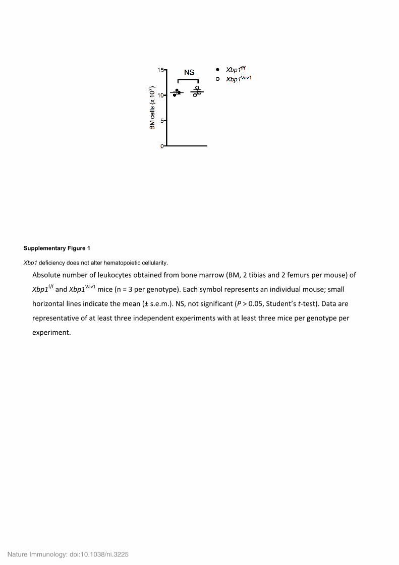
Supplementary Figure 1
Xbp1 deficiency does not alter hematopoietic cellularity.
Absolute number of leukocytes obtained from bone marrow (BM, 2 tibias and 2 femurs per mouse) of
Xbp1f/f and Xbp1Vav1 mice (n = 3 per genotype). Each symbol represents an individual mouse; small
horizontal lines indicate the mean (± s.e.m.). NS, not significant (P > 0.05, Student’s t-test). Data are
representative of at least three independent experiments with at least three mice per genotype per
experiment.
Nature Immunology: doi:10.1038/ni.3225

Supplementary Figure 2
Xbp1 deficiency minimally influences most leukocyte subsets.
(a) Flow cytometry of splenic CD3+ T cells and B220+ B cells in Xbp1f/f and Xbp1Vav1 mice. Numbers adjacent to
outlined areas indicate percent CD3+B220– T cells (top left) or CD3–B220+ B cells (bottom right). (b,c)
Frequency of T cells (b) and B cells (c) in spleens of Xbp1f/f and Xbp1Vav1 mice (n = 3 per genotype). (d) Flow
cytometry of splenic FcεRI+ basophils in Xbp1f/f and Xbp1Vav1 mice. Numbers adjacent to outlined areas
indicate percent SSClowFcεRI+ basophils gated on non-B and non-T cells. (e) Splenic basophil frequency in
Xbp1f/f and Xbp1Vav1 mice (n = 3 per genotype). (f) Flow cytometry of splenic CD11b+Ly6G+ neutrophils in
Xbp1f/f and Xbp1Vav1 mice. Numbers adjacent to outlined areas indicate percent CD11b+Ly6G+ neutrophils. (g)
Splenic neutrophil frequency in Xbp1f/f and Xbp1Vav1 mice (n = 3 per genotype). (h) Flow cytometry of splenic
CD11b+F4/80+ macrophages in Xbp1f/f and Xbp1Vav1 mice. Numbers adjacent to outlined areas indicate percent
CD11b+F4/80+ macrophages. (i) Splenic macrophage frequency in Xbp1f/f and Xbp1Vav1 mice (n = 3 per
genotype). (j) Flow cytometry of splenic CD11c+ dendritic cells (DCs) in Xbp1f/f and Xbp1Vav1 mice. Numbers
adjacent to outlined areas indicate percent B220–CD11c+ macrophages. (k) Splenic DC frequency in Xbp1f/f
and Xbp1Vav1 mice (n = 3 per genotype). Each symbol (b,c,e,g,i,k) represents an individual mouse; small
horizontal lines indicate the mean (± s.e.m.). NS, not significant (P > 0.05); *P < 0.05 (Student’s t-test). Data
are representative of at least three independent experiments with at least three mice per group in each.
Nature Immunology: doi:10.1038/ni.3225

Supplementary Figure 3
The Vav1-Cre transgene does not affect eosinophil differentiation.
Flow cytometry of CCR3+Siglec-F+ eosinophils from the blood of Xbp1f/f, Xbp1WT/WT Vav1-Cre+, and Xbp1Vav1
mice (n = 2 mice per group). Numbers adjacent to outlined areas indicate percent CCR3+Siglec-F+ eosinophils.
Data are representative of at least 2 independent experiments with at least two mice per group in each.
Nature Immunology: doi:10.1038/ni.3225

Supplementary Figure 4
Cre expressed from the Vav1-Cre transgene results in efficient deletion of Xbp1 in basophils, neutrophils and GMPs.
(a,b,c) Quantitative PCR analysis of the floxed Xbp1 exon 2 in bone marrow basophils (a), neutrophils (b), and
GMPs (c) sorted from Xbp1f/f and Xbp1Vav1 mice (n = 3 mice per genotype). Results were normalized to those of
Actb and are presented relative to those of Xbp1f/f cells. *P < 0.05 and **P < 0.01 (Student’s t-test). Data are
from one experiment (a,b; mean and s.e.m.) or are representative of more than three experiments with three
mice per genotype (c; mean and s.e.m.).
Nature Immunology: doi:10.1038/ni.3225

Supplementary Figure 5
Inhibition of IRE1α does not affect the survival of mature eosinophils.
(a) Flow cytometry of annexin-V and propidium iodide (PI) staining of Siglec-F+SSChi bone marrow cells after 24
h in vitro incubation with vehicle dimethyl sulfoxide (DMSO) or 20 μM of the IRE1α inhibitor 4μ8C. Numbers
adjacent to outlined areas indicate percent PI+Annexin-V+ late apoptotic eosinophils (top right), PI–Annexin-V+
early apoptotic eosinophils (bottom right), or PI–Annexin-V– live eosinophils. (b) Frequency of eosinophils (eos)
of total bone marrow cells after 24 h in vitro incubation with DMSO or 20 μM 4μ8C (n = 3 cultures per
condition). (c) Percent viability of eosinophils from bone marrow cultures after 24 h in vitro incubation with
DMSO or 20 μM 4μ8C (n = 3 cultures per condition). Each symbol (b,c) represents an individual mouse; small
horizontal lines indicate the mean (± s.e.m.). NS, not significant (P > 0.05, Student’s t-test). Data are
representative of two independent experiments with three independent biological replicates per condition.
Nature Immunology: doi:10.1038/ni.3225

Supplementary Figure 6
Overexpression of GATA-2 in GMPs drives commitment to the eosinophil lineage.
(a) Microscopy of GMPs transduced with GFP control (GFP-RV, ‘Control’) or GATA-2 expressing (GATA-2-RV,
‘GATA-2 TD’) retroviruses after indicated timepoints in culture. Scale bars, 5 μm. (b) Quantitative PCR analysis
of Prg2 and Epx in GMPs transduced as in a after 96 h in culture; results were normalized to those of Actb (n =
4 cultures per virus). (c) Microscopy of GATA-2-transduced Xbp1f/f GMPs and GATA-2-transduced Xbp1Vav1
GMPs after 96 h in culture. Scale bars, 5 μm. ***P < 0.0001 (Student’s t-test). Data are representative of two
independent experiments with at least three independent biological replicates per condition (a–c; mean and
s.e.m. in b).
Nature Immunology: doi:10.1038/ni.3225