Wound healing effect of topical Phenytoin on Rat … Abdolahi Fakhim, et al.pdf · out in the...
14
Int.J.Curr.Res.Aca.Rev.2015; 3(6):463-476 463 Wound healing effect of topical Phenytoin on Rat palatal mucosa Shahin Abdolahi Fakhim 1 , Hossein Babaei 2 , Masoud Zarringhalam 1 * and Javad Ashrafi 3 1 Department of ENT, Imam Reza Hospital, Faculty of medicine, Tabriz University of Medical Sciences, Iran 2 Drug Applied Research Center, Tabriz University of Medical Sciences, Tabriz, Iran. 3 Department of Pathology, Faculty of Veterinary Medicine, University of Tabriz, Tabriz, Iran *Corresponding author ABSTRACT Today, chronic wounds and non-remitting ulcers have become a health problem. Wound healing process involves several processes, including inflammatory response, regeneration of the epidermis, wrinkles, and wounds and eventually builds connective tissue and remodeling. The aim of this study was to evaluate the wound healing effect of topical Phenytoin on Rat palatal mucosa. In an experimental study that performed in the Drug Applied Research Center of Tabriz University of Medical Sciences, by examining the results of local restorative Phenytoin, wound healing effect of topical Phenytoin on Rat palatal mucosa were evaluated. In this study, 20 rats, which included 10 rats in the test group, 10 rats in the control group, were studied. The most of reepithelialization rate was occurred from forth to fifth day that more in test sample than placebo. On the ninth day, in the test samples, the damaged epithelium fully restored, while in the placebo group, until the eleventh day, hilling of damaged epithelium still remained incomplete. The formation of granulation tissue as low as started from the second day until eighth day have been rising trend and from ninth day, showed a downward trend that more in placebo samples than teat samples. The presence of inflammatory cells involved in the healing process was more in placebo samples than test samples. The angiogenesis rate in different days, in comparison with placebo was more in placebo samples than test samples. The thickness of the healed tissue showed an upward trend in all samples. Post operative wound infection was found in all samples with moderate to severe intensity. The formation of granulation tissue, the presence of inflammatory cells and angiogenesis in the test sample compared to placebo had a slower trend and at the end of eleven days, the result was almost the same. Because the sample testing, on the ninth day, lining was fully healed, granulation tissue deposition of collagen fiber organization more and more tests on samples from the placebo, Overall it can be concluded that in this study, Phenytoin more effective than placebo in the treatment of wound healing is completely rats. KEYWORDS Wound healing, Mouth ulcers, Phenytoin, Rats, Histology International Journal of Current Research and Academic Review ISSN: 2347-3215 Volume 3 Number 6 (June-2015) pp. 463-476 www.ijcrar.com
Transcript of Wound healing effect of topical Phenytoin on Rat … Abdolahi Fakhim, et al.pdf · out in the...
Int.J.Curr.Res.Aca.Rev.2015; 3(6):463-476
463
Wound healing effect of topical Phenytoin on Rat palatal mucosa
Shahin Abdolahi Fakhim1, Hossein Babaei
2, Masoud Zarringhalam
1* and Javad Ashrafi
3
1Department of ENT, Imam Reza Hospital, Faculty of medicine, Tabriz University of Medical
Sciences, Iran 2Drug Applied Research Center, Tabriz University of Medical Sciences, Tabriz, Iran.
3Department of Pathology, Faculty of Veterinary Medicine, University of Tabriz, Tabriz, Iran
*Corresponding author
A B S T R A C T
Today, chronic wounds and non-remitting ulcers have become a health
problem. Wound healing process involves several processes, including
inflammatory response, regeneration of the epidermis, wrinkles, and wounds and eventually builds connective tissue and remodeling. The aim of this study
was to evaluate the wound healing effect of topical Phenytoin on Rat palatal
mucosa. In an experimental study that performed in the Drug Applied Research Center of Tabriz University of Medical Sciences, by examining the
results of local restorative Phenytoin, wound healing effect of topical
Phenytoin on Rat palatal mucosa were evaluated. In this study, 20 rats, which
included 10 rats in the test group, 10 rats in the control group, were studied. The most of reepithelialization rate was occurred from forth to fifth day that
more in test sample than placebo. On the ninth day, in the test samples, the
damaged epithelium fully restored, while in the placebo group, until the eleventh day, hilling of damaged epithelium still remained incomplete. The
formation of granulation tissue as low as started from the second day until
eighth day have been rising trend and from ninth day, showed a downward
trend that more in placebo samples than teat samples. The presence of inflammatory cells involved in the healing process was more in placebo
samples than test samples. The angiogenesis rate in different days, in
comparison with placebo was more in placebo samples than test samples. The thickness of the healed tissue showed an upward trend in all samples. Post
operative wound infection was found in all samples with moderate to severe intensity. The formation of granulation tissue, the presence of inflammatory cells and angiogenesis in the test sample compared to placebo had a slower
trend and at the end of eleven days, the result was almost the same. Because
the sample testing, on the ninth day, lining was fully healed, granulation
tissue deposition of collagen fiber organization more and more tests on samples from the placebo, Overall it can be concluded that in this study,
Phenytoin more effective than placebo in the treatment of wound healing is
completely rats.
KEYWORDS
Wound healing,
Mouth ulcers,
Phenytoin,
Rats,
Histology
International Journal of Current Research and Academic Review
ISSN: 2347-3215 Volume 3 Number 6 (June-2015) pp. 463-476
www.ijcrar.com
Int.J.Curr.Res.Aca.Rev.2015; 3(6):463-476
464
Introduction
Today, chronic wounds and unremitting
wounds have become major health
problems. The healing process includes
inflammatory responses, regeneration of
epidermis, wound shrinkage, and formation
of connective tissues and re-modeling.
Proper wound treatment facilitates the
healing process and prevents infection and
chronicity of the wound (1).
So far, different methods have been
employed to reduce the length of the wound
healing process and one of these methods is
the administration of Phenytoin. Diphenyl-
hydantoin, which is known as Phenytoin,
was for the first time introduced as an oral
anti-seizure drug in 1937 (1). This medicine
affects the motor cortex of the brain similar
to barbiturates and leaves its anti-seizure
influence by inhibiting the activity of
sodium channels for the along time (2).
In 1939 it was found out that Phenytoin can
lead to gingival hyperplasia in patients who
use the drug for a long time. It was also
indicated that this medicine positively
contributes to the healing of wounds and
formation of granulation tissues.
Consequently, the idea of using this
medicine for the treatment of wounds was
formed (3).
Although the mechanism of the effect of
Phenytoin on wound healing is not known
yet, the resulting hyperplasia seems to be
caused by an increase in the growth factors
of connective tissues, B1 transforming
growth factor, platelet-driven growth
factors, type II fibroblast growth factors, and
epidermal growth factor (4, 5).
It has been reported that when Phenytoin is
applied on the skin and mucosal surfaces it
considerably contributes to the healing
process. It was also indicated that Phenytoin
is useful for the treatment of acute and
chronic lesions with different etiologies such
as decubitus ulcers, surgical wounds,
pressure ulcers, diabetic wounds, traumatic
injuries, scorch, war and bullet wounds,
venous stasis ulcers, abscess, epidermolysis
bullosa simplex, aphthous ulcers, and oral
lichen planus (6-8).
The best way for topical application of
Phenytoin is not known yet but in the study
by Modaghegh et al. the performance of four
different types of Phenytoin was compared
on the rats. It was also reported that pure
Phenytoin powder yields the best results (9).
Phenytoin powder is applied on single-layer
thin wounds, but powders obtained from
Phenytoin capsules can lead to the
emergence of a white scar on the wound,
which can be cured by mixing Phenytoin
with NaCl 0.9% (10).
It is not recommended to use the Phenytoin
syrup due to the additional compounds that
are present in the syrup. The injectable form
of Phenytoin also is not recommended for
topical use as it has a high PH (11).
The side effects of topical Phenytoin are
very rare and only a temporary burning
sensation has been reported. The resulting
sensation can be addressed with the use of
pure Phenytoin powder.
Hypertrophic granulation tissue was
observed in 10-36% of patients studied in
two studies. This complication can be cured
by stopping the treatment (12-13).
The systemic absorption of topic Phenytoin
is not significant and in most studies the
blood concentration of Phenytoin was
insignificant.
Int.J.Curr.Res.Aca.Rev.2015; 3(6):463-476
465
Not only has topical Phenytoin slight side
effects and gives insignificant systematic
absorption, but also is a safe, effective,
available and cheap medicine (6).
This study was an effort to explain the
negative effect of topical Phenytoin on
mouth ulcers in the rats. Since no study has
been so far conducted on the effect of
topical Phenytoin on the palatal region,
findings of this study can prove the
necessity of using this medicine on human
models.
Since so far no study has been conducted on
the topical administration of this medicine
on palatal mucosa, this study was carried out
to examine the topical application of this
drug on the palatal ulcers. A comparison
was also made between the results of the
topical administration of this medicine and a
placebo after treatment of cleft palate in
child patients. The objective of this study
was to examine the effect of topical
Phenytoin on the healing of palatal ulcers in
rats.
Materials and Methods
In an experimental study which was carried
out in the Medicinal Applied Research
Center of Tabriz University of Medical
Sciences the reconstructive results of topical
Phenytoin were examined to investigate the
effect of topical Phenytoin on the healing of
palatal wound in rats.
A total of 20 rats were selected and 10 rats
were classified in the experimental (test)
group and 10 were put in the control group.
The rats were randomly but equally divided
into the experimental and control groups.
This research was carried out on 20 healthy
male rats with a weight ranging from 200 to
300 grams. The rats were kept in a standard
environment with a temperature of
C 222 .
In order to include the rats into the study the
required permission was obtained from the
ethical committee and then all of the rats
were anaesthetized using a mix of
intramuscular ketamine hydrochloride (20
mg/kg) and xylazine (5 mg/kg). Next a full-
thickness round wound was created on the
mucoperiosteum area and on the hard palate
midline exactly on the anterior portion of the
second ruga of the hard palate and in front
of the rats’ molar teeth. The wounds had
equal shapes and sizes.
No medical treatment was employed in the
course of this study. Afterwards, the rats
were randomly divided into two groups: the
first group included 10 untreated rats (the
placebo group or Group A) and the second
group included 10 rats who received topical
Phenytoin (Group B).
All of the rats were exposed to mild
anesthesia and topical Phenytoin
(mucoadhesive gel) was applied once a day
on the wounds of patients in group B after
examining the wound, making an
microscopic observation of the healing
process, and taking images. In group A,
placebo was applied to the wound exactly
the same as the aforementioned topical
product of Phenytoin. The placebo,
however, had no effect.
The rats in the two groups were demolished
on days 3, 5, 7 and 9 (each day two rats
were killed) and tissue samples were
obtained from their palates. After obtaining
the tissue samples and staining them using
the H&E and Masson's-Trichrome methods,
histopathological studies were carried out on
the samples by two pathologists using
optical microscopes. The results were
reported subsequently. The histopathological
Int.J.Curr.Res.Aca.Rev.2015; 3(6):463-476
466
examinations included assays of the
epithelial tissue and granulation tissue of the
samples. The epithelial tissues were studied
for the connection of wound edges and
formation of integrated stratified squamous
tissues in cellular strata. The granulation
tissues of samples were examined for the
following factors: hyperemia; edema; the
extent and maturity of collagen fibers;
parallel or non-parallel arrangement of the
fibers, the wound surface, and the base
membrane of the epithelial tissue; the
maturity and extent of the resulting arteries;
and the orientation of the arteries (such as
parallel or perpendicular to the wound
surface and the base membrane of the
epithelial tissue); penetration of multinuclear
inflammatory cells and the degree of
penetration; and presence or absence of
bacterial micro-colonies.
It is worth mentioning that all of the
histological studies of this research were
single blind examination.
Ethical considerations
A written permission of the ethical
committee and an ethical code were
obtained to include the rats in the study.
Results and Discussion
Daily examination of the study samples
revealed the following results.
On day 2, bacterial micro-colonies were
observed on the infection of the damaged
epidermis of the experimental samples.
These colonies were not observed in other
samples.
On day 11, in addition to the definition and
grading of the post-operative infections in
the experimental samples, a severe infection
caused by the penetration of an external
object into the space between the
granulation tissue and periosteum of the
nasal cavity bone was also observed.
From day 5 of the test process, a decreasing
trend was observed in the tissue edema and
hyperemia. In all cases the healing of
epidermis was natural with a row of basal
cells and three to four rows of barbed cells.
From day 8, a decreasing trend was
observed in the inflammation and
angiogenesis factors. Moreover, the
resulting vessels also seemed mature from
the eighth day onward.
On day 11 and 12 of the test, slight
deposition of collagen fibers was observed.
The fibers were immature and perpendicular
to the tissues.
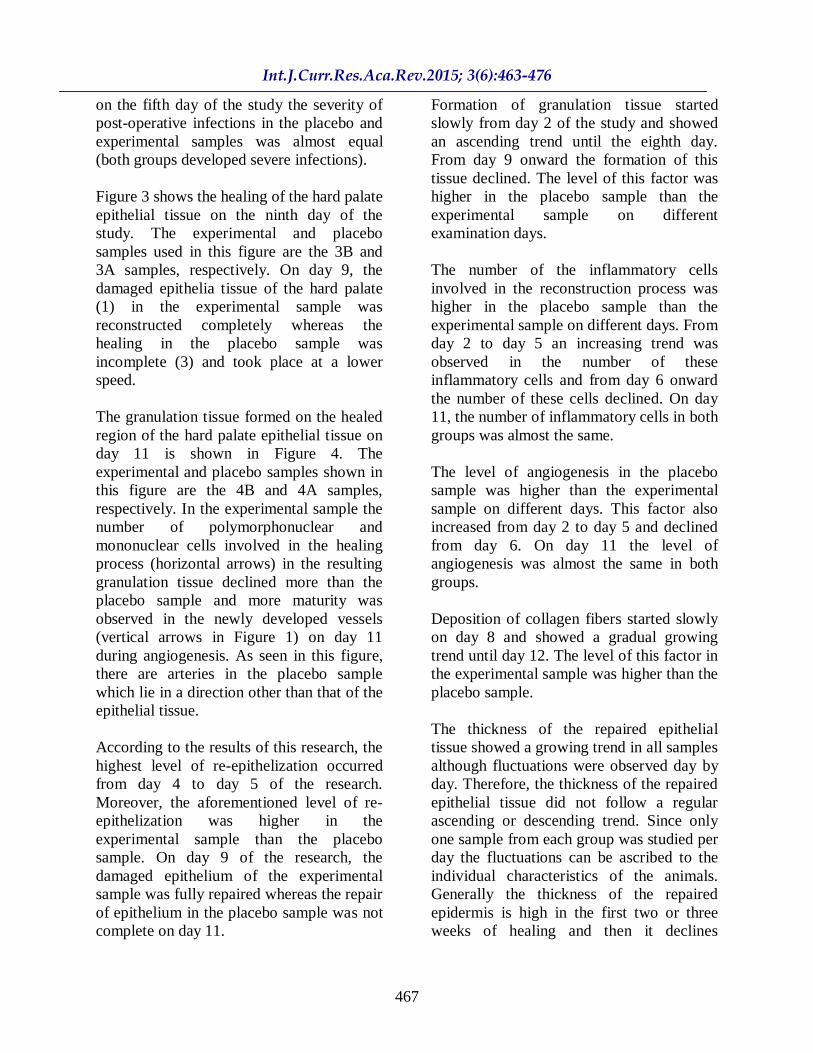
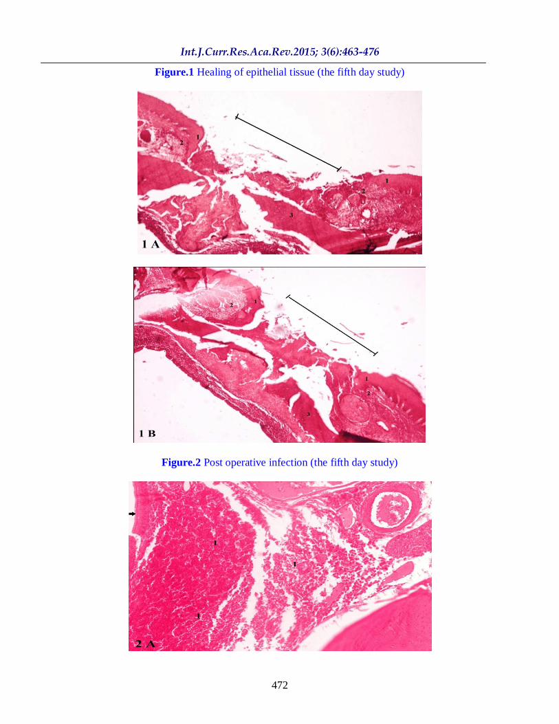
The healing of the epithelial tissues of the
hard palate on the fifth day is shown in
Figure -1. The experimental and placebo
(control) samples shown in this figure are
the 1B and 1A samples, respectively.
Formation of a new epidermis started on day
2 (1) and the epidermis of the two groups
had grown adequately on day 5. On the fifth
day, a slight difference was observed
between the untreated areas (the diagonal
line) in the experimental sample as
compared to the placebo sample.
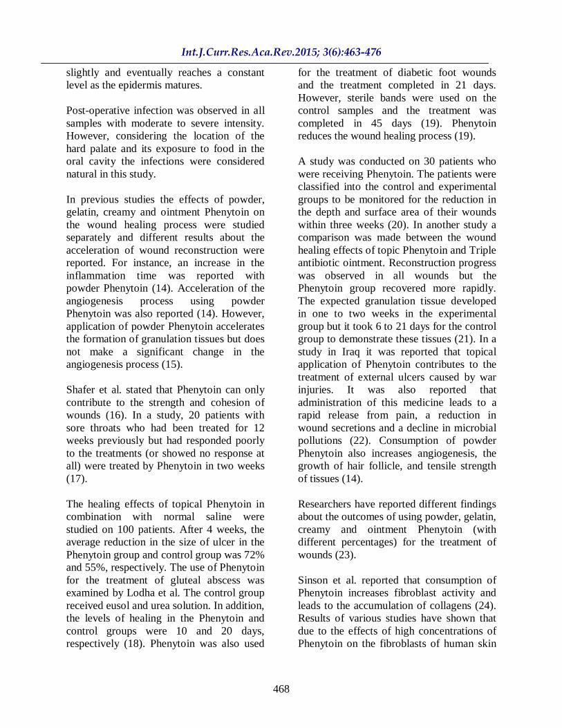
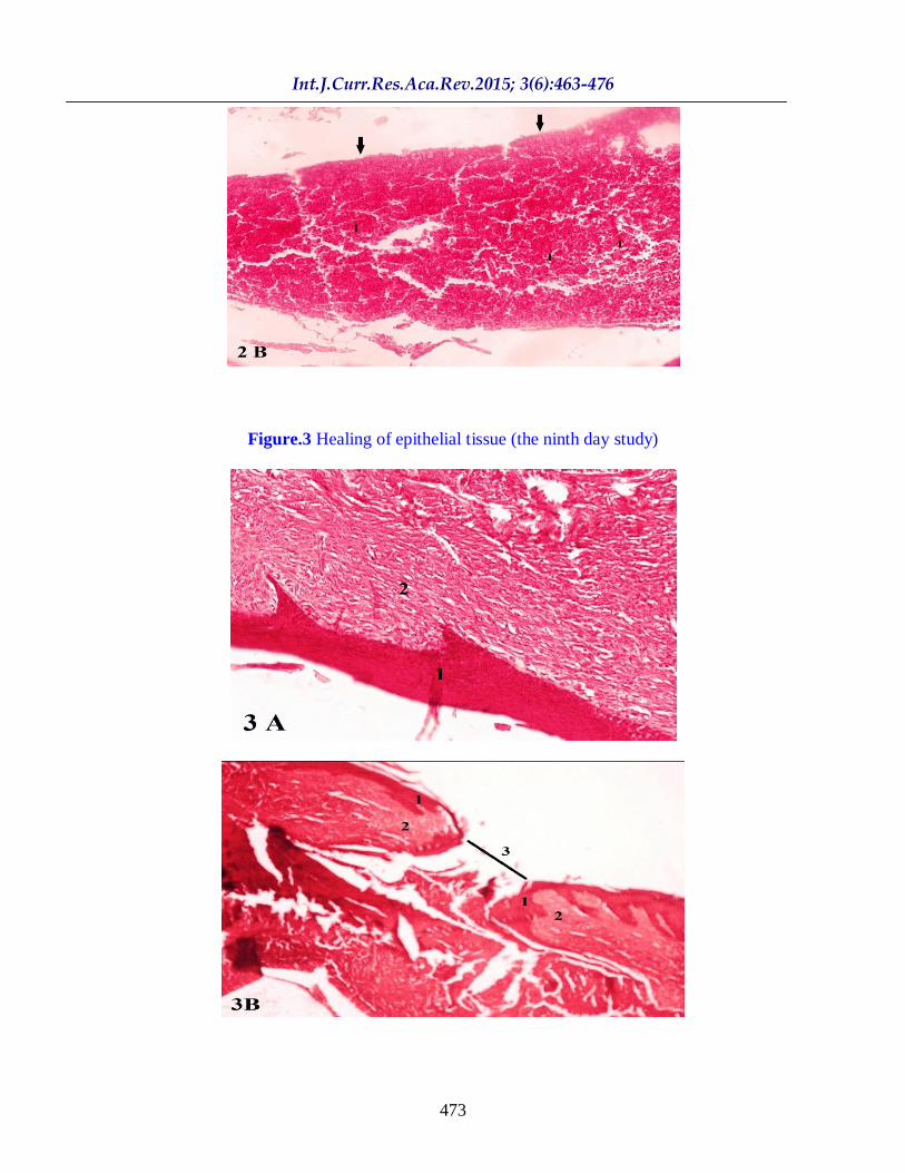
The post-operative infection on the fifth day
of the test is shown in Figure 2. The
experimental and placebo samples shown in
this image are the 2B and 2A samples,
respectively. The invasion of the
surroundings of the pseudo-stratified
cylindrical epithelial tissue (the arrow) of
the nasal bone by inflammatory cells in all
samples and on all days (with varying
severities) led to the emergence of post-
operative infections. As seen in this figure,
Int.J.Curr.Res.Aca.Rev.2015; 3(6):463-476
467
on the fifth day of the study the severity of
post-operative infections in the placebo and
experimental samples was almost equal
(both groups developed severe infections).
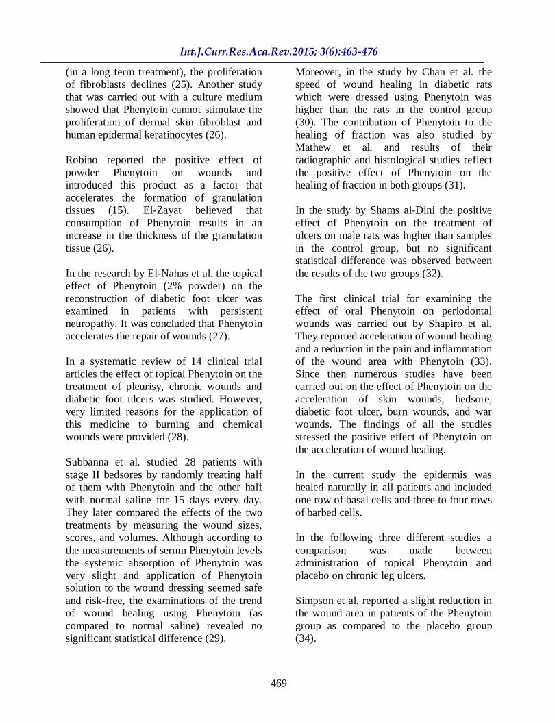
Figure 3 shows the healing of the hard palate
epithelial tissue on the ninth day of the
study. The experimental and placebo
samples used in this figure are the 3B and
3A samples, respectively. On day 9, the
damaged epithelia tissue of the hard palate
(1) in the experimental sample was
reconstructed completely whereas the
healing in the placebo sample was
incomplete (3) and took place at a lower
speed.
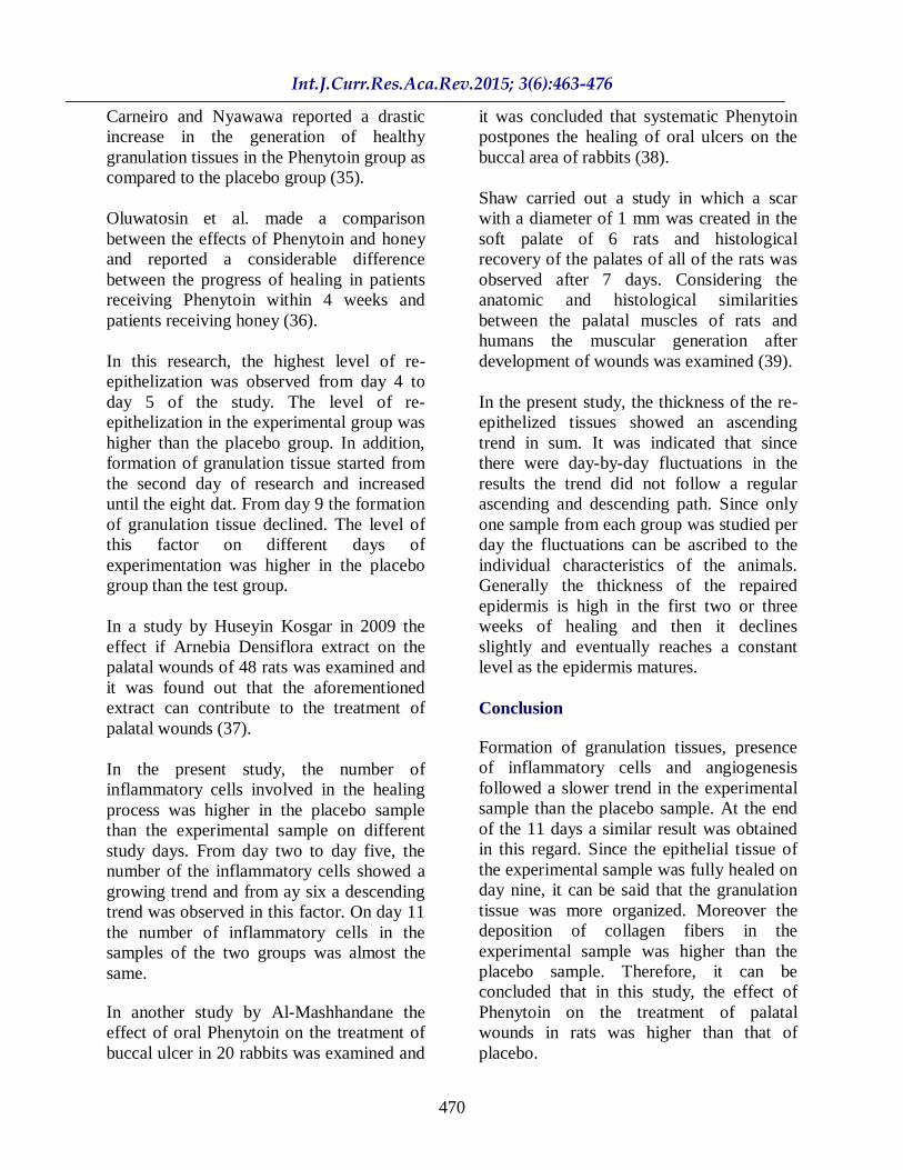
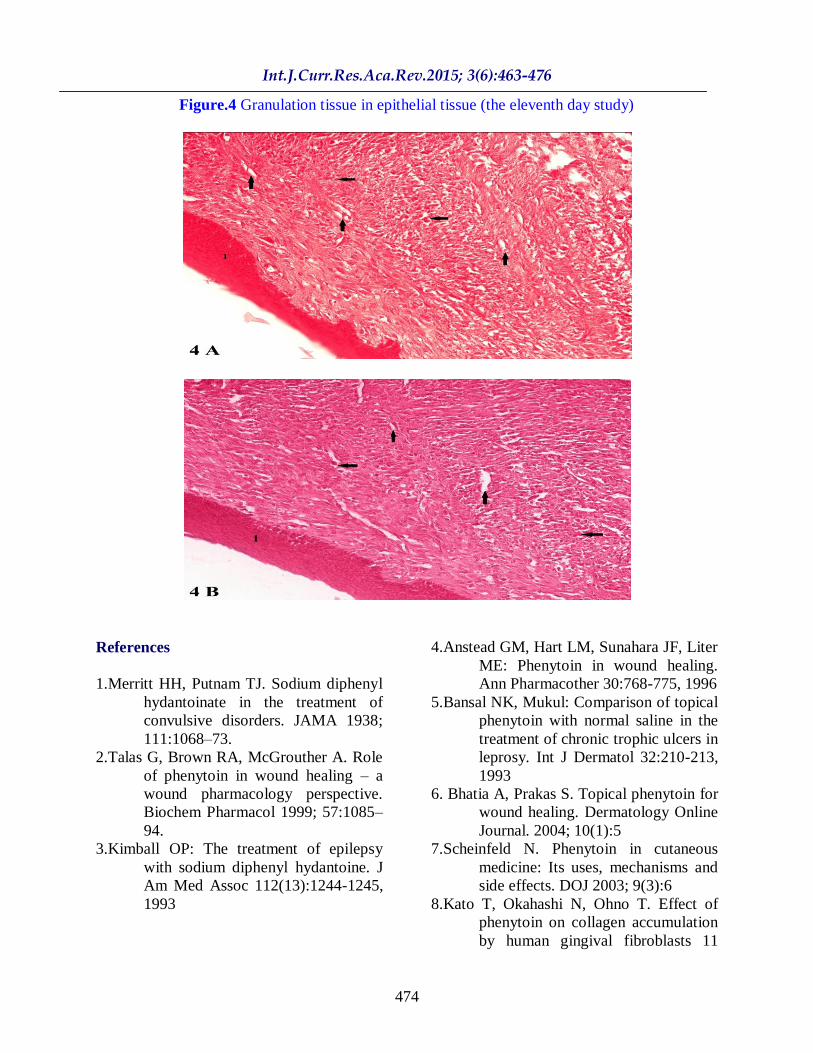
The granulation tissue formed on the healed
region of the hard palate epithelial tissue on
day 11 is shown in Figure 4. The
experimental and placebo samples shown in
this figure are the 4B and 4A samples,
respectively. In the experimental sample the
number of polymorphonuclear and
mononuclear cells involved in the healing
process (horizontal arrows) in the resulting
granulation tissue declined more than the
placebo sample and more maturity was
observed in the newly developed vessels
(vertical arrows in Figure 1) on day 11
during angiogenesis. As seen in this figure,
there are arteries in the placebo sample
which lie in a direction other than that of the
epithelial tissue.
According to the results of this research, the
highest level of re-epithelization occurred
from day 4 to day 5 of the research.
Moreover, the aforementioned level of re-
epithelization was higher in the
experimental sample than the placebo
sample. On day 9 of the research, the
damaged epithelium of the experimental
sample was fully repaired whereas the repair
of epithelium in the placebo sample was not
complete on day 11.
Formation of granulation tissue started
slowly from day 2 of the study and showed
an ascending trend until the eighth day.
From day 9 onward the formation of this
tissue declined. The level of this factor was
higher in the placebo sample than the
experimental sample on different
examination days.
The number of the inflammatory cells
involved in the reconstruction process was
higher in the placebo sample than the
experimental sample on different days. From
day 2 to day 5 an increasing trend was
observed in the number of these
inflammatory cells and from day 6 onward
the number of these cells declined. On day
11, the number of inflammatory cells in both
groups was almost the same.
The level of angiogenesis in the placebo
sample was higher than the experimental
sample on different days. This factor also
increased from day 2 to day 5 and declined
from day 6. On day 11 the level of
angiogenesis was almost the same in both
groups.
Deposition of collagen fibers started slowly
on day 8 and showed a gradual growing
trend until day 12. The level of this factor in
the experimental sample was higher than the
placebo sample.
The thickness of the repaired epithelial
tissue showed a growing trend in all samples
although fluctuations were observed day by
day. Therefore, the thickness of the repaired
epithelial tissue did not follow a regular
ascending or descending trend. Since only
one sample from each group was studied per
day the fluctuations can be ascribed to the
individual characteristics of the animals.
Generally the thickness of the repaired
epidermis is high in the first two or three
weeks of healing and then it declines
Int.J.Curr.Res.Aca.Rev.2015; 3(6):463-476
468
slightly and eventually reaches a constant
level as the epidermis matures.
Post-operative infection was observed in all
samples with moderate to severe intensity.
However, considering the location of the
hard palate and its exposure to food in the
oral cavity the infections were considered
natural in this study.
In previous studies the effects of powder,
gelatin, creamy and ointment Phenytoin on
the wound healing process were studied
separately and different results about the
acceleration of wound reconstruction were
reported. For instance, an increase in the
inflammation time was reported with
powder Phenytoin (14). Acceleration of the
angiogenesis process using powder
Phenytoin was also reported (14). However,
application of powder Phenytoin accelerates
the formation of granulation tissues but does
not make a significant change in the
angiogenesis process (15).
Shafer et al. stated that Phenytoin can only
contribute to the strength and cohesion of
wounds (16). In a study, 20 patients with
sore throats who had been treated for 12
weeks previously but had responded poorly
to the treatments (or showed no response at
all) were treated by Phenytoin in two weeks
(17).
The healing effects of topical Phenytoin in
combination with normal saline were
studied on 100 patients. After 4 weeks, the
average reduction in the size of ulcer in the
Phenytoin group and control group was 72%
and 55%, respectively. The use of Phenytoin
for the treatment of gluteal abscess was
examined by Lodha et al. The control group
received eusol and urea solution. In addition,
the levels of healing in the Phenytoin and
control groups were 10 and 20 days,
respectively (18). Phenytoin was also used
for the treatment of diabetic foot wounds
and the treatment completed in 21 days.
However, sterile bands were used on the
control samples and the treatment was
completed in 45 days (19). Phenytoin
reduces the wound healing process (19).
A study was conducted on 30 patients who
were receiving Phenytoin. The patients were
classified into the control and experimental
groups to be monitored for the reduction in
the depth and surface area of their wounds
within three weeks (20). In another study a
comparison was made between the wound
healing effects of topic Phenytoin and Triple
antibiotic ointment. Reconstruction progress
was observed in all wounds but the
Phenytoin group recovered more rapidly.
The expected granulation tissue developed
in one to two weeks in the experimental
group but it took 6 to 21 days for the control
group to demonstrate these tissues (21). In a
study in Iraq it was reported that topical
application of Phenytoin contributes to the
treatment of external ulcers caused by war
injuries. It was also reported that
administration of this medicine leads to a
rapid release from pain, a reduction in
wound secretions and a decline in microbial
pollutions (22). Consumption of powder
Phenytoin also increases angiogenesis, the
growth of hair follicle, and tensile strength
of tissues (14).
Researchers have reported different findings
about the outcomes of using powder, gelatin,
creamy and ointment Phenytoin (with
different percentages) for the treatment of
wounds (23).
Sinson et al. reported that consumption of
Phenytoin increases fibroblast activity and
leads to the accumulation of collagens (24).
Results of various studies have shown that
due to the effects of high concentrations of
Phenytoin on the fibroblasts of human skin
Int.J.Curr.Res.Aca.Rev.2015; 3(6):463-476
469
(in a long term treatment), the proliferation
of fibroblasts declines (25). Another study
that was carried out with a culture medium
showed that Phenytoin cannot stimulate the
proliferation of dermal skin fibroblast and
human epidermal keratinocytes (26).
Robino reported the positive effect of
powder Phenytoin on wounds and
introduced this product as a factor that
accelerates the formation of granulation
tissues (15). El-Zayat believed that
consumption of Phenytoin results in an
increase in the thickness of the granulation
tissue (26).
In the research by El-Nahas et al. the topical
effect of Phenytoin (2% powder) on the
reconstruction of diabetic foot ulcer was
examined in patients with persistent
neuropathy. It was concluded that Phenytoin
accelerates the repair of wounds (27).
In a systematic review of 14 clinical trial
articles the effect of topical Phenytoin on the
treatment of pleurisy, chronic wounds and
diabetic foot ulcers was studied. However,
very limited reasons for the application of
this medicine to burning and chemical
wounds were provided (28).
Subbanna et al. studied 28 patients with
stage II bedsores by randomly treating half
of them with Phenytoin and the other half
with normal saline for 15 days every day.
They later compared the effects of the two
treatments by measuring the wound sizes,
scores, and volumes. Although according to
the measurements of serum Phenytoin levels
the systemic absorption of Phenytoin was
very slight and application of Phenytoin
solution to the wound dressing seemed safe
and risk-free, the examinations of the trend
of wound healing using Phenytoin (as
compared to normal saline) revealed no
significant statistical difference (29).
Moreover, in the study by Chan et al. the
speed of wound healing in diabetic rats
which were dressed using Phenytoin was
higher than the rats in the control group
(30). The contribution of Phenytoin to the
healing of fraction was also studied by
Mathew et al. and results of their
radiographic and histological studies reflect
the positive effect of Phenytoin on the
healing of fraction in both groups (31).
In the study by Shams al-Dini the positive
effect of Phenytoin on the treatment of
ulcers on male rats was higher than samples
in the control group, but no significant
statistical difference was observed between
the results of the two groups (32).
The first clinical trial for examining the
effect of oral Phenytoin on periodontal
wounds was carried out by Shapiro et al.
They reported acceleration of wound healing
and a reduction in the pain and inflammation
of the wound area with Phenytoin (33).
Since then numerous studies have been
carried out on the effect of Phenytoin on the
acceleration of skin wounds, bedsore,
diabetic foot ulcer, burn wounds, and war
wounds. The findings of all the studies
stressed the positive effect of Phenytoin on
the acceleration of wound healing.
In the current study the epidermis was
healed naturally in all patients and included
one row of basal cells and three to four rows
of barbed cells.
In the following three different studies a
comparison was made between
administration of topical Phenytoin and
placebo on chronic leg ulcers.
Simpson et al. reported a slight reduction in
the wound area in patients of the Phenytoin
group as compared to the placebo group
(34).
Int.J.Curr.Res.Aca.Rev.2015; 3(6):463-476
470
Carneiro and Nyawawa reported a drastic
increase in the generation of healthy
granulation tissues in the Phenytoin group as
compared to the placebo group (35).
Oluwatosin et al. made a comparison
between the effects of Phenytoin and honey
and reported a considerable difference
between the progress of healing in patients
receiving Phenytoin within 4 weeks and
patients receiving honey (36).
In this research, the highest level of re-
epithelization was observed from day 4 to
day 5 of the study. The level of re-
epithelization in the experimental group was
higher than the placebo group. In addition,
formation of granulation tissue started from
the second day of research and increased
until the eight dat. From day 9 the formation
of granulation tissue declined. The level of
this factor on different days of
experimentation was higher in the placebo
group than the test group.
In a study by Huseyin Kosgar in 2009 the
effect if Arnebia Densiflora extract on the
palatal wounds of 48 rats was examined and
it was found out that the aforementioned
extract can contribute to the treatment of
palatal wounds (37).
In the present study, the number of
inflammatory cells involved in the healing
process was higher in the placebo sample
than the experimental sample on different
study days. From day two to day five, the
number of the inflammatory cells showed a
growing trend and from ay six a descending
trend was observed in this factor. On day 11
the number of inflammatory cells in the
samples of the two groups was almost the
same.
In another study by Al-Mashhandane the
effect of oral Phenytoin on the treatment of
buccal ulcer in 20 rabbits was examined and
it was concluded that systematic Phenytoin
postpones the healing of oral ulcers on the
buccal area of rabbits (38).
Shaw carried out a study in which a scar
with a diameter of 1 mm was created in the
soft palate of 6 rats and histological
recovery of the palates of all of the rats was
observed after 7 days. Considering the
anatomic and histological similarities
between the palatal muscles of rats and
humans the muscular generation after
development of wounds was examined (39).
In the present study, the thickness of the re-
epithelized tissues showed an ascending
trend in sum. It was indicated that since
there were day-by-day fluctuations in the
results the trend did not follow a regular
ascending and descending path. Since only
one sample from each group was studied per
day the fluctuations can be ascribed to the
individual characteristics of the animals.
Generally the thickness of the repaired
epidermis is high in the first two or three
weeks of healing and then it declines
slightly and eventually reaches a constant
level as the epidermis matures.
Conclusion
Formation of granulation tissues, presence
of inflammatory cells and angiogenesis
followed a slower trend in the experimental
sample than the placebo sample. At the end
of the 11 days a similar result was obtained
in this regard. Since the epithelial tissue of
the experimental sample was fully healed on
day nine, it can be said that the granulation
tissue was more organized. Moreover the
deposition of collagen fibers in the
experimental sample was higher than the
placebo sample. Therefore, it can be
concluded that in this study, the effect of
Phenytoin on the treatment of palatal
wounds in rats was higher than that of
placebo.
Int.J.Curr.Res.Aca.Rev.2015; 3(6):463-476
471
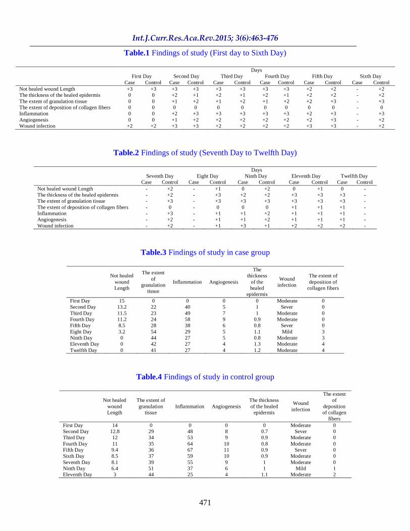
Table.1 Findings of study (First day to Sixth Day)
Days
First Day Second Day Third Day Fourth Day Fifth Day Sixth Day
Case Control Case Control Case Control Case Control Case Control Case Control
Not healed wound Length +3 +3 +3 +3 +3 +3 +3 +3 +2 +2 - +2
The thickness of the healed epidermis 0 0 +2 +1 +2 +1 +2 +1 +2 +2 - +2
The extent of granulation tissue 0 0 +1 +2 +1 +2 +1 +2 +2 +3 - +3
The extent of deposition of collagen fibers 0 0 0 0 0 0 0 0 0 0 - 0
Inflammation 0 0 +2 +3 +3 +3 +3 +3 +2 +3 - +3
Angiogenesis 0 0 +1 +2 +2 +2 +2 +2 +2 +3 - +2
Wound infection +2 +2 +3 +3 +2 +2 +2 +2 +3 +3 - +2
Table.2 Findings of study (Seventh Day to Twelfth Day)
Days
Seventh Day Eight Day Ninth Day Eleventh Day Twelfth Day
Case Control Case Control Case Control Case Control Case Control
Not healed wound Length - +2 - +1 0 +2 0 +1 0 -
The thickness of the healed epidermis - +2 - +3 +2 +2 +3 +3 +3 -
The extent of granulation tissue - +3 - +3 +3 +3 +3 +3 +3 -
The extent of deposition of collagen fibers - 0 - 0 0 0 +1 +1 +1 -
Inflammation - +3 - +1 +1 +2 +1 +1 +1 -
Angiogenesis - +2 - +1 +1 +2 +1 +1 +1 -
Wound infection - +2 - +1 +3 +1 +2 +2 +2 -
Table.3 Findings of study in case group
Not healed
wound
Length
The extent
of
granulation
tissue
Inflammation Angiogenesis
The
thickness
of the
healed
epidermis
Wound
infection
The extent of
deposition of
collagen fibers
First Day 15 0 0 0 0 Moderate 0
Second Day 13.2 22 40 5 1 Sever 0
Third Day 11.5 23 49 7 1 Moderate 0
Fourth Day 11.2 24 58 9 0.9 Moderate 0
Fifth Day 8.5 28 38 6 0.8 Sever 0
Eight Day 3.2 54 29 5 1.1 Mild 3
Ninth Day 0 44 27 5 0.8 Moderate 3
Eleventh Day 0 42 27 4 1.3 Moderate 4
Twelfth Day 0 41 27 4 1.2 Moderate 4
Table.4 Findings of study in control group
Not healed
wound
Length
The extent of
granulation
tissue
Inflammation Angiogenesis
The thickness
of the healed
epidermis
Wound
infection
The extent
of
deposition
of collagen
fibers
First Day 14 0 0 0 0 Moderate 0
Second Day 12.8 29 48 8 0.7 Sever 0
Third Day 12 34 53 9 0.9 Moderate 0
Fourth Day 11 35 64 10 0.8 Moderate 0
Fifth Day 9.4 36 67 11 0.9 Sever 0
Sixth Day 8.5 37 59 10 0.9 Moderate 0
Seventh Day 8.1 39 55 9 1 Moderate 0
Ninth Day 6.4 51 37 6 1 Mild 1
Eleventh Day 3 44 25 4 1.1 Moderate 2
Int.J.Curr.Res.Aca.Rev.2015; 3(6):463-476
472
Figure.1 Healing of epithelial tissue (the fifth day study)
Figure.2 Post operative infection (the fifth day study)
Int.J.Curr.Res.Aca.Rev.2015; 3(6):463-476
473
Figure.3 Healing of epithelial tissue (the ninth day study)
Int.J.Curr.Res.Aca.Rev.2015; 3(6):463-476
474
Figure.4 Granulation tissue in epithelial tissue (the eleventh day study)
References
1.Merritt HH, Putnam TJ. Sodium diphenyl
hydantoinate in the treatment of
convulsive disorders. JAMA 1938;
111:1068–73.
2.Talas G, Brown RA, McGrouther A. Role
of phenytoin in wound healing – a
wound pharmacology perspective.
Biochem Pharmacol 1999; 57:1085–
94.
3.Kimball OP: The treatment of epilepsy
with sodium diphenyl hydantoine. J
Am Med Assoc 112(13):1244-1245,
1993
4.Anstead GM, Hart LM, Sunahara JF, Liter
ME: Phenytoin in wound healing.
Ann Pharmacother 30:768-775, 1996
5.Bansal NK, Mukul: Comparison of topical
phenytoin with normal saline in the
treatment of chronic trophic ulcers in
leprosy. Int J Dermatol 32:210-213,
1993
6. Bhatia A, Prakas S. Topical phenytoin for
wound healing. Dermatology Online
Journal. 2004; 10(1):5
7.Scheinfeld N. Phenytoin in cutaneous
medicine: Its uses, mechanisms and
side effects. DOJ 2003; 9(3):6
8.Kato T, Okahashi N, Ohno T. Effect of
phenytoin on collagen accumulation
by human gingival fibroblasts 11
Int.J.Curr.Res.Aca.Rev.2015; 3(6):463-476
475
exposed to TNF- in vitro. Oral
Diseases, 2006; 12: 156- 162.
9.Modaghegh S, Salehian B, Tavassoli M, et
al. Use of phenytoin in healing of
war and non-war wounds. A pilot
study of 25 cases. Int J Dermatol
1989; 28:347-350.
10.Rhodes RS, Heyneman CA, Culbertson
VL, Wilson SE, Phatak HM. Topical
phenytoin treatment of stage II
decubitus ulcers in the elderly. Ann
Pharmacother 2001; 35:675-81.
11.Anstead GM, Hart LM, Sunahara JF,
Liter ME. Phenytoin in wound
healing. Ann Pharmacol 1996;
30:768-75.
12.Muthukumarasamy MG, Sivakumar G,
Manoharan G. Topical phenytoin in
diabetic foot ulcers. Diabetes Care
1991; 14:909-11.
13.Pendse AK, Sharma A, Sodani A, Hada
S. Topical phenytoin in wound
healing. Int J Dermatol 1993;
32:214-7.
14.Julia V, Dovi, Li-ke H, Louisa A,
Dipietro. Accelerated wound clouser
in neutrophil depleted mice. Journal
of leukocyte Biology 2003; 73:448-
55.
15.Kumar A, Michel F. Robbins basic
pathology. Tehran: Andisheh Rafih;
1387.
16.Helen J, Jonathan A. Keratinocyte
growth factor signaling:
Mathematical Biosciences 2000;
165:41-65.
17.Brown M, Perm P, Georgia R, Core S.
High voltage galvanic stimulation on
wound healing in guinea pigs: longer
term effect. Arch phys Med Rehabil
1995; 76: 1134-7.
18.Davidson J. Animal models for wound
repair. Arch Dermato Res 1998; 290
(supll):s1-s11.
19.Wayne K. Stadelman M, Alexander G.
Physiology and Healing Dynamics
Of chronic coetaneous Wounds. Am
J Surge 1998; 176: (suppl).
20.Izadyar B. Effect of fandermol ointment
on skin wound healing in male rat in
compare with normal saline,
Baghyatallh University; 1374.
21.Ehrlich HP, Keefer KA, Myers RL,
Passaniti A. Vanadate and the
absence of myofibroblasts in wound
contraction. Arch Surg 1999;
134(5):494-501.
22.Ehrlich HP, Buttle DJ. Comparative
studies of collagen lattice contraction
utilizing a normal and a transformed
cell line. J Cell Physiol 1983;
116(2):159-66.
23.Ehrlich HP, Rittenberg T. Free fatty acids
and dialyzed serum alterations of
fibroblast pulated collagen lattice
contraction. Tissue Cell 1992;
24(2):243-51.
24.Michael R, Udaya T, Gret M. Stimulation
of fibroblast proliferation and Matrix
Contraction by Wound Fluids. J
Biochem & cell Boil 1997;
29(1):231-39.
25.Noah S. Phenytoin in cutaneous
medicine: Its uses, mechanisms and
side effects. Dermatology on line
Journal; 9(3):6.
26.Thuy Vo, Pharm D. Topical phenytoin
for wound healing. Phrmacy News
Let 2001; 21: 5.
27.Scheinfeld N. Phenytoin in cutaneous
medicin: Its uses, mechnisms and
side effects. Dermatol On line 2004;
9(3): 6-15.
28.Turan M, Saraydyn SU, Bulut HE,
Elagoz S, Cetinkaya O, Karadayi K,
Canbay E, Sen M. Do vascular
endothelial growth factor and basic
fibroblast growth factor promote
phenytoins wound healing effect in
rat? An immunohistochemical and
histophatologic study. 2004; J
Dermatol Surg, 30(10): 1303-9.
Int.J.Curr.Res.Aca.Rev.2015; 3(6):463-476
476
29.Dacosta ML, Regan MC, Alsader M,
Leader M, Bouchier Hayes D.
Diphenylhydantion sodium promotes
early and marked angiogenesis and
results in creased collagen deposition
and tensile strength in healing
Wounds. J Surg 1998; 123(3):287-
93.
30.Wound healing (surgical wounds,
trauma, burns). Internet, http://
www.lef. Org/prerocols/prtcl-111.
html 2000; pp:1-5.
31.Michele H. Cameron. Physical agent in
rehabilitaion, W. B. Saunders Co
1999; pp: 126-173, 453-6, 1-38.
32.Albsoul-Younes A, Younes NA, Badran
DH. Topical phenytoin ointment
increases autograft acceptance in
rats. Saudi Med J 2006; 27(7): 962-6.
33.Shaw J, Hughes GM, Langan KM, Bell
PM. The clinical effect of topical
phenytoin on wound healing: a
systematic review. Br J Dermatol
2007; 157(5): 997-1004.
34.Chan FC, Kennedy C, Hanson RP,
O'Sullivan B, Kelly J, Bouchier-
Hayes D. Topical phenytion can
improve healing in a diabetic
incision animal wound model. J
Wound Care 2007; 16(8): 359-63.
35.Mehrvarz Sh, Tahmasebi MH, Asgari
AR. Effect of phenytoin powder on
open wound healing process in rat
skin. Kowsar Medical Journal 1998;
3(3): 177-82.
36.Pitiakoudis M, Giatromanolaki A,
Iliopoulos I, Tsaroucha AK,
Simopoulo SC, and Piperidou C.
Phenytoin induced lymphocytic
chemotaxixis angiogenesis and
accelerated healing of decubitus
ulcer in a patient with stroke. J Int
Med Res 2004 32(2): 201-5.
37.Rabio J. Diphenylhydantion in leprosy,
presented to the xII congress of
Dermatology, ooza, mexico. 1985,
pp 9-12.
38.Shafer W. Beatty R, Davis W. Effect of
dilantin sodium on tensile strength of
healing wounds. Proc.soc, Exp, Biol
1985; 98:348-50.
39.Oluwatsin OM, Olaabanji JK, Tijani LA.
A comparison of topical honey and
phenytoin in the treatment of chronic
ulcers. Afr J Med science 2000;
29(1):31-4.