Wilberforce Road, Cambridge, CB3 0WA, United Kingdom · Wilberforce Road, Cambridge, CB3 0WA,...
17
Bacterial Hydrodynamics * Eric Lauga † Department of Applied Mathematics and Theoretical Physics, Centre for Mathematical Sciences, University of Cambridge, Wilberforce Road, Cambridge, CB3 0WA, United Kingdom (Dated: September 9, 2015) Bacteria predate plants and animals by billions of years. Today, they are the world’s smallest cells yet they represent the bulk of the world’s biomass, and the main reservoir of nutrients for higher organisms. Most bacteria can move on their own, and the majority of motile bacteria are able to swim in viscous fluids using slender helical appendages called flagella. Low-Reynolds- number hydrodynamics is at the heart of the ability of flagella to generate propulsion at the micron scale. In fact, fluid dynamic forces impact many aspects of bacteriology, ranging from the ability of cells to reorient and search their surroundings to their interactions within mechanically and chemically-complex environments. Using hydrodynamics as an organizing framework, we review the biomechanics of bacterial motility and look ahead to future challenges. I. INTRODUCTION Bacteria constitute the bulk of the biomass of our planet. However, since we need a microscope to see them, we often forget their presence. Observing life in the ocean, we see fish and crustaceans, but miss the marine bacteria that outnumber them many times over. On land, we see animals and plants, but often forget that a hu- man body contains many more bacteria than mammalian cells. While they are responsible for many infectious diseases, bacteria play a critical role in the life of soils and higher organisms (including humans) by perform- ing chemical reactions and providing nutrients (Madigan et al., 2010). Ever since microscopes were invented, scientists have worked to decipher the rules dictating the behavior of bacteria. In addition to quantifying the manner in which bacteria shape our lives and teach us about our evolu- tionary history, biologists have long recognized that the behavior of bacteria is influenced by the physical con- straints of their habitat, and by their evolution to maxi- mize fitness under these constraints. The most prominent of these constraints is the presence of a surrounding fluid (Vogel, 1996). Not only are all bacteria found in fluids, but being typically less than a hundredth of a millime- ter in length, they experience much larger viscous forces than inertial forces, and have adapted their behavior in response to these forces. Most bacteria are motile (Jarrell & McBride, 2008; Kearns, 2010) and this motility is essential for the effec- tiveness of many pathogens (Ottemann & Miller, 1997). The most common form of bacterial motility is swim- ming. In order to swim, bacteria have evolved flag- ella (originally from secretory systems), which are slen- der helical appendages rotated by specialized motors, and whose motions in viscous environments propel the * to appear in: Annu. Rev. Fluid Mech. 48 (2016) † [email protected] cells forward (Bray, 2000). The surrounding fluid can be seen both as a constraint and an advantage. It is the presence of the fluid itself and its interactions with three-dimensional bacterial flagellar filaments which al- lows cells to move and sample their chemical environ- ment – a crucial step for the cells to display robust and adaptive chemotactic responses to both attractants and repellents, for nutrients, but also temperature, pH, and viscosity (Berg, 1993, 2004; Blair, 1995; Purcell, 1977). But as they self-propel, bacteria are subject to the external constraints set by the physical world, and in particular by hydrodynamics. In this review, we high- light the consequences of fluid dynamics relevant to the swimming of bacteria in viscous environments. Natu- rally, the biophysics of bacteria locomotion involves many aspects of soft matter physics, including nonlinear elas- ticity, screened electrostatics, and biochemical noise, and hence fluid dynamics represents but one feature of the complex balance of forces dictating the behavior of cells as they search their environment. The choice made here is to use hydrodynamics as an organizing framework to overview various aspects of cellular locomotion. We start by a biological overview of bacteria as cells, their geometry, the way they swim, and the variations among different species (§II). We then review the basic hydrodynamics features of flagellar propulsion (§III) fol- lowed by the flows induced by swimming bacteria (§IV). We next address the flagellar polymorphs, their compar- ative hydrodynamics, and the potential relevance to evo- lution of the flagellum (§V). With individual flagella un- derstood, we detail how fluid forces may play a role in the actuation of multiple flagella and be used by cells to reorient (§VI). The last three sections are devoted to lo- comotion near surfaces (§VII), in external flows (§VIII) and in complex fluids (§IX). Throughout this review, the focus will be on single- cell behavior. Our purpose is to understand the way a single bacterium exploits, and is subject to, the physi- cal constraints from the surrounding fluid. Other use- ful reviews can be used as complementary reading, in particular those highlighting the hydrodynamics of low- arXiv:1509.02184v1 [physics.flu-dyn] 7 Sep 2015
Transcript of Wilberforce Road, Cambridge, CB3 0WA, United Kingdom · Wilberforce Road, Cambridge, CB3 0WA,...
Bacterial Hydrodynamics∗
Eric Lauga†
Department of Applied Mathematics and Theoretical Physics, Centre for Mathematical Sciences, University of Cambridge,Wilberforce Road, Cambridge, CB3 0WA, United Kingdom
(Dated: September 9, 2015)
Bacteria predate plants and animals by billions of years. Today, they are the world’s smallestcells yet they represent the bulk of the world’s biomass, and the main reservoir of nutrients forhigher organisms. Most bacteria can move on their own, and the majority of motile bacteriaare able to swim in viscous fluids using slender helical appendages called flagella. Low-Reynolds-number hydrodynamics is at the heart of the ability of flagella to generate propulsion at the micronscale. In fact, fluid dynamic forces impact many aspects of bacteriology, ranging from the abilityof cells to reorient and search their surroundings to their interactions within mechanically andchemically-complex environments. Using hydrodynamics as an organizing framework, we reviewthe biomechanics of bacterial motility and look ahead to future challenges.
I. INTRODUCTION
Bacteria constitute the bulk of the biomass of ourplanet. However, since we need a microscope to seethem, we often forget their presence. Observing life in theocean, we see fish and crustaceans, but miss the marinebacteria that outnumber them many times over. On land,we see animals and plants, but often forget that a hu-man body contains many more bacteria than mammaliancells. While they are responsible for many infectiousdiseases, bacteria play a critical role in the life of soilsand higher organisms (including humans) by perform-ing chemical reactions and providing nutrients (Madiganet al., 2010).
Ever since microscopes were invented, scientists haveworked to decipher the rules dictating the behavior ofbacteria. In addition to quantifying the manner in whichbacteria shape our lives and teach us about our evolu-tionary history, biologists have long recognized that thebehavior of bacteria is influenced by the physical con-straints of their habitat, and by their evolution to maxi-mize fitness under these constraints. The most prominentof these constraints is the presence of a surrounding fluid(Vogel, 1996). Not only are all bacteria found in fluids,but being typically less than a hundredth of a millime-ter in length, they experience much larger viscous forcesthan inertial forces, and have adapted their behavior inresponse to these forces.
Most bacteria are motile (Jarrell & McBride, 2008;Kearns, 2010) and this motility is essential for the effec-tiveness of many pathogens (Ottemann & Miller, 1997).The most common form of bacterial motility is swim-ming. In order to swim, bacteria have evolved flag-ella (originally from secretory systems), which are slen-der helical appendages rotated by specialized motors,and whose motions in viscous environments propel the
∗to appear in: Annu. Rev. Fluid Mech. 48 (2016)†[email protected]
cells forward (Bray, 2000). The surrounding fluid canbe seen both as a constraint and an advantage. It isthe presence of the fluid itself and its interactions withthree-dimensional bacterial flagellar filaments which al-lows cells to move and sample their chemical environ-ment – a crucial step for the cells to display robust andadaptive chemotactic responses to both attractants andrepellents, for nutrients, but also temperature, pH, andviscosity (Berg, 1993, 2004; Blair, 1995; Purcell, 1977).
But as they self-propel, bacteria are subject to theexternal constraints set by the physical world, and inparticular by hydrodynamics. In this review, we high-light the consequences of fluid dynamics relevant to theswimming of bacteria in viscous environments. Natu-rally, the biophysics of bacteria locomotion involves manyaspects of soft matter physics, including nonlinear elas-ticity, screened electrostatics, and biochemical noise, andhence fluid dynamics represents but one feature of thecomplex balance of forces dictating the behavior of cellsas they search their environment. The choice made hereis to use hydrodynamics as an organizing framework tooverview various aspects of cellular locomotion.
We start by a biological overview of bacteria as cells,their geometry, the way they swim, and the variationsamong different species (§II). We then review the basichydrodynamics features of flagellar propulsion (§III) fol-lowed by the flows induced by swimming bacteria (§IV).We next address the flagellar polymorphs, their compar-ative hydrodynamics, and the potential relevance to evo-lution of the flagellum (§V). With individual flagella un-derstood, we detail how fluid forces may play a role inthe actuation of multiple flagella and be used by cells toreorient (§VI). The last three sections are devoted to lo-comotion near surfaces (§VII), in external flows (§VIII)and in complex fluids (§IX).
Throughout this review, the focus will be on single-cell behavior. Our purpose is to understand the way asingle bacterium exploits, and is subject to, the physi-cal constraints from the surrounding fluid. Other use-ful reviews can be used as complementary reading, inparticular those highlighting the hydrodynamics of low-
arX
iv:1
509.
0218
4v1
[ph
ysic
s.fl
u-dy
n] 7
Sep
201
5

2
FIG. 1 Atlas of flagellated bacteria. (a) Peritrichous Escherichia coli (Howard Berg, Harvard University); (b) Polar monotric-hous Pseudomonas aeruginosa (Fujii et al., 2008); (c) Polar lophotrichous Photobacterium fischeri (Allen & Baumann, 1971);(d) Polar amphitrichous Ectothiorhodospira hatochtoris (Imhoff & Truper, 1977); (e) Spirochetes with endoflagella Borreliaburgdorferi (Goldstein et al., 1996). All figures reproduced with permission.
Reynolds number swimming (Brennen & Winet, 1977;Lauga & Powers, 2009; Lighthill, 1975), the fluid dy-namics of plankton (Goldstein, 2015; Guasto et al., 2012;Pedley & Kessler, 1992), reproduction (Fauci & Dillon,2006; Gaffney et al., 2011), and collective cell locomotion(Koch & Subramanian, 2011).
II. BIOLOGICAL ATLAS
Bacteria come in many different shapes (Madiganet al., 2010). Those able to swim in fluids can be roughlydivided into two categories: bacteria whose propulsionis driven by helical flagellar filaments located outside anon-deforming cell body (the overwhelming majority, il-lustrated in Fig. 1a-d) and those with spiral-like bodyundergoing time-varying deformation (Fig. 1e).
Our understanding of how flagellated bacteria swimhas its roots in a series of investigations in the 1970sshowing conclusively that – unlike the active flagella ofeukaryotes, which are active and muscle-like (Bray, 2000)– bacterial flagellar filaments are passive organelles (Berg& Anderson, 1973; Silverman & Simon, 1974). Typicallya few to ten microns in length, and only 40 nm in di-ameter, bacterial flagellar filaments are helical polymerswhose structure is discussed in detail in §V. Each fila-ment is attached to a short flexible hook (≈ 60 nm inlength, see Fig. 2) acting as a universal joint. The hookis turned by a stepper motor driven by ion fluxes (bac-terial rotary motor, Fig. 2) which is well characterized
molecularly (Berg, 2003).Some flagellated cells are called peritrichous and have
many motors (and thus many flagella) located randomlyat various positions on the cell body (Fig. 1a). This isthe case for the bacterium Escherichia coli (or E. coli),chosen as a model organism to study the fundamen-tal biophysical aspects of cell motility and chemotaxis(Berg, 2004). In contrast, polar bacteria have motorsonly located at poles of the body. Of the polar bacteria,monotrichous bacteria only have one motor, and thusone filament (Fig. 1b), lophotrichous cells have a tuft offlagella originating from a single pole (Fig. 1c) while am-phitrichous bacteria have flagella at each pole (Fig. 1d).Note that variations in this classification exist; for exam-ple, some monotrichous or lophotrichous bacteria are notpolar. In all cases, the rotation of the motor is transmit-ted to the hook, which in turn transmits it to the heli-cal filament, leading to the generation of hydrodynamicpropulsive forces, as explained in detail in §III.
Fluid dynamics was used to determine the torque-frequency relationship for the rotary motor by linkingtethered bacterial flagella to beads of different sizes influids of varying viscosity (Chen & Berg, 2000). Theresulting change in the Stokes resistance to bead rota-tion allowed to show that the rotary motor works atconstant torque for a wide range of frequencies in thecounter-clockwise (CCW) directions (the frequency rangedepends strongly on temperature) before showing a lineardecrease of the torque at higher frequencies (Berg, 2003).In contrast, when rotating in the clockwise (CW) direc-

3
FIG. 2 Schematic representation of a peritrichous bacterium with three flagellar filaments showing a bacterial rotary motorembedded in the cell wall (the stator part of the motor is not shown) (Berg, 2003) and a hook with its junction to a flagellarfilament (K. Namba, Osaka University). All figures reproduced with permission.
tion, the motor torque linearly decreases with frequency(Yuan et al., 2010).
In the range relevant to locomotion, the motor can beassumed to behave as continuously rotating and produc-ing a constant torque. The value of this torque is how-ever still under debate and measurements are at oddswith theoretical predictions. While its stall torque hasbeen experimentally estimated to be on the order of4, 600 pN nm (Berry & Berg, 1997), experiments us-ing beads lead to the conclusion that the motor hasto be on the range 1260 ± 190 pN nm (Reid et al.,2006). In contrast, theoretical estimates using a sim-plified model for the fluid predict a significantly smallervalue of 370± 100 pN nm (Darnton et al., 2007).
While the majority of swimming bacteria are poweredby flagellar filaments rotating in the fluid outside thecells, two other modes of locomotion in fluids are notable.Bacteria of the spirochete family have a spiral shape andundergo whole-body undulations powered also by flag-ella (Fig. 1e). However, in this case, the filaments areconstrained in the small space between the cytoplasmicmembrane and outer membrane of the cell body, and it isthe rotation in this tight space which leads to wave-likewhole-body undulations and in turn leads to locomotionin a fluid (Vig & Wolgemuth, 2012). Swimming with-out flagella has also been reported for Spiroplasma, atype of helical bacterium whose body has no cell wall.In that case, a wavelike motion is induced by contrac-tion of the cell cytoskeleton leading to the propagation ofshape kinks along the body (Shaevitz et al., 2005; Wada& Netz, 2007). At least one other bacterium, the marineSynechococcus, swims in fluids in manner which is as yetundetermined (Ehlers & Oster, 2012).
To close this biological overview, we mention the othermode of fluid-based motility termed swarming (Jarrell &McBride, 2008; Kearns, 2010). A few bacterial families,when present in media-rich in nutrients, can undergo adifferentiation to a swarming state, where they becomemore elongated, grow more flagella and swim in closely-packed groups, the physics of which is only beginning tobe unraveled (Copeland & Weibel, 2009; Darnton et al.,2010).
III. HYDRODYNAMICS OF HELICAL PROPULSION
The most fundamental aspect of bacterial hydrody-namics is the ability of rotating flagella to producepropulsive forces (Chwang & Wu, 1971; Higdon, 1979;Lighthill, 1976). At the origin of this generation of forcesis the non-isotropic nature of hydrodynamic friction inthe Stokesian regime (Brennen & Winet, 1977; Lauga& Powers, 2009). At the low Reynolds numbers rele-vant to swimming bacteria (typically ranging from 10−4
to 10−2), the flows are governed by the incompressibleStokes equations, and drag forces on rigid bodies scalelinearly with their instantaneous velocities relative to thebackground fluid. Because flagellar filaments are slender(their aspect ratio is at least 100), their physics can beintuitively understood by analogy with the motion of arod. At a given velocity, a rod experiences a drag fromthe fluid about twice as large when translating perpen-dicular to its long axis as that when moving along it. Fora non-symmetric velocity/rod orientation, the hydrody-namic drag on the rod is thus not aligned with the di-rection of its velocity but possesses a nonzero componentperpendicular to it – hence the possibility of drag-based
4
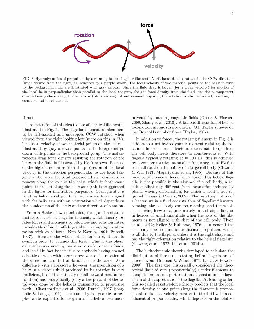
FIG. 3 Hydrodynamics of propulsion by a rotating helical flagellar filament. A left-handed helix rotates in the CCW direction(when viewed from the right) as indicated by a purple arrow. The local velocity of two material points on the helix relativeto the background fluid are illustrated with gray arrows. Since the fluid drag is larger (for a given velocity) for motion ofthe local helix perpendicular than parallel to the local tangent, the net force density from the fluid includes a componentdirected everywhere along the helix axis (black arrows). A net moment opposing the rotation is also generated, resulting incounter-rotation of the cell.
thrust.
The extension of this idea to case of a helical filament isillustrated in Fig. 3. The flagellar filament is taken hereto be left-handed and undergoes CCW rotation whenviewed from the right looking left (more on this in §V).The local velocity of two material points on the helix isillustrated by gray arrows: points in the foreground godown while points in the background go up. The instan-taneous drag force density resisting the rotation of thehelix in the fluid is illustrated by black arrows. Becauseof the higher resistance from the projection of the localvelocity in the direction perpendicular to the local tan-gent to the helix, the total drag includes a nonzero com-ponent along the axis of the helix, which in both casespoints to the left along the helix axis (this is exaggeratedin the figure for illustration purposes). Consequently, arotating helix is subject to a net viscous force alignedwith the helix axis with an orientation which depends onthe handedness of the helix and the direction of rotation.
From a Stokes flow standpoint, the grand resistancematrix for a helical flagellar filament, which linearly re-lates forces and moments to velocities and rotation rates,includes therefore an off-diagonal term coupling axial ro-tation with axial force (Kim & Karrila, 1991; Purcell,1997). Because the whole cell is force-free, it has toswim in order to balance this force. This is the physi-cal mechanism used by bacteria to self-propel in fluids,and it will in fact be intuitive to anybody having openeda bottle of wine with a corkscrew where the rotation ofthe screw induces its translation inside the cork. As adifference with a corkscrew however, the propulsion of ahelix in a viscous fluid produced by its rotation is veryinefficient, both kinematically (small forward motion perrotation) and energetically (only a few percent of the to-tal work done by the helix is transmitted to propulsivework) (Chattopadhyay et al., 2006; Purcell, 1997; Spag-nolie & Lauga, 2011). The same hydrodynamic princi-ples can be exploited to design artificial helical swimmers
powered by rotating magnetic fields (Ghosh & Fischer,2009; Zhang et al., 2010). A famous illustration of helicallocomotion in fluids is provided in G.I. Taylor’s movie onlow Reynolds number flows (Taylor, 1967).
In addition to forces, the rotating filament in Fig. 3 issubject to a net hydrodynamic moment resisting the ro-tation. In order for the bacterium to remain torque-free,the cell body needs therefore to counter-rotate. Withflagella typically rotating at ≈ 100 Hz, this is achievedby a counter-rotation at smaller frequency ≈ 10 Hz dueto small rotational mobility of a large cell body (Chwang& Wu, 1971; Magariyama et al., 1995). Because of thisbalance of moments, locomotion powered by helical flag-ella is not possible in the absence of a cell body, a re-sult qualitatively different from locomotion induced byplanar waving deformation, for which a head is not re-quired (Lauga & Powers, 2009). The resulting motion ofa bacterium in a fluid consists thus of flagellar filamentsrotating, the cell body counter-rotating, and the wholecell moving forward approximately in a straight line, orin helices of small amplitude when the axis of the fila-ments is not aligned with that of the cell body (Hyonet al., 2012; Keller & Rubinow, 1976). In general thecell body does not induce additional propulsion, whichis all due to the flagella, unless it is the right shape andhas the right orientation relative to the helical flagellum(Chwang et al., 1972; Liu et al., 2014b).
The hydrodynamic theories developed to calculate thedistribution of forces on rotating helical flagella are ofthree flavors (Brennen & Winet, 1977; Lauga & Powers,2009). The first one, historically, considered the theo-retical limit of very (exponentially) slender filaments tocompute forces as a perturbation expansion in the loga-rithm of the aspect ratio of the flagella. At leading order,this so-called resistive-force theory predicts that the localforce density at one point along the filament is propor-tional to its local velocity relative to the fluid with a co-efficient of proportionality which depends on the relative
5
orientation of the velocity with respect to the local tan-gent (Cox, 1970; Gray & Hancock, 1955; Lighthill, 1975).This is therefore the natural extension of the physicalpicture discussed above for rigid rods to deforming fila-ments. While this is the most widely used approach, andone which is is able to provide basic physical pictures, itis often not sufficiently accurate as the associated relativeerrors are only logarithmically small (Chattopadhyay &Wu, 2009; Johnson & Brokaw, 1979). More precise isthe second approach, termed slender-body theory, whichconsists in distributing appropriate flow singularities ofsuitable strengths in order to match the boundary con-dition on the surface of the filament at a required, alge-braically small, precision (Hancock, 1953; Johnson, 1980;Lighthill, 1976). In that case the equation relating thedistribution of velocity to the distribution of forces is non-local, because of long-range hydrodynamic interactions,and thus needs to be inverted numerically. A final op-tion is to use fully three-dimensional computations. Themost common method is an integration of the equationsof Stokes flows using either boundary elements (Liu et al.,2013; Phan-Thien et al., 1987), regularized flow singular-ities (Flores et al., 2005; Olson et al., 2013), or immersedboundaries (Fauci & Dillon, 2006). Mesoscopic particle-based methods have recently been proposed as an alter-native approach (Reigh et al., 2012).
IV. FLOWS INDUCED BY BACTERIA
The general fluid dynamics picture of a swimming bac-terium powered by the rotation of a flagellum is illus-trated in Fig. 4a. We use purple arrows to indicate the lo-cal forcing of the swimming cell on the surrounding fluid.Near the filament, the induced flow is a combination ofrotation and polar axial pumping directed away from thecell body. This is illustrated in Fig. 4b-c using slender-body theory for a helical waveform of diameter 400 nmand wavelength 2.3 µm (the so-called “normal” wave-form, see discussion in §V) (Spagnolie & Lauga, 2011).
Looking further away from the cell, because the swim-ming bacterium is force-free, it pushes on the fluid withan equal and opposite force to the one generated bythe rotating filament (Fig. 4a), and thus the leadingorder flow in the far-field is a force-dipole, or stresslet,as schematically illustrated in Fig. 4c (Batchelor, 1970;Ishikawa et al., 2007). The type of associated dipole istermed a pusher, in contrast with puller dipoles relevantto cells which swim flagella first, for example planktonicbi-flagellates (Guasto et al., 2012). This flow has beenmeasured for swimming E. coli using an experimentaltour-de-force (Drescher et al., 2011). The magnitude ofthe induced flow is shown in Fig. 4e (in µm/s), while thetheoretical dipolar prediction is shown in Fig. 4f, with adifference shown in Fig. 4g. The dipole strength is on theorder of 0.1−1 N µm (Berke et al., 2008; Drescher et al.,2011) and this dipolar picture is very accurate up to afew body lengths away, at which point the flow speeds
get overwhelmed by thermal noise.
While the leading-order flow is a stresslet decaying spa-tially as 1/r2, the velocity field around a bacterium alsoincludes higher-order (decaying as 1/r3) flow singulari-ties which are important in the near field. These includesource-dipoles and force quadrupoles (Chwang & Wu,1975). Force-quadrupoles, for example, dominate ve-locity correlations between swimming bacteria, becauseforce-dipoles are front-back symmetric (Liao et al., 2007).An important flow near a swimming bacterium is a rotlet-dipole. Since the flagellum and the cell body rotate rela-tive to the fluid in opposite directions (Fig. 4a), they acton the fluid as two point moments (or rotlets) of equaland opposite strengths, hence a rotlet-dipole, decayingspatially as 1/r3, and shown schematically in Fig. 4h (arotlet dipole is a particular force quadrupole). A com-putational illustration of the streamlines associated withthis flow is shown in Fig. 4i (Watari & Larson, 2010).Note that both the force-dipole and the rotlet-dipole canbe time-dependent, and in some cases induce instanta-neous flows stronger than their time averages (Watari &Larson, 2010).
The flows induced by swimming cells have been use-ful in understanding two problems. First, flows createdby populations of bacteria lead to enhanced transportin the fluid. In addition to Brownian motion, passivetracers (for example nutrient molecules, or much largersuspended particles) also feel the sum of all hydrody-namic flows from the population of swimming bacteria,and in general will thus display an increase in the rateat which they are transported. Enhanced diffusion byswimming bacteria was first reported experimentally forflagellated E. coli cells with non-Brownian particles (Wu& Libchaber, 2000), followed by work on molecular dyes(Kim & Breuer, 2004). Dilute theories and simulationsbased on binary collisions between swimmers (possiblywith a finite persistence time or finite run length) andpassive tracers (possibly Brownian) have allowed to pre-dict diffusion constants (Kasyap et al., 2014; Lin et al.,2011; Mino et al., 2013; Pushkin et al., 2013). In par-ticular, it was shown that the extra diffusivity remainslinear with the concentration of bacteria well beyondthe dilute regime. The effect, while negligible for smallmolecules with high Brownian diffusivity, can be signifi-cant for large molecules or non-Brownian tracers.
The second topic where swimming-induced flows arecritical is that of collective swimming. Suspension ofswimming bacteria display modes of locomotion quali-tatively different from individual cells, with long orien-tation and velocity correlation lengths, instabilities, andan intrinsic randomness – a process recently referred toas bacterial turbulence. Building on the standard theo-retical description for Brownian suspensions (Doi & Ed-wards, 1988), the modeling approach consists in describ-ing the populations of swimming bacteria as continuuausing a probability density function in orientation andposition. Conservation of probability then establishes abalance between changes in bacteria configurations and

6
FIG. 4 The flows produced by swimming bacteria. (a) Schematic representation of the forcing induced locally by the bacteriumon the surrounding fluid (purple arrows); (b) Rotational flow near a rotating helical filament (Spagnolie & Lauga, 2011); (c)Axial flow along the axis of the helical filament, directed away from the cell body; (d) In the far-field, the cell acts on the fluidas a force dipole; (e) Measurement of the flow induced by swimming E. coli (Drescher et al., 2011); (f) Theoretical predictionfrom force dipole; (g) Difference between measurement and theoretical prediction; (h) The rotation of the helical filament andthe counter rotation of the cell body acts on the fluid as a rotlet dipole; (i) Simulation of this rotational flow with streamlines(Watari & Larson, 2010). All figures reproduced with permission.
diffusion, with changes in both position and orientationwhich are brought about by the flows (and their gra-dients) induced by the swimming bacteria. While thecurrent article is focused on single-cell behaviour, we re-fer to the recent Annual Review on the topic (Koch &Subramanian, 2011) and book (Spagnolie, 2015), and ref-erences therein, for a more detailed overview.
V. POLYMORPHISM AND THE EVOLUTIONARY ROLEOF HYDRODYNAMICS
It is one of nature’s wonders that bacterial flagellarfilaments only exist in discrete helical shapes called flag-ellar polymorphs (Calladine, 1978; Hasegawa et al., 1998;Namba & Vonderviszt, 1997). Each filament is a polymercomposed of a single protein (flagellin) which assemblesin long protofilaments. Eleven of these protofilamentswrap around the circumference of the filament. Becauseflagellin (and thus each protofilament) exists in two dis-
tinct conformation states, an integer number of differentconformations is available for the flagellum, of which onlytwelve are molecularly and mechanically stable (Calla-dine, 1978; Srigiriraju & Powers, 2006). These are thehelical “polymorphic” shapes, illustrated in Fig. 5a. Ofthese 12 shapes, 2 are straight, and 10 are true helices.Of these 10 helices, 9 have been observed experimentally,induced by chemical, mechanical or temperature modi-fications (Hotani, 1980, 1982; Leifson, 1960). Of the 10non-straight shapes, 3 are left-handed – including themost common one, called “normal,” whose polymorphicnumber is #2 – and 7 are right handed – including theimportant “semi-coiled,” which is #4, and “curly,” whichis #5.
As an elastic body, the force-extension curve of individ-ual flagellar filaments and the transitions between the dif-ferent polymorphs have been precisely characterized ex-perimentally using optical tweezers pulling on filamentsattached to a glass surface (Darnton & Berg, 2007). Fit-ting the data to a Kirchoff elastic rod model with multiple

7
FIG. 5 (a) Illustration of the twelve polymorphic shapes of bacterial flagella, numbered #0 to #11 (Darnton et al., 2007;Hasegawa et al., 1998); (b) The intrinsic hydrodynamic efficiencies of each polymorph for Calladine’s theory (red) and forexperimentally-measured peritrichous (black) and polar (blue) flagellar polymorphs (Purcell, 1997; Spagnolie & Lauga, 2011).Left (resp. right)-handed helices are denoted with negative (resp. positive) wavelengths and the corresponding waveform withfilled (resp. empty) symbols, as seen in Fig. 5a. The circles allow differentiating peritrichous from polar polymorphs. All figuresreproduced with permission.
minima for curvature and twist allows then to determinethe elastic constants around polymorphic minima. Thebending rigidities so estimated are on the order of fewpN µm2, and the transition forces from one shape to thenext are on the order of a few pN (Darnton & Berg, 2007).A variety of associated modeling approaches for flagellarmechanics have been developed to reproduce these ex-perimental results (Vogel & Stark, 2010; Wada & Netz,2008). That flagellar filaments are elastic means theymight deform under rotation, but during normal swim-ming conditions this has been shown to be a small effect(Kim & Powers, 2005). It is however possible that themoments and forces from the fluid induce buckling of arotating filament, similar to elastic rods buckling undertheir own weight. While the threshold rotation rates ap-pear to be close to biological numbers (Vogel & Stark,2012), no sign of flagellar buckling has been reported sofar.
The fact that different shapes exist is important forlocomotion because, as we see in the next section, whena bacterium turns, the direction of rotation of one (ormore) bacterial motor is changing, which induces a rapidpolymorphic transformation for the associated filament,and in particular a flip of its chirality. This was mod-eled in a biophysical study for the bacterium Rhodobac-ter sphaeroides with two different polymorphs (Vogel &Stark, 2013). Similar chirality transformations have beenobtained in the lab using tethered flagella in externalflows (Coombs et al., 2002; Hotani, 1982). A helicalflagellum held in a uniform stream is subject to a flow-induced tension as well as hydrodynamic moments, whichare able to induce chirality reversal and cyclic transfor-
mations between right (curly or semi-coiled) and left-handed (normal) helices.
One aspect of polymorphism where fluid dynamicsplays a fascinating role is the propulsive performance ofeach flagellar waveforms. For each waveform, an intrin-sic propulsive efficiency may be defined from the mobilitymatrix of the helix, comparing the power used for locomo-tion to the power expanded by the rotary motor againstthe fluid (Purcell, 1997). Physically, efficient helices arethose which maximize the propulsive coupling betweenrotation and translation while minimizing the resistancein both translation and rotation. A near-rod helix, suchas polymorph #9 in Fig. 5a, or one with a small wave-length, such as polymorph #3, will be very inefficient.Which one of the different shapes is the most efficient?This is illustrated in Fig. 5b which plots the isovalues ofthe intrinsic hydrodynamic efficiency as a function of thehelix wavelength and the circumference of the cylinderon which it is coiled (Spagnolie & Lauga, 2011). Left-handed (resp. right-handed) helices are denoted with neg-ative (resp. positive) wavelengths. Superimposed on thecolor map are the geometrical measurements from theactual polymorphs for peritrichous (black symbols) andpolar flagella (blue symbols) together with the theoreti-cal shapes predicted by molecular theories (red symbols)(Calladine, 1978). Filled (resp. empty) symbols refer tothe left-handed (resp. right-handed) polymorphs shownin Fig. 5a. The two circles allow one to distinguish be-tween peritrichous and polar polymorphs. In all cases,the most efficient waveform is the left-handed normalpolymorph (#2 in Fig. 5a). This is the waveform dis-played by bacteria during forward locomotion. Further,
8
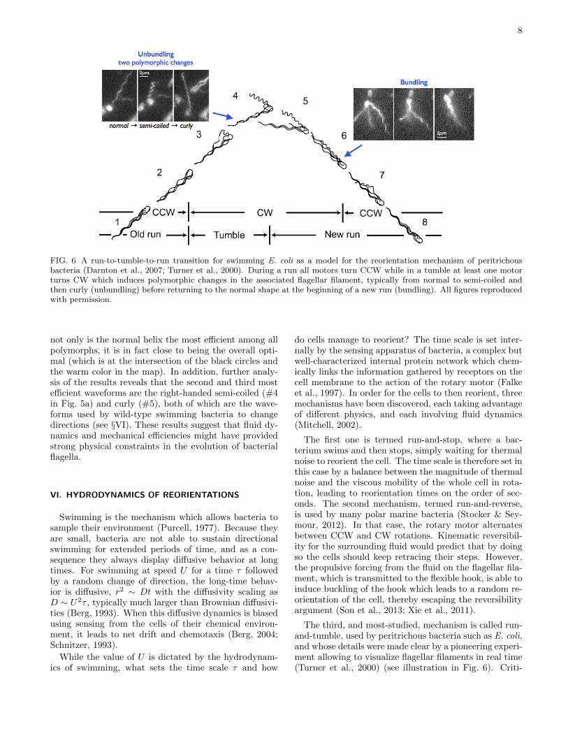
FIG. 6 A run-to-tumble-to-run transition for swimming E. coli as a model for the reorientation mechanism of peritrichousbacteria (Darnton et al., 2007; Turner et al., 2000). During a run all motors turn CCW while in a tumble at least one motorturns CW which induces polymorphic changes in the associated flagellar filament, typically from normal to semi-coiled andthen curly (unbundling) before returning to the normal shape at the beginning of a new run (bundling). All figures reproducedwith permission.
not only is the normal helix the most efficient among allpolymorphs, it is in fact close to being the overall opti-mal (which is at the intersection of the black circles andthe warm color in the map). In addition, further analy-sis of the results reveals that the second and third mostefficient waveforms are the right-handed semi-coiled (#4in Fig. 5a) and curly (#5), both of which are the wave-forms used by wild-type swimming bacteria to changedirections (see §VI). These results suggest that fluid dy-namics and mechanical efficiencies might have providedstrong physical constraints in the evolution of bacterialflagella.
VI. HYDRODYNAMICS OF REORIENTATIONS
Swimming is the mechanism which allows bacteria tosample their environment (Purcell, 1977). Because theyare small, bacteria are not able to sustain directionalswimming for extended periods of time, and as a con-sequence they always display diffusive behavior at longtimes. For swimming at speed U for a time τ followedby a random change of direction, the long-time behav-ior is diffusive, r2 ∼ Dt with the diffusivity scaling asD ∼ U2τ , typically much larger than Brownian diffusivi-ties (Berg, 1993). When this diffusive dynamics is biasedusing sensing from the cells of their chemical environ-ment, it leads to net drift and chemotaxis (Berg, 2004;Schnitzer, 1993).
While the value of U is dictated by the hydrodynam-ics of swimming, what sets the time scale τ and how
do cells manage to reorient? The time scale is set inter-nally by the sensing apparatus of bacteria, a complex butwell-characterized internal protein network which chem-ically links the information gathered by receptors on thecell membrane to the action of the rotary motor (Falkeet al., 1997). In order for the cells to then reorient, threemechanisms have been discovered, each taking advantageof different physics, and each involving fluid dynamics(Mitchell, 2002).
The first one is termed run-and-stop, where a bac-terium swims and then stops, simply waiting for thermalnoise to reorient the cell. The time scale is therefore set inthis case by a balance between the magnitude of thermalnoise and the viscous mobility of the whole cell in rota-tion, leading to reorientation times on the order of sec-onds. The second mechanism, termed run-and-reverse,is used by many polar marine bacteria (Stocker & Sey-mour, 2012). In that case, the rotary motor alternatesbetween CCW and CW rotations. Kinematic reversibil-ity for the surrounding fluid would predict that by doingso the cells should keep retracing their steps. However,the propulsive forcing from the fluid on the flagellar fila-ment, which is transmitted to the flexible hook, is able toinduce buckling of the hook which leads to a random re-orientation of the cell, thereby escaping the reversibilityargument (Son et al., 2013; Xie et al., 2011).
The third, and most-studied, mechanism is called run-and-tumble, used by peritrichous bacteria such as E. coli,and whose details were made clear by a pioneering experi-ment allowing to visualize flagellar filaments in real time(Turner et al., 2000) (see illustration in Fig. 6). Criti-

9
cal to the effective locomotion in this case is the abilityof multiple flagellar filaments to bundle when they all ro-tate in the same direction, and unbundle otherwise (Mac-nab, 1977; Magariyama et al., 2001). When a bacteriumruns, all its flagellar filamenta adopt the normal wave-form, turn in a CCW fashion (when viewed from behindthe cell looking in), and are gathered in a tight, synchro-nized helical bundle behind the cell. Based on chemicalclues from its environment, the cell might then initiate atumbling event (which is Poisson distributed), for whichat least one motor (but possibly more) switches its direc-tion rotation to CW, inducing a polymorphic change ofits flagellar filament from normal to semi-coiled (in mostcases) which at the same time flies out of the bundle,resulting in a change of orientation for the cell body. Asthe motor continues to rotate CW, the flagellum switchesto the curly shape, and then when the motor reverts backto a CCW rotation, comes back to the normal shape andreturns into the bundle. These short tumbles typicallylast τ ∼ 0.1s while the runs last about one second. Notethat many mutants of E. coli exist, either naturally oc-curring or genetically induced in the lab, displaying somevariation in geometry or behavior; for example, the mu-tant HCB-437 does not tumble and is smooth swimming(Wolfe et al., 1988).
The bundling and unbundling of flagellar filamentscompose a large-deformation fluid-structure interactionproblem at low Reynolds numbers combined with short-range flagella-flagella electrostatic repulsion (Lytle et al.,2002; Weibull, 1950). Due to this complexity, physicalstudies of bundling have taken two drastically differentapproaches. On one hand, investigations have been car-ried out on very simplified geometries and setups, eithernumerically (Kim & Powers, 2004; Reichert & Stark,2005) or experimentally at the macroscopic scale (Kimet al., 2003; Macnab, 1977; Qian et al., 2009), in orderto probe synchronization between driven identical helicesand their bundling. It was found that nonlocal hydro-dynamic interactions between helices were sufficient toinduce attraction, wrapping, and synchronization pro-vided the helices were of the right combination of chiral-ity (both left-handed and rotating CCW or their mirror-image equivalent) but some amount of elastic compliancewas required. Biologically, it was argued that this flexi-bility could be provided by the hook.
On the other hand, some groups have attempted totackle the complexity of the problem using realistic se-tups of deforming flagellar filaments from various (of-ten formidable) computational angles, including the useof regularized flow singularities (Flores et al., 2005),the immersed boundary method (Lim & Peskin, 2012),mesoscale methods (Reigh et al., 2012), boundary ele-ments (Kanehl & Ishikawa, 2014) and bead-spring mod-els adapted from polymer physics (Janssen & Graham,2011; Watari & Larson, 2010). The numerical resultsconfirm the physical picture of bundling and unbundlingdriven by a combination of flexibility and hydrodynamicinteractions, with a critical dependence on the geometri-
cal details, the relative configuration of the flagella, andthe value of the motor torques driving the rotation.
VII. BACTERIA SWIMMING NEAR SURFACES
Surfaces are known to affect the behavior of bacte-ria (Harshey, 2003; Vanloosdrecht et al., 1990). One ofthe most striking aspect of locomotion near boundariesis the tendency of swimming bacteria to be attractedto surfaces, with steady-state concentration showing 10fold-increases near surfaces (Berke et al., 2008). Thisis illustrated in Fig. 7a for smooth-swimming (i.e. non-tumbling) E. coli cells. The attraction mechanism wasfirst argued to be purely hydrodynamic in nature (Berkeet al., 2008; Hernandez-Ortiz et al., 2005), easily under-stood as an image effect. As seen in §IV, the flow pertur-bation induced by a swimming bacterium is (at leadingorder in the far field) a pusher force dipole. The hydrody-namic image system necessary to enforce a no-slip bound-ary condition on a flat surface includes a force dipole, aforce quadrupole, and a source dipole, all located on theother side of the surface (purple arrows in Fig. 7b) (Blake,1971). The net effect of these image singularities on theoriginal dipole is an hydrodynamic attraction toward thewall (black arrow). Intuitively, a pusher dipole drawsfluid in from its side, and therefore pulls itself toward asurface when swimming parallel to it. Similarly, the hy-drodynamic moment induced by the image system is ableto rotate pusher dipoles so that their stable orientationis swimming parallel to the surface (Berke et al., 2008),leading to kinematic asymmetries for bacteria switchingbetween pusher and puller modes (Magariyama et al.,2005).
Experimental measurements of the flow induced bybacteria showed, however, that this dipolar flow getsquickly overwhelmed by noise a few cell lengths away,and thus acts only when the cells are close to the sur-face, leading to long residence times (Drescher et al.,2011). For cells which swim toward a surface and gettrapped there, what determines the long-time equilib-rium distance between swimming cells and surfaces, andtheir orientation? Although simple models all predictbacteria eventually touching walls and therefore requireshort-range repulsion (Dunstan et al., 2012; Spagnolie& Lauga, 2012), boundary element computations with amodel polar bacterium demonstrated that hydrodynam-ics alone was able to select a stable orbit with swimmingat a finite distance (Fig. 7c) (Giacche et al., 2010).
A second remarkable feature of bacteria swimmingnear surface is a qualitative change in their trajecto-ries. Between reorientation events, flagellated bacteriafar from walls swim along straight or wiggly paths. Nearrigid surfaces, their trajectories are observed to becomecircular, typically CW when viewed from above the sur-face in the fluid (Berg & Turner, 1990; Frymier et al.,1995). This is illustrated in Fig. 7d for an individually-tracked E coli cell swimming near a glass surface (Vigeant

10
FIG. 7 (a) Attraction of smooth-swimming E. coli cells by two rigid surfaces located at y = 0 and y = 100 µm (symbols) andfit by a point-dipole model (lines) (Berke et al., 2008); (b) Hydrodynamic images for a pusher force dipole above a flat no-slipsurface (purple arrows) leading to attraction by the surface (black arrow); (c) Computations showing stable swimming at afinite distance from a rigid surface for a polar bacterium (distance to wall nondimensionalized by cell size as a function of timenondimensionalized by flagellar rotational frequency) (Giacche et al., 2010); (d) Tracking data for a smooth-swimming E. colicell near a glass surface (Vigeant & Ford, 1997); (e) Physical interpretation of wall-induced moment on bacterium with helicalflagellar filament leading to circular swimming: the rotating flagellum is subject to a surface-induced force to its left (whenviewed from above) and the counter-rotating body by a force in the opposite direction, leading to a moment and thereforerotation of the cell; (f) Rectification of fluorescent swimming E. coli in channels with asymmetric geometrical features (Galajdaet al., 2007). All figures reproduced with permission.
& Ford, 1997). Early computations for model polar bac-teria also predicted circular trajectories (Ramia et al.,1993) while recent experiments showed that the physic-ochemical nature of the interface plays a critical role se-lecting the rotation direction (Lemelle et al., 2013; Morseet al., 2013).
The switch from forward to circular swimming is afluid dynamics effect owing to new forces and momentsinduced near surfaces (Lauga et al., 2006). This is illus-trated in Fig. 7e for locomotion of model polar bacteriumabove of a rigid wall. A helix rotating parallel to a no-slipsurface is subject to a net force whose direction dependson the chirality of the helix and its direction of rotation
(to the left in Fig. 7d for a CCW rotation of a left-handedhelix). Similarly, as the cell body is counter-rotating, itis subject to a hydrodynamic force in the direction of itsrolling motion on the surface (to the right in Fig. 7d)(Kim & Karrila, 1991). These two forces are exactlyzero far from the surface and their magnitudes dependstrongly on the distance to the wall (Li et al., 2008).The net effect of these forces is a wall-induced momentacting on the cell. Since the cell is torque-free, it needsto rotate (here, CW when viewed from above), leadingto circular trajectories. Notably, the sign of the surface-induced forces are reversed if the rigid wall is replaced bya free surface, leading to circular swimming in the oppo-

11
FIG. 8 (a) Jeffery’s orbits for non-flagellated E. coli cells in a microchannel (Kaya & Koser, 2009); (b) Non-motile Bacillussubtilis cells show a uniform concentration under non-uniform shear (Rusconi et al., 2014); (c) Motile cells initially uniformlydistributed (black line) accumulate under non-uniform shear in the high-shear regions (red line); (d) Motile E. coli cellsswimming in circles near rigid surfaces in the absence of flow (Kaya & Koser, 2012); (e) Upstream swimming of the same cellsunder moderate shear; (f) Under strong shear, all cells are advected downstream. All figures reproduced with permission.
site direction (Di Leonardo et al., 2011), and a possibleco-existence between CW and CCW for cell populationsnear complex interfaces (Lemelle et al., 2013; Lopez &Lauga, 2014; Morse et al., 2013).
The interactions of bacteria with complex geometrieshave also lead to fascinating results. Swimming E. colicells in rectangular microchannels with three polymericwalls and one agar surface showed a strong preference toswim near the agar side of the channels (DiLuzio et al.,2005), a phenomenon still unexplained. The same bacte-ria are rectified by channels with asymmetric geometricalfeatures (Galajda et al., 2007), which can then be ex-ploited to passively increase cell concentrations (Fig. 7f).Similarly, bacteria can be harnessed to perform mechan-ical work and, for example, rotate asymmetric micro-gears (Di Leonardo et al., 2010; Sokolov et al., 2010).In strong confinement, helical flagella continue to inducepropulsion, but the swimming speed decreases for motiondriven at constant torque (Liu et al., 2014a). Related ex-periments show that the limit of flagella-driven motilityis reached when the cell body has clearance of about lessthan 30% of its size, although cells can continue to movein smaller channels through cell division (Mannik et al.,2009).
VIII. BACTERIA IN FLOWS
Similarly to colloidal particles, swimming bacteria re-spond to external flows by being passively advected androtated. In many relevant situations where the cells aremuch smaller than any of the flow length scales (e.g. inmarine environments), the flow can be assumed to belocally linear, and thus a superposition of uniform trans-lation, pure extension, and pure rotation. In that case,the dynamics of the (elongated) bacteria is described byJeffery’s equation which is exact for prolate spheroids inlinear flows (Pedley & Kessler, 1992). Physically, thecells move with the mean flow velocity and their rotationrate is the sum of the vorticity component (which alonewould predict rotation at a constant rate) and the (poten-tial) extensional component (which alone would predictsteady alignment with the principal axis of extension).The resulting tumbling trajectories, called Jeffery’s or-bits, are illustrated in Fig. 8a for non flagellated E. colicells under shear in a microfluidic device (Kaya & Koser,2009).
In addition to the classical Jeffery’s orbits, bacteria aresubject to an additional mechanism which results fromtheir chiral shapes. The hydrodynamics of a helical flag-ellum in a simple shear flow leads to a viscous torque be-
12
ing produced on the helix, which reorients it away fromthe plane of shear. Combined with motility, this is anexample of rheotaxis where cells responds to shear (here,purely physically) (Marcos et al., 2012). Beyond the as-sumption of linear flow, swimming bacteria respond tonon-uniform shears by developing non-uniform orienta-tion distributions, which, when coupled to motility, leadto cells accumulating in the high shear regions (Rusconiet al., 2014). This is illustrated in Fig. 8b-c for Bacil-lus subtilis in Poiseuille flow. Non-swimming cells have auniform distribution (Fig. 8b) whereas motile cells start-ing uniformly distributed (black line in Fig. 8c) developnon-uniform distributions and gather in the high-shearregions (red line Fig. 8c).
One compelling aspect of the response of bacteria toexternal flows is the feedback of swimming cells on thesurrounding fluid through the stresses they exert. Dueto their dipolar flows, swimming bacteria exert bulkstresses on the fluid – hence the term “stresslet” origi-nally coined by Batchelor in the context of passive sus-pensions (Batchelor, 1970). A group of swimming bac-teria subject to an external deformation will thus gener-ically respond by a state of stress with a rich dynamics.The most fundamental question relating stresses to de-formation in the fluid is: what is the effective viscosity ofa suspension of swimming bacteria? Experiments havebeen first carried out with the peritrichous Bacillus sub-tilis by estimating the viscosity in two different ways,first by looking at the unsteady decay of vortex and thenby measuring directly the torque on a rotating probe inthe cell suspension (Sokolov & Aranson, 2009). The re-sults showed a strong reduction in the effective viscosity(up to a factor of seven), while an increase was possi-ble at larger cell concentrations. A different setup wasproposed for E. coli where a suspension was flown in amicrochannel and the effective viscosity estimated usingpredictions for the unidirectional flow profile from multi-phase Newtonian fluid dynamics (Gachelin et al., 2013).In that case, the effective viscosity showed an initial de-crease, followed by shear-thickening up to relative vis-cosities above 1, followed by shear-thinning at high shearrates. In parallel, theoretical and computational work onmodel cells in both shear and extensional flows predictedthe viscosity decrease for pusher cells, together with thepossibility of negative viscosities and normal stress coeffi-cients of sign opposite to the ones for passive suspensions(Haines et al., 2009; Saintillan, 2010a,b).
Finally, bacteria swimming near boundaries in externalflows also have a propensity to swim upstream and henceagainst the flow (Cisneros et al., 2007; Hill et al., 2007).This is illustrated in Fig. 8d-f where motile E. coli cellswhich swim in circles in the absence of flow (Fig. 8d) areseen to move upstream under moderate shear (Fig. 8e)and are passively advected downstream at high shear(Fig. 8f) (Kaya & Koser, 2012). The physical originof this transition to upstream swimming has been pro-posed to be hydrodynamic, whereby a shear flow of mod-erate strength produces a torque on a downward-facing
cell which reorients it so that its stable equilibrium isto point upstream and thus swim against the flow (Hillet al., 2007; Kaya & Koser, 2012).
IX. LOCOMOTION IN COMPLEX FLUIDS
Bacteria who inhabit soils and higher organisms rou-tinely have to progress through complex environmentswith microstructures whose characteristic length scalesmight be similar to that of the cell. Some bacte-ria also have to endure chemically- and mechanically-heterogeneous environments. For example, Helicobacterpylori, which survives in the acidic environment of thestomach, creates an enzyme which locally increases pHand can induce rheological changes to the mucus pro-tecting the epithelium surface of the stomach, changingit from a gel to a viscous fluid in which it is able to swim(Bansil et al., 2013). In many instances, the surround-ing fluids might not be totally flowing but instead pos-sess their own relaxation dynamics, which might occuron time scales comparable to the intrinsic time scales ofthe locomotion. For example, during biofilm formation,bacteria produce an extracellular polymeric matrix forwhich they control the mechanical properties through wa-ter content (Costerton et al., 1995; Wilking et al., 2011)(Fig. 9a). A number of studies have attempted to quan-tify the impact of such complex fluids on the swimmingkinematics and energetics of bacteria.
Early studies were concerned with perhaps the simplestquestion possible: How is bacterial locomotion modifiedby a change in the viscosity of the surrounding (New-tonian) fluid? Flagellated bacteria display a systematictwo-phase response, where an increase in viscosity is seento first lead to a small increase in the swimming speedfollowed by a sharp decrease at large viscosities (Green-berg & Canale-Parola, 1977; Schneider & Doetsch, 1974;Shoesmith, 1960). The decrease has been rationalizedby assuming that the rotary motor is working at con-stant power output (Keller, 1974), an assumption wenow know is erroneous since it is the motor torque whichis constant (see §II). In stark contrast with flagellatedbacteria, spirochetes show a systematic enhancement oftheir swimming speed with an increase of the viscosity ofthe fluid (Kaiser & Doetsch, 1975). In a related study,the viscosity required to immobilize bacteria was mea-sured to be larger, by orders of magnitude, for spirochetescompared to flagellated bacteria (Greenberg & Canale-Parola, 1977).
How can the difference between flagellated bacteriaand spirochetes be rationalized? One argument broughtforward focused on the details of the microstructure ofthe fluid, arguing that all increases in viscosity are notcreated equal (Berg & Turner, 1979). Increases in viscos-ity are induced by dissolving polymers, but the nature ofthe polymer chains structure may be critical. Experi-ments showing large increases in swimming speeds usedsolution of long unbranched chain polymers (e.g., methyl-
13
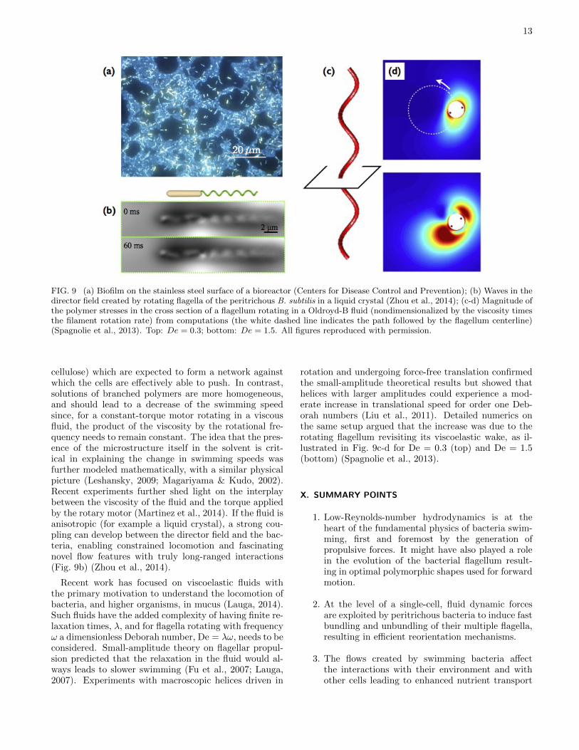
FIG. 9 (a) Biofilm on the stainless steel surface of a bioreactor (Centers for Disease Control and Prevention); (b) Waves in thedirector field created by rotating flagella of the peritrichous B. subtilis in a liquid crystal (Zhou et al., 2014); (c-d) Magnitude ofthe polymer stresses in the cross section of a flagellum rotating in a Oldroyd-B fluid (nondimensionalized by the viscosity timesthe filament rotation rate) from computations (the white dashed line indicates the path followed by the flagellum centerline)(Spagnolie et al., 2013). Top: De = 0.3; bottom: De = 1.5. All figures reproduced with permission.
cellulose) which are expected to form a network againstwhich the cells are effectively able to push. In contrast,solutions of branched polymers are more homogeneous,and should lead to a decrease of the swimming speedsince, for a constant-torque motor rotating in a viscousfluid, the product of the viscosity by the rotational fre-quency needs to remain constant. The idea that the pres-ence of the microstructure itself in the solvent is crit-ical in explaining the change in swimming speeds wasfurther modeled mathematically, with a similar physicalpicture (Leshansky, 2009; Magariyama & Kudo, 2002).Recent experiments further shed light on the interplaybetween the viscosity of the fluid and the torque appliedby the rotary motor (Martinez et al., 2014). If the fluid isanisotropic (for example a liquid crystal), a strong cou-pling can develop between the director field and the bac-teria, enabling constrained locomotion and fascinatingnovel flow features with truly long-ranged interactions(Fig. 9b) (Zhou et al., 2014).
Recent work has focused on viscoelastic fluids withthe primary motivation to understand the locomotion ofbacteria, and higher organisms, in mucus (Lauga, 2014).Such fluids have the added complexity of having finite re-laxation times, λ, and for flagella rotating with frequencyω a dimensionless Deborah number, De = λω, needs to beconsidered. Small-amplitude theory on flagellar propul-sion predicted that the relaxation in the fluid would al-ways leads to slower swimming (Fu et al., 2007; Lauga,2007). Experiments with macroscopic helices driven in
rotation and undergoing force-free translation confirmedthe small-amplitude theoretical results but showed thathelices with larger amplitudes could experience a mod-erate increase in translational speed for order one Deb-orah numbers (Liu et al., 2011). Detailed numerics onthe same setup argued that the increase was due to therotating flagellum revisiting its viscoelastic wake, as il-lustrated in Fig. 9c-d for De = 0.3 (top) and De = 1.5(bottom) (Spagnolie et al., 2013).
X. SUMMARY POINTS
1. Low-Reynolds-number hydrodynamics is at theheart of the fundamental physics of bacteria swim-ming, first and foremost by the generation ofpropulsive forces. It might have also played a rolein the evolution of the bacterial flagellum result-ing in optimal polymorphic shapes used for forwardmotion.
2. At the level of a single-cell, fluid dynamic forcesare exploited by peritrichous bacteria to induce fastbundling and unbundling of their multiple flagella,resulting in efficient reorientation mechanisms.
3. The flows created by swimming bacteria affectthe interactions with their environment and withother cells leading to enhanced nutrient transport
14
and collective locomotion. Similarly, complex mi-crostructures or external flows can have significantinfluence on the trajectories, concentrations, andorientations of swimming bacteria.
4. Although surrounding fluids play important rolesin the life of bacteria, hydrodynamic forces on flag-ellar filaments and cell bodies often have to be bal-anced with bending and twisting elasticity, short-range electrostatic interactions, and are subject toboth biochemical and thermal noise. Fluid dynam-ics is therefore only one of the complex physicalprocesses at play.
XI. FUTURE ISSUES
1. Why is the value of the experimentally-measuredtorque applied by the rotary motor so differentfrom that from hydrodynamic predictions (Darn-ton et al., 2007)? What are the other mechanicalforces at play?
2. Why does the flagellar hook of peritrichous bac-teria not buckle, as it does for polar bacteria un-der seemingly similar propulsive conditions (Sonet al., 2013)? Why is the flagellar hook so preciselytuned in size and rigidity that flagellar bundling isno longer possible if small changes are made to it(Brown et al., 2012)?
3. How exactly can a tight bundle of multiple flag-ella open up and close repeatedly without jammingunder the uncoordinated action of a few motors(Macnab, 1977)? How much of this process is trulydriven by hydrodynamic interactions vs. other pas-sive mechanisms?
4. Can we derive predictive whole-cell models ableto reproduce the full run-and-tumble statistics ofswimming bacteria based solely on our understand-ing on the behavior of rotary motors (Turner et al.,2000)?
Acknowledgements
I thank Tom Montenegro-Johnson for his help with thefigures. Discussions with Howard Berg and Ray Gold-stein are gratefully acknowledged. This work was fundedin part by the European Union (through a Marie-CurieCIG Grant) and by the Isaac Newton Trust (Cambridge).
References
Allen RD, Baumann P. 1971. Structure and arrangementof flagella in species of the genus Beneckea and Photobac-terium fischeri. J. Bacteriol. 107:295–302
Bansil R, Celli JP, Hardcastle JM, Turner BS. 2013. The influ-ence of mucus microstructure and rheology in Helicobacterpylori infection. Front. Immun. 4:310
Batchelor GK. 1970. The stress system in a suspension offorce-free particles. J. Fluid Mech. 41:545–570
Berg HC. 1993. Random Walks in Biology. New Jersey:Princeton University Press
Berg HC. 2003. The rotary motor of bacterial flagella. Annu.Rev. Biochem. 72:19–54
Berg HC. 2004. E. coli in Motion. New York, NY: Springer-Verlag
Berg HC, Anderson RA. 1973. Bacteria swim by rotatingtheir flagellar filaments. Nature 245:380–382
Berg HC, Turner L. 1979. Movement of microorganisms inviscous environments. Nature 278:349–351
Berg HC, Turner L. 1990. Chemotaxis of bacteria in glasscapillary arrays – Escherichia coli, motility, microchannelplate, and light scattering. Biophys. J. 58:919–930
Berke AP, Turner L, Berg HC, Lauga E. 2008. Hydrodynamicattraction of swimming microorganisms by surfaces. Phys.Rev. Lett. 101:038102
Berry RM, Berg HC. 1997. Absence of a barrier to backwardsrotation of the bacterial flagellar motor demonstrated withoptical tweezers. Proc. Natl. Acad. Soc., USA 94:14433–14437
Blair DF. 1995. How bacteria sense and swim. Annu. Rev.Microbiol. 49:489–520
Blake JR. 1971. A note on the image system for a stokesletin a no-slip boundary. Proc. Camb. Phil. Soc. 70:303–310
Bray D. 2000. Cell Movements. New York, NY: GarlandPublishing
Brennen C, Winet H. 1977. Fluid mechanics of propulsion bycilia and flagella. Annu. Rev. Fluid Mech. 9:339–398
Brown MT, Steel BC, Silvestrin C, Wilkinson DA, DelalezNJ, et al. 2012. Flagellar hook flexibility is essential forbundle formation in swimming Escherichia coli cells. J.Bacter. 194:3495–3501
Calladine CR. 1978. Change of waveform in bacterial flagella: the role of mechanics at the molecular level. J. Mol. Biol.118:457–479
Chattopadhyay S, Moldovan R, Yeung C, Wu XL. 2006.Swimming efficiency of bacterium Escherichia coli. Proc.Natl. Acad. Sci. USA 103:13712–13717
Chattopadhyay S, Wu X. 2009. The effect of long-range hy-drodynamic interaction on the swimming of a single bac-terium. Biophys. J. 96:2023–2028
Chen X, Berg HC. 2000. Torque-speed relationship of the flag-ellar rotary motor of Escherichia coli. Biophys. J. 78:1036–1041
Chwang AT, Wu TY. 1971. Helical movement of microorgan-isms. Proc. Roy. Soc. Lond. B 178:327–346
Chwang AT, Wu TY. 1975. Hydromechanics of low-Reynolds-number flow. Part 2. Singularity method for Stokes flows.J. Fluid Mech. 67:787–815
Chwang AT, Wu TY, Winet H. 1972. Locomotion of spirilla.Biophys. J. 12:1549
Cisneros LH, Cortez R, Dombrowski C, Goldstein RE, KesslerJO. 2007. Fluid dynamics of self-propelled microorganisms,from individuals to concentrated populations. Exp. Fluids43:737–753
Coombs D, Huber G, Kessler JO, Goldstein RE. 2002. Pe-riodic chirality transformations propagating on bacterialflagella. Phys. Rev. Lett. 89:118102–1–118102–4
Copeland MF, Weibel DB. 2009. Bacterial swarming: a model
15
system for studying dynamic self-assembly. Soft Matt.5:1174–1187
Costerton JW, Lewandowski Z, Caldwell DE, Korber DR,Lappinscott HM. 1995. Microbial biofilms. Annu. Rev.Microbiol. 49:711–745
Cox RG. 1970. The motion of long slender bodies in a viscousfluid. Part 1. General theory. J. Fluid Mech. 44:791–810
Darnton NC, Berg HC. 2007. Force-extension measurementson bacterial flagella: Triggering polymorphic transforma-tions. Biophys. J. 92:2230–2236
Darnton NC, Turner L, Rojevsky S, Berg HC. 2007. On torqueand tumbling in swimming Escherichia coli. J. Bacteriol.189:1756–1764
Darnton NC, Turner L, Rojevsky S, Berg HC. 2010. Dynamicsof bacterial swarming. Biophys. J. 98:2082–2090
Di Leonardo R, Angelani L, DellArciprete D, Ruocco G, IebbaV, et al. 2010. Bacterial ratchet motors. Proc. Natl. Acad.Sci. U.S.A. 107:9541–9545
Di Leonardo R, Dell’Arciprete D, Angelani L, Iebba V. 2011.Swimming with an image. Phys. Rev. Lett. 106:038101
DiLuzio WR, Turner L, Mayer M, Garstecki P, Weibel DB,et al. 2005. Escherichia coli swim on the right-hand side.Nature 435:1271–1274
Doi M, Edwards SF. 1988. The Theory of Polymer Dynamics.Oxford, U.K.: Oxford University Press
Drescher K, Dunkel J, Cisneros LH, Ganguly S, GoldsteinRE. 2011. Fluid dynamics and noise in bacterial cell-celland cell-surface scattering. Proc. Natl. Acad. Sci. U.S.A.108:10940–10945
Dunstan J, Mino G, Clement E, Soto R. 2012. A two-spheremodel for bacteria swimming near solid surfaces. Phys.Fluids 24:011901
Ehlers K, Oster G. 2012. On the mysterious propulsion ofSynechococcus. PLOS One 7:e36081
Falke JJ, Bass RB, Butler SL, Chervitz SA, Danielson MA.1997. The two-component signaling pathway of bacterialchemotaxis: A molecular view of signal transduction byreceptors, kinases, and adaptation enzymes. Annu. Rev.Cell Dev. Biol. 13:457
Fauci LJ, Dillon R. 2006. Biofluidmechanics of reproduction.Annu. Rev. Fluid Mech. 38:371–394
Flores H, Lobaton E, Mendez-Diez S, Tlupova S, Cortez R.2005. A study of bacterial flagellar bundling. Bull. Math.Biol. 67:137–168
Frymier PD, Ford RM, Berg HC, Cummings PT. 1995. Three-dimensional tracking of motile bacteria near a solid planarsurface. Proc. Natl. Acad. Sci. USA 92:6195–6199
Fu HC, Powers TR, Wolgemuth HC. 2007. Theory of swim-ming filaments in viscoelastic media. Phys. Rev. Lett.99:258101–258105
Fujii M, Shibata S, Aizawa SI. 2008. Polar, peritrichous, andlateral flagella belong to three distinguishable flagellar fam-ilies. J. Mol. Biol. 379:273–283
Gachelin J, Mino G, Berthet H, Lindner A, Rousselet A,Clement E. 2013. Non-newtonian viscosity of echerichiacoli suspensions. Phys. Rev. Lett. 110:268103
Gaffney EA, Gadelha H, Smith DJ, Blake JR, Kirkman-Brown JC. 2011. Mammalian sperm motility: Observationand theory. Annu. Rev. Fluid Mech. 43:501–528
Galajda P, Keymer JE, Chaikin P, Austin RH. 2007. A wallof funnels concentrates swimming bacteria. J. Bacteriol.189:8704–8707
Ghosh A, Fischer P. 2009. Controlled propulsion of artificialmagnetic nanostructured propellers. Nano Lett. 9:2243–
2245Giacche D, Ishikawa T, Yamaguchi T. 2010. Hydrodynamic
entrapment of bacteria swimming near a solid surface.Phys. Rev. E 82:056309
Goldstein RE. 2015. Green algae as model organisms for bi-ological fluid dynamics. Annu. Rev. Fluid Mech. 47:34375
Goldstein SF, Buttle KF, Charon NW. 1996. Structural anal-ysis of the Leptospiraceae and Borrelia burgdorferi by high-voltage electron microscopy. J. Bacteriol. 178:6539–6545
Gray J, Hancock GJ. 1955. The propulsion of Sea-urchinspermatozoa. J. Exp. Biol. 32:802–814
Greenberg EP, Canale-Parola E. 1977. Motility of flagellatedbacteria in viscous environments. J. Bacteriol. 132:356–358
Guasto JS, Rusconi R, Stocker R. 2012. Fluid mechanicsof planktonic microorganisms. Annu. Rev. Fluid Mech.44:373400
Haines BM, Sokolov A, Aranson IS, Berlyand L, Karpeev DA.2009. Three-dimensional model for the effective viscosityof bacterial suspensions. Phys. Rev. E 80:041922
Hancock GJ. 1953. The self-propulsion of microscopic organ-isms through liquids. Proc. Roy. Soc. Lond. A 217:96–121
Harshey RM. 2003. Bacterial motility on a surface: Manyways to a common goal. Annu. Rev. Microbiol. 57:249–273
Hasegawa K, Yamashita I, Namba K. 1998. Quasi- andnonequivalence in the structure of bacterial flagellar fila-ment. Biophys. J. 74:569–575
Hernandez-Ortiz JP, Stoltz CG, Graham MD. 2005. Trans-port and collective dynamics in suspensions of confinedswimming particles. Phys. Rev. Lett. 95:204501
Higdon JJL. 1979. Hydrodynamics of flagellar propulsion –Helical waves. J. Fluid Mech. 94:331–351
Hill J, Kalkanci O, McMurry JL, Koser H. 2007. Hydrody-namic surface interactions enable Escherichia coli to seekefficient routes to swim upstream. Phys. Rev. Lett. 98:1–4
Hotani H. 1980. Micro-video study of moving bacterial flagel-lar filaments. II. Polymorphic transition in alcohol. Biosyst.12:325–330
Hotani H. 1982. Micro-video study of moving bacterial flag-ellar filaments III. Cyclic transformation induced by me-chanical force. J. Mol. Biol. 156:791
Hyon Y, Marcos, Powers TR, Stocker R, Fu HC. 2012. Thewiggling trajectories of bacteria. J. Fluid Mech. 705:58–76
Imhoff JF, Truper HG. 1977. Ectothiorhodospira halochlo-ris sp. nov., a new extremely halophilic phototrophic bac-terium containing bacteriochlorophyll b∗. Arch. Microbiol.114:115–121
Ishikawa T, Sekiya G, Imai Y, Yamaguchi T. 2007. Hydro-dynamic interaction between two swimming bacteria. Bio-phys. J. 93:2217–2225
Janssen PJA, Graham MD. 2011. Coexistence of tight andloose bundled states in a model of bacterial flagellar dy-namics. Phys. Rev. E 84
Jarrell KF, McBride MJ. 2008. The surprisingly diverse waysthat prokaryotes move. Nature Rev. Microbiol. 6:466–476
Johnson RE. 1980. An improved slender body theory forStokes flow. J. Fluid Mech. 99:411–431
Johnson RE, Brokaw CJ. 1979. Flagellar hydrodynamics.a comparison between resistive-force theory and slender-body theory. Biophys. J. 25:113
Kaiser GE, Doetsch RN. 1975. Enhanced translational motionof Leptospira in viscous environments. Nature 255:656–657
Kanehl P, Ishikawa T. 2014. Fluid mechanics of swimmingbacteria with multiple flagella. Phys. Rev. E 89:042704
Kasyap TV, Koch DL, Wu M. 2014. Hydrodynamic tracer
16
diffusion in suspensions of swimming bacteria. Phys. Fluids26:081901
Kaya T, Koser H. 2009. Characterization of hydrodynamicsurface interactions of Escherichia coli cell bodies in shearflow. Phys. Rev. Lett. 103:138103
Kaya T, Koser H. 2012. Direct upstream motility in Es-cherichia coli. Biophys. J. 102:1514–1523
Kearns DB. 2010. A field guide to bacterial swarming motility.Nature Rev. Microbiol. 8:634–644
Keller JB. 1974. Effect of viscosity on swimming velocity ofbacteria. Proc. Natl. Acad. Sci. USA 71:3253–3254
Keller JB, Rubinow SI. 1976. Swimming of flagellated mi-croorganisms. Biophys. J. 16:151–170
Kim M, Bird JC, Parys AJV, Breuer KS, Powers TR. 2003.A macroscopic scale model of bacterial flagellar bundling.Proc. Natl. Acad. Sci. USA 100:15481–15485
Kim M, Powers TR. 2004. Hydrodynamic interactions be-tween rotating helices. Phys. Rev. E 69:061910
Kim MJ, Breuer KS. 2004. Enhanced diffusion due to motilebacteria. Phys. Fluids 16:L78–L81
Kim MJ, Powers TR. 2005. Deformation of a helical fila-ment by flow and electric or magnetic fields. Phys. Rev. E71:021914–1–10
Kim S, Karrila JS. 1991. Microhydrodynamics: Principlesand Selected Applications. Boston, MA: Butterworth-Heinemann
Koch DL, Subramanian G. 2011. Collective hydrodynamicsof swimming microorganisms: Living fluids. Annu. Rev.Fluid Mech. 43:637 – 659
Lauga E. 2007. Propulsion in a viscoelastic fluid. Phys. Fluids19:083104
Lauga E. 2014. Locomotion in complex fluids: Integral theo-rems. Phys. Fluids 26:081902
Lauga E, DiLuzio WR, Whitesides GM, Stone HA. 2006.Swimming in circles: Motion of bacteria near solid bound-aries. Biophys. J. 90:400–412
Lauga E, Powers TR. 2009. The hydrodynamics of swimmingmicroorganisms. Rep. Prog. Phys. 72:096601
Leifson E. 1960. Atlas of Bacterial Flagellation. New Yorkand London: Academic Press
Lemelle L, Palierne JF, Chatre E, Vaillant C, Place C. 2013.Curvature reversal of the circular motion of swimming bac-teria probes for slip at solid/liquid interfaces. Soft Matter9:9759–9762
Leshansky AM. 2009. Enhanced low-Reynolds-numberpropulsion in heterogeneous viscous environments. Phys.Rev. E 80:051911
Li G, Tam LK, Tang JX. 2008. Amplified effect of brown-ian motion in bacterial near-surface swimming. Proc. Natl.Acad. Sci. USA 105:18355–18359
Liao Q, Subramanian G, DeLisa MP, Koch DL, Wu MM.2007. Pair velocity correlations among swimming Es-cherichia coli bacteria are determined by force-quadrupolehydrodynamic interactions. Phys. Fluids 19:061701
Lighthill J. 1975. Mathematical Biofluiddynamics. Philadel-phia: SIAM
Lighthill J. 1976. Flagellar hydrodynamics—The John vonNeumann lecture, 1975. SIAM Rev. 18:161–230
Lim S, Peskin CS. 2012. Fluid-mechanical interaction of flex-ible bacterial flagella by the immersed boundary method.Phys. Rev. E 85:036307
Lin Z, Thiffeault JL, Childress S. 2011. Stirring by squirmers.J. Fluid Mech. 669:167–177
Liu B, Breuer KS, Powers TR. 2013. Helical swimming in
stokes flow using a novel boundary-element method. Phys.Fluids 25:061902
Liu B, Breuer KS, Powers TR. 2014a. Propulsion by a helicalflagellum in a capillary tube. Phys. Fluids 26:011701
Liu B, Gulino M, Morse M, Tang JX, Powers TR, Breuer KS.2014b. Helical motion of the cell body enhances caulobac-ter crescentus motility. Proc. Natl. Acad. Sci. U.S.A.111:11252–11256
Liu B, Powers TR, Breuer KS. 2011. Force-free swimming ofa model helical flagellum in viscoelastic fluids. Proc. Natl.Acad. Sci. USA 108:19516–19520
Lopez D, Lauga E. 2014. Dynamics of swimming bacteria atcomplex interfaces. Phys. Fluids 26:071902
Lytle DA, Johnson CH, Rice EW. 2002. A systematic compar-ison of the electrokinetic properties of environmentally im-portant microorganisms in water. Colloid. Surf. B 24:91–101
Macnab RM. 1977. Bacterial flagella rotating in bundles:A study in helical geometry. Proc. Natl. Acad. Sci. USA74:221
Madigan MT, Martinko JM, Stahl D, Clark DP. 2010. BrockBiology of Microorganisms (13th Edition). San Francisco,CA: Benjamin Cummings
Magariyama Y, Ichiba M, Nakata K, Baba K, Ohtani T, et al.2005. Difference in bacterial motion between forward andbackward swimming caused by the wall effect. Biophys. J.88:3648–3658
Magariyama Y, Kudo S. 2002. A mathematical explanationof an increase in bacterial swimming speed with viscosityin linear-polymer solutions. Biophys. J. 83:733–739
Magariyama Y, Sugiyama S, Kudo S. 2001. Bacterial swim-ming speed and rotation rate of bundled flagella. FemsMicrobiol. Lett. 199:125–129
Magariyama Y, Sugiyama S, Muramoto K, Kawagishi I, ImaeY, Kudo S. 1995. Simultaneous measurement of bacterialflagellar rotation rate and swimming speed. Biophys. J.69:2154–2162
Mannik J, Driessen R, Galajda P, Keymer JE, Dekker C.2009. Bacterial growth and motility in sub-micron con-strictions. Proc. Natl. Acad. Sci. USA 106:14861–14866
Marcos, Fu HC, Powers TR, Stocker R. 2012. Bacterial rheo-taxis. Proc. Natl. Acad. Sci. U.S.A. 109:4780–4785
Martinez VA, Schwarz-Linek J, Reufer M, Wilson LG, Mo-rozov AN, Poon WCK. 2014. Flagellated bacterial motil-ity in polymer solutions. Proc. Natl. Acad. Sci. U.S.A.111:17771–17776
Mino GL, Dunstan J, Rousselet A, Clement E, Soto R. 2013.Induced diffusion of tracers in a bacterial suspension: The-ory and experiments. J. Fluid Mech. 729:423–444
Mitchell JG. 2002. The energetics and scaling of search strate-gies in bacteria. Am. Natur. 160:727–740
Morse M, Huang A, Li G, Maxey MR, Tang JX. 2013. Molec-ular adsorption steers bacterial swimming at the air/waterinterface. Biophys. J. 105:21 – 28
Namba K, Vonderviszt F. 1997. Molecular architecture ofbacterial flagellum. Q. Rev. Biophys. 30:1–65
Olson SD, Lim S, Cortez R. 2013. Modeling the dynamicsof an elastic rod with intrinsic curvature and twist using aregularized Stokes formulation. J. Comp. Phys. 238:169–187
Ottemann KM, Miller JF. 1997. Roles for motility inbacterial-host interactions. Mol. Microbiol. 24:1109–1117
Pedley TJ, Kessler JO. 1992. Hydrodynamic phenomenain suspensions of swimming microorganisms. Annu. Rev.
17
Fluid Mech. 24:313–358Phan-Thien N, Tran-Cong T, Ramia M. 1987. A boundary-
element analysis of flagellar propulsion. J. Fluid Mech.184:533–549
Purcell EM. 1977. Life at low Reynolds number. Am. J. Phys.45:3–11
Purcell EM. 1997. The efficiency of propulsion by a rotatingflagellum. Proc. Natl. Acad. Soc., USA 94:1130711311
Pushkin DO, Shum H, Yeomans JM. 2013. Fluid transportby individual microswimmers. J. Fluid Mech. 726:5–25
Qian B, Jiang H, Gagnon DA, Breuer KS, Powers TR. 2009.Minimal model for synchronization induced by hydrody-namic interactions. Phys. Rev. E 80:061919
Ramia M, Tullock DL, Phan-Thien N. 1993. The role of hy-drodynamic interaction in the locomotion of microorgan-isms. Biophys. J. 65:755–778
Reichert M, Stark H. 2005. Synchronization of rotating helicesby hydrodynamic interactions. Eur. Phys. J. E 17:493–500
Reid SW, Leake MC, Chandler JH, Lo CJ, Armitage JP,Berry RM. 2006. The maximum number of torque-generating units in the flagellar motor of Escherichia coliis at least 11. Proc. Natl. Acad. Sci. USA 103:8066–8071
Reigh SY, Winkler RG, Gompper G. 2012. Synchronizationand bundling of anchored bacterial flagella. Soft Matt.8:4363–4372
Rusconi R, Guasto JS, Stocker R. 2014. Bacterial transportsuppressed by fluid shear. Nature Phys. 10:212–217
Saintillan D. 2010a. Extensional rheology of active suspen-sions. Phys. Rev. E 81:056307
Saintillan D. 2010b. The dilute rheology of swimming suspen-sions: A simple kinetic model. J. Exp. Mech. 50:1275
Schneider W, Doetsch RN. 1974. Effect of viscosity on bac-terial motility. J. Bacteriol. 117:696–701
Schnitzer MJ. 1993. Theory of continuum random walks andapplication to chemotaxis. Phys. Rev. E 48:2553
Shaevitz JW, Lee JY, Fletcher DA. 2005. Spiroplasma swimby a processive change in body helicity. Cell 122:941
Shoesmith JG. 1960. The measurement of bacterial motility.J. Gen. Microbiol. 22:528–535
Silverman M, Simon M. 1974. Flagellar rotation and themechanism of bacterial motility. Nature 249:73–74
Sokolov A, Apodaca MM, Grzybowski BA, Aranson IS. 2010.Swimming bacteria power microscopic gears. Proc. Natl.Acad. Sci. USA 107:969–974
Sokolov A, Aranson IS. 2009. Reduction of viscosity in sus-pension of swimming bacteria. Phys. Rev. Lett. 103:148101
Son K, Guasto JS, Stocker R. 2013. Bacteria can exploit aflagellar buckling instability to change direction. NaturePhys. 9:494–498
Spagnolie SE, ed. 2015. Complex Fluids in Biological Systems.Biological and Medical Physics, Biomedical Engineering.Springer, New York
Spagnolie SE, Lauga E. 2011. Comparative hydrodynamicsof bacterial polymorphism. Phys. Rev. Lett. 106:058103
Spagnolie SE, Lauga E. 2012. Hydrodynamics of self-propulsion near a boundary: predictions and accuracy offar-field approximations. J. Fluid Mech. 700:105–147
Spagnolie SE, Liu B, Powers TR. 2013. Locomotion of helicalbodies in viscoelastic fluids: Enhanced swimming at large
helical amplitudes. Phys. Rev. Lett. 111:068101Srigiriraju SV, Powers TR. 2006. Model for polymorphic tran-
sitions in bacterial flagella. Phys. Rev. E 73:011902Stocker R, Seymour JR. 2012. Ecology and physics of bac-
terial chemotaxis in the ocean. Microbiol. Mol. Biol. Rev.
76:792–812Taylor GI. 1967. Low Reynolds number flows. US Natl.
Comm. Fluid Mech. Films videoTurner L, Ryu WS, Berg HC. 2000. Real-time imaging of
fluorescent flagellar filaments. J. Bacteriol. 182:2793–2801Vanloosdrecht MCM, Lyklema J, Norde W, Zehnder AJB.
1990. Influence of interfaces on microbial activity. Micro-biol. Rev. 54:75–87
Vig DK, Wolgemuth CW. 2012. Swimming dynamics of thelyme disease spirochete. Phys. Rev. Lett. 109:218104
Vigeant MAS, Ford RM. 1997. Interactions between motileEscherichia coli and glass in media with various ionicstrengths, as observed with a three-dimensional trackingmicroscope. Appl. Environ. Microbiol. 63:3474–3479
Vogel R, Stark H. 2010. Force-extension curves of bacterialflagella. Eur. Phys. J. E 33:259–271
Vogel R, Stark H. 2012. Motor-driven bacterial flagella andbuckling instabilities. Eur. Phys. J. E 35:1–15
Vogel R, Stark H. 2013. Rotation-induced polymorphic tran-sitions in bacterial flagella. Phys. Rev. Lett. 110:158104
Vogel S. 1996. Life in Moving Fluids. Princeton, NJ: Prince-ton University Press
Wada H, Netz RR. 2007. Model for self-propulsive helical fil-aments: Kink-pair propagation. Phys. Rev. Lett. 99:108102
Wada H, Netz RR. 2008. Discrete elastic model for stretching-induced flagellar polymorphs. EPL 82
Watari N, Larson RG. 2010. The hydrodynamics of a run-and-tumble bacterium propelled by polymorphic helical flagella.Biophys. J. 98:12–17
Weibull C. 1950. Electrophoretic and titrimetric measure-ments on bacterial flagella. Acta. Chim. Scandinav. 4:260–267
Wilking JN, Angelini TE, Seminara A, Brenner MP, WeitzDA. 2011. Biofilms as complex fluids. MRS Bull. 36:385–391
Wolfe AJ, Conley MP, Berg HC. 1988. Acetyladenylate playsa role in controlling the direction of flagellar rotation. Proc.Natl. Acad. Sci. U.S.A. 85:6711–6715
Wu XL, Libchaber A. 2000. Particle diffusion in a quasi-two-dimensional bacterial bath. Phys. Rev. Lett. 84:3017–3020
Xie L, Altindal T, Chattopadhyay S, Wu XL. 2011. Bacte-rial flagellum as a propeller and as a rudder for efficientchemotaxis. Proc. Natl. Acad. Sci. USA 108:2246–2251
Yuan J, Fahrner KA, Turner L, Berg HC. 2010. Asymmetry inthe clockwise and counterclockwise rotation of the bacterialflagellar motor. Proc. Natl. Acad. Sci. U.S.A. 107:12846–12849
Zhang L, Peyer KE, Nelson BJ. 2010. Artificial bacterial flag-ella for micromanipulation. Lab Chip 10:2203–2215
Zhou S, Sokolov S, Lavrentovich OD, Aranson IS. 2014. Livingliquid crystals. Proc. Natl. Acad. Sci. U.S.A. 111:1265–1270