Wide Angle Geometry EDXRF Spectrometers with Secondary Target and Direct … · ·...
9
WIDE ANGLE GEOMETRY EDXRF’ SPECTROMETERS WITH SECONDARY TARGET AND DIRECT EXCITATION MODES B. Yokhin Jordan Valley A R, POB 103, Migdal Haemek, 23100, Israel ABSTRACT A novel Energy Dispersive X-Ray Fluorescence optical scheme was developed in order to provide the users with a high-end in performance, medium in price, high luminosity laboratory spectrometer allowing both secondary target mode of excitation and direct mode of excitation (with/without filters) with one stationary medium power X-ray tube. The secondary target mode is known for many years as a method for obtaining quasi- monochromatic excitation, which helps to decrease dramatically the background and therefore to lower the detection limits. Another advantage is a possibility to conduct selective excitation in order to excite some minor elements without exciting the major ones. In the past the secondary target approach involved either the physical repositioning of the X-ray tube, or using two stationary X-ray tubes for switching between secondary and direct modes. The new JVAR instrument switches between the modes with one stationary tube, and at the same time provides 3 to 7 times better efficiency in both modes, compared to existing models of the commercially available EDXRF spectrometers capable of operating in both modes. Such an improvement was achieved by implementing the so called Wide Angle Geometry ( WAG - patent pending ), which is based on a combination of utilizing of the wide cone X-ray beam from the special X-ray tube and the proprietary X-ray beam distributor. Better efficiency in the direct mode opens some new applications for light element analysis, while better efficiency in secondary target mode allows use practically at substantially lower power of the X-ray tube. The instrument offers a choice of 8 secondary targets, plus the direct channel with 8 filters. The full package of friendly software includes qualitative and quantitative analysis with multiacquisition empirical regressions, various types of fundamental parameters (with or without standards), and the possibility of setting up one-button operation routine analysis. A special modification of the spectrometer was designed in order to allow handling very large samples. This version simplifies non-destructive measurements of the thickness of thin film coatings on big metal parts. Copyright(C)JCPDS-International Centre for Diffraction Data 2000, Advances in X-ray Analysis, Vol.42 11 Copyright(C)JCPDS-International Centre for Diffraction Data 2000, Advances in X-ray Analysis, Vol.42 11 ISSN 1097-0002
Transcript of Wide Angle Geometry EDXRF Spectrometers with Secondary Target and Direct … · ·...

WIDE ANGLE GEOMETRY EDXRF’ SPECTROMETERS WITH SECONDARY TARGET AND DIRECT EXCITATION MODES
B. Yokhin
Jordan Valley A R, POB 103, Migdal Haemek, 23100, Israel
ABSTRACT
A novel Energy Dispersive X-Ray Fluorescence optical scheme was developed in order to provide the users with a high-end in performance, medium in price, high luminosity laboratory spectrometer allowing both secondary target mode of excitation and direct mode of excitation (with/without filters) with one stationary medium power X-ray tube.
The secondary target mode is known for many years as a method for obtaining quasi- monochromatic excitation, which helps to decrease dramatically the background and therefore to lower the detection limits. Another advantage is a possibility to conduct selective excitation in order to excite some minor elements without exciting the major ones.
In the past the secondary target approach involved either the physical repositioning of the X-ray tube, or using two stationary X-ray tubes for switching between secondary and direct modes. The new JVAR instrument switches between the modes with one stationary tube, and at the same time provides 3 to 7 times better efficiency in both modes, compared to existing models of the commercially available EDXRF spectrometers capable of operating in both modes. Such an improvement was achieved by implementing the so called Wide Angle Geometry ( WAG - patent pending ), which is based on a combination of utilizing of the wide cone X-ray beam from the special X-ray tube and the proprietary X-ray beam distributor.
Better efficiency in the direct mode opens some new applications for light element analysis, while better efficiency in secondary target mode allows use practically at substantially lower power of the X-ray tube. The instrument offers a choice of 8 secondary targets, plus the direct channel with 8 filters. The full package of friendly software includes qualitative and quantitative analysis with multiacquisition empirical regressions, various types of fundamental parameters (with or without standards), and the possibility of setting up one-button operation routine analysis.
A special modification of the spectrometer was designed in order to allow handling very large samples. This version simplifies non-destructive measurements of the thickness of thin film coatings on big metal parts.
Copyright(C)JCPDS-International Centre for Diffraction Data 2000, Advances in X-ray Analysis, Vol.42 11Copyright(C)JCPDS-International Centre for Diffraction Data 2000, Advances in X-ray Analysis, Vol.42 11ISSN 1097-0002

This document was presented at the Denver X-ray Conference (DXC) on Applications of X-ray Analysis. Sponsored by the International Centre for Diffraction Data (ICDD). This document is provided by ICDD in cooperation with the authors and presenters of the DXC for the express purpose of educating the scientific community. All copyrights for the document are retained by ICDD. Usage is restricted for the purposes of education and scientific research. DXC Website – www.dxcicdd.com
ICDD Website - www.icdd.com
ISSN 1097-0002

OBJECTIVE
The purpose of this work was to improve the performance of a certain class of X-Ray Fluorescence (XRF) spectrometers: Energy Dispersive (ED) instruments based on semiconductor detectors and X-ray tubes.
Among all the EDXRF spectrometers these instruments represent the high end, having energy resolution good enough to resolve the lines of the neighbor elements and excitation flux strong enough to allow filtering or conversion on secondary targets in order to produce optimal spectrum for excitation in a specific analytical application.
Another objective was to achieve this improvement without increasing the cost.
As a result, the analytical market obtains a new general purpose laboratory EDXRF machine with the highest performance/price ratio.
SECONDARY TARGETS
EDXRF analyzers are well known and widely used for analytical control in industry and other spheres. Based on high-resolution semiconductor X-ray detectors, they became popular scientific tools in late 60’s, and were introduced to the analytical market in mid-70’s. Those using X-ray tubes for excitation have enough powerful flux to allow a variety of excitation conditions working at different voltages and emission currents of the X-ray tube, with different collimators and filters [ 11.
At the same time, the bremsstrahlung component of the X-ray tube spectrum makes non- monochromatic excitation source, which leads to a background increase, due to the scattering of this component by the sample. In order to overcome this difficulty, the high end ED analyzers provide a so-called secondary target mode of excitation [2], by which the X-rays from the tube first hit some target (usually a coin of pure metal) and the characteristic radiation of this target excites the fluorescence of the elements in the sample. In this mode the sample is blocked from the direct radiation of the tube.
Such a mode provides with a quasi-monochromatic excitation (mainly Ka and KP lines of the secondary target, plus some minor residual bremsstrahlung due to the scattering by it), and therefore allows the reduction of the background and improvement of the detection limits. Another advantage is a possibility to conduct selective excitation, exciting some minor elements of a sample without exciting the major ones. This becomes possible if the lines of chosen secondary target are less energetic than the absorption edge of the major component, but more so than the absorption edge of the element of interest (Fig. 1).
Copyright(C)JCPDS-International Centre for Diffraction Data 2000, Advances in X-ray Analysis, Vol.42 12Copyright(C)JCPDS-International Centre for Diffraction Data 2000, Advances in X-ray Analysis, Vol.42 12ISSN 1097-0002
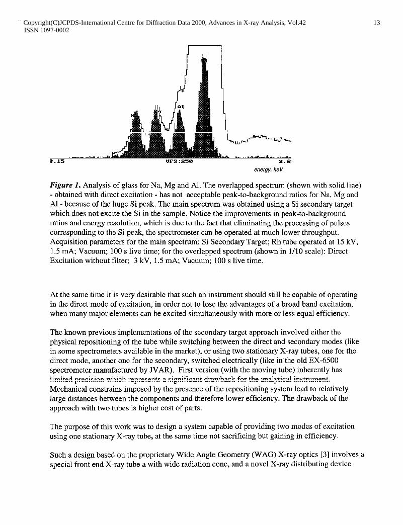
energy, keV
Figure I. Analysis of glass for Na, Mg and Al. The overlapped spectrum (shown with solid line) - obtained with direct excitation - has not acceptable peak-to-background ratios for Na, Mg and Al - because of the huge Si peak. The main spectrum was obtained using a Si secondary target which does not excite the Si in the sample. Notice the improvements in peak-to-background ratios and energy resolution, which is due to the fact that eliminating the processing of pulses corresponding to the Si peak, the spectrometer can be operated at much lower throughput. Acquisition parameters for the main spectrum: Si Secondary Target; Rh tube operated at 15 kV, 1.5 mA; Vacuum; 100 s live time; for the overlapped spectrum (shown in l/10 scale): Direct Excitation without filter; 3 kV, 1.5 mA; Vacuum; 100 s live time.
At the same time it is very desirable that such an instrument should still be capable of operating in the direct mode of excitation, in order not to lose the advantages of a broad band excitation, when many major elements can be excited simultaneously with more or less equal efficiency.
The known previous implementations of the secondary target approach involved either the physical repositioning of the tube while switching between the direct and secondary modes (like in some spectrometers available in the market), or using two stationary X-ray tubes, one for the direct mode, another one for the secondary, switched electrically (like in the old EX-6500 spectrometer manufactured by JVAR). First version (with the moving tube) inherently has limited precision which represents a significant drawback for the analytical instrument. Mechanical constrains imposed by the presence of the repositioning system lead to relatively large distances between the components and therefore lower efficiency. The drawback of the approach with two tubes is higher cost of parts.
The purpose of this work was to design a system capable of providing two modes of excitation using one stationary X-ray tube, at the same time not sacrificing but gaining in efficiency.
Such a design based on the proprietary Wide Angle Geometry (WAG) X-ray optics [3] involves a special front end X-ray tube a with wide radiation cone, and a novel X-ray distributing device
Copyright(C)JCPDS-International Centre for Diffraction Data 2000, Advances in X-ray Analysis, Vol.42 13Copyright(C)JCPDS-International Centre for Diffraction Data 2000, Advances in X-ray Analysis, Vol.42 13ISSN 1097-0002

capable of utilizing this radiation cone in order to switch between two modes of excitation and within each mode, to switch between different filters in the direct mode and different targets in the secondary mode.
The X-ray distributor has 8 positions for the filters (the standard set is : None, None + Collimator, Ti, Fe, Cu, MO, Rh, Sn), and 8 positions for the secondary targets (Al, Si, Ti, Fe, Ge, MO, Sn, Gd as the standard set). The standard X-ray tube anode material is Rh. The thin Beryllium window of the tube (75 cc) enhances the performance for light elements ( Z I 15).
This arrangement allows an extremely close position of the sample relative to the secondary target currently in use. The front part of the X-ray tube housing is designed to allow a close position of the tube anode to the secondary target, and to block the sample and the detector from the direct radiation of the tube. Therefore, the overall efficiency of the new instrument is even a few times higher compared to the known previous implementations - systems with one moving tube and systems with two tubes. For example, the new EX-6600 has efficiency 5 to 13 times higher for direct excitation (Fig.2) and 5 to 7 times higher for secondary excitation (Fig.3) , compared to the EX-6500 spectrometer that was equipped with two tubes, i.e. it can provide with better performance at a lower cost.
- scattering of RhL lines of the tube
UFS : 32688 5.0
keV
Figure 2. Comparison of intensities obtained with a new spectrometer vs. the previous model, for the direct excitation mode. Measuring Fluorine in teflon. Main spectrum was obtained with a new Ex6600 model; Overlapped (shown with solid line) was obtained with previous EX6500 EDXRF machine. Acquisition parameters were the same: Direct excitation without filter; Rh tube operated at 4 kV, 4 mA; in Vacuum; 200 s live time.
Copyright(C)JCPDS-International Centre for Diffraction Data 2000, Advances in X-ray Analysis, Vol.42 14Copyright(C)JCPDS-International Centre for Diffraction Data 2000, Advances in X-ray Analysis, Vol.42 14ISSN 1097-0002
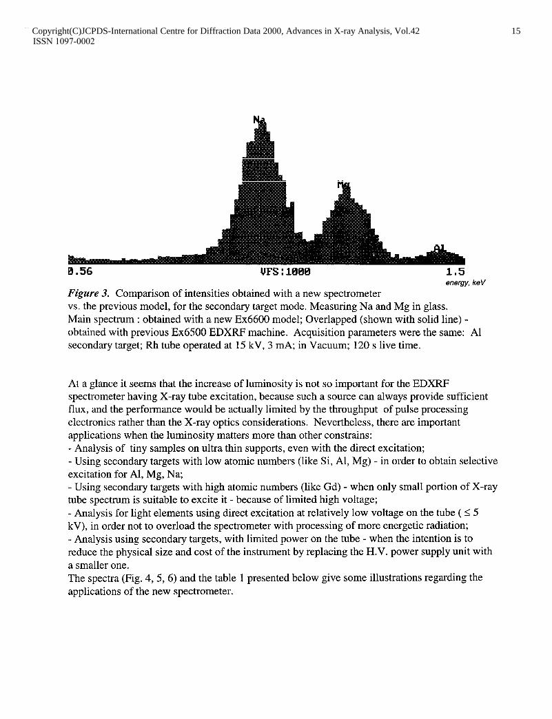
0.56 UFS : 10Ml 1.5 energy, ke V
Figure 3. Comparison of intensities obtained with a new spectrometer vs. the previous model, for the secondary target mode. Measuring Na and Mg in glass. Main spectrum : obtained with a new Ex6600 model; Overlapped (shown with solid line) - obtained with previous Ex6500 EDXRF machine. Acquisition parameters were the same: Al secondary target; Rh tube operated at 15 kV, 3 mA; in Vacuum; 120 s live time.
At a glance it seems that the increase of luminosity is not so important for the EDXRF spectrometer having X-ray tube excitation, because such a source can always provide sufficient flux, and the performance would be actually limited by the throughput of pulse processing electronics rather than the X-ray optics considerations. Nevertheless, there are important applications when the luminosity matters more than other constrains: - Analysis of tiny samples on ultra thin supports, even with the direct excitation; - Using secondary targets with low atomic numbers (like Si, Al, Mg) - in order to obtain selective excitation for Al, Mg, Na; - Using secondary targets with high atomic numbers (like Gd) - when only small portion of X-ray tube spectrum is suitable to excite it - because of limited high voltage; - Analysis for light elements using direct excitation at relatively low voltage on the tube ( 5 5 kV), in order not to overload the spectrometer with processing of more energetic radiation; - Analysis using secondary targets, with limited power on the tube - when the intention is to reduce the physical size and cost of the instrument by replacing the H.V. power supply unit with a smaller one. The spectra (Fig. 4,5,6) and the table 1 presented below give some illustrations regarding the applications of the new spectrometer.
Copyright(C)JCPDS-International Centre for Diffraction Data 2000, Advances in X-ray Analysis, Vol.42 15Copyright(C)JCPDS-International Centre for Diffraction Data 2000, Advances in X-ray Analysis, Vol.42 15ISSN 1097-0002

“FS : .l.emEla l. -9 energy, ke V
Figure 4. Measuring Na content in geological sample. Direct excitation, without filter, the voltage on the X-ray tube was set to 3 kV only in order to decrease the efficiency of excitation for dominant components - Mg, Al & Si. Nevertheless, the Na peak is drowning in the background tails from other peaks.
energy, keV
Figure 5. The same sample, but excitation with Al secondary target. This target doesn’t excite Al, but still excites Mg, so the Na peak hardly can be seen, sitting on the tail of Mg.
energy, ke V
Figure 6. The same sample, but excitation with Mg secondary target, which selectively excites Na (other peaks are due to residual Bremsstrahlung scattering by Mg target). Na peak can be clearly detected. Minimum Detection Limit of NazO in such matrix is 390 ppm.
Copyright(C)JCPDS-International Centre for Diffraction Data 2000, Advances in X-ray Analysis, Vol.42 16Copyright(C)JCPDS-International Centre for Diffraction Data 2000, Advances in X-ray Analysis, Vol.42 16ISSN 1097-0002

Table 1. Detection Limits obtained on the real samples
Element / Matrix
Na20 / geological
Na20 / cement
MgO / geological
MgO / cement
Zn / soil
Ni / soil
Cr I soil
Sr I soil
Rb I soil
Pb I soil
Mn I oil
Fe I oil
cu I oil
Pb / oil
DL, ppm time, s sample / conditions
390
415 to 680
190
400
0.8
0.9
1.4
0.5
0.5
0.9
0.23
0.23
0.27
0.54
1,200
1,200
1,200
1,200
700
700
300
700
700
700
700
700
700
700
MRGl Mg ST
SRM1888 & 1885 Mg ST
MRGl Al ST
SRM1888 & 188.5 Al ST
GBW07403 Ge ST
as above
GBW07403
GBW07404
GBW07308
GBW07308
conostan oil
as above
as above
conostan oil
Fe ST
MO ST
MO ST
MO ST
Ge ST
MO ST
Apart from the basic configuration described above, a few other modifications were developed, one of them featuring the sample chamber suitable for very large and heavy samples. This model is provided -with laser pointing device for targeting specific analysis points on a large sample. The non-contacting down-looking geometry prevents sample surface contamination.
Copyright(C)JCPDS-International Centre for Diffraction Data 2000, Advances in X-ray Analysis, Vol.42 17Copyright(C)JCPDS-International Centre for Diffraction Data 2000, Advances in X-ray Analysis, Vol.42 17ISSN 1097-0002
CONCLUSIONS
A novel EDXRF Analyzer can provide both modes of excitation (direct excitation with/without filters, and secondary target mode) using one stationary medium power X-ray tube. This was achieved not sacrificing efficiency but gaining in both modes.
The instrument has a sample chamber which allows operation in Air atmosphere, Vacuum or Helium. The Si(Li)-detector with ultra thin organic window allows detection of light elements down to Oxygen. A special modification features a chamber for very large samples, with the geometry preventing sample surface contamination.
The variety of possible excitation conditions, in a combination with the friendly software which can handle multi-acquisition regressions or standardless algorithms [4], allows expanding the area of possible applications.
REFERENCES
[l] B. Yokhin, “Efficient Sub-ppm EDXRF Analyzer”, Pittsburgh Conf on Anal. Chem. & Appl. Spectrometry, 1992, New Orleans, Book of Abstracts, 093P.
[2] B. Yokhin, R.C.Tisdale, “High-sensitivity EDXRF Technology”, American Laboratory, 25 (1993) 24C - 28C.
[3] X-ray Fluorescence Analyzer. U.S. patent application filed Feb. 24, 1998. [4] B. Yokhin, “Standardless Fundamental Parameters Program for EDXRF Analyzers”,
Pittsburgh ConJ on Anal. Chem. & Appl. Spectrometry, 1993, Atlanta, Book of Abstracts, 014P.
Copyright(C)JCPDS-International Centre for Diffraction Data 2000, Advances in X-ray Analysis, Vol.42 18Copyright(C)JCPDS-International Centre for Diffraction Data 2000, Advances in X-ray Analysis, Vol.42 18ISSN 1097-0002