Volume 32 Number 5 October 2012 - armatys.fr · were monitored under long-term occlusal function....
11
0 Volume 32 Number 5 October 20 12 9081
Transcript of Volume 32 Number 5 October 2012 - armatys.fr · were monitored under long-term occlusal function....

0
Volume 32 Number 5
October 2012
9081

A Histologic and Clinical Evaluation of Ridge Preservation Following Grafting with Demineralized Bone Matrix, Cancellous Bone Chips, an.d Resorbable Extracellular Matrix Membrane
E. Todd Scheyer, DDS, MS* Peter Schupbach, PhD** Michael K. McGuire, DDS*
In an attempt to reduce postextraction alveolar bone resorption, ridge
preservation grafting procedures with or without resorbable membranes have
become standard-of-care treatments following tooth removal. This prospective
case series examined histologic and clinical outcomes following socket grafting
with a syringeable paste allograft and a resorbable extracellular matrix membrane
at three different time periods following postextraction grafting: 6, 12, and 24
weeks. At each time period, bone core specimens were retrieved for microscopic
examination, and implants were placed. Following prosthetic restoration, implants
were monitored under long-term occlusal function. At all three time periods,
histologic results revealed active bone regeneration. At 6 weeks, localized
areas of woven bone were evident, although nonmineralized osteoid was the
dominant feature. At 12 and 24 weeks, regenerated woven bone dominated the
histologic landscape, with increasing amounts evident in the latter specimens.
Regardless of when implants were placed following grafting, implant survival
under function occurred. (lnt J Periodontics Restorative Dent 2012;32:543-552.)
*Private Practice, Houston, Texas. **Schupbach Ltd, Service and Research Laboratory for Biomaterials, Histology, and Imaging,
Horgen, Switzerland.
Correspondence to: Dr E. Todd Scheyer, 3400 S. Gessner Rd, Suite 102, Houston, TX 77063;
fax: 713-952-0614; email: [email protected].
543
Successful implant placement re
quires adequate alveolar ridge
volume. Effective management
of postextraction sockets and any
associated defects is necessary
to provide sufficient bone for im
plant placement. Multiple causes
of postextraction bone resorption
are known, including bundle bone
resorption, thin and often dehisced
labial or buccal bone, preexisting
periodontal disease/ traumatic ex
tractions, and soft tissue flap reflec
tion during tooth removal. 1- 10
Crestal bone resorption is an
unavoidable consequence of tooth
loss, with much of the remodeling
occurring within the first 3 months
following tooth removal. 1•2 In the
maxillary esthetic zone, volumet
ric bone loss is particularly severe
during the first 6 months follow
ing tooth extraction and contin
ues over time, with as much as an
additional 11% of volumetric loss
during the following 5 years. 11 •12
After 1 year without treatment, up
to 50% loss of alveolar crestal bone
width will likely occur, especially
in the esthetically critical anterior
maxilla.5•13- 15 In a recent systematic
Volume 32, Number 5, 2012
544
review/ alveolar bony dimensional implant survival related to different
changes following tooth removal implant insertion times following
were examined. Across multiple ridge preservation procedures.
studies/ reductions in alveolar The primary purpose of this
width and height consistently oc- case series was to examine the bone
curred/ with loss of width being regenerative outcomes at three dif-
more severe than loss of height. 3 ferent time intervals following ridge
Postextraction bone resorp..:: preservation grafting with DynaBiast
tion can significantly compromise... (Keystone Dental)/ an FDA-cleared
implant placement/ implant-relatedT<;,·.· ... demineraliz;;;d allograft bone rna-
esthetics/ and long-term implant trix/ and DynaMatrix extracellular
survival under function. Although matrix resorbable membrane (Key-
immediate placement of implants stone Dental). DynaBiast (DBM) is
following tooth removal is an ac- a composite graft material of de-
cepted protocol when warranted/ mineralized bone and mineralized
various ridge preservation proto- cancellous bone chips derived from
cols are often indicated prior to the same donor and configured
implant insertion. Multiple case as either a putty or an injectable
reports and case series have ex- paste in a reverse phase resorbable
amined the effectiveness of medium. The extracellular matrix
these protocols in diminishing membrane is obtained from the
postextraction bone resorption submucosa of the small intestine of
and preserving the hard tissue pigs using a process that retains the
morphology critical for effective natural composition of matrix mol-
implant placement. 1•5•8•13 ·16- 22 Com- ecules/ such as types 1/ Ill/ IV and
man to these protocols is the use VI collagens/ gylcoproteins/ proteo-
of osteoconductive and/ at times/ glycans/ gylcosaminoglycans/ and
osteoinductive matrices/ including growth factors.36•37 The secondary
autogenous bone/ mineralized and purpose of this study was to exam-
demineralized freeze-dried or irra- ine the survival of implants placed at
diated allograft/ bovine bone min- each of the different time intervals
eral/ alloplasts/ and others. 16,20,23-34
Along with varying graft matrices/
time from graft placement to im
plant insertion varies widely and
is generally reported to be from 2
to 12 months.26•35 Although robust
bone regeneration is a shared goal
of all postextraction grafting proto
cols/ sparse information is available
describing histologic differences
at various time intervals following
ridge preservation grafting. In ad
dition/ little is known regarding
of abutment connection and at later
time points under occlusal function.
Method and materials
Seventeen patients (5 men/ 12
women; age range/ 32 to 69 years)
who met the inclusion criteria were
included in this study. All patients
either had no significant systemic
disease or were medically well
controlled and taking no steroids/
The International Journal of Periodontics & Restorative Dentistry
bisphosphonates/ or chemothera
peutic drugs and had stopped
cigarette/ cigar/ or pipe smoking at
least 3 weeks prior to study initia
tion. Each patient required a single
tooth extraction followed by sock
et grafting for ridge preservation.
Third molar and mandibular inci
sor teeth were excluded/ as were.
acutely infected teeth. Patients
were assigned randomly to one of
three treatment groups depend
ing on the time interval between
socket grafting and implant place
ment and core retrieval: group A
(6 weeks)/ group B (12 weeks)/ or
group C (24 weeks). Implant sur
vival was examined at abutment
connection and under function at
various long-term time intervals.
Following thorough discussion
of all surgical procedures as well as
potential risks/ each patient agreed
to proceed/ and an informed con
sent form was signed based on the
Helsinki Declaration of 1975/ as re
vised in 2000.
Defect characterization and initial examination
Treated sites were limited to single
maxillary or mandibular extraction
sockets. The majority of sites had
intact buccal walls with adjacent
teeth that had little to no interden
tal bone loss. Two sites exhibited
severe buccal cortical bone loss
and two had the coronal third miss
ing. Tooth extractions in all cases
were considered minimally trau
matic/ with mucoperiosteal flap
reflection allowed as needed to
position the membrane 2 to 3 mm
over the bone margin and under
the soft tissue margin. Those sites
with more extensive bone loss re
quired greater mucoperiosteal flap
reflection to facilitate proper mem
brane placement over the graft
and bone margins. No attempt was
made to obtain primary soft tissue
closure.
For all patients, a standard
head, neck, and oral clinical exami
nation was performed. Periapical
radiographs, computed tomogra
phy scans, and preoperative photo
graphs of the proposed extraction
site were obtained at the screening
visit and at later time points during
the study.
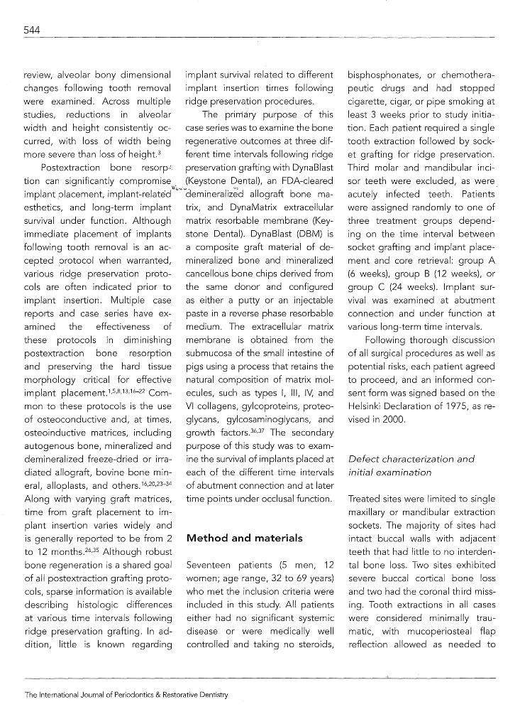
Fig 1 (left) DBM paste was syringed to the alveolar crest immediately following tooth extraction.
Fig 2 (right) Grafted sites were covered with an extracellular resorbable membrane.
Tooth extraction, ridge preservation, and core retrieval procedures
Following administration of local an
esthesia, teeth were removed atrau
matically, followed by thorough
socket debridement. Using a no.
1/4 round bur, internal cortical sock
et walls were perforated to induce
moderate venous bleeding. DBM
paste was then syringed into the
socket to the alveolar crest (Fig 1 ).
An extracellular membrane was
then placed to cover the grafted
sites and host bone margin, and
the flaps were repositioned, but not
closed primarily, via multiple 4.0 re
sorbable sutures (Fig 2).
545
Volume 32, Number 5, 2012

546
Depending on the randomly
selected group, bone core removal,
including the overlying epithelium,
and implant placement were com
pleted at 6, 12, or 24 weeks± 7 days
following socket grafting (Fig 3). Re
trieved bone cores remained in the
trephines and were placed imme
diately into formaldehyde and sent
for microscopic examination. Bone
and tissue level implant insertion
(Keystone Dental) was facilitated by
the EasyGuide (Keystone Dental)
computer-generated implant place
ment system, which is capable of
generating a rigid surgical implant
osteotomy guide. A primary im
plant stability of at least 35 Ncm was
achieved for all 17 implants.
Abutment connection and im
plant restoration time intervals var
ied from 2.5 to 6 months following
implant placement. Figure 4 shows
the 6-month radiograph of a group
B subject in whom abutment con
nection and implant restoration
occurred 3 months following im
plant placement. In this example,
the total time for tooth extraction,
socket grafting, and definitive
implant-supported restoration de
livery was 6 months.
Fig 3 (left) Core specimens, including the overlying epithelium, were obtained at 6, 12, or 24 weeks following extraction and DBM grafting.
Fig 4 (right) Six months after tooth extraction and socket grafting, radiographic evidence suggests good bone-to-implant contact and sLiccessful osseointegration in this group B patient.
Light microscopy
All cores from groups A, B, and C
were fixed in formaldehyde, de
hydrated in an increasing series of
alcohol rinses, infiltrated with Tec
novit 7200 + BPO resin (Heraeus
Kulzer), and embedded. Unde
calcified ground sections (one for
each trephine) were made with a
thickness of approximately 50 fJm
and stained with Sanderson's Rapid
Bone Stain (Dorn & Hart Micro
edge). Slides were digitally photo
graphed using a Leica DM 6000B
light microscope.
Backscatter scanning electron microscopy
The remaining resin block contain
ing the longitudinally cut trephine
was polished and sputter-coated
with a 6-nm carbon layer using a
Bai-Tec SCD 500 sputter device.
The specimens were evaluated
with the backscatter detector us
ing a Zeiss 40BP scanning electron
microscope. The resulting images
were compiled to create an over
view image.
The International Journal of Periodontics & Restorative Dentistry
Results
Clinical findings
Immediate postgrafting healing for
all patients was uneventful, with
minimal swelling and inflamma
tion and no signs of postoperative
infection. No adverse events oc
curred during the interval between
ridge preservation grafting and im
plant insertion.
At implant placement, re-epi
thelialization at the alveolar crest
was evident in all patients. Soft tis
sues remained healthy, with mini
mal to no inflammation and without
signs of infection. Following con
servative mucoperiosteal flap re
flection, clinical evidence of bone
regeneration was readily evident in
groups B and C. At both 12 and 24
weeks, implant insertion proceed
ed uneventfully. Group A patients
at 6 weeks exhibited less volume
of regenerated coronal bone, al
though implant placement with pri
mary apical fixation in native bone
proceeded without complication in
each patient in group A.
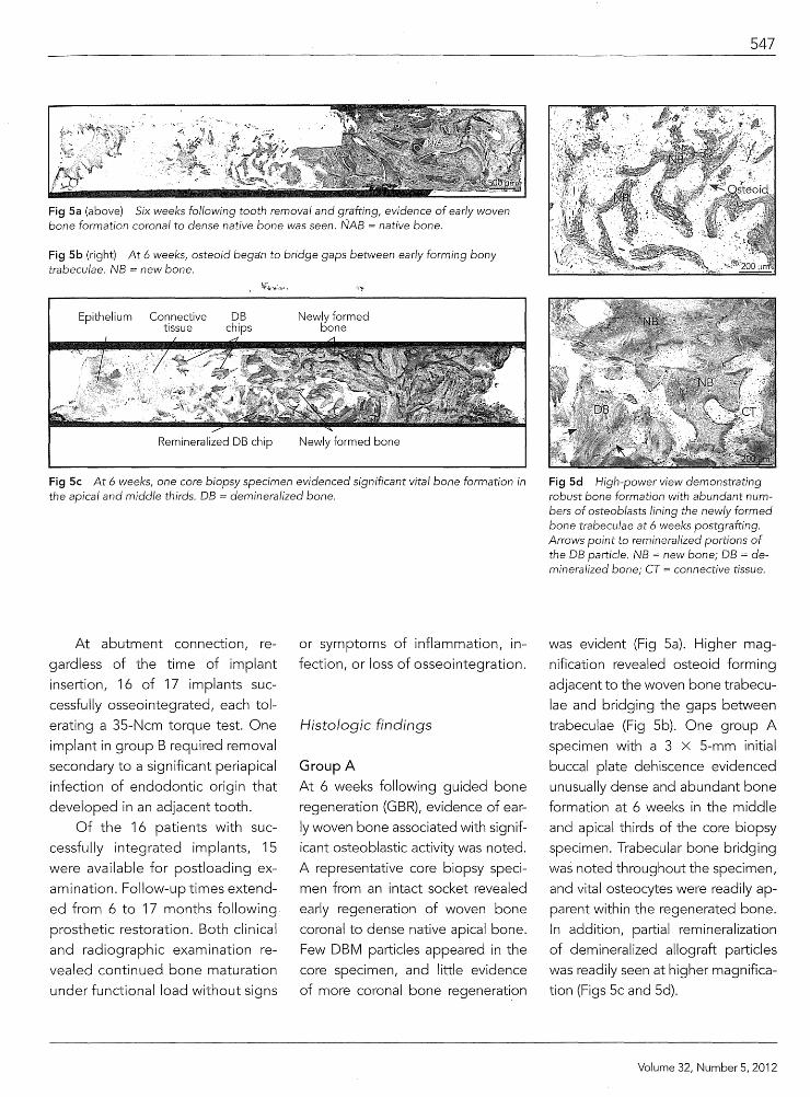
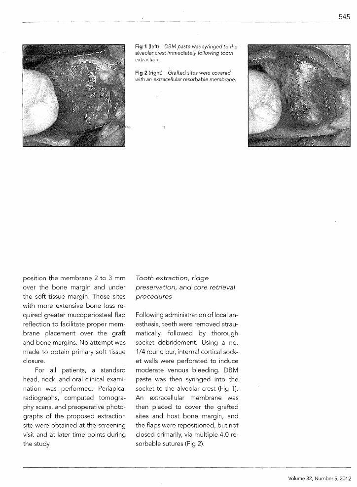
Fig Sa (above) Six weeks following tooth removal and grafting, evidence of early woven bone formation coronal to dense native bone was seen. NAB native bone.
Fig Sb (right) At 6 weeks, osteoid bega·n to bridge gaps between early forming bony trabeculae. NB new bone.
Epithelium Connective tissue
DB chips
Newly formed oone
Fig Sc At 6 weeks, one core biopsy specimen evidenced significant vital bone formation in the apical and middle thirds. DB = demineralized bone.
At abutment connection, re
gardless of the time of implant
insertion, 16 of 17 implants suc
cessfully osseointegrated, each tol
erating a 35-Ncm torque test. One
implant in group B required removal
secondary to a significant periapical
infection of endodontic origin that
developed in an adjacent tooth.
Of the 16 patients with suc
cessfully integrated implants, 15
were available for postloading ex
amination. Follow-uptimes extend
ed from 6 to 17 months following
prosthetic restoration. Both clinical
and radiographic examination re
vealed continued bone maturation
under functional load without signs
or symptoms of inflammation, in
fection, or loss of osseointegration.
Histologic findings
Group A
At 6 weeks following guided bone
regeneration (GBR), evidence of ear
ly woven bone associated with signif
icant osteoblastic activity was noted.
A representative core biopsy speci
men from an intact socket revealed
early regeneration of woven bone
coronal to dense native apical bone.
Few DBM particles appeared in the
core specimen, and little evidence
of more coronal bone regeneration
547
Fig Sd High-power view demonstrating robust bone formation with abundant numbers of osteoblasts lining the newly formed bone trabeculae at 6 weeks postgrafting. Arrows point to remineralized portions of the DB particle. NB = new bone; DB = demineralized bone; CT = connective tissue.
was evident (Fig Sa). Higher mag
nification revealed osteoid forming
adjacent to the woven bone trabecu
lae and bridging the gaps between
trabeculae (Fig 5b). One group A
specimen with a 3 X 5-mm initial
buccal plate dehiscence evidenced
unusually dense and abundant bone
formation at 6 weeks in the middle
and apical thirds of the core biopsy
specimen. Trabecular bone bridging
was noted throughout the specimen,
and vital osteocytes were readily ap
parent within the regenerated bone.
In addition, partial remineralization
of demineralized allograft particles
was readily seen at higher magnifica
tion (Figs 5c and 5d).
Volume 32, Number 5, 2012

548
Group B
At 12 weeks following GBR/ sig
nificant vital bone formation had
occurred. An intact core from a
mandibular second molar site
with an initial 3-mm buccal furca
tion bone loss revealed actively
regenerating woven bone in both
the middle and apical thirds of
the retrieved core (Fig 6a). Higher
CBC
Fig 6b (left) Higher magnification demonstrated newly formed bone surrounding and replacing particles of demineralized allograft. Arrows point to newly formed bone replacing portions of a large DBM particle. NB = new bone; DB = demineralized bone.
Fig 6c (right) In this magnified view, new bone is seen forming along pore surfaces found within the demineralized allograft. CT = connective tissue; DB = demineralized bone; NB = new bone.
Fig 7 At 12 weeks, mono/ayers of osteob/asts were seen actively secreting osteoid that bridged the gaps between grafted DBM particles and surrounding newly formed woven bone. CT = connective tissue; DB demineralized bone; NB new bone.
magnification demonstrated newly
formed woven bone surrounding
and replacing particles of deminer
alized allograft (Fig 6b). At higher
power/ regenerated bone was seen
forming against surfaces of pores
found within DBM particles (Fig 6c).
In a mandibular first molar site
with an initial Class Ill furcation
defect/ intense osteogenesis was
The International Journal of Periodontics & Restorative Dentistry
Fig 6a Twelve weeks following DBM grafting, woven bone regeneration was readily seen in the apical and middle thirds of the retrieved bone core. E = epithelium; CT =
connective tissue; CBC = cancellous bone chips; NB = new bone; DB = demineralized bone.
readily apparent at 12 weeks. Ad
vancing fronts of osteoblasts ac
tively secreted significant amounts
of osteoid that bridged the gaps
between grafted particles/ suggest
ing ongoing regeneration of vital
bone. As in the 6-week specimens/
partial remineralization of deminer
alized allograft particles was noted
at higher power (Fig 7).
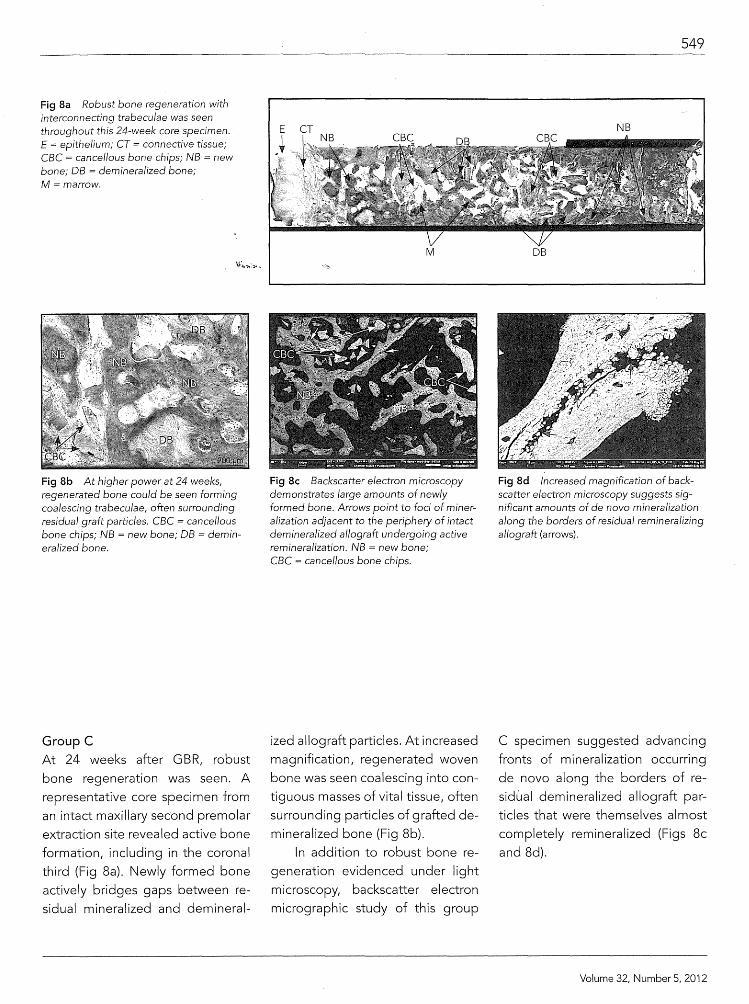
Fig 8a Robust bone regeneration with interconnecting trabeculae was seen throughout this 24-week core specimen. E = epithelium; CT = connective tissue; CBC = cancellous bone chips; NB = new bone; DB = demineralized bone; M =marrow.
Fig 8b At higher power at 24 weeks, regenerated bone could be seen forming coalescing trabeculae, often surrounding residual graft particles. CBC cancellous bone chips; NB = new bone; DB = demineralized bone.
Group C At 24 weeks after GBR, robust
bone regeneration was seen. A
representative core specimen from
an intact maxillary second premolar
extraction site revealed active bone
formation, including in the coronal
third (Fig 8a). Newly formed bone
actively bridges gaps between re
sidual mineralized and demineral-
M
Fig 8c Backscatter electron microscopy demonstrates large amounts of newly formed bone. Arrows point to foci of mineralization adjacent to the periphery of intact demineralized allograft undergoing active remineralization. NB = new bone; CBC cancellous bone chips.
ized allograft particles. At increased
magnification, regenerated woven
bone was seen coalescing into con
tiguous masses of vital tissue, often
surrounding particles of grafted de
mineralized bone (Fig 8b).
In addition to robust bone re
generation evidenced under light
microscopy, backscatter electron
micrographic study of this group
549
DB
Fig 8d Increased magnification of backscatter electron microscopy suggests significant amounts of de novo mineralization along the borders of residual remineralizing allograft (arrows).
C specimen suggested advancing
fronts of mineralization occurring
de novo along the borders of re
sidual demineralized allograft par
ticles that were themselves almost
completely remineralized (Figs 8c
and 8d).
Volume 32, Number 5, 2012
550
Discussion
Following tooth removal, crestal
bone resorption begins almost
immediately and is the inevitable
consequence of alveolar bone's
dependence on teeth to main
tain anatomically driven volumet
ric stability. 1•2
·11
•12 In an attempt
to minimize postextraction bone
resorption and maintain essential
crestal bone morphology prior to
implant placement, ridge preserva
tion procedures have become the
standard of care following tooth
removal. However, few studies
have examined human histologic
healing of grafted extraction sock
ets at very early time points, ie, 6
weeks postgrafting, followed by
long-term clinical follow-up of im
plant survival.28•38 This case series
was one such attempt at examining
these parameters.
In this study, new bone for
mation was examined histologi
cally at three different time points
after tooth extraction and socket
grafting with a composite demin
eralized and mineralized allograft
matrix used in conjunction with
an overlying resorbable collagen
membrane. Significant differences
in bone regeneration were seen
at each of the three time points.
Core specimens at 6 weeks (group
A) revealed significant osteoblas
tic activity leading to nonmineral
ized osteoid with areas of early
woven bone formation. One early
responder in group A exhibited a
more advanced regenerative re
sponse compared to other group
A subjects at 6 weeks. At both 12
and 24 weeks, osteoid production
was also accompanied by signifi
cant woven bone formation. At 12
weeks postgraftir1g (group B), new
bone regeneration, while pres
ent elsewhere, was rarely noted
at the coronal third in the core bi-
. opsy specimens. In contrast, by 24
~~NYgeks (group .C), evidence of new ~ ~,., ....... ~ ·.;f.'
vital bone formation was apparent
throughout the core specimen.
Of particular interest was the
finding of dot-like foci of mineraliza
tion noted via backscatter electron
microscopy in one group C speci
men occurring along the borders
of remineralizing demineralized al
lograft particles. First described by
Groenveld et al, 39 acellular remin
eralization within demineralized
allograft particles in areas remote
from vital bone appears as a nec
essary prerequisite for subsequent
remodeling and replacement of al
lograft with newly regenerated vital
bone. The dot-like foci of mineral
ization occurring along the borders
of the allograft particles appear
spatially related to varying fronts
of active mineralization (ie, oste
oid to mineralized bone, predentin
to dentin). Active mineralization of
demineralized allograft particles
was also noted in the 12-week his
tologic sections of group B and in
the regenerative advanced 6-week
group A specimen.
Regardless of the duration
between socket grafting and im
plant placement, 16 of 17 implants
survived at abutment connec
tion as well as under full occlusal
function, with the one unsuccess
ful implant resulting from acute
The International Journal of Periodontics & Restorative Dentistry
periapical infection of an adjacent
tooth. Osseointegration occurred
independent of when grafting oc
curred and showed no evidence of
compromise throughout the study.
The presence of intense osteoblas
tic activity, especially at earlier time
points, suggests that active ongoing
bone regeneration may have con
tributed to implant survival. Results
of this study suggest that staged
protocols of tooth extraction, GBR
with effective graft matrices and
barrier membranes, and delayed
implant placement may allow for
earlier implant insertion times with
out compromising long-term im
plant survival. Other variables such
as adequate primary stability, the
type of implant surface at the bone
to-implant interface, and vectors of
occlusal force are equally important
determinants for long-term implant
survival, especially in the accelerat
ed implant placement and loading
protocols highlighted in this study.
The bone matrix examined in
this case series along with the re
sorbable extracellular matrix mem
brane appeared effective for GBR
ridge preservation procedures at
all three time periods, followed by
stable long-term implant survival.
This case series study, while prom
ising, was insufficiently powered to
allow for quantitative histomorpho
metric analysis. Further prospective
randomized trials with both test
and control groups and sufficient
power to allow for both qualitative
and quantitative examination are
needed to fully evaluate the po
tential of both the graft matrix and
membrane used in this study.

References
1. Schropp L, Wenzel A, Kostopoulos L, Karring T. Bone healing and soft tissue contour changes following single-tooth extraction: A clinical and radiographic 12-month prospective study. lnt J Periodontics Restorative Dent 2003;23:313-323.
2. Cardaropoli G, Araujo M, Lindhe J. Dynamics of bone tissue formation in to9th extraction sites. An experimental study" in dogs. J Clin Periodontol2003;30:809-818.
3. Vander Weijden F, Deii'Acqua F, Slot c51~:··--·· Alveolar bone dimensional changes of post-extraction sockets in humans: A systematic review. J Clin Periodontal 2009; 36:1048-1058.
4. Araujo MG, Lindhe J. Dimensional ridge alterations following tooth extraction. An experimental study in the dog. J Clin Periodontal 2005;32:212-218.
5. Nevins M, Camelo M, De Paoli S, et al. A study of the fate of the buccal wall of extraction sockets of teeth with prominent roots. lnt J Periodontics Restorative Dent 2006;26:19-29.
6. Wood DL, Hoag PM, Donnenfeld OW, Rosenfeld LD. Alveolar crest reduction following full and partial thickness flaps. J Periodontal 1972;43: 141-144.
7. Bragger U, Pasquali L, Kornman KS. Remodeling of interdental alveolar bone after periodontal flap procedures assessed by means of computer-assisted densitometric image analysis (CADIA). J Clin Periodontal 1988;15:558-564.
8. Araujo MG, Lindhe J. Ridge alterations following tooth extraction with and without flap elevation: An experimental study in the dog. Clin Oral Implants Res 2009; 20:545-549.
9. Fickl S, Zuhr 0, Wachtel H, Bolz W, Huerzeler M. Tissue alterations after tooth extraction with and without surgical trauma: A volumetric study in the beagle dog. J Clin Periodontal 2008;35:356-363.
10. Fickl S, Schneider D, Zuhr 0, et al. Dimensional changes of the ridge contour after socket preservation and buccal overbuilding: An animal study. J Clin Periodontal 2009;36:442-448.
11. Nemcovsky CE, Serfaty V. Alveolar ridge preservation following extraction of maxillary anterior teeth. Report on 23 consecutive cases. J Periodontal 1996;67: 390-395.
12. Artzi Z, Nemcovsky CE. The application of deproteinized bovine bone mineral for ridge preservation prior to implantation. Clinical and histological observations in a case. report. J Periodontal 1998;69: 1062:-1067.
13. Lekovic V, Camargo PM, Klokkevold PR, et al. Preservation of alveolar bone in extraction sockets using bioabsorbable membranes. J Periodontal 1998;69: 1044-10:49.
14. Boyne PJ. Osseous repair of the postextraction alveolus in man. Oral Surg Oral Med Oral Pathol 1966;21 :805-813.
15. Sevor JJ, Meffert R. Placement of implants into fresh extraction sites using a resorbable collagen membrane: Case reports. Pract Periodontics Aesthet Dent 1992; 4(3):35-41 .
16. Bartee BK. Extraction site reconstruction for alveolar site preservation. Part 1: Rationale and materials selection. J Oral lmplantol 2001 ;27:187-193.
17. Bartee BK. Extraction site reconstruction for alveolar site preservation. Part 2: Membrane-assisted surgical technique. J Orallmplantol 2001 ;27:194-197.
18. ArtziZ, Tal H, Dayan D. Porous bovine bone mineral in healing of human extraction sockets. Part 1: Histomorphometric evaluations at 9 months. J Periodontal 2000; 71:1015-1023.
19. Fugazzotto PA. GBR using bovine bone matrix and resorbable and nonresorbable membranes. Part 2: Clinical results. lnt J Periodontics Restorative Dent 2003;23: 599-605.
20. Serino G, Biancu S, Iezzi G, Piattelli A. Ridge preservation following tooth extraction using a polylactide and polyglycolide sponge as space filler: A clinical and histological study in humans. Clin Oral Implants Res 2003;14:651-658.
21. Wang HL, Kiyonobu K, Neiva RF. Socket augmentation: Rationale and technique. Implant Dent 2004;13:286-296.
22. Barone A, Aldini NN, Fini M, Giardino R, Calvo Guirado JL, Covani U. Xenograft versus extraction alone for ridge preservation after tooth removal: A clinical and histomorphometric study. J Periodontal 2008;79:1370-1377.
551
Volume 32, Number 5, 2012

552
23. lasella JM, Greenwell H, Miller RL, et al. Ridge preseNation with freeze-dried bone allograft and a collagen membrane compared to extraction alone for implant site development: A clinical and histologic study in humans. J Periodontal 2003;74: 990-999.
24. Froum SJ, Wallace SS, Elian N, Cho SC, Tarnow DP. Comparison of mineralized cancellous bone allograft (Puros) and anorganic bovine bone matrix (Bio-Oss). for sinus augmentation: Histomorphom
29. Vasilic N, Henderson R, Jorgenson T, Sutherland E, Carson R. The use of bovine porous bone mineral in combination with collagen membrane or autologous fibrinogenlfibrpnectin system for ridge preseNation following tooth extraction. J Okla Dent Assoc 2003;93(4):33-38.
30. Sclar AG. Ridge preseNation for optimum esthetics and function. Postgrad Dent 1999;6(2):3-11.
etry at 26 to 32 weeks after grafting. lntw;.-~:,. .. J Periodontics Restorative Dent 2006;26: 543-551.
31. Sclar AG. Strategies for management of single-tooth extraction sites in aesthetic implant thwapy. J Oral Maxillofac Surg 2004;62(suppl 2):90-1 05.
32. Fowler EB, Breault LG, Rebitski G. Ridge preseNation utilizing an acellular dermal allograft and demineralized freeze-dried bone allograft: Part II. Immediate endosseous implant placement. J Periodontal 2000;71 :1360-1364 [erratum 2000; 71 :1670].
25. Wang HL, Tsao YP. Histologic evaluation of socket augmentation with mineralized human allograft. lnt J Periodontics Restorative Dent 2008;28:231-237.
26. Beck TM, Mealey BL. Histologic analy-sis of healing after tooth extraction with ridge preseNation using mineralized human bone allograft. J Periodontal 201 0; 81:1765-1772.
27. Carmagnola D, Adriaens P, Berglundh T. Healing of human extraction sockets filled with Bio-Oss. Clin Oral Implants Res 2003;14:137-143.
28. Heberer S, AI-Chawaf B, Hidebrand D, Nelson JJ, Nelson K. Histomorphometric analysis of extraction sockets augmented with Bio-Oss Collagen after a 6-week healing period: A prospective study. Clin Oral Implants Res 2008;19:1219-1225.
33. lrinakis T, Tabesh M. PreseNing the socket dimensions with bone grafting in single sites: An esthetic surgical approach when planning delayed implant placement. J Orallmplantol 2007;33:156-163.
34. Sandor GK, Kainulainen VT, Oueiroz JO, Carmichael RP, Oikarinen KS. PreseNation of ridge dimensions following grafting with coral granules of 48 post-traumatic and post-extraction dento-alveolar defects. Dent Traumatol 2003;19:221-227.
The International Journal of Periodontics & Restorative Dentistry
35. Darby I, Chen ST, Buser D. Ridge preseNation techniques for implant therapy. lnt J Oral Maxillofac Implants 2009;24 (suppl):260-271.
36. Nihsen ES, Johnson CE, Hiles MC. Bioactivity of small intestinal submucosa and oxidized regenerated cellulose/collagen. Adv Skin Wound Care 2008;21 :479-486.
37. Hodde J, Janis A, Ernst D, Zopf D, Sherman D, Johnson C. Effects of sterilization on an extracellular matrix scaffold: Part 1. Composition and matrix architecture. J Mater Sci Mater Med 2007;18:537-543 ..
38. Chen ST, Wilson TG Jr, Hammerle CH. Immediate or early placement of implants following tooth extraction: Review of biologic basis, clinical procedures, and outcomes. lnt J Oral Maxillofac Implants 2004;19(supp1):12-25.
39. Groenveld EHJ, van den Bergh JPA, Holzmann P, ten Bruggenkate CM, Tuinzing DB, Burger EH. Mineralization processes in demineralized bone matrix grafts in human maxillary sinus floor elevations. J Biomed Mater Res 1999;48:393-402.