Nucleic Acids and Protein Synthesis. What are nucleic acids?
Volume 17 Number 15 1989 Nucleic Acids Research
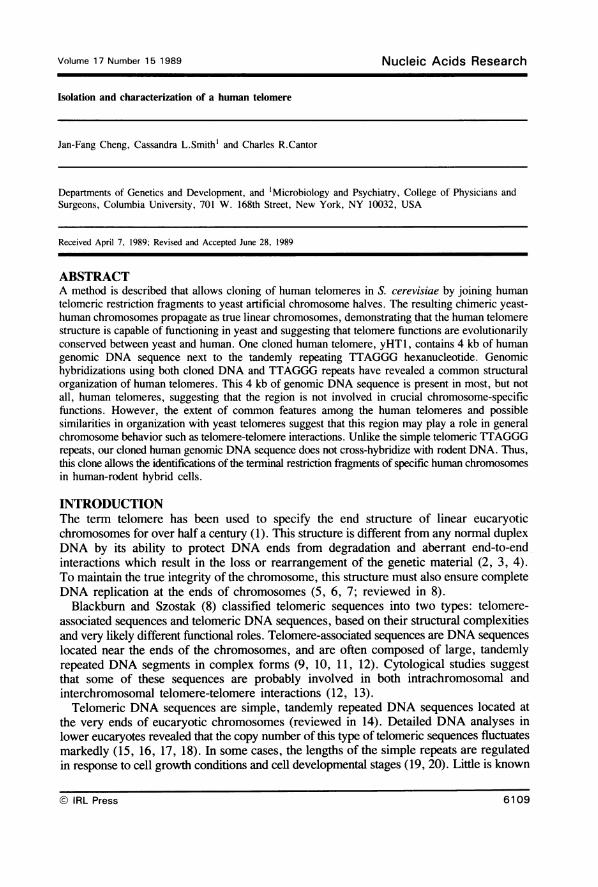
Isolation and characterization of a human telomere
Jan-Fang Cheng, Cassandra L.Smithl and Charles R.Cantor
Departments of Genetics and Development, and 'Microbiology and Psychiatry, College of Physicians andSurgeons, Columbia University, 701 W. 168th Street, New York, NY 10032, USA
Received April 7, 1989; Revised and Accepted June 28, 1989
ABSTRACTA method is described that allows cloning of human telomeres in S. cerevisiae by joining humantelomeric restriction fragments to yeast artificial chromosome halves. The resulting chimeric yeast-human chromosomes propagate as true linear chromosomes, demonstrating that the human telomerestructure is capable of functioning in yeast and suggesting that telomere functions are evolutionarilyconserved between yeast and human. One cloned human telomere, yHTl, contains 4 kb of humangenomic DNA sequence next to the tandemly repeating TTAGGG hexanucleotide. Genomichybridizations using both cloned DNA and TTAGGG repeats have revealed a common structuralorganization of human telomeres. This 4 kb of genomic DNA sequence is present in most, but notall, human telomeres, suggesting that the region is not involved in crucial chromosome-specificfunctions. However, the extent of common features among the human telomeres and possiblesimilarities in organization with yeast telomeres suggest that this region may play a role in generalchromosome behavior such as telomere-telomere interactions. Unlike the simple telomeric TTAGGGrepeats, our cloned human genomic DNA sequence does not cross-hybridize with rodent DNA. Thus,this clone allows the identifications of the terminal restriction fragments of specific human chromosomesin human-rodent hybrid cells.
INTRODUCTIONThe term telomere has been used to specify the end structure of linear eucaryoticchromosomes for over half a century (1). This structure is different from any normal duplexDNA by its ability to protect DNA ends from degradation and aberrant end-to-endinteractions which result in the loss or rearrangement of the genetic material (2, 3, 4).To maintain the true integrity of the chromosome, this structure must also ensure completeDNA replication at the ends of chromosomes (5, 6, 7; reviewed in 8).Blackburn and Szostak (8) classified telomeric sequences into two types: telomere-
associated sequences and telomeric DNA sequences, based on their structural complexitiesand very likely different functional roles. Telomere-associated sequences are DNA sequenceslocated near the ends of the chromosomes, and are often composed of large, tandemlyrepeated DNA segments in complex forms (9, 10, 11, 12). Cytological studies suggestthat some of these sequences are probably involved in both intrachromosomal andinterchromosomal telomere-telomere interactions (12, 13).Telomeric DNA sequences are simple, tandemly repeated DNA sequences located at
the very ends of eucaryotic chromosomes (reviewed in 14). Detailed DNA analyses inlower eucaryotes revealed that the copy number of this type of telomeric sequences fluctuatesmarkedly (15, 16, 17, 18). In some cases, the lengths of the simple repeats are regulatedin response to cell growth conditions and cell developmental stages (19, 20). Little is known
© IRL Press
Volume 17 Number 15 1989 Nucleic Acids Research
6109
n a~~~~~~~)
-EE EEco
-CO-CO-c r(00 000 CY0
00
o
0
Ecoco cc c
o : : d cc co0'00 j0 - - ---~
.0~~~E00000 a jj=e' c.Q-C
0~00
0 000 0o0ou,Z qZZZZ0u = c X = i X e = o°Ue I -
oo0D 0
0 0rC) C\
ac c
0)n ~~~~L
U)
' <> E Eo co 0
E E E E E E00
CCCoo CoCC
C0 0 0 0
-N
Cs 0
_ .. _-
Cs
+. I.C"..r
w- C.D -
x 0 xx 4- Y
,v _
U) U) U) U
0- 0-U)-00n0n 0n no:oo--.oC, o, CLDE DE D E E
CC CC C CC C C
E E E E E E E E EI=I 11I I I I
co r- CO0o aw"tC (OIlILn*Do.C
.FaU)0-rC
0 CC O00..9-0 _
c _
O u
E-0
C -0O. 'Q M-O ,c_ _
E E0oo oxIIZ Z L) L
cs cm cw N
E E E E0 en 0 0
CM~ ~ ~ U U) U)U)CW ;0 @@ @ @ z c Qc0 0
cm cn CNsn sn ns 0E E E E
0 0 0 0 0 E E E E
.c .C .C .C .C v oC CCC0 C C
E E E E Er c c -' -C > -__
0 0 _ _ _
cm CD 4m co 0 _ 0 _c c c c c zo c c cc 0 c c c ctc cC CC 00CC
._ ._ ._ ._ ._ ._
0 0 0 0) 0) 0) 0) 0) 0)
0 0 0) 0 0) 0 0 U~ UC C CC CC CCC0 0 0 0 0 0 000.000000000Y X
Dc>: *0_ _
-D>
0 0
E E°E a 'E
E
IcDc
CC
0CD clOLn _- < Is {
C C. U.awIZ O 3. LL , I
' 0n n> > > >.C C c c
c c c c
E E E E3 :: I I
I- If,v I rl
ICCCC00006EEEEz'IUII
CD
n N- 0
I. C# LL
>
2
cr
I rl- ccC: CM
cQ.0C.)
C~00
.2
as
0
0
2
U)
ccYa
0C
ZY
0
c
U)
cn0co
z
-
Nucleic Acids Research
0
cT)00r0
C2
a0
C
0
C
0.I.0
0
0
L)
0C
0
-6U)
00E0-i
f-g0n
n
0c
0C.)'0c
c'C
0
U)0
E0c
0E
0
-
-j
0
0
6110
I
I
I
Nucleic Acids Research
about the cellular signals that regulate the length variation of this telomeric sequence. DNAsequence comparisons and cross-hybridization experiments with telomeric DNAs showremarkable evolutionary con-servation among lower eucaryotes (14), monocot and dicotplants (21), human (22), and many other organisms (23). In yeast, Tetrahymena telomericsequences allow a linear plasmid DNA to act as a linear self-replicating DNA molecule.This linear DNA was also used as a vector to clone yeast chromosomal telomeres (24).In a similar study, Pluta et al. (25) showed that the terminal restriction fragments fromOxytricha chromosomes, whose telomeres share no common structural feature with yeastexcept short repeating sequences, are still capable of converting a yeast plasmid to actas an extrachromosomal linear DNA molecule. These results demonstrate two importantfacts that encouraged the cloning of human telomeres described in this paper: First, thesetelomeric simple repeating sequences contain the structural information essential for therecognition of telomere functions. Second, these telomere functions are highly conservedeven in nonhomologous systems.
Isolation of human telomeres and adjacent DNA sequences is of great interest for severalreasons. First, it will accelerate the physical mapping of human chromosomes becausethese regions define the ends of the physical maps. Second, it provides critical probesto hunt for disease genes located near the telomeres, such as the Huntington's Diseasegene on the short arm of chromosome 4 (26) and the polycystic kidney disease gene onthe short arm of chromosome 16 (27). Third, since high frequency recombination occursat the ends of chromosomes, it will be intriguing to learn what types of DNA structuresor genes are located near the telomeres.
Clones containing telomeres are apparently not present in ordinary genomic DNA librariesbecause telomere end sequences are incompatible with conventional cloning. Most recenttelomere studies have been limited to the lower eucaryotes or organisms with a high ratioof telomeres to total DNA content (21). We wished to set up a standard procedure forcloning telomeric restriction fragments from higher eucaryotes that contain low ratios oftelomeres to DNA content. Chimeric yeast-human artificial chromosomes were generatedby taking advantage of the yeast artificial chromosome (YAC) system developed by Burkeet al. (28). These clones contain both the telomeric TTAGGG repeating sequences andDNA sequences adjacent to these repeats. Our studies on one cloned human telomere andits adjacent DNA sequence show that the human telomeric conserved sequences extendmany kb beyond the simple repeats. One DNA segment within this cloned region appearsto be human specific. This finding provides a useful probe for identifying the most distalrestriction fragment of a specific human chromosome in human-rodent hybrid cells.
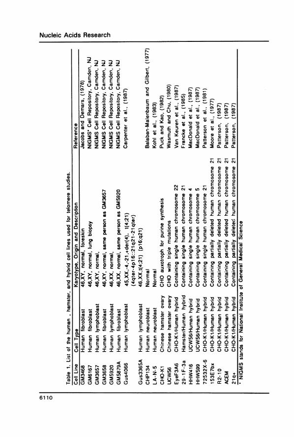
EXPERIMENTAL PROCEDURESCell LinesAll cell lines used in this paper are listed in Table 1. Cell lines GM3468, GM6167, GM3657,GM3658, GM5920, GM5879A, CHP134, LA-N-5, 29-lF-3a and CHO-KI were grownin RPMI 1640 medium supplemented with 10% fetal bovine serum (FBS), 100 units/mlpenicillin and 100 mg/ml streptomycin (PS). EyeF3A6 cells were grown in the same RPMImedium using 10% dialyzed FBS. Cell lines Gus4066, Gus3365, UCW56, HHW416 andHHW599 were grown in Iscove's Modified Delbecco's Medium (IMDM) supplementedwith 10% FBS and PS. Cell lines 72532X-6, 153E7bx, R2-10 and ACEM were grownin MEM Alpha and HAM F12 (1:1), supplemented with 10% dialyzed fetal calf serum(FCS) and PS. Cell line 21q+ was grown in MEM (without glycine) supplemented with
6111
Nucleic Acids Research
10% FCS, PS, 2 mM glutamine and 2 mM proline. All media and supplements werepurchased from GIBCO and Cellgro. The incubation temperatures for HHW416 andHHW599 were 34WC and 39°C, respectively. All other cell lines were grown at 37°C.Pulsed Field Gel ElectrophoresisYeast transformants were grown overnight in a 5 ml synthetic medium lacking tryptophanat 30°C, and cell concentrations were determined using a hemocytometer. Cells wereharvested at 3000 rpm for 10 min in a clinical centrifuge, and washed once with 3 mlof 50 mM EDTA (pH 7.5). Cells were pelleted, resuspended and incubated at 37°C in1 ml of 1 M sorbitol, 0.1 M EDTA (pH 7.5), and 0.1 mg/ml zymolyase 60T (SeikagakuAmerica, Inc.) for 1 hr. Spheroplasts were harvested at 1000 rpm for 5 min, and washedonce with 3 ml 1 M sorbitol and 0.1 M EDTA (pH 7.5). Yeast DNAs were preparedin low gelling-temperature agarose at a concentration of 108 cells/ml (29). PFGelectrophoresis of yeast DNAs was carried out using the Pharmacia-LKB Pulsaphorapparatus with 10-sec pulse times at an electric field strength of 10 V/cm for 30 hr.Mammalian DNAs were prepared in low gelling-temperature agarose and digested with
Not I restriction enzyme (29). DNA fragments were then separated by PFG using a 100-secpulse time at 10 V/cm for 40 hr.Genomic HybridizationSouthern blotting and hybridization of yeast and mammalian genomic DNAs employednytran filters (Schleicher and Schuell) and a modified procedure of Church and Gilbert(30). The hybridization reactions were done at 65°C for 14-16 hr in 0.5 M NaHPO4(pH 7.2), 7% SDS, 1 mM EDTA, 100 mg/ml sheared salmon sperm DNA and approximate5 ng/ml radiolabeled DNA probes. (TTAGGG)7 was radiolabeled with [-y-32P]ATP (3000Ci/mM, Amersham) and T4 polynucleotide kinase (New England Biolabs, Inc.) to a specificactivity of 1 x 10⁷ cpm/mg. All other probes were labeled with [ca-32P]dCTP (3000Ci/mM) in random priming reactions (31) to specific activities ranging from 108 to 109cpm/yg. Filters were washed twice at 650C in 2 x SSC (l x SSC = 0.15 M NaCl, 0.015M sodium citrate, pH 7.5) and 1 % SDS. Filters were reused after stripping off the probesby incubating them in H20 for 30 min at 68°C and then in 50% deionized formamidefor another 30 min at 68°C.Molecular Cloning of Human TelomeresHigh molecular weight genomic DNA was prepared from a human fibroblastoid cell line,GM3468 (32, 33), in 0.5% low gelling-temperature agarose (Seaplaque, FMC, Inc.) tominimize DNA breakage (29, 34). DNA prepared from 106 cells was completely digestedwith EcoR I in agarose. The agarose was melted at 650C, and removed by phenol extractionsand phenol/chloroform/isoamyl alcohol (25:24: 1) extractions. The DNA fragments wereprecipitated with 2 M ammonium acetate and ethanol. Linearized EcoR I fragments werecircularized at a concentration of 0.5 Alg/lm with 5 units/ml T4 DNA ligase (New EnglandBiolabs, Inc.) at 4°C for 12 hr, and precipitated with ethanol. The telomeric EcoR Ifragments should retain their linear configurations.pYAC4 is a bacterial plasmid containing all necessary DNA sequences for constructing
yeast artificial chromosome (28). Plasmid pYAC4 was digested with EcoR I and BamHI to generate a 6.0 kb EcoR I-BamH I fragment. This fragment was isolated on a DEAEmembrane (NA45, Schleicher and Schuell) following gel electrophoresis. The 5' phosphateswere removed by treatment with calf intestinal phosphatase (Boehringer MannheimBiochemicals). Ligation of this 6.0 kb fragment with human EcoR I fragments was carriedout at 10°C for 12 hr using 20 units/ml T4 DNA ligase at a human DNA concentration
6112

Nucleic Acids Research
DNAI
|pYAC4 DNAl
BamH
Tel
human genomic DNA
4 digested with EcoRI
Tel
recircularize thelamHl internal fragments in a
digested with low DNA concentrationEcoRI and BamHI
isolate the 6 kb ( )j
Trp CEN
igate the recircularizedhuman DNA to the YAC vector
Tel Trp CEN Tel
4 select for Trp + clones
| screen the transformantswith (TTAGGG)7 probe
human telomere clones
Figure 1. Human Telomere Cloning SchemeA complete EcoR I digest of high molecular weight human genomic DNA was prepared in agarose (55). Theresulting fragments were recovered, and treated with T4 DNA ligase at low DNA concentration. Fragments withtwo EcoR I ends are circularized by ligation while fragments with one EcoR I end and one telomere end (Tel)remain linear. A 6 kb EcoR I-BamH I fragment containing a yeast centromere (CEN), a tryptophan-selectablemarker (Trp), and one telomere end (Tel) was generated from plasmid pYAC4 (28) and ligated to the humanDNA fragments to form yeast-human chimeric artificial chromosomes. These molecules were transformed intoa Trp- yeast strain AB1380 (28, 60) and clones were selected on a growth medium lacking tryptophan. TheTrp+ yeast cells were further screened by colony hybridization with (TTAGGG)7 to detect human telomericclones.
of 100 Atg/ml. 0.5 Ag of the ligated DNA was transformed into S. cerevisiae strain AB1380(28) by the lyticase method of Burgers and Percival (35). All transformants were selectedon plates containing synthetic medium lacking tryptophan (36). Tryptophan-positive yeastswere screened by colony hybridization using the protocol of Sherman et al. (36).Subclones of the Human TelomereChromosomal DNAs of yeast cells containing cloned human telomeres were separatedby PFG electrophoresis (37). The artificial chromosome, yHT1, was visualized by ethidium
6113
Nucleic Acids Research
bromide staining, and purified by electroelution (38). DNA fragments of gel-purifiedartificial chromosome were subcloned into a bacterial vector, Bluescript (Stratagene).Plasmid pPst2.7 contains a 2.7 kb Pst I fragment located next to the (TTAGGG)n simplerepeating sequence at the end of the human telomere yHTl. Plasmid pEBgl.2 containsan 1.2 kb EcoR I-Bgl II fragment located distal to the human telomere end of yHT1.Bal31 ReactionsHigh molecular weight genomic DNA from yeast and 29-IF-3a hybrid cells (39) wasprepared as described, respectively, by Sherman et al. (36) and Maniatis et al. (38). Onemg of yeast genomic DNA was digested with 30 units/ml of Bal3 1 nuclease (New EnglandBiolabs, Inc.) at 30°C in 10 tdl of 0.6 M NaCl, 12 mM CaC12, 12 mM MgCl2, 20 mMTris-HCl (pH 8.0) and 1 mM EDTA. In case of 29-lF-3a DNA, 1 unit/ml of Bal31 nucleaseand 10 ,ug/ml genomic DNA were incubated at 30°C. Reactions were stopped at the timesindicated by addition ofEGTA to a final concentration of 20 mM. DNA was then extractedwith phenol and chloroform, and precipitated with ethanol.
RESULTSCloning Human Telomeres in YeastOur protocol for cloning human telomeres is based on the results of Szostak and Blackburn(24) and Pluta et al. (25) who showed that the telomeric sequences of Tetrahymena andOxytricha macronuclear DNAs can support telomere formation in yeasts. The protocolwas designed by assuming that human telomeres would serve the same functions in yeastas did the ciliated protozoan sequences.The scheme for cloning telomeric DNA fragments from a human fibroblast cell line,
GM3468 (32), is described in Figure 1. Human genomic DNA was prepared in low gelling-temperature agarose to minimize the mechanical breakage of large DNA molecules beforeenzymatic cleavage. This preparation step limits the number of DNA molecules carryingrandomily broken ends which will interfere with the circularization step described below.The human DNA was digested in agarose with the restriction enzyme EcoR I, and theresulting fragments were isolated from the agarose plug as described in ExperimentalProcedures. Internal (non-telomeric) DNA molecules were circularized by ligation at alow DNA concentration so that self-ligation was more efficient than intermolecular ligation.This step removed most of the ligatable ends provided by the internal EcoR I fragments,and thus enriched the sample for ligatable telomeric EcoR I fragments. Plasmid, pYAC4,constructed by Burke et al. (28) for cloning large DNA fragments as yeast artificialchromosomes, contains a convenient 6 kb EcoR I-BamH I fragment which includes oneyeast centromere, an ARS sequence, a selectable Trp marker and one telomere end. Ligationof a human telomeric EcoR I fragment with this 6 kb EcoR I-BamH I fragment will forma chimeric yeast-human artificial chromosome (Fig. 1). In this construct, the humantelomeric fragment provides the other telomere end and protects this linear DNA moleculefrom integrating into yeast chromosomes. In principle, a yeast library containing differenthuman telomeric fragments can be constructed using this protocol.Moyzis et al. (22) hybridized human metaphase chromosomes with (TTAGGG)7 and
showed that all human chromosomes carry the simple repeating sequence (TTAGGG)nat their telomeres. We used this same oligonucleotide as a probe in colony hybridizationsto screen for yeast transformants carrying human telomeres. One YAC clone, HYI,containing two 0.7 kb Tetrahymena telomeric TTGGGG repeats (28) was included as acontrol in the hybridizations to rule out possible cross-hybridizations between (TTAGGG)7
6114
Nucleic Acids Research
1 2 3 4 5 6 7
_a -50kb
do _ -11kb
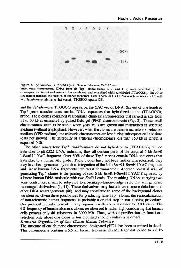
Figure 2. Hybridization of (TTAGGG)7 to Human Telomeric YAC ClonesIntact yeast chromosomal DNAs from six Trp+ clones (lanes 1, 2, and 4-7) were separated by PFGelectrophoresis, transferred onto a nylon membrane, and hybridized with radiolabeled (TTAGGG)7. The 50 kbsize marker indicates the position of lambda monomer. Lane 3 contains HYl DNA which includes a YAC withtwo Tetrahymena telomeres that contain TTGGGG repeats (28).
and the Tetrahymena TTGGGG repeats on the YAC vector DNA. Six out of one hundredTrp+ yeast transformants carried DNA sequences that hybridized to the (TTAGGG)7probe. These clones contained yeast-human chimeric chromosomes that ranged in size from11 to 50 kb as estimated by pulsed field gel (PFG) electrophoresis (Fig. 2). These smallchromosomes seem to be stable when yeast cells are grown and maintained in selectivemedium (without tryptophan). However, when the clones are transferred into non-selectivemedium (YPD medium), the chimeric chromosomes are lost during subsequent cell divisions(data not shown). The instability of artificial chromosomes less than 150 kb in length isexpected (40).The other ninety-four Trp+ transformants do not hybridize to (TTAGGG)7 but do
hybridize to pBR322 DNA, indicating they all contain parts of the original 6 kb EcoRI-BamH I YAC fragment. Over 50% of these Trp+ clones contain DNA sequences thathybridize to a human Alu probe. These clones have not been further characterized: theymay have been generated by random integration of the 6 kb EcoR I-BamH I YAC fragmentand linear human DNA fragments into yeast chromosomes. Another potential way ofgenerating Trp+ clones is the joining of two 6 kb EcoR I-BamH I YAC fragments bya linear human DNA molecule with two EcoR I ends. The resulting DNAs, carrying twoyeast centromeres, will be subjected to a breakage-fusion-bridge cycle that will generaterearranged derivatives (1, 41). These derivatives may include centromere deletions andother DNA rearrangements (40), and may contribute to some of the background cloneswe observe. Given these possibilities for producing false Trp+ clones, the recircularizationof non-telomeric human fragments is probably a crucial step in our cloning procedure.Our protocol is likely to work in any organism with a low telomere to DNA ratio. The6% frequency ofhuman telomeric clones we observed is rather high considering that humancells possess only 46 telomeres in 3000 Mb. Thus, without purification or functionalselection only about one clone in ten thousand should contain a telomere.Structural Organization of One Cloned Human TelomereThe structure of one chimeric chromosome, designated yHTl, has been examined in detail.This chromosome contains a 5.5 kb human telomeric EcoR I fragment joined to a 6 kb
6115
Nucleic Acids Research ~~~~~~~~~~~~~~~~~... .. .. ... .. ..
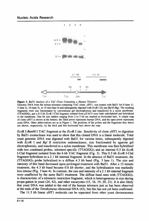
Figure 3. Ba131 Analysis of a YAC Clone Containing a Human TelomereGenomic DNA from the human telomere-containing YAC clone, yHTI, was treated with Bal31 for 0 (lane 1),5 (lane 2), 10 (lane 3), or 15 min (lane 4) and subsequently digested with EcoR I (E) and Bgl II (Bg). The resultingfragments were size fractionated by conventional gel electrophoresis and transferred to a nylon membrane.(TTAGGG)7 and a 0.5 kb EcoR I-Clal fragment isolated from pYAC4 were both radiolabeled and hybridizedto the membrane. One kb size ladders ranging from 2 to 5 kb are marked as horizontal bars. A simple mapof clone yHTI is shown at the bottom; the filled arrow represents human DNA, and the open arrow representsyeast DNA. Other abbreviations are as in Figure 1. The positions of the probes and the fragments they detectare shown, respectively, by the thick and thin horizontal bars above the map.
EcoR I-BamH I YAC fragment at the EcoR I site. Sensitivity of clone yHT1 to digestionby Bal31 exonuclease was used to show that this cloned DNA is a linear molecule. Totalyeast genomic DNA was digested with Bal31 for various times, subsequently digestedwith EcoR I and Bgl II restriction endonucleases, size fractionated by agarose gelelectrophoresis, and transferred to a nylon membrane. This membrane was then hybridizedwith two combined probes, telomere-specific (TTAGGG)7 and an internal 0.5 kb EcoRI-ClaI fragment isolated from the 6 kb YAC fragment (Fig. 3). This 0.5 kb EcoR I-Clalfragment hybridizes to a 2.1 kb internal fragment. In the absence of Bal3 1 treatment, the(TTAGGG)7 probe hybridized to a diffuse 4.3 kb band (Fig. 3 lane 1). The size andintensity of this band decreased upon prolonged treatment with Bal31. After a 15 minutetreatment, the 4.3 kb band became 0.6 kb shorter, and the hybridization was markedlyless intense (Fig. 3 lane 4). In contrast, the size and intensity of a 2.1 kb internal fragmentwere unaffected by the same Bal31 treatment. The diffuse band seen with (TTAGGG)7is characteristic of a telomeric restriction fragment, which is heterogeneous in size duringpropa-gation in yeast (24, 42), and other eucaryotes (15, 16, 19, 20, 21). It is also likelythat yeast DNA was added to the end of the human telomere just as has been observedat the ends of the Tetrahymena ribosomal DNA (43), but this has not yet been confirmed.The 11.5 kb linear yHTI molecule can be separated from other yeast chromosomes
6116

Nucleic Acids Research
YAC Human DNA 1 jKb
E Bg Ps Pu Pu K Ps
segment B
segment6 kb YAC porhnn AT-rich (-90%) Alu This regbn is oommon in ut kbTTAGG
ot the chromosome region repeat most of the human chromosomes repeating sequence
Figure 4. Schematic Structure of the Chimeric Yeast-Hunan Clone yHTJClone yHT1 is a chimeric minichromosome containing human DNA (filled arrow) and yeast DNA (open arrow).Arrowheads represent regions of telomeric simple repeats. The open circle represents a yeast centromere. Locationsof EcoR I (E), Bgl II (Bg), Pst I (Ps), Pvu II (Pv), and Kpn I (K) restriction sites are shown above the map.The position of an Alu repetitive element is indicated by a solid arrowhead below the map, which points in thedirection of the poly(dA) stretch. The positions of segments A and B used in subsequent experiments are indicatedby the horizontal bars below the map.
by PFG and directly isolated from the gel. Several restriction enzymes have been usedto map this cloned human telomere (Fig. 4). Both Bgl II and Kpn I have unique cleavagesites in the human 5.5 kb EcoR I fragment, located 4.3 kb and 1.4 kb from the tip ofthe chromosome, respectively. Two DNA segments of yHTl were subcloned for detailedstructural analysis. They are a 2.7 kb Pst I fragment located less than 400 bp from theterminal TTAGGG repeats, and a 1.2 kb EcoR I-Bgl II fragment located distal to the end
Figure 5. Hybrdizations of Human, Hamster, and Hybnd Cell DNAs with Human Telomeric Probes(A). Genomic DNAs isolated from cell lines GM3468 (lane 1 and 2), Gus4066 (lane 3 and 4), EyeF3A6 (lane5 and 6), CHO-KI (lane 7 and 8), 29-IF-3a (lane 9 and 10), UCW56 (lane 11 and 12), HHW416 (lane 13 and14), and HHW599 (lane 15 and 16) were digested with EcoR I (odd numbered lanes) or Bgl II (even numberedlanes). The resulting fragments were size fractionated by conventional gel electrophoresis and transferred to anylon membrane. The membrane was then hybridized with radiolabeled segment A (Fig. 4). Sizes markers areshown by horizontal bars, positioned every 2 kb. (B). The same membrane shown in (A) was rehybridized withradiolabeled segment B.
6117

Nucleic Acids Research
(Fig. 4). Both of these sequences were subcloned into a bacterial vector, and designatedas pPst2.7 and pEBgl .2, respectively. Preliminary DNA sequencing data show that a long,tandemly repeated, AT-rich sequence is located in the middle of the 1.2 kb EcoR I-BglII fragment. In fact, the whole 1.2 kb EcoR I-Bgl II DNA segment contains AT-richsequences. We estimate that the DNA in this region is over 80% A+T. Clone yHTlcontains one Alu repeat located 3.8 kb from the chromosome end. This proves,unequivocally, the human origin of the cloned sequence. DNA sequence analysis revealedthat the poly(dA) stretch of this Alu repeat is oriented proximal to the telomere (unpublishedresults).Clone yHTJ Contains DNA Sequences that Selectively Recognize Human Telomeres inHamster-Human Hybrid CellsTwo DNA segments isolated from plasmid pPst2.7 were used as probes in hybridizationexperiment to study the genomic organization of yHT 1. These segments are a 1.8 kb PvuII-Pst I and a 0.6 kb Pvu II fragment, designated as segment A and segment B, respectively(Fig. 4). Segment A was used as a hybridization probe to a genomic blot containing DNAsfrom two human cultured cell lines, two hamster cell lines and a collection of hybrid cellscontaining single human chromosomes (see Table 1 for a description of cell lines). ManyEcoR I and Bgl II fragments were detected in the human cell lines, indicating that multiplecopies of segment A are present in the human genome. The sets of EcoR I and Bgl Irestriction fragments that hybridized to segment A are quite similar in the two human celllines, GM3468 and Gus4066 (Fig. 5A, lanes 1 and 3; lanes 2 and 4). This suggests thatcopies of segment A are located in regions of chromosomes highly conserved betweenindividuals. Segment A cross-hybridized weakly with hamster DNA and detectedheterogeneous size fragments (Fig. 5A, lanes 7, 8, 11, 12). This means a similar DNAsequence is also present in multiple copies in hamster genomes. Hamster-human hybridcells show extra bands that are not present in their parental hamster cells. Thus, thesebands must come from the human chromosomes.A simpler hybridization pattern was seen when the same blot was probed with segment
B. (Fig. SB). Fewer restriction fragments hybridized to segment B than segment A in humanDNA samples (Fig. 5A and SB, lanes 1-4). This shows that the human genome containsmore copies of segment A than segment B. No cross-hybridization was found betweensegment B and hamster DNA (Fig.5B, lanes 7, 8, 11, 12). Two fragments were detectedin hybrid cells containing only human chromosome 22 (Fig. 5B, lanes 5, 6). A single,slightly diffuse hybridizing fragment was detected in hybrid cells containing only humanchromosome 21 (Fig. SB, lanes 9, 10), only human chromosome 4 (Fig. 5B, lanes 13,14), or only human chromosome 5 (Fig. SB, lanes 15, 16). It is possible that only oneend of each of these human chromosomes contains sequences that hybridize to segmentB. Another possibility is that sequences that hybridize to segment B are located on thesame size restriction fragments at both ends of each of these chromosomes. Whatever theexplanation, these results suggest that segment B DNA can be used to detect humanrestriction fragments in hybrid cells. Other results will show that these restriction fragmentsare telomeric (see below). Thus, segment B is particularly useful in mapping humanchromosomes in hybrid cells because it can detect the end fragments of specific humanchromosomes.The telomeric location of segment B DNA was demonstrated by its sensitivity to Bal3 1
exonuclease treatment, a characteristic of telomeric sequences (17, 18, 21, 44, 45, 46).Bal31 sensitivity was first measured on a hybrid cell instead of a human cell because of
6118

Nucleic Acids Research
A 1 2 3 4 5 6
17 kbt
14 kb
B 1 2 3 4 5 6
-6 3 kib ^*.
Figure 6. Bal31 Analysis of EcoR I Fragment Containing Human Chromosome 21 TelomeresGenomic DNA isolated from the hybrid cell line, 29-lF-3a, was treated with Bal3l for 0, 20, 40, 60, 120, and140 min (lanes 1 to 6, respectively) and subsequently digested with EcoR I. The resulting fragments were sizefractionated by gel electrophoresis and transferred to a nylon membrane. The membrane was then probed withboth radiolabeled segment B and pGSB3 a single copy probe assigned to human chromosome 21 (47). DNAsizes are shown in kb.
its simpler and predictable hybridization pattern to segment B. Genomic DNA preparedfrom cell line 29-1F-3a (Table 1), which contains a single human chromosome 21, was
first digested with Bal3 1 for various lengths of time and then digested with the restrictionenzyme EcoR I. The fragments were fractionated by conventional gel electrophoresis,transferred to a nylon membrane and hybridized with segment B DNA (Fig. 6). In theabsence of Bal31 treatment, segment B hybridized to a slightly diffuse fragment about17 kb in length. The size of this EcoR I fragment decreased upon prolonged treatmentwith Bal31. The rate of exonuclease digestion was roughly linear during the first 60 minutesof digestion (Fig. 6 lane 1-4). Approximately 3 kb of the sequence was removed aftera 2 hour Bal3l treatment. The same membrane was then hybridized with pGSB DNA.This cloned interstitial single-copy chromosome 21 sequence has been mapped to 21q22.3which is the distal portion of the human chromosome 21 q arm (47, 48, 49). The probehybridized to a 6.4 kb EcoR I fragment which was unaffected by Bal31 digestion (Fig.6). This result demonstrates that under the conditions used, the Bal31 digestion was telomerespecific.Structural Organization of Other Human TelomeresAdditional evidence that yHTI is a telomeric clone came from comparative genomichybridizations with (TTAGGG)7 and yHTI subclones. Not I restriction fragments ofvarious human cultured cells were fractionated by PFG and blotted onto a nytran membrane.These human samples includes two neuroblastoma cell lines (CHP134 and LA-N-5), four
6119

Nucleic Acids Research
Figure 7. Not I Telomeric Fragments of Various Human Cell LinesGenomic DNAs isolated from human cells CHP134 (lane 1), LA-N-5 (lane 2), Gus3365A (lane 3), Gus 4066(lane 4), GM3468 (lane 5), GM3657 (lane 6), GM3658 (lane 7), GM5920 (lane 8), GM5879A (lane 9), andGM6167 (lane 10) were digested with Not I, size fractionated by PFG eletrophoresis, and blotted onto a nylonmembrane. The membrane was (A) probed with radiolabeled (TTAGGG)7; (B), reprobed with radiolabeledsegment A; (C), reprobed with radiolabeled segment B. Sizes markers are shown by horizontal bars positionedevery 250 kb.
lymphoblastoid cell lines (Gus3365A, Gus4066, GM5920 and GM3657), and four fibroblastcell lines (GM3468, GM3658, GM5879A and GM6167; see Table 1 for details). The pairsof lines, GM3657 plus GM3658, and GM5920 plus GM5879, are samples prepared fromdifferent tissues of the same individuals. Radiolabeled (TTAGGG)7 was used to detectall the human telomeric Not I fragments in this genomic blot (Fig. 7A). At least 15 distincttelomeric Not I fragments (not including overlapping bands) were detected in each cellline ranging in size from less than 50 to 850 kb. Each cell line displayed a unique pattern
6120

A
EcoRt 8gin1
24 .
9 4 .
Nucleic Acids Research
PvuitPstI PstI
(; 1.... [23~~~~231
94
44
*.:::2.7t2.3
Us t 20
0._.
0.5
f) 1 kb,..................................
EcoRt 8glit Pvull Pvuil.~~~~~~~~~~~~~~~~~~~~~~~~
Psil
Figure 8. Restriction Fragments Identified by Telomeric Probes(A). Genomic DNA isolated from a human cell line, GM3468, was digested with various restriction enzymes,
size fractionated by conventional gel electrophoresis, and blotted onto a nylon membrane. Each enzyme-digestedlane was sliced into two strips. The strips shown on the left were hybridized to radiolabeled segment B whilethe strips shown on the right were hybridized to radiolabeled (T1AGGG)7. Arrowheads indicate the correspondingfragments in yHT1. Sizes are given in kb. (B). A proposed general structure distal to the human telomeric simple(TTAGGG)n repeats (consecutive arrowheads); five restriction sites indicated appear fairly well conserved inhuman chromosomes.
of Not I fragments detected by (TTAGGG)7. Cell lines derived from different tissues ofthe same person also had unique hybridization patterns (lane 6 vs lane 7, lane 8 vs lane 9).When the same blot was probed with segment A, fewer fragments were detected than
with (TTAGGG)7 (Fig. 7B). For example, the bands designated as Ni and N2 in Fig.7A did not hybridize to segment A in Fig. 7B. Fig. 7C shows the same blot hybridizedwith segment B. Even fewer Not I fragments were detected. For example, the smallestNot I fragment, designated as N3 in Fig. 7A, was missing in Fig. 7C. However, all thefragments that hybridized to segment B also contain sequences that hybridized to segmentA, and all the fragments that hybridized to segment A also contain sequences that hybridizedto (TTAGGG)7. These results suggest that all copies of segments A and B in the humangenome are located on telomeric Not I fragments. They also indicates that DNA sequencesproximal to the telomeric ends are somewhat more conserved in human chromosomes thandistal DNA sequences. However, the number of telomeres that lack segment A and segmentB sequences is not yet known. Hybridizations of genomic DNA from a panel of hybrid
6121
B
.: ^W,
Nucleic Acids Research
cell lines with segment A and B or in situ hybridizations of human metaphase chromosomeswith these probes are needed to identify which telomeres do not contain this conservedsequence.
Six yeast transformants hybridized to the (TTAGGG)7 probe in the initial screening forhuman telomere clones. Since clone yHTl contains DNA sequences also present in manyother human telomeres, some other clones would be expected to share homologoussequences with clone yHT 1. However, no cross hybridization was detected between cloneyHT1 and the other five clones. One possibility is that these clones represent a subgroupof human telomeres that contain no sequence similarities to clone yHT . Future work willdistinguish whether or not these clones are true telomeric clones by measuring their Bal3 1sensitivities.The properties of the cloned telomeric sequences were investigated further by comparing
four different genomic restriction patterns revealed by hybridizations with segment B and(TTAGGG)7 (Fig. 8A). Most of the EcoR I and Bgl II fragments detected by segmentB and (TTAGGG)7 are heterogeneous in size; they range between 9.4 and 23.1 kb.Nevertheless, a few EcoR I and Bgl II fragments, such as the 5.5 kb EcoR I and 4.3 kbBgl H fragments, are uniform in size (Figs 5 and 8). This suggests that not all of the segmentB and TTAGGG repeats are located on telomeric EcoR I and Bgl H fragments. More EcoRI and Bgl II bands are detected by (TTAGGG)7 than by segment B (Fig. 8A). This isconsistent with the Not I digestion results where a number of Not I telomeric fragmentsdid not contain segment B sequences.The Pst I and Pvu II fragments, which hybridize to segment B, are smaller and more
uniform in size than the EcoR I and Bgl II fragments (Fig. 8A). In contrast, the Pst Iand Pvu H fragments that hybridize to (TTAGGG)7 remain large and heterogeneous. Thissuggests that most of the human telomeres contain at least one Pst I and one Pvu II sitelocated between the segment B sequence and the TTAGGG repeat. A 0.6 kb Pvu II fragmentis the predominant genomic band detected by segment B. This suggests that the two PvuII sites in yHT1 are also conserved in other telomeres. The internal 2.7 kb Pst I and 0.6kb Pvu II fragments, present in yHTI, are seen in genomic hybridizations with segmentB probe (arrowheads in Fig. 8) but not with (TTAGGG)7 probe. These results suggestthat no DNA rearrangement has occurred within this 2.7 kb Pst I fragment during thecloning of yHT 1. However, the corresponding EcoR I and Bgl H fragments are not foundin this genomic hybridization because most of the length of the human TTAGGG repeatswere trimmed by yeast during DNA replication.Most Bgl H fragments containing the TTAGGG repeats are approximately 1 kb shorter
than most EcoR I fragments. The 1 kb size difference is seen even more distinctly withfragments that hybridize to segment B. These differences match with the distance seenbetween the EcoR I and Bgl H restriction sites in the yHT1 clone. This suggests that mosthuman telomeres contain these conserved EcoR I and Bgl H sites. Similarly, most PstI fragments detected by (TTAGGG)7 were approximately 3 kb shorter than most Bgl IIfragments. This difference in size matches the distance seen between the Bgl H site andthe distal Pst I site in the yHTI clone. This observation suggests that the distal Pst I siteis also conserved in most telomeres.A model for the organization of human telomeric sequences which is consistent with
all of these observations is shown in Fig. 8B. The model applies to most, but not all ofthe human telomeres. In this model, conserved human telomeric sequences extend at least4.4 kb beyond the TTAGGG repeats. The distance between the conserved Pst I site and
6122
Nucleic Acids Research
xN
YC .0
2 I N o N C)(5 C N CEM < N'e a s a: a C0
__a_ _ _
'1IF
-._-.... 2
_ ....
*<X_ _.
-v
_
_r
xC4
t1x.0w:U)In
I-0I
.S -w 750kb
2 +W rii< ,1
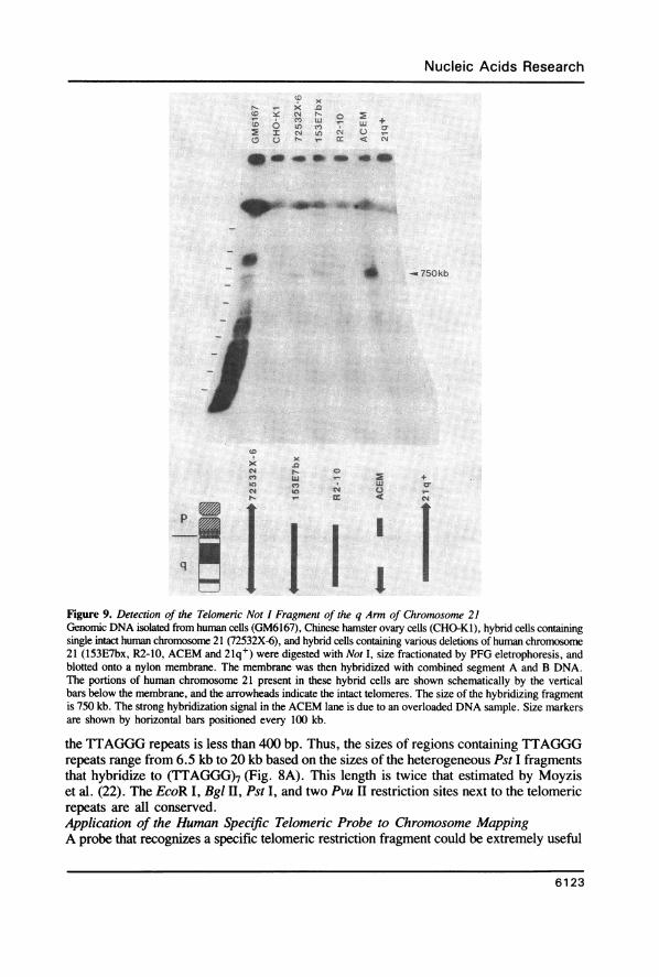
Figure 9. Detection of the Telomeric Not I Fragment of the q Arm of Chromosome 21Genomic DNA isolated from human cells (GM6167), Chinese hamster ovary cells (CHO-KI), hybrid cells containingsingle intact human chromosome 21 (72532X-6), and hybrid cells containing various deletions of human chromosome21 (153E7bx, R2-10, ACEM and 21q+) were digested with Not I, size fractionated by PFG eletrophoresis, andblotted onto a nylon membrane. The membrane was then hybridized with combined segment A and B DNA.The portions of human chromosome 21 present in these hybrid cells are shown schematically by the verticalbars below the membrane, and the arrowheads indicate the intact telomeres. The size of the hybridizing fragmentis 750 kb. The strong hybridization signal in the ACEM lane is due to an overloaded DNA sample. Size markersare shown by horizontal bars positioned every 100 kb.
the TTAGGG repeats is less than 400 bp. Thus, the sizes of regions containing TTAGGGrepeats range from 6.5 kb to 20 kb based on the sizes of the heterogeneous Pst I fragmentsthat hybridize to (TTAGGG)7 (Fig. 8A). This length is twice that estimated by Moyziset al. (22). The EcoR I, Bgl H, Pst I, and two Pvu II restriction sites next to the telomericrepeats are all conserved.Application of the Human Specific Telomeric Probe to Chromosome MappingA probe that recognizes a specific telomeric restriction fragment could be extremely useful
6123

Nucleic Acids Research
for physical mapping of human chromosomes. The clone yHTl contains sequences thatrecognize only human telomeres in rodent-human hybrid cells, but it potentially detectsboth ends of each human chromosome. However, hybrid cells containing partial deletionsof human chromosomes can be used to distinguish one particular human chromosome endfrom the other.One example of such an experiment, detecting the telomeric Not I fragment from the
q arm of human chromosome 21, is shown in Fig. 9. A blot containing Not I digests ofgenomic DNA from a human cell line (GM6167), a Chinese hamster ovary cell line (CHO-KI), and five hybrid cell lines (72532X-6, 153E7bx, R2-10, ACEM, 21q+; see Table1) was hybridized with a combined probe of segments A and B. Since this probe cross-hybridizes weakly to rodent DNA, the parental (CHO-KI) hybridization signals must besubtracted from that seen in the hybrid cells. Two hybrid cells, 153E7bx and ACEM,containing human chromosome 21 with deletions of the tip of the p arm showed a stronglyhybridizing fragment of approximately 750 kb in length (Fig. 9). This fragment was notdetected in the hybrid cell lines 21q+ and R2-10 which contain deletions of the end ofthe q arm of chromosome 21. The 750 kb fragment was also detected in 72532X-6 cellswhich contain intact human chromosome 21. The reason that a hybridization signal fromthe p arm of chromosome 21 was not detected in any of these cells is not known. Perhaps,this Not I fragment is too large or too small to be resolved by our particular PFG runningconditions. Alternatively, the p arm telomere may not contain any segment A and Bsequences.
DISCUSSIONA method is described for cloning human telomeric restriction fragments. In principle,
this method should be applicable to other eucaryotic organisms since telomere functionsappear to be conserved in all eucaryotes from yeast to human. Unlike other strategies forcloning telomeres from higher eucaryotes (21, 22), this method does not require exonucleasetreatment, so that the cloned telomeres should retain their original end sequences. We donot know, however, whether poly(dGdT)-(dCdA) tracts were added to the ends of thehuman telomeres as was observed with Tetrahymena ribosomal DNAs cloned in yeast (43,50).A 5.5 kb human EcoR I fragment containing human telomeric simple repeats can form
a 11.5 kb chimeric minichromosome with half of a YAC vector DNA. Apparently, cellsmust be kept under selective pressure in order to maintain an artificial chromosome ofthis size. Short yeast artificial chromosomes (< 42 kb) are known to be unstable becausethey segregate randomly at mitosis rather than disjoining from each other like naturalchromosomes. The stability of these linear chromosomes increases with length (51). Thus,the telomere cloning scheme can presumably be improved by ligating larger restrictionfragments, such as those generated by Not I or Mlu I, to the YAC DNA. Cloning of largerfragments should also allow isolation of single copy DNAs adjacent to the conservedtelomere regions.
Isolation of single copy DNAs that recognize specific telomeres is important for severalreasons. First, they can be used directly to identify the ends of chromosome restrictionmaps. Second, they can be used to detect chromosomal translocations between small DNAsegments at chromosome ends, which can not be seen by low resolution chromosomebanding (48). Third, they can be used to find restriction fragment length polymorphismsthat can serve as genetic markers to determine linkage relationships of some inherited
6124
Nucleic Acids Research
disorders located at the tips of chromosomes. For example, special efforts have been madeto isolate DNA probes from the short arm of chromosome 4, and to create a linkage mapto position the gene responsible for Huntington's Disease near the telomere (26, 52, 53).Yet no distal, flanking marker for this disease has been identified. Isolation of clone yHTlhas allowed us to identify sequences next to the telomere of 4p (unpublished results). Thisnarrows down the possible location of the Huntington's Disease gene considerably.
In clone yHTl, a 4.4 kb conserved DNA sequence is located next to the telomericTTAGGG repeats. Part of this 4.4 kb DNA sequence (segment A) hybridizes weakly torodent DNAs (Fig. 5A). These weak and heterodisperse cross-hybridizations could reflectshort sequences that are evolutionally conserved because they serve similar functions. Cross-hybridizations may be due to a conserved family of autonomous replicating sequences
(ARS). In yeast, a family of highly conserved ARS are located near the telomeres as partof the subtelomeric conserved sequences (54). These particular telomeric ARS elementsreplicate at the end of S phase (55). Similarly, telomeres in higher eucaryotes replicatelate in S phase (56); presumably they are also initiated by a family of late-replicating ARSelements located near the telomeres. Identification of the DNA sequences responsible forthe cross-hybridization and testing the ability of the sequences to self-replicate will benecessary to prove that a human ARS is part of this conserved telomeric region.Another intriguing structural feature of the 4.4 kb telomeric conserved sequence is the
greater than 1 kb segment of AT-rich DNA sequences. Such sequences are usually foundin the non-transcribed spacer regions of 5S rDNA (57), transfer RNA genes (58), ribosomalRNA genes (59), and histone genes (60). Other data suggest that AT-rich sequences are
chromosomal structures preferentially attached to the nuclear scaffold (61) or the nuclearmatrix (62). It is not clear whether these subtelomeric AT-rich sequences serve any suchfunctions in cells. However, the finding of these sequences close to telomeres is consistentwith the observations that both AT-rich heterochromatin and telomeric DNA replicate latein S phase (63).The yHTl clone contains a human telomeric EcoR I fragment. The chromosomal origin
of this telomere has not yet been determined because the DNA sequences in yHTI are
also present in other human telomeres. Genomic hybridizations and preliminary DNAsequence data of the region adjacent to TTAGGG repeats in yHTI suggest a model forthe DNA structures near the tips of human chromosomes. This model resembles theorganization of yeast telomeres proposed by Chan and Tye (54) in several aspects. First,the DNA sequences next to the telomeric simple repeats are conserved in most, but notall, of the chromosomes in yeast (64) and in human (this work). Thus, these conservedregions do not appear to play any important roles in individual chromo-some-specificfunctions (64). Second, DNA sequences located further away from the telomeres are lessconserved. For example, the telomere distal X elements in yeast are less conserved thanmore proximal Y elements (54). Although not enough is known yet to group the humantelomeric sequences in a similar way, segment B which is less distal than segment A detectsfewer distinct human sequences. These similar DNA organizations of telomeres in yeastand human may be general telomere characteristics of all eucaryotes.
ACKNOWLEDGEMENTSWe are grateful to Drs. David Patterson, James F. Gusella, Uta Francke and Maggie L.Van Keuren for providing various cell lines, and Drs. Robert K. Moyzis and Gordon D.
6125

Nucleic Acids Research
Stewart for providing DNA probes. This work is supported by grants from the NIH(GM14825), the NCI (CA39782), and the DOE (DE-FG02-87ER-GD852).
REFERENCES1. Muller, H. J. (1938) The Collecting Net-Woods Hole 13, 181-198.2. McClintock, B. (1941) Genetics 26, 234-282.3. McClintock, B. (1942) Proc. Natl. Acad. Sci. USA 28, 458-463.4. Orr-Weaver, T. L., Szostak, J. W., and Rothstein, R. J. (1981). Proc. Natl. Acad. Sci. USA 78, 6354-6358.5. Cavalier-Smith, T. (1974) Nature 250, 467-470.6. Bateman, A. J. (1975) Nature 253, 379.7. Dancis, B. M., and Holmquist, G. P. (1979) J. Theor. Biol. 78, 211-224.8. Blackburn, E. H., and Szostak, J. W. (1984) Ann. Rev. Biochem. 53, 163-194.9. Rubin, G. M. (1978) Cold Spring Harbor Symp. Quant. Biol. 42, 1041-1046.
10. Bedbrook, J. R., Jones, J., O'Dell, M., Thompson, R. D., and Flavell, R. B. (1980) Cell 19, 545-560.11. Jones, J. D. G., and Flavell, R. B. (1983) Cold Spring Harbor Symp. Quant. Biol. 47, 1209-1213.12. Young, B. S., Pession, A., Traverse, K. L., French, C., and Pardue, M. L. (1983) Cell 34, 85-94.13. Ashley, T. (1979) J. Cell. Sci. 38, 357-367.14. Blackburn, E. H. (1984) Cell 37, 7-8.15. Boswell, R. E., Klobutcher, L. A., and Prescott, D. M. (1982) Proc. Natl. Acad. Sci. USA 79, 3255-3259.16. Bernards, A., Michels, P. A. M., Lincke, C. R., and Borst, P. (1983) Nature 303, 592-597.17. Van der Ploeg, L. H. T., Liu, A. Y. C., and Borst, P. (1984) Cell 36, 459-468.18. Shampay, J., Szostak, J. W., and Blackburn, E. H. (1984) Nature 310, 154-157.19. Roth, M., and Prescott, D. M. (1985) Cell 41, 411-417.20. Larson, D. D., Spangler, E. A., and Blackburn, E. H. (1987) Cell 50, 477-483.21. Richards, E. J., and Ausubel, F. M. (1988). Cell 53, 127-136.22. Moyzis, R. K., Buckingham, J. M., Cram, L. S., Dani, M., Deaven, L. L., Jones, M. D., Meyne, J.,
Ratliff, R. L., and Wu, J.-R. (1988) Proc. Natl. Acad. Sci. USA 85, 6622-6626.23. Allshire, R. C., Gosden, J. R., Cross, S. H., Cranston, G., Rout, D., Sugawara, N., Szostak, J. W., Fantes,
P. A., and Hastie, N. D. (1988) Nature 332, 656-659.24. Szostak, J. W., and Blackburn, E. H. (1982) Cell 29, 245-255.25. Pluta, A. F., Dani, G. M., Spear, B. B., and Zakian, V. A. (1984) Proc. Natl. Acad. Sci. USA 81, 1475-1479.26. Gilliam, T. C., Tanzi, R. E., Haines, J. L., Bonner, T. I., Faryniarz, A. G., Hobbs, W. J., MacDonald,
M. E., Cheng, S. V., Folstein, S. E., Conneally, P. M., Wexler, N. S., and Gusella, J. F. (1987) Cell50, 565-571.
27. Harris, P., Lalande, M., Stroh, H., Bruns, G., Flint, A., and Latt, S. A. (1987) Genetics 77, 95-103.28. Burke, D. T., Carle, G. F., and Olson, M. V. (1987) Science 236, 806-812.29. Smith, C. L., Klco, S. R., and Cantor, C. R. (1988) In Davies, K. (ed.), Genome Analysis: A Practical
Approach IRL Press, McLean, VA, pp. 41-72.30. Church, G. M., and Gilbert, W. (1984) Proc. Natl. Acad. Sci. USA 81, 1991-1995.31. Feinberg, A. P., and Vogelstein, B. (1983) Anal. Biochem. 132, 6-13.32. Jacobs, L., and Demars, R. (1978) Mutation Research 53, 29-53.33. Jacobs, L., Bean, C. L., and Marx, J. A. (1983) Environmental Mutagenesis 5, 717-731.34. Smith, C. L., and Cantor, C. R. (1987) Methods in Enzymol. 155, 449-467.35. Burgers, P. M. J., and Percival, K. J. (1987) 163, 391-397.36. Sherman, F., Fink, G., and Lawrence, C. (1986) Methods in Yeast Genetics, pp. I-186. Cold Spring Harbor
Lab., Cold Spring Harbor, NY.37. Schwartz, D. C., Saffran, W., Welsh, J., Haas, R., Goldenberg, M., and Cantor, C. R. (1983) Cold Spring
Harbor Symp. Quant. Biol. 47, 189-195.38. Maniatis, T., Fritsch,E. F., and Sambrook, J. (1982). Molecular Cloning: A Laboratory Manual, pp. 1-545.
Cold Spring Harbor Lab., Cold Spring Harbor, NY.39. Francke, U., Ochs, H. D., de Martinville, B., Giacalone, J., Lindgren, V., Disteche, C., Pagon, R. A.,
Hofker, M. H., van Ommen, G.-J. B., Pearson, P. L., and Wedgwood, R. J. (1985) Am. J. Hum. Genet.37, 250-267.
40. Murray, A. W., Schultes, N. P., and Szostak, J. W. (1986) Cell 45, 529-536.41. Haber, J. E., Thorburn, P. C., and Roger, D. (1984). Genetics 106, 185-205.42. Murray, A. W., Claus, T. E., and Szostak, J. W. (1988) Mol. Cell. Biol. 8, 4642-4650.43. Walmsley, R. W., Szostak, J. W., and Petes, T. D. (1983) Nature 302, 84-86.
6126
Nucleic Acids Research
44. Yao, M.-C., and Yao, C.-H. (1981) Proc. Natl. Acad. Sci. USA 78, 7436-7439.45. Blackburn, E. H., and Challoner, P. B. (1984) Cell 36, 447-457.46. Ponzi, M., Pace, T., Dore, E., and Frontali, C. (1985) EMBO J. 4, 2991-2995.47. Stewart, G. D., Tanzi, R. E., and Gusella, J. F. (1985) Nucl. Acids Res. 13, 7168.48. Phelan, M. C., Morton, C. C., Stevenson, R. E., Tanzi, R. E., Stewart, G. D., Watkins, P. C., Gusella,
J. F., and Amos, J. A. (1988) Am. J. Hum. Genet. 43, 511-519.49. Tanzi, R. E., Haines, J. L., Watkins, P. C., Stewart, G. D., Wallace, M. R., Hallewell, R., Sacchi, N.,
Wong, C., Wexler, N. S., Conneally, P. M., and Gusella, J. F. (1988) Genomics 3, 129-136.50. Walmsley, R. W., Chan, C. S. M., Tye, B.-K., and Petes, T. D. (1984) Nature 310, 157-160.51. Murray, A. W., and Szostak. J. W. (1983) Nature 305, 189-193.52. MacDonald, M. E., Anderson, M. A., Gilliam, T. C., Tranebjaerg, L., Carpenter, N. J., Magenis, E.,
Hayden, M. R., Healey, S. T., Bonner, T. I., and Gusella, J. F. (1987) Genomics 1, 29-34.53. Wasmuth, J. J., Hewitt, J., Smith, B., Allard, D., Haines, J. L., Skarecky, D., Partlow, R., and Hayden,
M. R. (1988) Nature 332, 734-736.54. Chan, C. S. M., and Tye, B.-K. (1983) Cell 33, 563-573.55. McCarroll, R. M., and Fangman, W. L. (1988) Cell 54, 505-513.56. Crossen, P. E., Pathak, S., and Arrighi, F. E. (1975) Chromosoma 52, 339-347.57. Fedoroff, N. V., and Brown, D. D. (1978) Cell 13, 701-716.58. Valenzuela, P., Venegas, A., Weinberg, F., Bishop, R., and Rotter, W. J. (1978) Proc. Natl. Acad. Sci.
USA. 75, 190-194.59. Sollner-Webb, B., and Reeder, R. H. (1979) Cell 18, 485-499.60. Moss, T., and Birnstiel, M. (1979) 6, 3733-3743.61. Mirkovitch, J., Mirault, M.-E., and Laemmli, U. K. (1984) Cell 39, 223-232.62. Flickinger, R. A. (1986) Cell Biol. Int. Rep. 10, 415-420.63. Comings, D. E. (1972) 71, 106-112.64. Zakian, V. A., and Blanton, H. M. (1988) Mol. Cell. Biol. 8, 2257-2260.65. Carpenter, N. J., Mayes, J. S., Say, B., and Wilson, D. P. (1987) J. Med. Genet. 24, 706-709.66. Balaban-Malenbaum, G., and Gilbert, F. (1977) Science 198, 739-741.67. Kohl, N. E., Kanda, N., Schreck, R. R., Bruns, G., Latt, S. A., Gilbert, F., and Alt, F. W. (1983) Cell
35, 359-367.68. Puck, T. T., and Kao, F.-T. (1982) Ann. Rev. Genet. 16, 225-271.69. Wasmuth, J. J., and Chu, L.-Y. (1980) J. Cell Biol. 87, 697-702.70. Van Keuren, M. L., Hart, I. M., Kao, F.-T., Neve, R. L., Bruns, G. A. P., Kurnit, D. M., and Patterson,
D. (1987) Cytogenet. Cell Genet. 44, 142-147.71. Patterson, D., Graw, S., and Jones C. (1981) Proc. Natl. Acad. Sci. USA 78, 405-409.72. Moore, E. E., Jones, C., Kao, F. T., Oates, D. (1977) Am. J. Hum. Genet. 29, 389-396.73. Patterson, D. (1987) Somatic Cell and Mol. Genetics 13, 365-371.
6127
This article, submitted on disc, has been automaticallyconverted into this typeset format by the publisher.