Nucleic Acids and Protein Synthesis. What are nucleic acids?
1
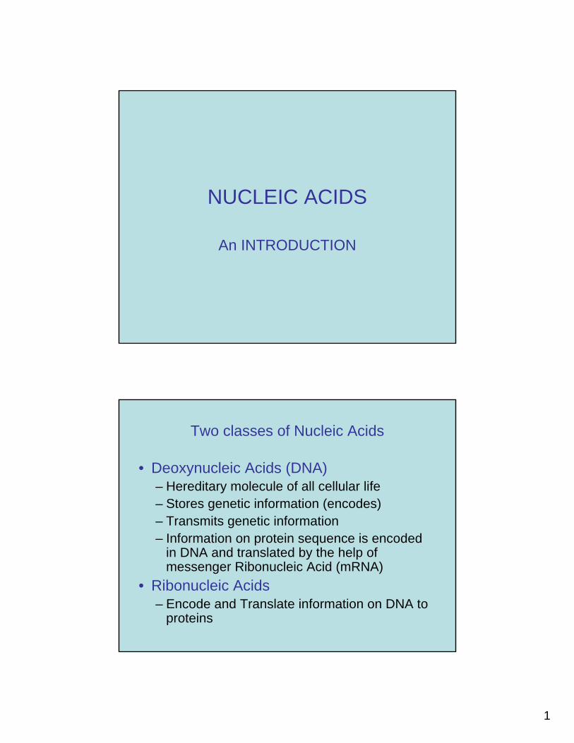
NUCLEIC ACIDS
An INTRODUCTION
Two classes of Nucleic Acids
• Deoxynucleic Acids (DNA)– Hereditary molecule of all cellular life– Stores genetic information (encodes)– Transmits genetic information – Information on protein sequence is encoded
in DNA and translated by the help of messenger Ribonucleic Acid (mRNA)
• Ribonucleic Acids– Encode and Translate information on DNA to
proteins
2
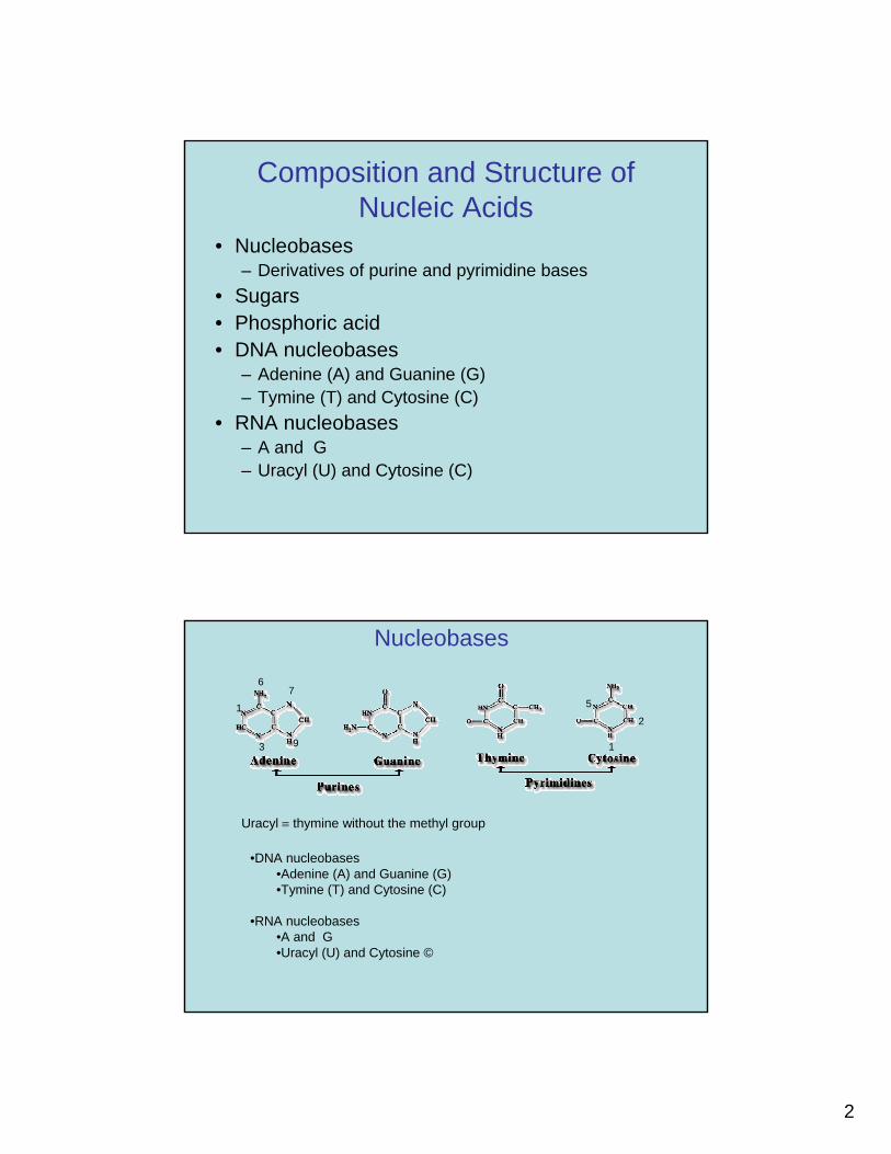
Composition and Structure of Nucleic Acids
• Nucleobases– Derivatives of purine and pyrimidine bases
• Sugars• Phosphoric acid• DNA nucleobases
– Adenine (A) and Guanine (G)– Tymine (T) and Cytosine (C)
• RNA nucleobases– A and G– Uracyl (U) and Cytosine (C)
Nucleobases
Uracyl ≡ thymine without the methyl group
•DNA nucleobases•Adenine (A) and Guanine (G)•Tymine (T) and Cytosine (C)
•RNA nucleobases•A and G•Uracyl (U) and Cytosine ©
1
7
3 9 1
6
2
5
3
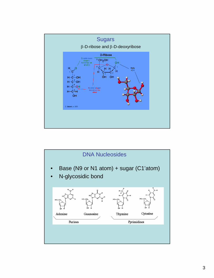
Sugarsβ-D-ribose and β-D-deoxyribose
DNA Nucleosides
• Base (N9 or N1 atom) + sugar (C1’atom) • N-glycosidic bond
4
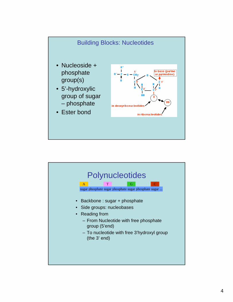
Building Blocks: Nucleotides
• Nucleoside + phosphate group(s)
• 5’-hydroxylic group of sugar – phosphate
• Ester bond
Polynucleotides
• Backbone : sugar + phosphate• Side groups: nucleobases• Reading from
– From Nucleotide with free phosphate group (5’end)
– To nucleotide with free 3’hydroxyl group (the 3’ end)
A T G C sugar phosphate sugar phosphate sugar phosphate sugar ...
5
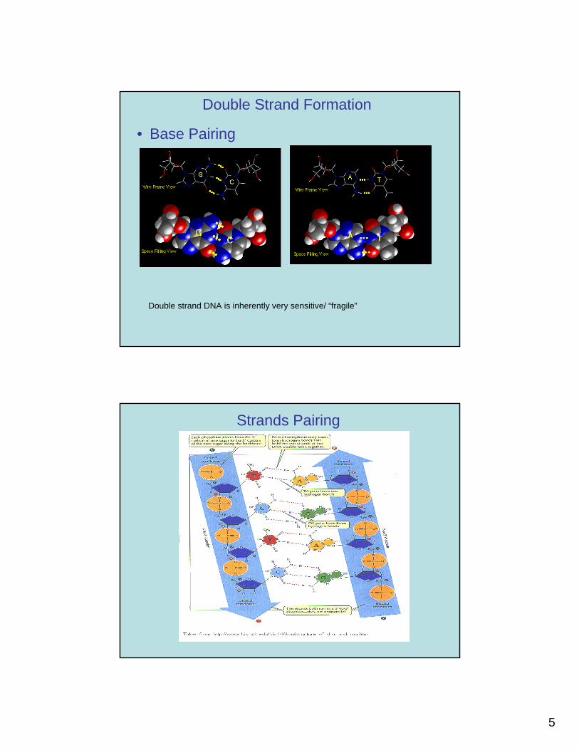
Double Strand Formation
• Base Pairing
Double strand DNA is inherently very sensitive/ “fragile”
Strands Pairing
6
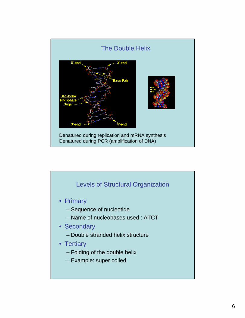
The Double Helix
Denatured during replication and mRNA synthesisDenatured during PCR (amplification of DNA)
Levels of Structural Organization
• Primary– Sequence of nucleotide– Name of nucleobases used : ATCT
• Secondary– Double stranded helix structure
• Tertiary– Folding of the double helix– Example: super coiled
7
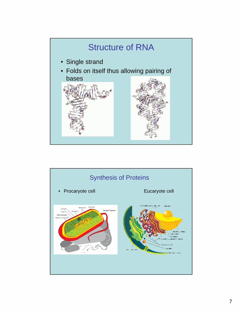
Structure of RNA• Single strand• Folds on itself thus allowing pairing of
bases
Synthesis of Proteins
• Procaryote cell Eucaryote cell
8
Synthesis of Proteins
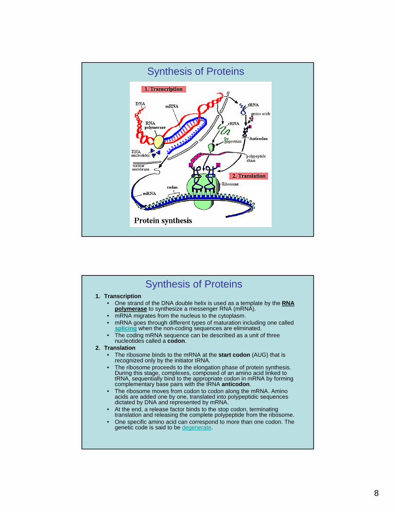
Synthesis of Proteins1. Transcription
• One strand of the DNA double helix is used as a template by the RNA polymerase to synthesize a messenger RNA (mRNA).
• mRNA migrates from the nucleus to the cytoplasm.• mRNA goes through different types of maturation including one called
splicing when the non-coding sequences are eliminated. • The coding mRNA sequence can be described as a unit of three
nucleotides called a codon.2. Translation
• The ribosome binds to the mRNA at the start codon (AUG) that is recognized only by the initiator tRNA.
• The ribosome proceeds to the elongation phase of protein synthesis. During this stage, complexes, composed of an amino acid linked to tRNA, sequentially bind to the appropriate codon in mRNA by forming complementary base pairs with the tRNA anticodon.
• The ribosome moves from codon to codon along the mRNA. Amino acids are added one by one, translated into polypeptidic sequences dictated by DNA and represented by mRNA.
• At the end, a release factor binds to the stop codon, terminating translation and releasing the complete polypeptide from the ribosome.
• One specific amino acid can correspond to more than one codon. The genetic code is said to be degenerate.
9
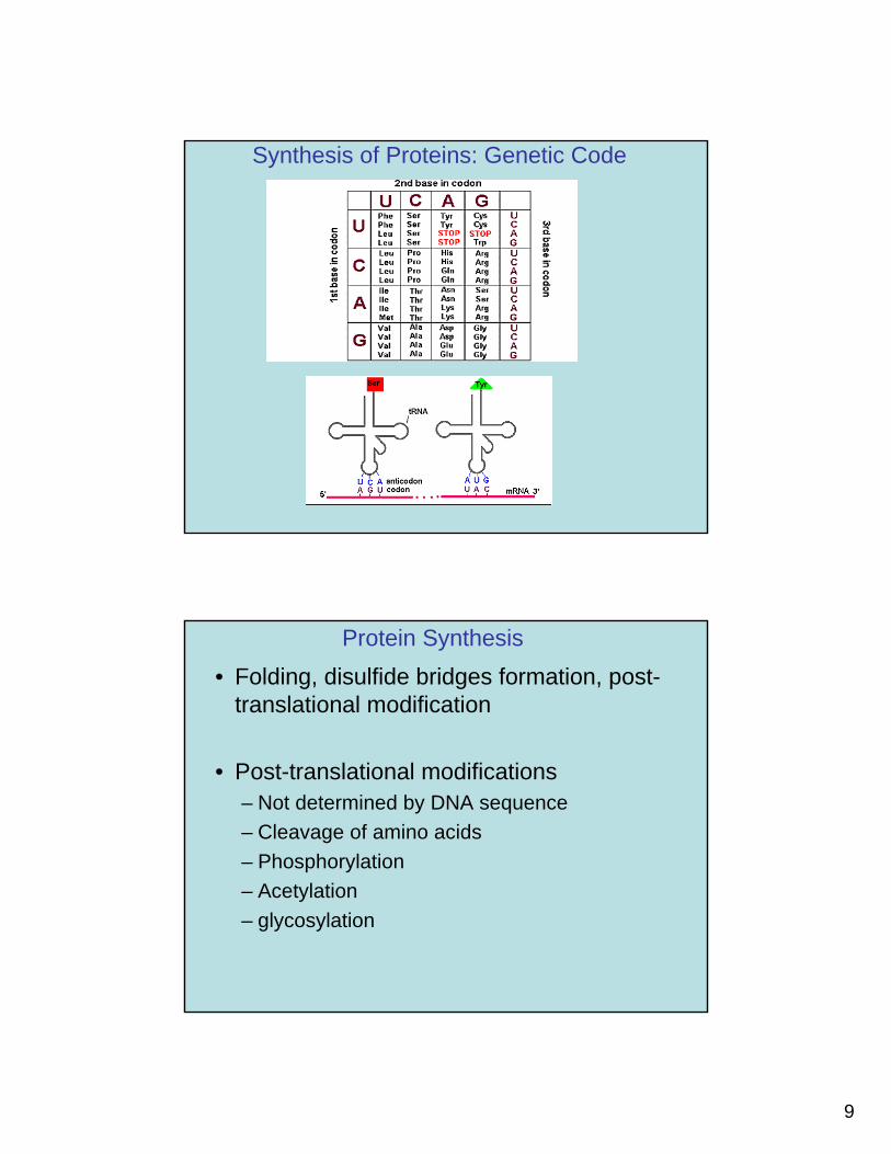
Synthesis of Proteins: Genetic Code
Protein Synthesis• Folding, disulfide bridges formation, post-
translational modification
• Post-translational modifications– Not determined by DNA sequence– Cleavage of amino acids– Phosphorylation– Acetylation– glycosylation
10
Analytical Challenges for the Analysis of Biological Macromolecules
• Inherent sensitivity
– Both proteins and nucleic acid 3D structure are very sensitive to environmental conditions such as pH, ionic strength, solvent composition
– Why? stabilized by H-bonds, other dipole-dipole interactions, electrostatic interactions, van der Waals interactions
– Problems• Denaturation• Crystallization challenges
Analytical Challenges for the Analysis of Biological Macromolecules
• Size of macromolecules– Several signals that must be resolved and analyzed– Complex three dimensional Structure– NMR challenges
• Number of macromolecules in real samples– Separation methods– Methods for selective detection
• Amount of sample– Sensitivity must be high– Amplification