yeditepeanatomy1.files.wordpress.com · Web viewUnlike the lower limb, which is used for support,...
49
BONES & JOINTS OF THE UPPER LIMB 30 10. 2012 Kaan Yücel M.D., Ph.D. http://yeditepeanatomy1.org A TOTAL OF 24 FIGURES IN THE TEXT
-
Upload
nguyenthuan -
Category
Documents
-
view
215 -
download
2
Transcript of yeditepeanatomy1.files.wordpress.com · Web viewUnlike the lower limb, which is used for support,...

BONES & JOINTS OF
THE UPPER LIMB
30 10. 2012
Kaan YücelM.D., Ph.D.
http://yeditepeanatomy1.org
A TOTAL OF 24 FIGURES IN THE TEXT
Dr.Kaan Yücel http://yeditepeanatomy1.org Bones & joints of the upper limb
http://www.youtube.com/yeditepeanatomy
BONES OF THE UPPER LIMBThe upper limb is characterized by its mobility and ability to conduct fine motor skills (manipulation).
These characteristics are especially marked in the hand. One of the major functions of the hand is to grip and manipulate objects. Based on the position of its major joints and component bones, the upper limb is divided into shoulder, arm, forearm, and hand for precise description.
The superior appendicular skeleton articulates with the axial skeleton only at the sternoclavicular joint, allowing great mobility. The clavicles and scapulae of the pectoral girdle are supported, stabilized, and moved by axioappendicular muscles that attach to the relatively fixed ribs, sternum, and vertebrae of the axial skeleton.
The clavicle is the only bony attachment between the trunk and the upper limb. The medial end is called “sternal end". The lateral end is called “acromial” end”.
The scapula lies on the posterolateral aspect of the thorax. It has medial, lateral, and superior borders and superior, lateral, and inferior angles. The posterior surface of the scapula is unevenly divided by spine of the scapula, into a small supraspinous fossa and a much larger infraspinous fossa. The spine continues laterally as the flat expanded acromion, which forms the subcutaneous point of the shoulder and articulates with the acromial end of the clavicle. The concave costal surface of most of the scapula forms a large subscapular fossa.
The skeletal support for the arm is the humerus. The humerus (arm bone) articulates with the scapula at the glenohumeral joint and the radius and ulna at the elbow joint. The proximal end of the humerus has a head, surgical and anatomical necks, and greater and lesser tubercles. The distal end of the humerus has a condyle, two epicondyles, and three fossae.
Ulna is the medial and longer of the two forearm bones.It is the stabilizing bone of the forearm. Radius is the lateral and shorter of the two forearm bones.
The hand is the region of the upper limb distal to the wrist joint. It is subdivided into three parts: wrist, (carpus); metacarpus; digits (five fingers including the thumb).
JOINTS OF THE UPPER LIMBMovement of the pectoral girdle involves the sternoclavicular, acromioclavicular, and glenohumeral joints,
usually all moving simultaneously. Functional defects in any of the joints impair movements of the pectoral girdle. Mobility of the scapula is essential for free movement of the upper limb. The clavicle forms a strut that holds the scapula, and hence the glenohumeral joint, away from the thorax so it can move freely.Sternoclavicular joint: the only articulation between the upper limb and the axial skeleton. The sternal end of the clavicle articulates with the manubrium and a small part of the 1st costal cartilage. Saddle type, but functions as a ball-and-socket joint.Acromioclavicular joint: The acromial end of the clavicle articulates with the acromion of the scapula. Plane type. Glenohumeral (Shoulder) joint: More freedom of movement than any other joint in the body. Humeral head articulates with the relatively shallow glenoid cavity of the scapula, which is deepened slightly but effectively by the glenoid labrum (L., lip). Ball-and-socket type of synovial joint.Elbow joint: The spool-shaped trochlea and spheroidal capitulum of the humerus articulate with the trochlear notch of the ulna and the slightly concave superior aspect of the head of the radius, respectively; therefore, there are humeroulnar and humeroradial articulations. Hinge typeProximal (Superior) radio-ulnar joint: The head of the radius articulates with the radial notch of the ulna. Pivot type.Distal (Inferior) radio-ulnar joint: The head of the ulna articulates with the ulnar notch on the medial side of the distal end of the radius.Pivot type.Wrist (Radiocarpal) joint: The wrist (carpus), the proximal segment of the hand, is a complex of eight carpal bones, articulating proximally with the forearm via the wrist joint and distally with the five metacarpals. The ulna does not participate in the wrist joint. The distal end of the radius and the articular disc of the distal radio-ulnar joint articulate with the proximal row of carpal bones, except for the pisiform. Condyloid (ellipsoid) type.Intercarpal joints: Carpal bones (the intercarpal joints interconnect the carpal bones). Plante type.Carpometacarpal joints: The distal surfaces of the carpals of the distal row articulate with the carpal surfaces of the bases of the metacarpals. The important carpometacarpal joint of the thumb is between the trapezium and the base of the 1st metacarpal; it has a separate articular cavity. Like the carpals, adjacent metacarpals articulate with each other. The carpometacarpal and intermetacarpal joints are the plane type of synovial joint, except for the carpometacarpal joint of the thumb, which is a saddle joint. The metacarpophalangeal joints are the condyloid type of synovial joint that permit movement in two planes: flexion-extension and adduction-abduction. The interphalangeal joints are the hinge type of synovial joint that permit flexion-extension only.The heads of the metacarpals articulate with the bases of the proximal phalanges, and the heads of the phalanges articulate with the
2
Dr.Kaan Yücel http://yeditepeanatomy1.org Bones & joints of the upper limb
The upper limb is characterized by its mobility and ability to conduct fine motor skills (manipulation).
These characteristics are especially marked in the hand. One of the major functions of the hand is to grip and
manipulate objects. Efficiency of hand function results in large part from the ability to place it in the proper
position by movements at the scapulothoracic, glenohumeral, elbow, radio-ulnar, and wrist joints.
Unlike the lower limb, which is used for support, stability, and locomotion, the upper limb is highly
mobile for positioning the hand in space.
The upper limb is associated with the lateral aspect of the lower portion of the neck and with the
thoracic wall. It is suspended from the trunk by muscles and a small skeletal articulation between the clavicle
and the sternum: the sternoclavicular joint. Based on the position of its major joints and component bones,
the upper limb is divided into shoulder, arm, forearm, and hand for precise description:
• Shoulder: is the proximal segment of the limb and the area of upper limb attachment to the trunk. The
pectoral (shoulder) girdle is a bony ring, incomplete posteriorly, formed by the scapulae and clavicles and
completed anteriorly by the manubrium of the sternum (part of the axial skeleton).
• Arm (L. brachium): first segment of the free upper limb (more mobile part of the upper limb independent
of the trunk) and the longest segment of the limb. The arm is the part of the upper limb between the
shoulder and the elbow joint. It extends between and connects the shoulder and the elbow and consists of
anterior and posterior regions of the arm, centered around the humerus.
• Forearm (L. antebrachium): second longest segment of the limb. The forearm is between the elbow joint
and the wrist joint. The forearm includes anterior and posterior regions of the forearm overlying the radius
and ulna.
• Hand (L. manus): is the part of the upper limb distal to the forearm. The hand is formed around the
carpus, metacarpus, and phalanges. It is composed of the wrist, palm, dorsum of hand, and digits (fingers,
including an opposable thumb).
http://twitter.com/yeditepeanatomy3
1. UPPER LIMB

Dr.Kaan Yücel http://yeditepeanatomy1.org Bones & joints of the upper limb
Figure 1. Regions of the body and the upper limbhttp://psychology.wikia.com/wiki/Mouth_(human)
The pectoral girdle and bones of the free part of the upper limb form the superior appendicular
skeleton; the pelvic girdle and bones of the free part of the lower limb form the inferior appendicular
skeleton. The superior appendicular skeleton articulates with the axial skeleton only at the sternoclavicular
joint, allowing great mobility. The clavicles and scapulae of the pectoral girdle are supported, stabilized, and
moved by axioappendicular muscles that attach to the relatively fixed ribs, sternum, and vertebrae of the
axial skeleton.
Figure 2. Bones of the upper limbhttps://encrypted-tbn2.gstatic.com/images?q=tbn:ANd9GcSW8LslcjBie9q3v6c-iF5_yVmA8HzibdIxevO5SvdpHeTI8MDa
http://www.youtube.com/yeditepeanatomy 4
2. BONES OF THE UPPER LIMB
3. BONES OF THE PECTORAL GIRDLE

Dr.Kaan Yücel http://yeditepeanatomy1.org Bones & joints of the upper limb
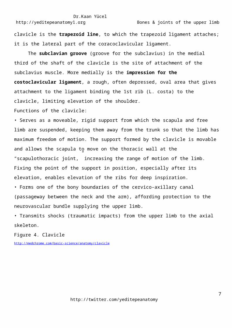
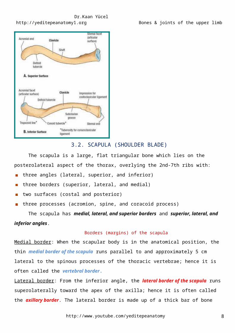
3.1. CLAVICLE (COLLAR BONE)The clavicle is the only bony attachment between the trunk and the upper limb. It is palpable along
its entire length and has a gentle S-shaped contour, with the forward-facing convex part medial and the
forward-facing concave part lateral.
The medial end is called “sternal end". The sternal end is enlarged and triangular where it articulates with
the manubrium of the sternum at the sternoclavicular joint.
The lateral end is called “acromial” end”. The acromial end is flat where it articulates with the acromion of
the scapula at the acromioclavicular joint.
Figure 3. The location of the claviclehttp://www.daviddarling.info/images/clavicle.gif
The medial two thirds of the shaft of the clavicle are convex anteriorly, whereas the lateral third is flattened
and concave anteriorly. These curvatures increase the resilience of the clavicle and give it the appearance of
an elongated capital S.
Although designated as a long bone, the clavicle has no medullary (marrow) cavity. It consists of spongy
(trabecular) bone with a shell of compact bone.
The inferior surface of the clavicle is rough because strong ligaments bind it to the 1st rib near its
sternal end and suspend the scapula from its acromial end. The conoid tubercle, near the acromial end of
the clavicle, gives attachment to the conoid ligament, the medial part of the coracoclavicular ligament. Also,
near the acromial end of the clavicle is the trapezoid line, to which the trapezoid ligament attaches; it is the
lateral part of the coracoclavicular ligament.
The subclavian groove (groove for the subclavius) in the medial third of the shaft of the clavicle is the
site of attachment of the subclavius muscle. More medially is the impression for the costoclavicular
ligament, a rough, often depressed, oval area that gives attachment to the ligament binding the 1st rib (L.
costa) to the clavicle, limiting elevation of the shoulder.
http://twitter.com/yeditepeanatomy5

Dr.Kaan Yücel http://yeditepeanatomy1.org Bones & joints of the upper limb
Functions of the clavicle:
• Serves as a moveable, rigid support from which the scapula and free limb are suspended, keeping them
away from the trunk so that the limb has maximum freedom of motion. The support formed by the clavicle
is movable and allows the scapula to move on the thoracic wall at the “scapulothoracic joint,” increasing the
range of motion of the limb. Fixing the point of the support in position, especially after its elevation, enables
elevation of the ribs for deep inspiration.
• Forms one of the bony boundaries of the cervico-axillary canal (passageway between the neck and the
arm), affording protection to the neurovascular bundle supplying the upper limb.
• Transmits shocks (traumatic impacts) from the upper limb to the axial skeleton.
Figure 4. Claviclehttp://medchrome.com/basic-science/anatomy/clavicle
3.2. SCAPULA (SHOULDER BLADE)The scapula is a large, flat triangular bone which lies on the posterolateral aspect of the thorax,
overlying the 2nd-7th ribs with:
three angles (lateral, superior, and inferior)
three borders (superior, lateral, and medial)
two surfaces (costal and posterior)
three processes (acromion, spine, and coracoid process)
The scapula has medial, lateral, and superior borders and superior, lateral, and inferior angles.
Borders (margins) of the scapula
http://www.youtube.com/yeditepeanatomy 6
Dr.Kaan Yücel http://yeditepeanatomy1.org Bones & joints of the upper limb
Medial border: When the scapular body is in the anatomical position, the thin medial border of the scapula
runs parallel to and approximately 5 cm lateral to the spinous processes of the thoracic vertebrae; hence it is
often called the vertebral border.
Lateral border: From the inferior angle, the lateral border of the scapula runs superolaterally toward the
apex of the axilla; hence it is often called the axillary border. The lateral border is made up of a thick bar of
bone that prevents buckling of this stressbearing region of the scapula.The lateral border terminates in the
truncated lateral angle of the scapula, the thickest part of the bone that bears the broadened head of the
scapula. The glenoid cavity is the primary feature of the head. The shallow constriction between the head
and the body defines the neck of the scapula.
Superior border: is marked near the junction of its medial two thirds and lateral third by the suprascapular
notch. The suprascapular notch is located where the superior border joins the base of the coracoid process.
The superior border is the thinnest and shortest of the three borders.
The scapula is capable of considerable movement on the thoracic wall at the physiological
scapulothoracic joint, providing the base from which the upper limb operates.
Posterior surface of the scapulaAcromionDeltoid tubercle of the scapular spineSpine of the scapulaSupraspinous and infraspinous fossae
The convex posterior surface of the scapula is unevenly divided by a thick projecting ridge of bone;
spine of the scapula, into a small supraspinous fossa and a much larger infraspinous fossa. The spine
continues laterally as the flat expanded acromion (G. akros, point), which forms the subcutaneous point of
the shoulder and articulates with the acromial end of the clavicle. The deltoid tubercle of the scapular spine
is the prominence indicating the medial point of attachment of the deltoid. The spine and acromion serve as
levers for the attached muscles, particularly the trapezius.
Anterior (Costal) surface of the scapulaSubscapular fossaThe concave costal surface of most of the scapula forms a large subscapular fossa. The broad bony surfaces
of the three fossae provide attachments for fleshy muscles.
Lateral surface of the scapulaCoracoid processFossa ovalis
http://twitter.com/yeditepeanatomy7
Dr.Kaan Yücel http://yeditepeanatomy1.org Bones & joints of the upper limb
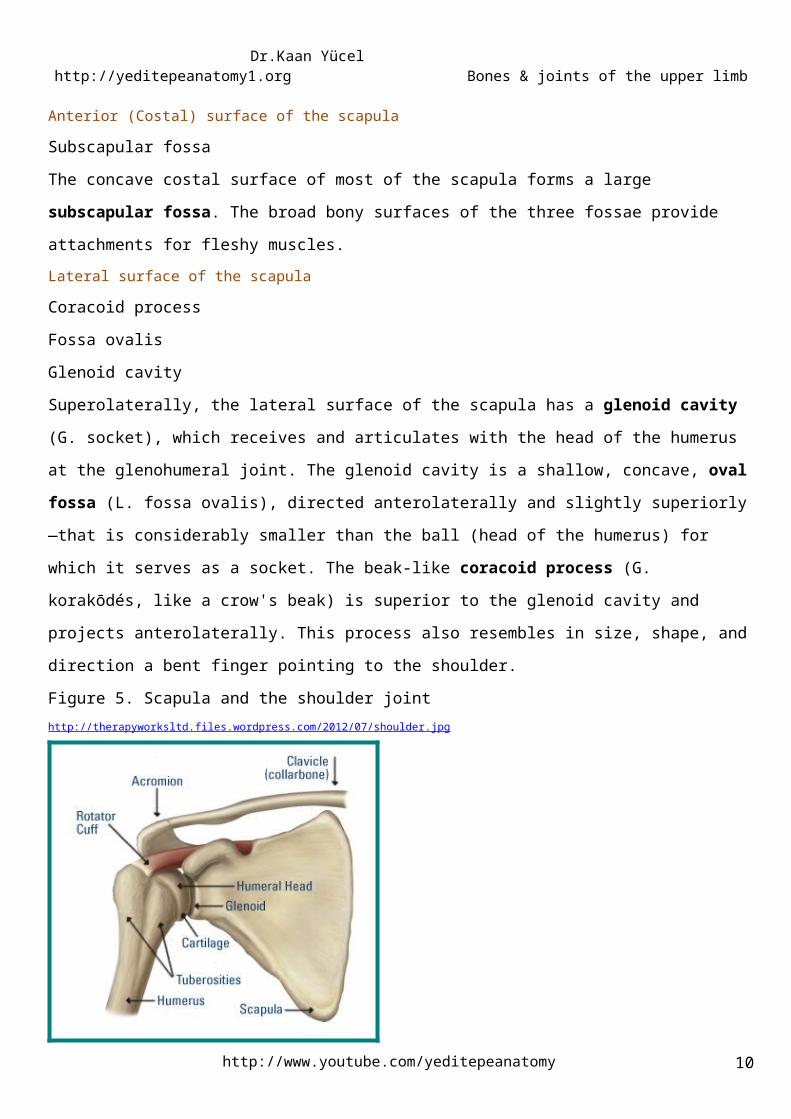
Glenoid cavitySuperolaterally, the lateral surface of the scapula has a glenoid cavity (G. socket), which receives and
articulates with the head of the humerus at the glenohumeral joint. The glenoid cavity is a shallow, concave,
oval fossa (L. fossa ovalis), directed anterolaterally and slightly superiorly—that is considerably smaller than
the ball (head of the humerus) for which it serves as a socket. The beak-like coracoid process (G. korakōdés,
like a crow's beak) is superior to the glenoid cavity and projects anterolaterally. This process also resembles
in size, shape, and direction a bent finger pointing to the shoulder.
Figure 5. Scapula and the shoulder jointhttp://therapyworksltd.files.wordpress.com/2012/07/shoulder.jpg
Figure 6. Scapula- costal surface, lateral view, and posterior surfacehttp://conornordengren.com/2011/10/14/the-shoulder-girdle-part-1-bones-and-joints-2
Fractures of the Clavicle
http://www.youtube.com/yeditepeanatomy 8
CLINICAL ANATOMY

Dr.Kaan Yücel http://yeditepeanatomy1.org Bones & joints of the upper limb
The clavicle is one of the most frequently fractured bones. Clavicular fractures are especially common
in children and are often caused by an indirect force transmitted from an outstretched hand through the
bones of the forearm and arm to the shoulder during a fall. A fracture may also result from a fall directly on
the shoulder. The weakest part of the clavicle is the junction of its middle and lateral thirds.
Fractures of the ScapulaFracture of the scapula is usually the result of severe trauma, as occurs in pedestrian-vehicle
accidents. Usually there are also fractured ribs. Most fractures require little treatment because the scapula is
covered on both sides by muscles.
.
4.1. HUMERUS (ARM BONE)the largest bone in the upper limb
The skeletal support for the arm is the humerus. The humerus (arm bone) articulates with the
scapula at the glenohumeral joint and the radius and ulna at the elbow joint.
In cross-section, the shaft of the humerus is somewhat triangular with:
anterior, lateral, and medial borders
anterolateral, anteromedial, and posterior surfaces
Intermuscular septa, which separate the anterior compartment from the posterior compartment, attach to
the medial and lateral borders.
Proximal end of the humerusAnatomical neck of the humerusGreater & lesser tuberclesHead of the humerusIntertubercular (bicipital) grooveSurgical neck of the humerus
The proximal end of the humerus has a head, surgical and anatomical necks, and greater and lesser
tubercles. The spherical head of the humerus articulates with the glenoid cavity of the scapula. The
anatomical neck of the humerus is formed by the groove circumscribing the head and separating it from the
greater and lesser tubercles. It indicates the line of attachment of the glenohumeral joint capsule. The
surgical neck of the humerus, a common site of fracture, is the narrow part distal to the head and tubercles.
http://twitter.com/yeditepeanatomy9
4. BONE OF THE ARM
Dr.Kaan Yücel http://yeditepeanatomy1.org Bones & joints of the upper limb
The junction of the head and neck with the shaft of the humerus is indicated by the greater and lesser
tubercles, which provide attachment and leverage to some scapulohumeral muscles. The greater tubercle is
at the lateral margin of the humerus, whereas the lesser tubercle projects anteriorly from the bone. The
intertubercular (bicipital) groove separates the tubercles.
Shaft of the humerusDeltoid tuberosityMedial and lateral supraepicondylar (supracondylar) ridgesRadial grooveThe shaft of the humerus has two prominent features: the deltoid tuberosity laterally, for attachment of the
deltoid muscle, and the oblique radial groove (groove for radial nerve, spiral groove) posteriorly.
Distally, the bone becomes flattened, and the medial and lateral borders expand inferiorly as the lateral
supraepicondylar ridge (lateral supracondylar ridge) and the medial supraepicondylar ridge (medial
supracondylar ridge). The lateral supraepicondylar ridge is more pronounced than the medial and is
roughened for the attachment of muscles found in the posterior compartment of the forearm.
Distal end of the humerusCondyle of the humerus; Trochlea+Capitulum Coronoid fossaMedial and lateral epicondylesOlecranon fossaThe distal end of the humerus has a condyle, two epicondyles, and three fossae:
Condyle
The two articular parts of the condyle, the capitulum and the trochlea, articulate with the two bones of the
forearm.
Capitulum (L. little head) articulates with the radius of the forearm. Lateral in position and hemispherical in
shape, is not visible when the humerus is viewed from the posterior aspect.
Trochlea (L. pulley) articulates with the ulna of the forearm. It is pulley shaped and lies medial to the
capitulum. Its medial edge is more pronounced than its lateral edge and, unlike the capitulum, it extends
onto the posterior surface of the bone.
Epicondyles
The two epicondyles lie adjacent, and somewhat superior, to the trochlea and capitulum.
The medial epicondyle, a large bony protuberance, is the major palpable landmark on the medial side of the
elbow, and projects medially from the distal end of the humerus. It is the attachment site for the muscles of
the anterior (flexor) compartment of the forearm.
http://www.youtube.com/yeditepeanatomy 10

Dr.Kaan Yücel http://yeditepeanatomy1.org Bones & joints of the upper limb
The lateral epicondyle is much less pronounced than the medial epicondyle. It is lateral to the capitulum and
the attachment site for the muscles of the posterior (extensor) compartment of the forearm.
Fossae
Coronoid fossa adjacent to the radial fossa superior to the trochlea. It receives the coronoid process of the
ulna during full flexion of the elbow.
Olecranon fossa largest fossa, immediately superior to the trochlea on the posterior surface of the distal
end of the humerus. It accommodates the olecranon of the ulna during full extension of the elbow.
Radial fossa a shallow fossa immediately superior to the capitulum on the anterior surface of the humerus.
It accommodates the edge of the head of the radius when the forearm is fully flexed.
Figure 7. Humerus Figure 8. Distal end of the humerushttp://www.edoctoronline.com/medical-atlas.asp?c=4&id=22082 http://www.medpreponline.com/2009/06/supracondylar-fractures-of-humerus-in.html
Fractures of the humerus
http://twitter.com/yeditepeanatomy11
CLINICAL ANATOMY

Dr.Kaan Yücel http://yeditepeanatomy1.org Bones & joints of the upper limb
Most injuries of the proximal end of the humerus are fractures of the surgical neck. These injuries are
especially common in elderly people with osteoporosis. The injuries usually result from a minor fall on the
hand, with the force being transmitted up the forearm bones of the extended limb.
The two forearm bones serve together to form the second unit of an articulated mobile strut (the
first unit being the humerus), with a mobile base formed by the shoulder, that positions the hand. However,
because this unit is formed by two parallel bones, one of which (the radius) can pivot about the other (the
ulna), supination and pronation are possible. This makes it possible to rotate the hand when the elbow is
flexed.
5.1. ULNAmedial and longer of the two forearm bones
The ulna is the stabilizing bone of the forearm.
Proximal end of the ulnaCoronoid process OlecranonRadial notchTrochlear notch (Semilunar notch)Tuberosity of the ulnaThe proximal end of the ulna is larger than the proximal end of the radius. This end is specialized for
articulation with the humerus proximally and the head of the radius laterally. For articulation with the
humerus, the ulna has two prominent projections:
1) Olecranon projects proximally from its posterior aspect (forming the tip of the elbow) and serves as a
short lever for extension of the elbow.
2) Coronoid process projects anteriorly. Its superolateral surface is articular and participates, with the
olecranon, in forming the trochlear notch. The lateral surface is marked by a smooth rounded cavity; radial
notch for articulation with the head of the radius.
Inferior to the coronoid process is the tuberosity of the ulna.
The olecranon and coronoid processes form the walls of the trochlear notch (semilunar notch). The
trochlear in profile resembles the jaws of a Crescent wrench as it “grips” (articulates with) the trochlea of
the humerus.
Shaft of the ulnaSupinator crest Supinator fossa
http://www.youtube.com/yeditepeanatomy 12
5. BONES OF THE FOREARM

Dr.Kaan Yücel http://yeditepeanatomy1.org Bones & joints of the upper limb
The shaft of the ulna is broad superiorly where it is continuous with the large proximal end and narrow
distally to form a small distal head. The shaft of the ulna is triangular in cross-section and has:
three borders (anterior, posterior, and interosseous)
three surfaces (anterior, posterior, and medial).
The anterior border is smooth and rounded. The posterior border is sharp and palpable along its entire
length. The interosseous border is also sharp and is the attachment site for the interosseous membrane,
which joins the ulna to the radius.
Inferior to the radial notch on the lateral surface of the ulnar shaft is a prominent ridge, the supinator crest.
On the lateral surface of proximal end of ulna, just below the radial notch is a concavity, the supinator fossa.
Distal end of the ulnaHead of the ulnaUlnar styloid processAt the narrow distal end of the ulna is a small but abrupt enlargement, the disc-like head of the ulna with a
small, conical ulnar styloid process. The ulna does not reach—and therefore does not participate in—the
wrist (radiocarpal) joint.
5.2.RADIUSlateral and shorter of the two forearm bones
Proximal end of the radius A short head Neck Radial tuberosityHead of the radiusProximally, the smooth superior aspect of the discoid head of the radius is concave for articulation with the
capitulum of the humerus during flexion and extension of the elbow joint. The head also articulates
peripherally with the radial notch of the ulna; thus the head is covered with articular cartilage.
Neck of the radiusThe neck of the radius is a constriction distal to the head. The oval radial tuberosity is distal to the medial
part of the neck and demarcates the proximal end (head and neck) of the radius from the shaft. The radial
tuberosity is medially directed.
Shaft of the radiusOblique line of the radius
http://twitter.com/yeditepeanatomy13

Dr.Kaan Yücel http://yeditepeanatomy1.org Bones & joints of the upper limb
The shaft of the radius, in contrast to that of the ulna, gradually enlarges as it passes distally. Throughout
most of its length, like the ulna, the shaft of the radius is triangular in cross-section, with:
three borders (anterior, posterior, and interosseous)
three surfaces (anterior, posterior, and lateral).
The anterior border begins on the medial side of the bone as a continuation of the radial tuberosity. In the
superior third of the bone, it crosses the shaft diagonally, from medial to lateral, as the oblique line of the
radius. The posterior border is distinct only in the middle third of the bone. The interosseous border is sharp
and is the attachment site for the interosseous membrane, which links the radius to the ulna.
Distal end of the radius Dorsal tubercle of the radius Radial styloid process Ulnar notchThe medial aspect of the distal end of the radius forms a concavity, the ulnar notch, which accommodates
the head of the ulna. Its lateral aspect becomes increasingly ridge-like, terminating distally in the radial
styloid process. The radial styloid process is larger than the ulnar styloid process and extends farther
distally. This relationship is of clinical importance when the ulna and/or the radius is fractured.
Projecting dorsally, the dorsal tubercle of the radius lies between otherwise shallow grooves for the passage
of the tendons of forearm muscles.
The distal end of the bone is marked by two facets for articulation with two carpal bones (the scaphoid and
lunate).
Figures 9 & 10. Radius & Ulnahttp://www.bcnlp.ac.th/Anatomy/page/apichat/bone/page/ulna.html http://www.daviddarling.info/images/radius_and_ulna.jpg
http://www.youtube.com/yeditepeanatomy 14

Dr.Kaan Yücel http://yeditepeanatomy1.org Bones & joints of the upper limb
Fractures of the Radius and UlnaFractures of both the radius and the ulna are usually the result of severe injury. A direct injury usually
produces transverse fractures at the same level, usually in the middle third of the bones. Isolated fractures
of the radius or ulna also occur. Because the shafts of these bones are firmly bound together by the
interosseous membrane, a fracture of one bone is likely to be associated with dislocation of the nearest
joint.
Fracture of the distal end of the radius is a common fracture in adults > 50 years of age and occurs
more frequently in women because their bones are more commonly weakened by osteoporosis. A complete
transverse fracture of the distal 2 cm of the radius, called a Colles’ fracture, is the most common fracture of
the forearm. The distal fragment is displaced dorsally and is often comminuted (broken into pieces). The
fracture usually occurs as the result of trying to ease a fall by outstretching the upper limb. The force drives
the distal fragment posteriorly and superiorly, and the distal articular surface is inclined posteriorly. This
posterior displacement produces a posterior bump, sometimes referred to as the “dinner-fork deformity”
because the forearm and wrist resemble the shape of a fork.
Fractures of the olecranon process can result from a fall on the flexed elbow or from a direct blow.
Olecranon BursitisA small subcutaneous bursa is present over the olecranon process of the ulna, and repeated trauma
often produces chronic bursitis.
http://twitter.com/yeditepeanatomy15
CLINICAL ANATOMY

Dr.Kaan Yücel http://yeditepeanatomy1.org Bones & joints of the upper limb
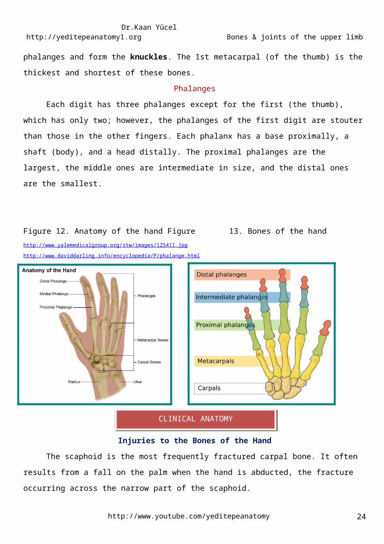
The hand is the region of the upper limb distal to the wrist joint. It is subdivided into three parts:
wrist (carpus);
metacarpus;
digits (five fingers including the thumb).
Carpal bonesThe wrist, or carpus, is composed of eight carpal bones (carpals) arranged in proximal and distal
rows of four. These small bones give flexibility to the wrist. The carpus is markedly convex from side to side
posteriorly and concave anteriorly. Augmenting movement at the wrist joint, the two rows of carpals glide
on each other; in addition, each bone glides on those adjacent to it.
From lateral to medial, the four bones in the proximal row of carpals• Scaphoid (G. skaphé, skiff, boat): a boat-shaped bone that articulates proximally with the radius and has
a prominent scaphoid tubercle; it is the largest bone in the proximal row of carpals.
• Lunate (L. luna, moon): a crescent (or moon) -shaped bone between the scaphoid and the triquetral
bones; it articulates proximally with the radius and is broader anteriorly than posteriorly.
• Triquetrum (L. triquetrus, three-cornered): a pyramidal bone on the medial side of the carpus; it
articulates proximally with the articular disc of the distal radio-ulnar joint.
• Pisiform (L. pisum, pea): a small, pea-shaped bone that lies on the palmar surface of the triquetrum.
From lateral to medial, the four bones in the distal row of carpals• Trapezium (G. trapeze, table): a four-sided bone on the lateral side of the carpus; it articulates with the
1st and 2nd metacarpals, scaphoid, and trapezoid bones.
• Trapezoid: a wedge-shaped bone that resembles the trapezium; it articulates with the 2nd metacarpal,
trapezium, capitate, and scaphoid bones.
• Capitate (L. caput, head): a head-shaped bone with a rounded extremity and the largest bone in the
carpus; it articulates primarily with the 3rd metacarpal distally and with the trapezoid, scaphoid, lunate, and
hamate.
• Hamate (L. hamulus, a little hook): a wedge-shaped bone on the medial side of the hand; it articulates
with the 4th and 5th metacarpal, capitate, and triquetral bones; it has a distinctive hooked process, the hook
of the hamate, that extends anteriorly.
Carpal arch The carpal bones do not lie in a flat plane; rather, they form an arch, whose base is directed anteriorly. The
lateral side of this base is formed by the tubercles of the scaphoid and trapezium. The medial side is formed
by the pisiform and the hook of hamate. http://www.youtube.com/yeditepeanatomy 16
6. BONES OF THE HAND

Dr.Kaan Yücel http://yeditepeanatomy1.org Bones & joints of the upper limb
Figure 11. Carpal boneshttp://www.daviddarling.info/encyclopedia/C/carpal.html
The proximal surfaces of the distal row of carpals articulate with the proximal row of carpals, and their
distal surfaces articulate with the metacarpals.
MetacarpusThe metacarpus forms the skeleton of the palm of the hand between the carpus and the phalanges.
It is composed of five metacarpal bones (metacarpals). Each metacarpal consists of a base, shaft, and head.
The proximal bases of the metacarpals articulate with the carpal bones, and the distal heads of the
metacarpals articulate with the proximal phalanges and form the knuckles. The 1st metacarpal (of the
thumb) is the thickest and shortest of these bones.
PhalangesEach digit has three phalanges except for the first (the thumb), which has only two; however, the
phalanges of the first digit are stouter than those in the other fingers. Each phalanx has a base proximally, a
shaft (body), and a head distally. The proximal phalanges are the largest, the middle ones are intermediate
in size, and the distal ones are the smallest.
Figure 12. Anatomy of the hand Figure 13. Bones of the handhttp://www.yalemedicalgroup.org/stw/images/125411.jpg http://www.daviddarling.info/encyclopedia/P/phalange.html
http://twitter.com/yeditepeanatomy17
Dr.Kaan Yücel http://yeditepeanatomy1.org Bones & joints of the upper limb
Injuries to the Bones of the HandThe scaphoid is the most frequently fractured carpal bone. It often results from a fall on the palm
when the hand is abducted, the fracture occurring across the narrow part of the scaphoid.
Dislocation of the lunate bone occasionally occurs in young adults who fall on the outstretched hand
in a way that causes hyperextension of the wrist joint.
Fractures of the metacarpal bones can occur as a result of direct violence, such as the clenched fist
striking a hard object. The fracture always angulates dorsally. The “boxer’s fracture” commonly produces an
oblique fracture of the neck of the fifth and sometimes the fourth metacarpal bones.
Fractures of the phalanges are common and usually follow direct injury.
Joints of the pectoral girdle
Acromioclavicular joint & Sternoclavicular joint
Glenohumeral (Shoulder) joint
Elbow joint
Proximal (Superior) and Distal (Inferior) radio-
ulnar joints
Radiocarpal (Wrist) joint
Intercarpal joints
Carpometacarpal and intermetacarpal joints
Figure 14. Joints of the upper limbhttp://home.comcast.net/~wnor/lesson5jointsofupperlimb.htm
http://www.youtube.com/yeditepeanatomy 18
7. JOINTS OF THE UPPER LIMB
CLINICAL ANATOMY
Dr.Kaan Yücel http://yeditepeanatomy1.org Bones & joints of the upper limb
1. Articulation between Sternum and clavicle
2. Distinct feature of the joint the only articulation between the upper limb and the axial skeleton. It can be
readily palpated because the sternal end of the clavicle lies superior to the manubrium of the sternum.
3. Synovial joint type Saddle type, but functions as a ball-and-socket joint
4. Articular disc Yes. The articular disc divides the sternoclavicular joint into two compartments.
5. Articular surfaces The sternoclavicular joint occurs between the sternal end of the clavicle and the
clavicular notch of the manubrium of sternum together with a small part of the first costal cartilage.
6. Ligaments of the sternoclavicular joint
The sternoclavicular joint is surrounded by a joint capsule and is reinforced by four ligaments:
Anterior and posterior sternoclavicular ligamentsCostoclavicular ligamentInterclavicular ligamentThe anterior and posterior sternoclavicular ligaments are anterior and posterior, respectively, to the joint.
The interclavicular ligament links the ends of the two clavicles to each other and to the superior surface of
the manubrium of sternum.
http://twitter.com/yeditepeanatomy19
STERNOCLAVICULAR JOINT

Dr.Kaan Yücel http://yeditepeanatomy1.org Bones & joints of the upper limb
The costoclavicular ligament is positioned laterally to the joint and links the proximal end of the clavicle to
the first rib and related costal cartilage.
Anterior and posterior sternoclavicular ligaments reinforce the joint capsule anteriorly and
posteriorly. The interclavicular ligament strengthens the capsule superiorly. The costoclavicular ligament
anchors the inferior surface of the sternal end of the clavicle to the 1st rib and its costal cartilage, limiting
elevation of the pectoral girdle.
The strength of the sternoclavicular joint depends on ligaments and its articular disc. The disc is firmly
attached to the anterior and posterior sternoclavicular ligaments, thickenings of the fibrous layer of the joint
capsule, as well as the interclavicular ligament. The great strength of the sternoclavicular joint is a
consequence of these attachments. Thus, although the articular disc serves as a shock absorber of forces
transmitted along the clavicle from the upper limb, dislocation of the clavicle is rare, whereas fracture of the
clavicle is common.
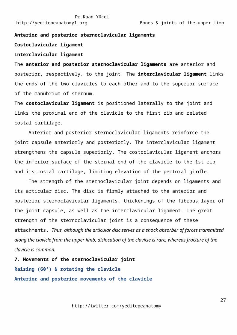
7. Movements of the sternoclavicular joint
Raising (60°) & rotating the clavicleAnterior and posterior movements of the clavicle
The sternoclavicular joint allows movement of the clavicle, predominantly in the anteroposterior and
vertical planes, although some rotation also occurs. Although the sternoclavicular joint is extremely strong, it
is significantly mobile to allow movements of the pectoral girdle and upper limb. During full elevation of the
limb, the clavicle is raised to approximately a 60° angle. When elevation is achieved via flexion, it is
accompanied by rotation of the clavicle around its longitudinal axis.
Figure 15. Sternoclavicular jointhttp://www.daviddarling.info/encyclopedia/S/sternoclavicular_joint.html
http://www.youtube.com/yeditepeanatomy 20

Dr.Kaan Yücel http://yeditepeanatomy1.org Bones & joints of the upper limb
Sternoclavicular Joint InjuriesThe strong costoclavicular ligament firmly holds the medial end of the clavicle to the first costal
cartilage. Violent forces directed along the long axis of the clavicle usually result in fracture of that bone, but
dislocation of the sternoclavicular joint takes place occasionally. If the costoclavicular ligament ruptures
completely, it is difficult to maintain the normal position of the clavicle once reduction has been
accomplished.
1. Articulation between Acromion of scapulae and clavicle
2. Distinct feature of the joint Located 2-3 cm from the “point” of the shoulder formed by the lateral part of
the acromion
3. Synovial joint type Plane type
4. Articular disc Yes. The articular surfaces are separated by an incomplete wedge-shaped articular disc.
5. Articular surfaces The acromial end of the clavicle articulates with the acromion of the scapula. The
articular surfaces, covered with fibrocartilage.
6. Ligaments of the acromioclavicular joint
Intrinsic ligament of the acromioclavicular joint
Acromioclavicular ligamentExtrinsic ligament of the acromioclavicular joint
Coracoclavicular ligament- conoid ligament and trapezoid ligament
http://twitter.com/yeditepeanatomy21
ACROMIOCLAVICULAR JOINT
CLINICAL ANATOMY
Dr.Kaan Yücel http://yeditepeanatomy1.org Bones & joints of the upper limb
The acromioclavicular ligament is a fibrous band extending from the acromion to the clavicle that
strengthens the acromioclavicular joint superiorly. However, the integrity of the joint is maintained by
extrinsic ligaments, distant from the joint itself.
The much larger coracoclavicular ligament is a strong pair of bands that unite the coracoid process of
the scapula to the clavicle, anchoring the clavicle to the coracoid process.
The coracoclavicular ligament is not directly related to the joint. However,it is an important strong
accessory ligament, providing much of the weightbearing support for the upper limb on the clavicle. It
maintains the position of the clavicle on the acromion. It spans the distance between the coracoid process of
the scapula and the inferior surface of the acromial end of the clavicle.
The coracoclavicular ligament consists of two ligaments, the conoid and trapezoid ligaments, which
are often separated by a bursa.
The vertical conoid ligament is an inverted triangle (cone), is attached to the root of the coracoid process. Its
wide attachment (base of the triangle) is to the conoid tubercle on the inferior surface of the clavicle. The
nearly horizontal trapezoid ligament is attached to the superior surface of the coracoid process and extends
laterally to the trapezoid line on the inferior surface of the clavicle.
In addition to augmenting the acromioclavicular joint, the coracoclavicular ligament provides the
means by which the scapula and free limb are (passively) suspended from the clavicular strut.
7. Movements of the acromioclavicular joint
The acromioclavicular joint allows movement in the anteroposterior and vertical planes together with
some axial rotation. The acromion of the scapula rotates on the acromial end of the clavicle. These
movements are associated with motion at the physiological scapulothoracic joint.
Figure 16. Acromioclavicular jointhttp://www.rahulgladwin.com/medimages/index.php?level=picture&id=96
http://www.youtube.com/yeditepeanatomy 22
CLINICAL ANATOMY

Dr.Kaan Yücel http://yeditepeanatomy1.org Bones & joints of the upper limb
Acromioclavicular Joint DislocationA severe blow on the point of the shoulder, as is incurred during blocking or tackling in football or
any severe fall, can result in the acromion being thrust beneath the lateral end of the clavicle, tearing the
coracoclavicular ligament. This condition is known as shoulder separation. The displaced outer end of the
clavicle is easily palpable.
1. Articulation between Humerus and scapula
2. Distinct feature of the joint Ball-and-socket type of synovial joint that permits a wide range of movement;
however, its mobility makes the joint relatively unstable. The glenohumeral joint has more freedom of
movement than any other joint in the body.
3. Synovial joint type Ball-and-socket type
4. Articular disc No.
5. Articular surfaces The large, round humeral head articulates with the relatively shallow glenoid cavity of
the scapula, which is deepened slightly but effectively by the ring-like, fibrocartilaginous glenoid labrum (L.,
lip). Both articular surfaces are covered with hyaline cartilage. The glenoid cavity accepts little more than a
third of the humeral head, which is held in the cavity by the tonus of the musculotendinous rotator cuff
muscles.
6. Ligaments of the glenohumeral joint
Glenohumeral ligamentsCoracohumeral ligamentTransverse humeral ligamentCoracoacromial ligamentThe fibrous membrane of the joint capsule is thickened:
Glenohumeral ligaments three fibrous bands, evident only on the internal aspect of the capsule
anterosuperiorly in three locations to form superior, middle, and inferior glenohumeral ligaments
pass from the superomedial margin of the glenoid cavity to the lesser tubercle and inferiorly related
anatomical neck of the humerus.
strengthen the anterior aspect of the joint capsule of the joint.
Coracohumeral ligament strong broad band
http://twitter.com/yeditepeanatomy23
GLENOHUMERAL (SHOULDER) JOINT - ARTICULATIO HUMERI
Dr.Kaan Yücel http://yeditepeanatomy1.org Bones & joints of the upper limb
passes from the base of the coracoid process to the anterior aspect of the greater tubercle of the
humerus.
strengthens the capsule superiorly.
Transverse humeral ligament a broad fibrous band
between the greater and lesser tubercles of the humerus, bridging over the intertubercular sulcus
converts the groove into a canal, which holds the synovial sheath and tendon of the biceps brachii in
place during movements of the glenohumeral joint.
The coraco-acromial arch is an extrinsic, protective structure formed by the smooth inferior aspect of
the acromion and the coracoid process of the scapula, with the coracoacromial ligament spanning between
them. This osseoligamentous structure forms a protective arch that overlies the humeral head, preventing
its superior displacement from the glenoid cavity. The coraco-acromial arch is so strong that a forceful
superior thrust of the humerus will not fracture it; the humeral shaft or clavicle fractures first.
Transmitting force superiorly along the humerus (e.g., when standing at a desk and partly supporting the
body with the outstretched limbs), the humeral head presses against the coraco-acromial arch.
Joint stability is provided by surrounding muscle tendons and a skeletal arch formed superiorly by the
coracoid process and acromion and the coracoacromial ligament.
7. Movements of the glenohumeral joint
Flexion of the humerusExtension of the humerusAbduction of the humerusAdduction of the humerusMedial and lateral rotation of the humerusCircumduction
The glenohumeral joint has more freedom of movement than any other joint in the body. This
freedom results from the laxity of its joint capsule and the large size of the humeral head compared with the
small size of the glenoid cavity. The glenohumeral joint allows movements around three axes and permits
flexion-extension, abduction-adduction, rotation (medial and lateral) of the humerus, and circumduction.
Lateral rotation of the humerus increases the range of abduction. When the arm is abducted without
rotation, available articular surface is exhausted and the greater tubercle contacts the coraco-acromial arch,
preventing further abduction. If the arm is then laterally rotated 180°, the tubercles are rotated posteriorly
and more articular surface becomes available to continue elevation.
Circumduction at the glenohumeral joint is an orderly sequence of flexion, abduction, extension, and
adduction—or the reverse. Unless performed over a small range, these movements do not occur at the http://www.youtube.com/yeditepeanatomy 24

Dr.Kaan Yücel http://yeditepeanatomy1.org Bones & joints of the upper limb
glenohumeral joint in isolation; they are accompanied by movements at the two other joints of the pectoral
girdle (sternoclavicular and acromioclavicular joints).
8. Bursae around the glenohumeral joint
Several bursae (sac-like cavities), containing capillary films of synovial fluid secreted by the synovial
membrane, are situated near the glenohumeral joint. Bursae are located where tendons rub against bone,
ligaments, or other tendons and where skin moves over a bony prominence. The bursae around the
glenohumeral joint are of special clinical importance because some of them communicate with the joint
cavity (e.g., the subscapular bursa). Consequently, opening a bursa may mean entering the cavity of the
glenohumeral joint.
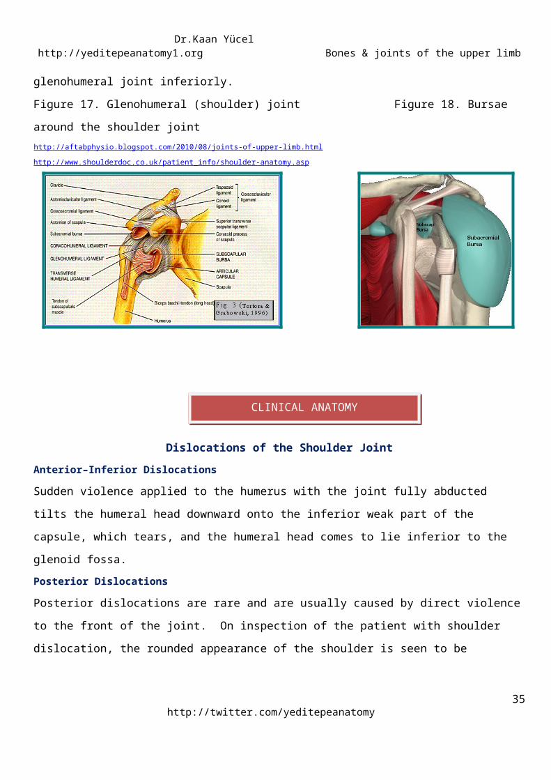
Subscapular Bursa: is located between the tendon of the subscapularis and the neck of the scapula. The
bursa protects the tendon where it passes inferior to the root of the coracoid process and over the neck of
the scapula.
Subacromial Bursa (Subdeltoid bursa): is located between the acromion, coraco-acromial ligament,
superiorly and joint capsule of the glenohumeral joint inferiorly.
Figure 17. Glenohumeral (shoulder) joint Figure 18. Bursae around the shoulder jointhttp://aftabphysio.blogspot.com/2010/08/joints-of-upper-limb.html http://www.shoulderdoc.co.uk/patient_info/shoulder-anatomy.asp
Dislocations of the Shoulder JointAnterior–Inferior Dislocations
http://twitter.com/yeditepeanatomy25
CLINICAL ANATOMY
Dr.Kaan Yücel http://yeditepeanatomy1.org Bones & joints of the upper limb
Sudden violence applied to the humerus with the joint fully abducted tilts the humeral head downward onto
the inferior weak part of the capsule, which tears, and the humeral head comes to lie inferior to the glenoid
fossa.
Posterior DislocationsPosterior dislocations are rare and are usually caused by direct violence to the front of the joint. On
inspection of the patient with shoulder dislocation, the rounded appearance of the shoulder is seen to be
lost because the greater tuberosity of the humerus is no longer bulging laterally beneath the deltoid muscle.
.
1. Articulation between Humerus, ulna and radius
2. Distinct feature of the joint Located 2-3 cm inferior to the epicondyles of the humerus
3. Synovial joint type Hinge type
4. Articular disc No.
5. Articular surfaces The spool-shaped trochlea and spheroidal capitulum of the humerus articulate with the
trochlear notch of the ulna and the slightly concave superior aspect of the head of the radius, respectively;
therefore, there are humeroulnar and humeroradial articulations.
6. Joint capsule of elbow joint The fibrous layer of the joint capsule surrounds the elbow joint. The joint
capsule is weak anteriorly and posteriorly but is strengthened on each side by collateral ligaments.
7. Ligaments of the elbow joint
Medial (ulnar) and lateral (radial) collateral ligamentsThe collateral ligaments of the elbow joint are strong triangular bands that are medial and lateral thickenings
of the fibrous layer of the joint capsule.
The lateral, fan-like radial collateral ligament extends from the lateral epicondyle of the humerus
and blends distally with the anular ligament of the radius, which encircles and holds the head of the radius in
the radial notch of the ulna. The anular ligament forms the proximal radio-ulnar joint and permits pronation
and supination of the forearm.
The medial, triangular ulnar collateral ligament extends from the medial epicondyle of the humerus
to the coronoid process and olecranon of the ulna and consists of three bands: (1) the anterior cord-like
band is the strongest, (2) the posterior fan-like band is the weakest, and (3) the slender oblique band
deepens the socket for the trochlea of the humerus.
8. Movements of the elbow joint
Flexion and extension
http://www.youtube.com/yeditepeanatomy 26
RADIOCARPAL JOINTELBOW JOINT
Dr.Kaan Yücel http://yeditepeanatomy1.org Bones & joints of the upper limb
Flexion and extension occur at the elbow joint. The long axis of the fully extended ulna makes an angle of
approximately 170° with the long axis of the humerus. This angle is called the carrying angle, named for the
way the forearm angles away from the body when something is carried. The obliquity of the ulna and thus of
the carrying angle is more pronounced (the angle is approximately 10° more acute) in women than in men. It
is said to enable the swinging limbs to clear the wide female pelvis when walking. In the anatomical position,
the elbow is against the waist.
9. Bursae of the elbow joint Only some of the bursae around the elbow joint are clinically important.
The three olecranon bursae are the:
•Intratendinous olecranon bursa, which is sometimes present in the tendon of triceps brachii.
•Subtendinous olecranon bursa, which is located between the olecranon and the triceps tendon, just
proximal to its attachment to the olecranon.
•Subcutaneous olecranon bursa, which is located in the subcutaneous connective tissue over the olecranon.
Figure 19. Elbow jointhttp://www.aaos75th.org/stories/patient_story.htm?id=11
Figure 20. Bursae around the elbow jointhttp://www.joint-pain-expert.net/olecranon-bursitis.html
http://twitter.com/yeditepeanatomy27
Dr.Kaan Yücel http://yeditepeanatomy1.org Bones & joints of the upper limb
Stability of the Elbow JointThe elbow joint is stable because of the wrench-shaped articular surface of the olecranon and the
pulley-shaped trochlea of the humerus; it also has strong medial and lateral ligaments. When examining the
elbow joint, the physician must remember the normal relations of the bony points. In extension, the medial
and lateral epicondyles and the top of the olecranon are in a straight line; in flexion, the bony points form
the boundaries of an equilateral triangle.
Dislocations of the Elbow JointElbow dislocations are common, and most are posterior. Posterior dislocation usually follows falling
on the outstretched hand. Posterior dislocations of the joint are common in children because the parts of
the bones that stabilize the joint are incompletely developed. Avulsion of the epiphysis of the medial
epicondyle is also common in childhood because then the medial ligament is much stronger than the bond
of union between the epiphysis and the diaphysis.
1. Articulation between Radius and ulna proximally
2. Distinct feature of the joint -
3. Synovial joint type Pivot type
4. Articular disc No.
5. Articular surfaces The head of the radius articulates with the radial notch of the ulna.
6. Ligaments of the proximal radio-ulnar joint
http://www.youtube.com/yeditepeanatomy 28
PROXIMAL(SUPERIOR) RADIO-ULNAR JOINT
CLINICAL ANATOMY
Dr.Kaan Yücel http://yeditepeanatomy1.org Bones & joints of the upper limb
Annular ligamentThe radial head is held in position by the anular ligament of the radius. The strong anular ligament is
attached to the ulna anterior and posterior to its radial notch. It surrounds the articulating bony surfaces and
forms a collar that, with the radial notch, creates a ring that completely encircles the head of the radius.
7. Movements of the proximal radio-ulnar joint
Supination & pronationThe proximal (superior) radio-ulnar joint is a pivot type of synovial joint that allows movement of the head of
the radius on the ulna.
During pronation and supination, it is the radius that rotates; its head rotates within the cup-shaped
collar formed by the anular ligament and the radial notch on the ulna. Distally, the end of the radius rotates
around the head of the ulna. Almost always, supination and pronation are accompanied by synergistic
movements of the glenohumeral and elbow joints that produce simultaneous movement of the ulna, except
when the elbow is flexed.
Figures 21 & 22. Proximal radio-ulnar jointhttp://teachmeanatomy.net/upper-limb-2/joints-of-the-upper-limb/radioulnar-joints http://www.msdlatinamerica.com/ebooks/HandSurgery/sid484870.html
1. Articulation between Radius and ulna distally
2. Distinct feature of the joint -
3. Synovial joint type Pivot type
4. Articular disc Yes. A fibrocartilaginous, triangular articular disc of the distal radioulnar joint (sometimes
referred to by clinicians as the “triangular ligament”) binds the ends of the ulna and radius together and is
http://twitter.com/yeditepeanatomy29
DISTAL (INFERIOR) RADIO-ULNAR JOINT

Dr.Kaan Yücel http://yeditepeanatomy1.org Bones & joints of the upper limb
the main uniting structure of the joint. The articular disc separates the cavity of the distal radio-ulnar joint
from the cavity of the wrist joint.
5. Articular surfaces The rounded head of the ulna articulates with the ulnar notch on the medial side of the
distal end of the radius.
6. Ligaments of the distal radio-ulnar joint
Anterior and posterior ligamentsAnterior and posterior ligaments strengthen the fibrous layer of the joint capsule of the distal radio-ulnar
joint. These relatively weak transverse bands extend from the radius to the ulna across the anterior and
posterior surfaces of the joint.
7. Movements of the distal radio-ulnar joint
The radius moves around the relatively fixed distal end of the ulna. The distal radio-ulnar joint allows the
distal end of the radius to move anteromedially over the ulna. During pronation of the forearm and hand,
the distal end of the radius moves (rotates) anteriorly and medially, crossing over the ulna anteriorly. During
supination, the radius uncrosses from the ulna, its distal end moving (rotating) laterally and posteriorly so
the bones become parallel.
Figure 23. Distal radio-ulnar jointhttp://en.wikipedia.org/wiki/File:Gray334.png
Radioulnar Joint Disease
http://www.youtube.com/yeditepeanatomy 30
CLINICAL ANATOMY

Dr.Kaan Yücel http://yeditepeanatomy1.org Bones & joints of the upper limb
The proximal radioulnar joint communicates with the elbow joint, whereas the distal radioulnar joint
does not communicate with the wrist joint. In practical terms, this means that infection of the elbow joint
invariably involves the proximal radioulnar joint.
The strength of the proximal radioulnar joint depends on the integrity of the strong anular ligament.
Rupture of this ligament occurs in cases of anterior dislocation of the head of the radius on the capitulum of
the humerus. In young children, in whom the head of the radius is still small and undeveloped, a sudden jerk
on the arm can pull the radial head down through the anular ligament.
1. Articulation between Radius and carpal bones
2. Distinct feature of the joint
3. Synovial joint type Condyloid (ellipsoid) type
4. Articular disc Yes.
5. Articular surfaces The wrist (carpus), the proximal segment of the hand, is a complex of eight carpal
bones, articulating proximally with the forearm via the wrist joint and distally with the five metacarpals. The
ulna does not participate in the wrist joint. The distal end of the radius and the articular disc of the distal
radio-ulnar joint articulate with the proximal row of carpal bones, except for the pisiform.
6. Ligaments of the wrist joint
Dorsal and palmar radiocarpal ligamentsUlnar collateral ligamentRadial collateral ligament
The fibrous layer of the joint capsule is strengthened by strong dorsal and palmar radiocarpal
ligaments. The palmar radiocarpal ligaments pass from the radius to the two rows of carpals. They are strong
and directed so that the hand follows the radius during supination of the forearm. The dorsal radiocarpal
ligaments take the same direction so that the hand follows the radius during pronation of the forearm.
The joint capsule is also strengthened medially by the ulnar collateral ligament, which is attached to
the ulnar styloid process and triquetrum. The joint capsule is also strengthened laterally by the radial
collateral ligament, which is attached to the radial styloid process and scaphoid.
7. Movements of the wrist joint
FlexionExtension Abduction (Radial deviation)
http://twitter.com/yeditepeanatomy31
WRIST JOINT (RADIOCARPAL JOINT)

Dr.Kaan Yücel http://yeditepeanatomy1.org Bones & joints of the upper limb
Adduction (Ulnar deviation)Circumduction
The movements at the wrist joint may be augmented by additional smaller movements at the
intercarpal and midcarpal joints. The movements are flexion—extension, abduction—adduction (radial
deviation-ulnar deviation), and circumduction. The hand can be flexed on the forearm more than it can be
extended; these movements are accompanied (actually, are initiated) by similar movements at the
midcarpal joint between the proximal and the distal rows of carpal bones. Adduction of the hand is greater
than abduction. Most adduction occurs at the wrist joint. Abduction from the neutral position occurs at the
midcarpal joint. Circumduction of the hand consists of successive flexion, adduction, extension, and
abduction.
8. Surface anatomy of the wrist joint The position of the joint is indicated approximately by a line joining
the styloid processes of the radius and ulna, or by the proximal wrist crease.
Figure 24. Wrist (Radiocarpal) jointhttp://teachmeanatomy.net/upper-limb-2/joints-of-the-upper-limb/wrist-joint
Wrist Joint InjuriesA fall on the outstretched hand can strain the anterior ligament of the wrist joint, producing synovial
effusion, joint pain, and limitation of movement. These symptoms and signs must not be confused with
those produced by a fractured scaphoid or dislocation of the lunate bone, which are similar.
Falls on the Outstretched HandIn falls on the outstretched hand, forces are transmitted from the scaphoid to the distal end of the
radius, from the radius across the interosseous membrane to the ulna, and from the ulna to the humerus;
thence, through the glenoid fossa of the scapula to the coracoclavicular ligament and the clavicle, and
finally, to the sternum. If the forces are excessive, different parts of the upper limb give way under
the strain. The area affected seems to be related to age. In a young child, for example, there may be a
posterior displacement of the distal radial epiphysis; in the teenager the clavicle might fracture; in the young http://www.youtube.com/yeditepeanatomy 32
INTERCARPAL JOINTS
CLINICAL ANATOMY

Dr.Kaan Yücel http://yeditepeanatomy1.org Bones & joints of the upper limb
adult the scaphoid is commonly fractured; and in the elderly the distal end of the radius is fractured about 1
in. (2..5 cm) proximal to the wrist joint (Colles’ fracture).
1. Articulation between carpal bones (the intercarpal joints interconnect the carpal bones)
2. Distinct feature of the joint -
3. Synovial joint type Plane type
4. Articular disc No.
5. Articular surfaces
Joints between the carpal bones of the proximal row.
Joints between the carpal bones of the distal row.
The midcarpal joint, a complex joint between the proximal and distal rows of carpal bones.
The pisotriquetral joint, formed from the articulation of the pisiform with the palmar surface of the
triquetrum.
6. Ligaments of the intercarpal joints
Anterior, posterior, and interosseous ligaments7. Movements of the intercarpal joints
GlidingFlexion & extension of the handAbduction & adduction of the handThe gliding movements possible between the carpals occur concomitantly with movements at the wrist
joint, augmenting them and increasing the overall range of movement. Flexion and extension of the hand
are actually initiated at the midcarpal joint, between the proximal and the distal rows of carpals. Most
flexion and adduction occur mainly at the wrist joint, whereas extension and abduction occur primarily at
the midcarpal joint. Movements at the other intercarpal joints are small, with the proximal row being more
mobile than the distal row.
1. Articulation between carpal bones & metacarpals, between metacarpals
2. Distinct feature of the joint -
http://twitter.com/yeditepeanatomy33
CARPOMETACARPAL & INTERMETACARPAL JOINTS

Dr.Kaan Yücel http://yeditepeanatomy1.org Bones & joints of the upper limb
3. Synovial joint type Plane type plane except for the carpometacarpal joint of the thumb, which is a saddle
joint.
4. Articular disc No.
5. Articular surfaces The distal surfaces of the carpals of the distal row articulate with the carpal surfaces of
the bases of the metacarpals at the carpometacarpal joints. The important carpometacarpal joint of the
thumb is between the trapezium and the base of the 1st metacarpal; it has a separate articular cavity. Like
the carpals, adjacent metacarpals articulate with each other; intermetacarpal joints occur between the
radial and ulnar aspects of the bases of the metacarpals.
6. Ligaments of the carpometacarpal and intermetacarpal joints
Palmar and dorsal carpometacarpal and palmar and dorsal intermetacarpal ligamentsInterosseus metacarpal ligamentsSuperficial and deep transverse metacarpal ligamentsThe bones are united in the region of the joints by palmar and dorsal carpometacarpal and palmar and
dorsal intermetacarpal ligaments and by interosseous intermetacarpal ligaments. In addition, the
superficial and deep transverse metacarpal ligaments (the former part of the palmar aponeurosis),
associated with the distal ends of the metacarpals, play a role in limiting movement at these two joints as
they limit separation of the metacarpal heads.
7. Movements of the carpometacarpal and intermetacarpal joints
The carpometacarpal joint of the thumb permits angular movements in any plane (flexion-extension,
abduction-adduction, or circumduction) and a restricted amount of axial rotation. Most important, the
movement essential to opposition of the thumb occurs here.
Almost no movement occurs at the carpometacarpal joints of the 2nd and 3rd digits, that of the 4th
digit is slightly mobile, and that of the 5th digit is moderately mobile, flexing and rotating slightly during a
tight grasp. When the palm of the hand is “cupped” (as during pad-to-pad opposition of thumb and little
finger), two thirds of the movement occur at the carpometacarpal joint of the thumb, and one third occurs
at the carpometacarpal and intercarpal joints of the 4th and 5th fingers.
1. Articulation between metacarpals and phalanges and between phalanges
2. Distinct feature of the joint -
http://www.youtube.com/yeditepeanatomy 34
METACARPOPHALANGEAL & INTERPHALANGEAL JOINTS
Dr.Kaan Yücel http://yeditepeanatomy1.org Bones & joints of the upper limb
3. Synovial joint type The metacarpophalangeal joints are the condyloid type of synovial joint that permit
movement in two planes: flexion-extension and adduction-abduction. The interphalangeal joints are the
hinge type of synovial joint that permit flexion-extension only.
4. Articular disc No.
5. Articular surfaces The heads of the metacarpals articulate with the bases of the proximal phalanges in the
metacarpophalangeal (MP) joints, and the heads of the phalanges articulate with the bases of more distally
located phalanges in the interphalangeal (IP) joints.
6. Ligaments of the carpometacarpal and intermetacarpal joints
Medial and lateral collateral ligamentsPalmar ligamentsThe fibrous layer of each MC and IP joint capsule is strengthened by two (medial and lateral) collateral
ligaments. These ligaments have two parts:
• Denser cord-like parts pass distally from the heads of the metacarpals and phalanges to the bases of the
phalanges.
• Thinner fan-like parts pass anteriorly to attach to thick, densely fibrous or fibrocartilaginous plates, the
palmar ligaments (plates), which form the palmar aspect of the joint capsule.
The fan-like parts of the collateral ligaments cause the palmar ligaments to move like a visor over the
underlying metacarpal or phalangeal heads.
The palmar ligaments of the 2nd-5th MP joints are united by deep transverse metacarpal ligaments that
hold the heads of the metacarpals together.
7. Movements of the carpometacarpal and intermetacarpal joints
Flexion-extension, abduction-adduction, and circumduction of the 2nd-5th digits occur at the 2nd-5th MP
joints. Movement at the MP joint of the thumb is limited to flexion-extension. Only flexion and extension
occur at the IP joints.
http://twitter.com/yeditepeanatomy35