Vessels and Circulation. 2 Some embryology first There are at first six pairs of aortic arches In...
67
Vessels and Circulation
-
Upload
debra-ball -
Category
Documents
-
view
214 -
download
0
Transcript of Vessels and Circulation. 2 Some embryology first There are at first six pairs of aortic arches In...

Vessels and Circulation

2
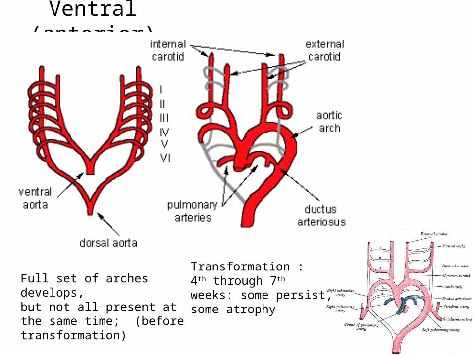
Some embryology first
There are at first six pairs of aortic arches
In fish these are connected to the gills
They undergo a transformation in mammals Birds use the right
arch of the fourth pair Mammals use the
left arch of the fourth pair
3
Ventral (anterior) view
Full set of arches develops,but not all present at the same time; (beforetransformation)
Transformation :4th through 7th
weeks: some persist,some atrophy

4
Right common carotid a ------------------------------.Right subclavian a. --------------------------Brachiocephalic trunk-----------------------------------
4th arches become: Left side: aortic arch Right side: brachiocephalic trunk

5
What the aortic arches become…
Right common carotid a ---------------------------.Right subclavian a. ---------------------------Brachiocephalic trunk-------------------------------

6
3 Major types of blood vessels
Body RA RV Lungs LA LV Boby
1.Arteries2.Capillaries 3.Veins
Arteries carry blood away from the heart-”branch,” “diverge” or “fork”
Veins carry blood toward the heart-”join”, “merge,” “converge”

7
General characteristics of vessels
Three layers (except for the smallest)1. Tunica intima - AKA intima
2. Tunica media – smooth muscle
3. Tunica externa - AKA adventitia
Lumen is the central blood filled space

8
Intima is endothelium (simple squamous epithelium) May have subendothelial layer if 1mm or larger
Tunica media: layers of circular smooth muscles Lamina (layers) of elastin and collagen internal and external Thicker in arteries than veins (maintain blood pressure)
Smooth muscle contraction: vasoconstriction
Smooth muscle relaxation: vasodilation
Sympathetic vasomotor nerves of autonomic nervous system regulate

9
Adventitia (t. externa) – longitudinally running collagen and elastin for strength and recoil

10“muscular” middle sized artery

11
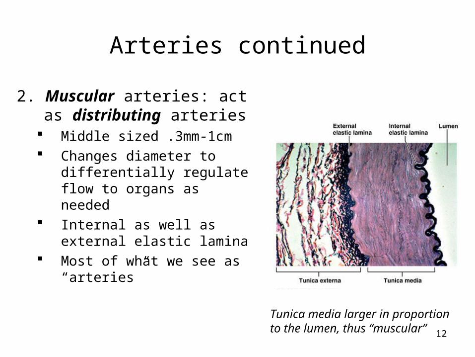
Arteries Carry blood away from the heart From big to small, these are the categories:
1. Elastic 2. Muscular 3. Arterioles (then these to capillaries)
Pressure diminishes along the route
1. Elastic arteries: act as conduits 2.5-1 cm diameter Expand with surge
of blood from heart Recoil and continue
the propagation of blood Elastin is thick in media:
dampens the surge of blood pressure
Aorta and its branches
12
Arteries continued
2. Muscular arteries: act as distributing arteries
Middle sized .3mm-1cm Changes diameter to
differentially regulate flow to organs as needed
Internal as well as external elastic lamina
Most of what we see as “arteries”
Tunica media larger in proportion to the lumen, thus “muscular”

13
Arteries continued3. Arterioles
Smallest: .3mm-10um Only larger ones have all 3
layers Regulated 2 ways:
Locally in the tissues Sympathetic control
Systemic blood pressure (the “BP” we measure) can be regulated through them
Send blood into capillaries
Tunica media has only a few layers of smooth muscle cells

14
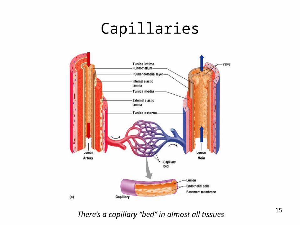
Capillaries
Heart to arteries to capillaries to veins to heart
Capillaries are smallest 8-10um Just big enough for single file erythrocytes Composed of: single layer of endothelial cells surrounded
by basement membrane
Universal function Oxygen and nutrient delivery to tissues CO2 and nitrogenous waste (protein break-down product)
removal Some also have tissue specific functions
15
Capillaries
There’s a capillary “bed” in almost all tissues

16
17
Capillary permeability
Direct diffusion through endothelial cell membranes Only O2 and CO2
Other molecules by various other methods Blood brain barrier: complete tight junctions
Selective transport of necessary molecules Lipid soluble agents (like anesthetics) get
through, as do O2 and CO2

18
Veins
Pressure has been lowered so capillaries can tolerate
With lower pressure, walls (of veins) can be thinner
From smallest to large:Capillaries to postcapillary venules to venules to
veins Veins are larger than arteries, plus
Tunica externa is thicker There is less elastin

19
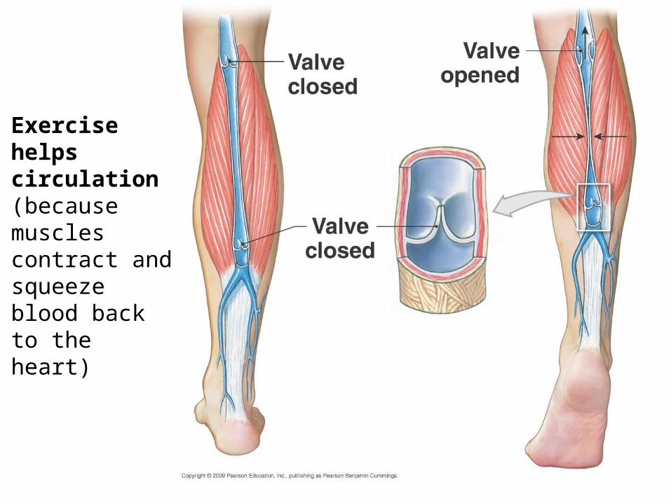
Special features of veins
Valves Prevent backflow Most abundant in legs (where
blood has to travel against gravity)
Muscular contraction Aids the return of blood to heart in
conjunction with valves
Mechanical issues…
(really good to know)
20
Exercise helps circulation (because muscles contract and squeeze blood back to the heart)

21
Vascular anastomoses
Vessels communicating with each other Veins have more than arteries Form alternative pathways or collateral channels Protect organs from being supplied by just one
route Poor anastomoses & therefore vulnerable: central
artery of retina, kidneys, spleen, bone diaphyses
Vasa vasorum Means vessels of the vessels Blood supply to vessel itself Smallest vessels don’t need

22
Vascular System (Blood vessels of the body)
Two circulations Systemic Pulmonary
Arteries and veins usually run together Often nerves run with them Sometimes the systems do not have bilateral
symmetry In head and limbs, most are bilaterally symmetrical

23
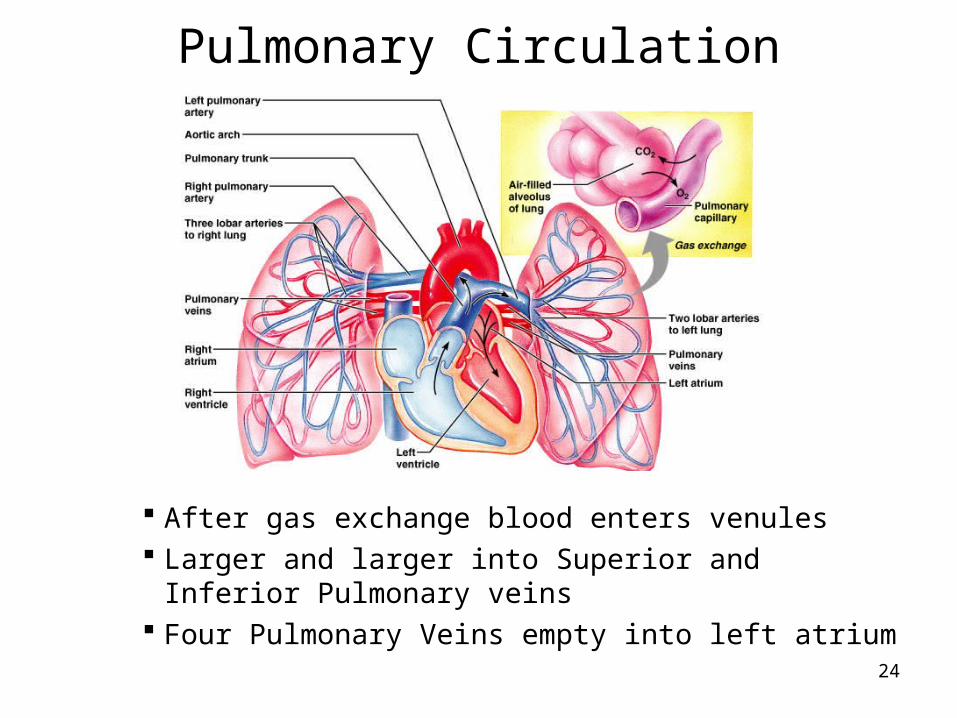
Pulmonary Circulation
Pulmonary trunk branches Right and left pulmonary arteries Division into lobar arteries
3 on right 2 on left
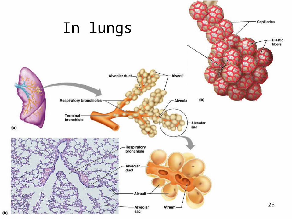
Smaller and smaller arterioles, into capillaries surrounding alveoli Gas exchange
Pulmonary system pressure is only 1/6 of systemic blood pressure
24
Pulmonary Circulation
After gas exchange blood enters venules Larger and larger into Superior and Inferior
Pulmonary veins Four Pulmonary Veins empty into left atrium

25
26
In lungs

27
Systemic Circulation Oxygenated blood to body Leaves LV through Ascending Aorta
Only branches are the 2 coronary arteries to the heart Aortic Arch has three arteries branching from it:
1. Brachiocephalic trunk, has 2 branches: Right common carotid a. Right subclavian a.
2. Left common carotid a.3. Left subclavian a.
Ligamentum arteriosumconnecting to pulmonary a.
remember aortic arches…
28
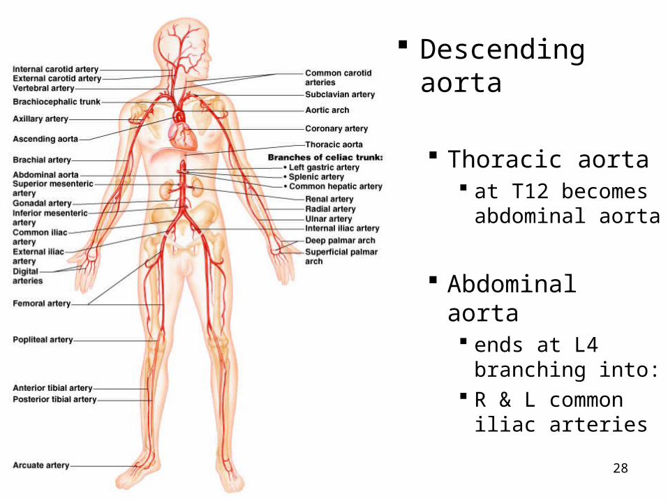
Descending aorta
Thoracic aorta at T12 becomes
abdominal aorta
Abdominal aorta ends at L4
branching into: R & L common
iliac arteries

29
Common carotids branch: Internal carotids External
carotids
Subclavian: 3 branches Vertebral
arteries Thyrocerical
trunk Costocervical
trunk
30
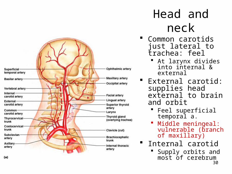
Head and neck Common carotids just
lateral to trachea: feel At larynx divides into
internal & external External carotid:
supplies head external to brain and orbit Feel superficial
temporal a. Middle meningeal:
vulnerable (branch of maxillary)
Internal carotid Supply orbits and
most of cerebrum

31
Internal carotid a.
Enters skull through carotid canal
Gives off: Ophthalmic artery
Then divides into anterior and middle cerebral arteries (see next slides):
together they supply 80% of cerebrum

32

33

34
Angiogram
35
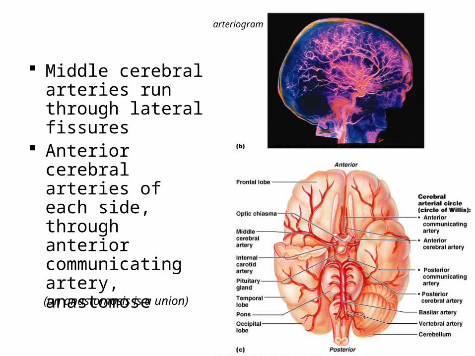
Middle cerebral arteries run through lateral fissures
Anterior cerebral arteries of each side, through anterior communicating artery, anastomose
(an anastomosis is a union)
arteriogram

36
R and L vertebral arteries* (from subclavians) Ascend through vertebral foramina of C6-C1
transverse processes Through foramen magnum into skull Join to form one Basilar artery*
*
**
*

37
Basilar artery: branches Divides into posterior cerebral arteries
Posterior communicating arteries connect to middle cerebral arteries
CIRCLE OF WILLIS(now called “cerebral arterial circle”)
Note how it loops around pituitary gland & optic chiasm

38

39
Upper limb
Subclavian runs laterally onto 1st rib, under clavicle
Enters axilla as axillary artery Sends branches
Continues as brachial artery in upper arm Splits into radial &
ulnar arteries See hand supply
Feel brachial & radial pulses

40

41
overview

42
Thorax
Anterior intercostals branch off Internal thoracic* (branch of subclavian)
Posterior intercostals branch off Thoracic aorta
Intercostal arteries, veins and nerves run just UNDER the ribs
Small bronchial arteries supply the lung structures
*
43
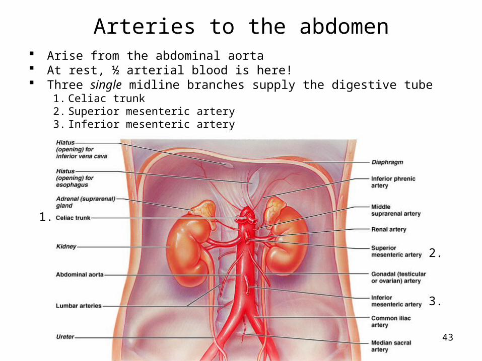
Arteries to the abdomen Arise from the abdominal aorta At rest, ½ arterial blood is here! Three single midline branches supply the digestive tube
1. Celiac trunk2. Superior mesenteric artery3. Inferior mesenteric artery
1.
2.
3.
44
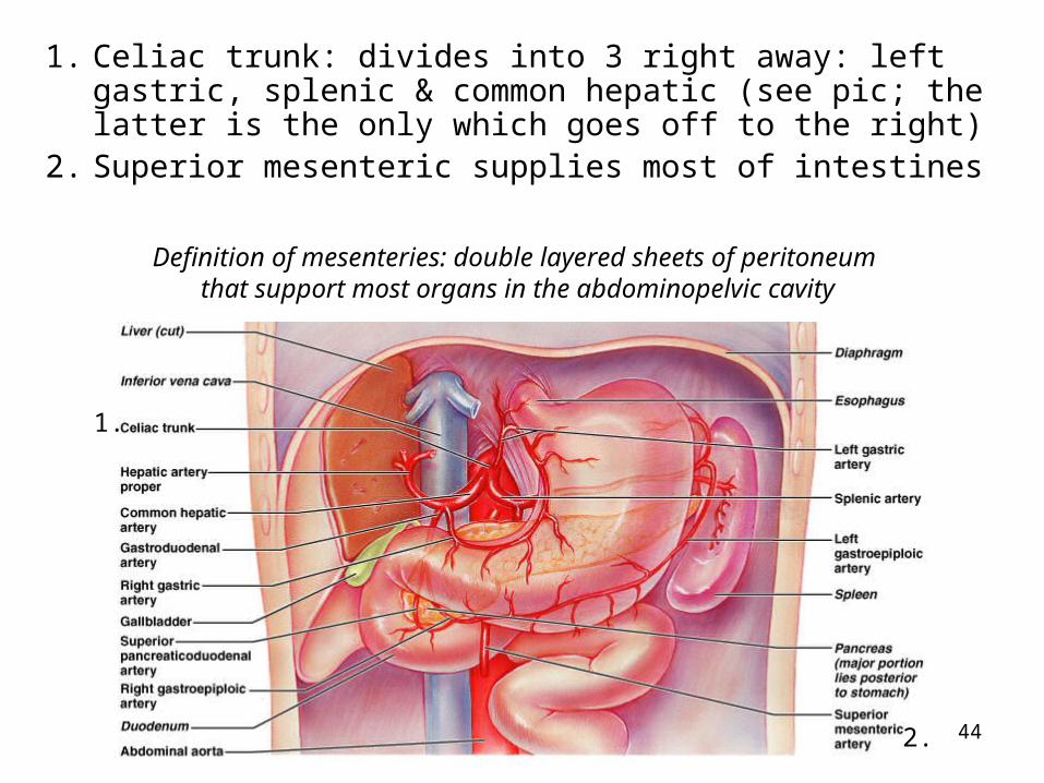
1. Celiac trunk: divides into 3 right away: left gastric, splenic & common hepatic (see pic; the latter is the only which goes off to the right)
2. Superior mesenteric supplies most of intestines
1.
2.
Definition of mesenteries: double layered sheets of peritoneum that support most organs in the abdominopelvic cavity

45
3. Inferior mesenteric supplies distal half of large intestine
2.
3.
1.
Know what these terms mean: phrenic, gastric, hepatic, renal, colic
(The 1, 2 and 3 are branches of the abdominal aorta)

46
Arteries to the abdomen
Paired branches off the abdominal aorta supply adrenal glands, kidneys, gonads and abdominal body wall
supply diaphragm
3.
supply adrenals
to kidney

47
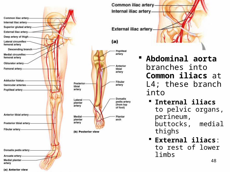
48
Abdominal aorta branches into Common iliacs at L4; these branch into Internal iliacs to
pelvic organs, perineum, buttocks, medial thighs
External iliacs: to rest of lower limbs
49
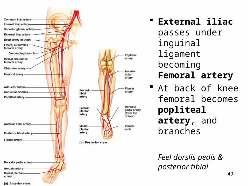
External iliac passes under inguinal ligament becoming Femoral artery
At back of knee femoral becomes popliteal artery, and branches
Feel dorslis pedis & posterior tibial

50

51
review

52
review
53
Systemic Veins
3 major vessels enter Right Atrium: SVC (superior vena cava) IVC (inferior vena cava) Coronary sinus
Many veins are very superficial (unlike arteries)
Venous plexuses (networks of anastomoses and parallel veins) are very common
Head and hepatic portal systems are unusual

54
Dural sinuses Drain the veins of
the brain Cavernous sinuses
Carotid arteries and some cranial nerves run within them
Dangerous if trauma Come together as
sigmoid sinus – becomes Internal Jugular vein Exits skull through
jugular foramen

55
Internal jugular veins Drain most of blood from brain Run lateral to internal then common carotid At base of neck joins subclavian v. to form brachiocephalic v.
External jugulars – drain some of scalp & face

56
Vein overview
Azygos system drains the thorax:
Note that unlike the arteries, the veins have a brachiocephalic on the right and left sides
57
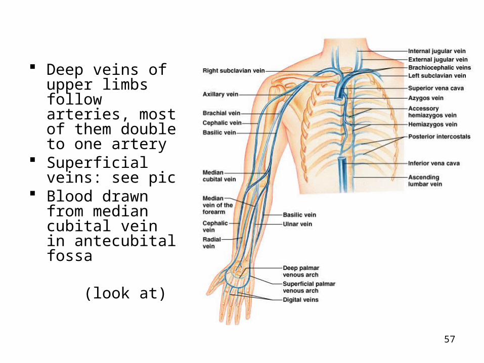
Deep veins of upper limbs follow arteries, most of them double to one artery
Superficial veins: see pic
Blood drawn from median cubital vein in antecubital fossa
(look at)

58
Tributaries of IVC: note asymmetry Left gonadal and suprarenal veins drain into left renal
vein On right they drain directly into IVC Right and left hepatic veins enter superior part of IVC

59
Hepatic portal system Picks up digested nutrients from stomach & intestines and delivers them
to liver for processing and storage Storage of nutrients Detoxification of toxins, drugs, etc.
Two capillary beds Route: artery to capillaries of gut to hepatic portal vein to liver’s
capillaries to hepatic vein to IVC
Don’t confuse hepatic vein with hepatic portal vein

60
Kind of confusing…
Superior mesenteric and splenic veins join to form hepatic portal vein, which goes up into liver
Inferior mesenteric empties into the splenic vein
*
*

61
Hepatic portal system Picks up digested nutrients from stomach & intestines
and delivers them to liver for processing and storage Storage of nutrients Detoxification of toxins, drugs, etc.
Two capillary beds Route: artery to capillaries of gut to hepatic portal
vein to liver’s capillaries to hepatic vein to IVC
(same info with different pic)
Tributaries of hepatic portal vein:-superior mesenteric vein-splenic vein-inferior mesenteric vein

62

63
Leg veins Names similar to
arteries Femoral becomes
external iliac after crossing under inguinal ligament
External iliac joins with internal iliac to form common iliac vein
_________used for grafting in coronaryartery bypass grafts: is the longest vein in the body

64
Fetal Circulation
The one umbilical vein brings blood which has been to the placenta for oxygenation (by gas diffusion from mom’s blood)
The pair of umbilical arteries (branches from baby’s internal iliac arteries) carry blood to placenta to pick up oxygen and nutrients
Fetal heart starts beating at 21 days post conception

65

66
67
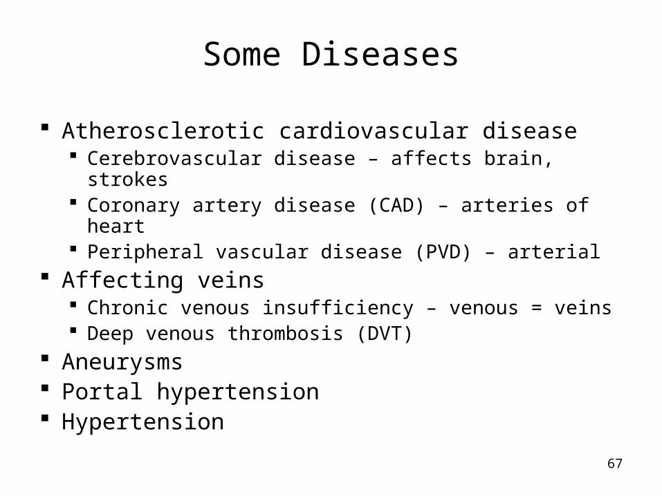
Some Diseases
Atherosclerotic cardiovascular disease Cerebrovascular disease – affects brain, strokes Coronary artery disease (CAD) – arteries of heart Peripheral vascular disease (PVD) – arterial
Affecting veins Chronic venous insufficiency – venous = veins Deep venous thrombosis (DVT)
Aneurysms Portal hypertension Hypertension