Vascular-targeted therapies for Duchenne - Skeletal Muscle
12
REVIEW Open Access Vascular-targeted therapies for Duchenne muscular dystrophy James P Ennen 1,2,3 , Mayank Verma 1,2,3,4 and Atsushi Asakura 1,2,3* Abstract Duchenne muscular dystrophy (DMD) is the most common muscular dystrophy and an X-linked recessive, progressive muscle wasting disease caused by the absence of a functional dystrophin protein. Dystrophin has a structural role as a cytoskeletal stabilization protein and protects cells against contraction-induced damage. Dystrophin also serves a signaling role through mechanotransduction of forces and localization of neuronal nitric oxide synthase (nNOS), which produces nitric oxide (NO) to facilitate vasorelaxation. In DMD, the signaling defects produce inadequate tissue perfusion caused by functional ischemia due to a diminished ability to respond to shear stress induced endothelium- dependent dilation. Additionally, the structural defects seen in DMD render myocytes with an increased susceptibility to mechanical stress. The combination of both defects is necessary to generate myocyte damage, which induces successive rounds of myofiber degeneration and regeneration, loss of calcium homeostasis, chronic inflammatory response, fibrosis, and myonecrosis. In individuals with DMD, these processes inevitably cause loss of ambulation shortly after the first decade and an abbreviated life with death in the third or fourth decade due to cardio-respiratory anomalies. There is no known cure for DMD, and although the culpable gene has been identified for more than twenty years, research on treatments has produced few clinically relevant results. Several recent studies on novel DMD therapeutics are vascular targeted and focused on attenuating the inherent functional ischemia. One approach improves vasorelaxation capacity through pharmaceutical inhibition of either phosphodiesterase 5 (PDE5) or angiotensin- converting enzyme (ACE). Another approach increases the density of the underlying vascular network by inducing angiogenesis, and this has been accomplished through either direct delivery of vascular endothelial growth factor (VEGF) or by downregulating the VEGF decoy-receptor type 1 (VEGFR-1 or Flt-1). The pro-angiogenic approaches also seem to be pro-myogenic and could resolve the age-related decline in satellite cell (SC) quantity seen in mdx models through expansion of the SC juxtavascular niche. Here we review these four vascular targeted treatment strategies for DMD and discuss mechanisms, proof of concept, and the potential for clinical relevance associated with each therapy. Keywords: Duchenne muscular dystrophy, VEGF, Flt-1, Flk-1, Nitric oxide, PDE5 inhibitor, ACE inhibitor, Satellite cell, Muscle regeneration, Myofiber damage Review Duchenne muscular dystrophy (DMD) is an X-linked re- cessive, progressive muscle wasting disease caused by muta- tions in the DMD gene that lead to absence of a functional dystrophin protein [1,2]. Both fatal and devastating, DMD is the most common muscular dystrophy seen in children and has an annual incidence affecting one in every 3600– 6000 newborn males [3]. Normally, dystrophin serves as the bridge in the dystrophin-associated glycoprotein com- plex (DAPC), connecting the cytoskeleton, via attachments to subsarcolemmal F-actin, to the extracellular matrix through an association with plasma membrane bound β- dystroglycan [4]. In the DAPC, dystrophin has a structural role as a cytoskeletal stabilization protein and protects cells against contraction-induced damage. Dystrophin also serves signaling roles, including mechanotransduction of forces and localization of signaling proteins, such as neu- ronal nitric oxide synthase (nNOS), which synthesizes ni- tric oxide (NO) to facilitate vasorelaxation [5-7]. Without * Correspondence: [email protected] 1 Stem Cell Institute, University of Minnesota Medical School, McGuire Translational Research Facility, Room 4-220, 2001 6th Street SE, Minneapolis, MN 55455, USA 2 Paul and Shelia Wellstone Muscular Dystrophy Center, University of Minnesota Medical School, Wallin Medical Biosciences Building, 2101 6th Steet SE, Minneapolis, MN 55455, USA Full list of author information is available at the end of the article © 2013 Ennen et al.; licensee BioMed Central Ltd. This is an Open Access article distributed under the terms of the Creative Commons Attribution License (http://creativecommons.org/licenses/by/2.0), which permits unrestricted use, distribution, and reproduction in any medium, provided the original work is properly cited. Ennen et al. Skeletal Muscle 2013, 3:9 http://www.skeletalmusclejournal.com/content/3/1/9
Transcript of Vascular-targeted therapies for Duchenne - Skeletal Muscle

Ennen et al. Skeletal Muscle 2013, 3:9http://www.skeletalmusclejournal.com/content/3/1/9
REVIEW Open Access
Vascular-targeted therapies for Duchennemuscular dystrophyJames P Ennen1,2,3, Mayank Verma1,2,3,4 and Atsushi Asakura1,2,3*
Abstract
Duchenne muscular dystrophy (DMD) is the most common muscular dystrophy and an X-linked recessive, progressivemuscle wasting disease caused by the absence of a functional dystrophin protein. Dystrophin has a structural role as acytoskeletal stabilization protein and protects cells against contraction-induced damage. Dystrophin also serves asignaling role through mechanotransduction of forces and localization of neuronal nitric oxide synthase (nNOS), whichproduces nitric oxide (NO) to facilitate vasorelaxation. In DMD, the signaling defects produce inadequate tissueperfusion caused by functional ischemia due to a diminished ability to respond to shear stress induced endothelium-dependent dilation. Additionally, the structural defects seen in DMD render myocytes with an increased susceptibilityto mechanical stress. The combination of both defects is necessary to generate myocyte damage, which inducessuccessive rounds of myofiber degeneration and regeneration, loss of calcium homeostasis, chronic inflammatoryresponse, fibrosis, and myonecrosis. In individuals with DMD, these processes inevitably cause loss of ambulationshortly after the first decade and an abbreviated life with death in the third or fourth decade due to cardio-respiratoryanomalies. There is no known cure for DMD, and although the culpable gene has been identified for more than twentyyears, research on treatments has produced few clinically relevant results. Several recent studies on novel DMDtherapeutics are vascular targeted and focused on attenuating the inherent functional ischemia. One approach improvesvasorelaxation capacity through pharmaceutical inhibition of either phosphodiesterase 5 (PDE5) or angiotensin-converting enzyme (ACE). Another approach increases the density of the underlying vascular network by inducingangiogenesis, and this has been accomplished through either direct delivery of vascular endothelial growth factor(VEGF) or by downregulating the VEGF decoy-receptor type 1 (VEGFR-1 or Flt-1). The pro-angiogenic approaches alsoseem to be pro-myogenic and could resolve the age-related decline in satellite cell (SC) quantity seen in mdx modelsthrough expansion of the SC juxtavascular niche. Here we review these four vascular targeted treatment strategies forDMD and discuss mechanisms, proof of concept, and the potential for clinical relevance associated with each therapy.
Keywords: Duchenne muscular dystrophy, VEGF, Flt-1, Flk-1, Nitric oxide, PDE5 inhibitor, ACE inhibitor, Satellite cell,Muscle regeneration, Myofiber damage
ReviewDuchenne muscular dystrophy (DMD) is an X-linked re-cessive, progressive muscle wasting disease caused by muta-tions in the DMD gene that lead to absence of a functionaldystrophin protein [1,2]. Both fatal and devastating, DMDis the most common muscular dystrophy seen in children
* Correspondence: [email protected] Cell Institute, University of Minnesota Medical School, McGuireTranslational Research Facility, Room 4-220, 2001 6th Street SE, Minneapolis,MN 55455, USA2Paul and Shelia Wellstone Muscular Dystrophy Center, University ofMinnesota Medical School, Wallin Medical Biosciences Building, 2101 6thSteet SE, Minneapolis, MN 55455, USAFull list of author information is available at the end of the article
© 2013 Ennen et al.; licensee BioMed CentralCommons Attribution License (http://creativecreproduction in any medium, provided the or
and has an annual incidence affecting one in every 3600–6000 newborn males [3]. Normally, dystrophin serves asthe bridge in the dystrophin-associated glycoprotein com-plex (DAPC), connecting the cytoskeleton, via attachmentsto subsarcolemmal F-actin, to the extracellular matrixthrough an association with plasma membrane bound β-dystroglycan [4]. In the DAPC, dystrophin has a structuralrole as a cytoskeletal stabilization protein and protectscells against contraction-induced damage. Dystrophin alsoserves signaling roles, including mechanotransduction offorces and localization of signaling proteins, such as neu-ronal nitric oxide synthase (nNOS), which synthesizes ni-tric oxide (NO) to facilitate vasorelaxation [5-7]. Without
Ltd. This is an Open Access article distributed under the terms of the Creativeommons.org/licenses/by/2.0), which permits unrestricted use, distribution, andiginal work is properly cited.

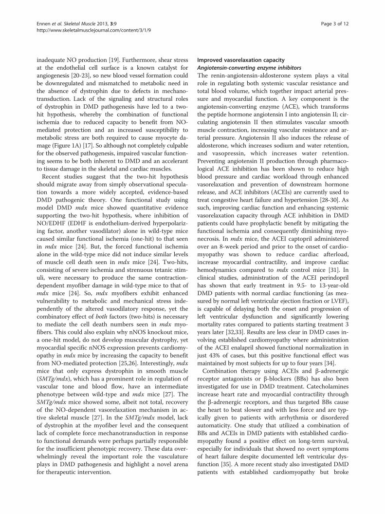
Functional ischemia from reduced NO-
mediated protection
Greater cellularsusceptibility
to metabolic stress
Combined effect of both factors
Myofiber damage
A
Greater cellularsusceptibility
to metabolic stress
Reduced myofiber damage
Reduced functional ischemia
Reduced net damage from
combining both factors
B
Vascular Therapy Treatment Target
Figure 1 The two-hit hypothesis for myocyte damage and theproposed outcome of functional ischemia attenuation inDuchenne muscular dystrophy (DMD). (A) The combined effectsfrom functional ischemia due to reduced nitric oxide (NO)-mediatedprotection and greater cellular susceptibility to metabolic stress arenecessary to produce the myofiber damage observed in DMD [17].(B) Attenuating functional ischemia by administering a vasculartargeted treatment can reduce the net-combined effect of bothtwo-hit factors and consequently curtail myofiber damage.
Ennen et al. Skeletal Muscle 2013, 3:9 Page 2 of 12http://www.skeletalmusclejournal.com/content/3/1/9
dystrophin, the DAPC cannot completely assemble, andthe supportive link between the cytoskeleton and theextracellular matrix becomes destabilized [8]. Despite nor-mal development, the membrane in dystrophin-deficientcells is easily damaged. Membrane microlesions facilitatean influx of calcium ions, which activate proteases tobegin auto-digestion of the musculature sarcoplasm[9-11]. Macrophages later arrive at the tissue to removecellular debris, and satellite cells (SCs) are activated andproliferate to induce myofiber regeneration. This causessuccessive rounds of myofiber degeneration and regener-ation that is exacerbated by continual membrane damageand ensuing myonecrosis. In addition, cytokines releasedin the process of myonecrosis recruit inflammatory cells,which release inflammatory cytokines to activate fibro-blasts that lay down extracellular matrix proteins and leadto fibrosis [12]. Skeletal muscle regenerative capacity laterdiminishes with advancing age and decreasing numbers ofSCs, and muscle tissue is steadily replaced by adipose andconnective tissues [13].The previously described cellular events manifest them-
selves clinically in a devastating and progressive manner.Despite continuous contractions by the myocardium, theskeletal muscles deteriorate first in individuals with DMD,and most permanently lose ambulatory abilities shortlyafter the first decade [14]. Myocardial problems presentlater, and clinically relevant cardiomyopathy is seen in 90%of patients over 18 years old, namely due to the onset ofcardiac fibrosis in addition to rhythm and conduction ab-normalities [14]. Respiratory problems are also inevitabledue to muscle wasting in the diaphragm and the onset ofscoliosis [14]. Even with improvements in treatment,notably multidisciplinary care, the combined cardio-respiratory anomalies mean that most individuals withDMD die in their third or fourth decade of life [15,16].Despite knowledge of the responsible gene for over twentyyears, a DMD cure remains to be found, and research ontreatments has produced few clinically relevant results.Current treatment options, such as corticosteroid admin-istration, physical therapy, nocturnal ventilation, and sur-gical interventions aim for symptomatic management andhave been shown to improve lifespan and quality of life[16]. The clinical utility and feasibility of gene therapy andcell therapy remain to be elucidated, and other treatmentareas must be sought. Our current, more holistic under-standing of DMD pathogenesis, especially with morerecent knowledge of the vascular role of dystrophin, im-plies that vascular-targeted therapies are strong candi-dates for future investigation. Specifically, attenuatingfunctional ischemia could reduce myocyte damage, in-crease tissue perfusion, reduce cardiac workload, and pre-vent cardiac and skeletal muscle remodeling (Figure 1B).This review will focus on vascular-targeted treatmentavenues aimed at either improving vasorelaxation capacity
or increasing the underlying vascular density in order toreduce the functional ischemia and improve the DMDphenotype.
Defect of nitric oxide-mediated vasodilation contributesto Duchenne muscular dystrophy phenotypeThe DMD pathogenesis is partially explained by the lack ofthe signaling role of dystrophin, which normally localizesnNOS to the sarcolemma through binding to the C-terminal region of dystrophin [6]. The nNOS is responsiblefor NO production to facilitate smooth muscle vasodila-tion in response to increased metabolic demands. Duringmuscle contraction, NO-mediated vasodilation is import-ant to help offset the α-adrenergic vasoconstriction in re-sponse to sympathetic activation, which optimizes muscleperfusion [18]. This functional response is intact in healthychildren, but in children with DMD the sympathetic vaso-constriction in skeletal muscle is unopposed due to lack ofNO-mediated vasodilation [18].The nNOS is absent from the sarcolemma and is
greatly downregulated in the cytoplasm of dystrophin-deficient muscle, which results in muscle vasoconstric-tion and abnormal blood flow during skeletal musclecontraction [6,18,19]. Specifically, loss of dystrophin inthe smooth muscle results in a decreased capacity ofthe vasculature to respond to shear stress inducedendothelium-dependent dilation, probably related to thesignaling defects seen in both force transduction and
Ennen et al. Skeletal Muscle 2013, 3:9 Page 3 of 12http://www.skeletalmusclejournal.com/content/3/1/9
inadequate NO production [19]. Furthermore, shear stressat the endothelial cell surface is a known catalyst forangiogenesis [20-23], so new blood vessel formation couldbe downregulated and mismatched to metabolic need inthe absence of dystrophin due to defects in mechano-transduction. Lack of the signaling and structural rolesof dystrophin in DMD pathogenesis have led to a two-hit hypothesis, whereby the combination of functionalischemia due to reduced capacity to benefit from NO-mediated protection and an increased susceptibility tometabolic stress are both required to cause myocyte da-mage (Figure 1A) [17]. So although not completely culpablefor the observed pathogenesis, impaired vascular function-ing seems to be both inherent to DMD and an accelerantto tissue damage in the skeletal and cardiac muscles.Recent studies suggest that the two-hit hypothesis
should migrate away from simply observational specula-tion towards a more widely accepted, evidence-basedDMD pathogenic theory. One functional study usingmodel DMD mdx mice showed quantitative evidencesupporting the two-hit hypothesis, where inhibition ofNO/EDHF (EDHF is endothelium-derived hyperpolariz-ing factor, another vasodilator) alone in wild-type micecaused similar functional ischemia (one-hit) to that seenin mdx mice [24]. But, the forced functional ischemiaalone in the wild-type mice did not induce similar levelsof muscle cell death seen in mdx mice [24]. Two-hits,consisting of severe ischemia and strenuous tetanic stim-uli, were necessary to produce the same contraction-dependent myofiber damage in wild-type mice to that ofmdx mice [24]. So, mdx myofibers exhibit enhancedvulnerability to metabolic and mechanical stress inde-pendently of the altered vasodilatory response, yet thecombinatory effect of both factors (two-hits) is necessaryto mediate the cell death numbers seen in mdx myo-fibers. This could also explain why nNOS knockout mice,a one-hit model, do not develop muscular dystrophy, yetmyocardial specific nNOS expression prevents cardiomy-opathy in mdx mice by increasing the capacity to benefitfrom NO-mediated protection [25,26]. Interestingly, mdxmice that only express dystrophin in smooth muscle(SMTg/mdx), which has a prominent role in regulation ofvascular tone and blood flow, have an intermediatephenotype between wild-type and mdx mice [27]. TheSMTg/mdx mice showed some, albeit not total, recoveryof the NO-dependent vasorelaxation mechanism in ac-tive skeletal muscle [27]. In the SMTg/mdx model, lackof dystrophin at the myofiber level and the consequentlack of complete force mechanotransduction in responseto functional demands were perhaps partially responsiblefor the insufficient phenotypic recovery. These data over-whelmingly reveal the important role the vasculatureplays in DMD pathogenesis and highlight a novel arenafor therapeutic intervention.
Improved vasorelaxation capacityAngiotensin-converting enzyme inhibitorsThe renin-angiotensin-aldosterone system plays a vitalrole in regulating both systemic vascular resistance andtotal blood volume, which together impact arterial pres-sure and myocardial function. A key component is theangiotensin-converting enzyme (ACE), which transformsthe peptide hormone angiotensin I into angiotensin II; cir-culating angiotensin II then stimulates vascular smoothmuscle contraction, increasing vascular resistance and ar-terial pressure. Angiotensin II also induces the release ofaldosterone, which increases sodium and water retention,and vasopressin, which increases water retention.Preventing angiotensin II production through pharmaco-logical ACE inhibition has been shown to reduce highblood pressure and cardiac workload through enhancedvasorelaxation and prevention of downstream hormonerelease, and ACE inhibitors (ACEIs) are currently used totreat congestive heart failure and hypertension [28-30]. Assuch, improving cardiac function and enhancing systemicvasorelaxation capacity through ACE inhibition in DMDpatients could have prophylactic benefit by mitigating thefunctional ischemia and consequently diminishing myo-necrosis. In mdx mice, the ACEI captopril administeredover an 8-week period and prior to the onset of cardio-myopathy was shown to reduce cardiac afterload,increase myocardial contractility, and improve cardiachemodynamics compared to mdx control mice [31]. Inclinical studies, administration of the ACEI perindoprilhas shown that early treatment in 9.5- to 13-year-oldDMD patients with normal cardiac functioning (as mea-sured by normal left ventricular ejection fraction or LVEF),is capable of delaying both the onset and progression ofleft ventricular dysfunction and significantly loweringmortality rates compared to patients starting treatment 3years later [32,33]. Results are less clear in DMD cases in-volving established cardiomyopathy where administrationof the ACEI enalapril showed functional normalization injust 43% of cases, but this positive functional effect wasmaintained by most subjects for up to four years [34].Combination therapy using ACEIs and β-adrenergic
receptor antagonists or β-blockers (BBs) has also beeninvestigated for use in DMD treatment. Catecholaminesincrease heart rate and myocardial contractility throughthe β-adrenergic receptors, and thus targeted BBs causethe heart to beat slower and with less force and are typ-ically given to patients with arrhythmia or disorderedautomaticity. One study that utilized a combination ofBBs and ACEIs in DMD patients with established cardio-myopathy found a positive effect on long-term survival,especially for individuals that showed no overt symptomsof heart failure despite documented left ventricular dys-function [35]. A more recent study also investigated DMDpatients with established cardiomyopathy but broke

Ennen et al. Skeletal Muscle 2013, 3:9 Page 4 of 12http://www.skeletalmusclejournal.com/content/3/1/9
treatment groups down into ACEI (lisinopril) alone orACEI plus BB (metoprolol) [36]. Both treatment groupsdisplayed improvements in cardiac function compared topre-therapy measurements, but no significant differencein cardiac function was seen between groups [36]. Futurestudies should address treatment using ACEI alone orACEI plus BB in DMD cases where cardiomyopathy hasnot been fully established to assess the potential forprophylactic benefit as this could definitively rule out theneed for a BB. Additionally, with regard to the two-hit hy-pothesis, studies that address functional ischemia attenu-ation to mitigate myonecrosis through enhanced tissueperfusion by ACEI-mediated vasorelaxation have not yetbeen performed.Another therapeutic strategy that targets the renin-
angiotensin-aldosterone system to improve vasorelax-ation capacity utilizes the antihypertensive drug losartan,which is an angiotensin II type I receptor antagonist orangiotensin receptor blocker (ARB). Long-term adminis-tration of losartan in mdx mice showed improvementsin myocardial function, but not skeletal muscle function,and reductions in mortality compared to control [37,38].Explanations as to why losartan could only amelioratethe function of cardiac muscle remain limited, but theprimary mechanism could be the significant reduction inafterload seen in the hearts of losartan treated mdx mice[38]. Decreased afterload certainly reduces cardiac work-load, and this may minimize mechanical injury and sub-sequent fibrosis to the sensitive cardiomyocytes in mdxhearts. Still, the use of losartan as a prophylactic treatmentagainst DMD-related cardiomyopathy seems promisingbased on these pre-clinical studies and the current clinicalavailability of losartan (COZAAR™) for its use in hyper-tension. Future investigation should be directed at evaluat-ing losartan in DMD patients but also, owing to pathwaysimilarity, at comparing the effectiveness of losartan to themany FDA-approved ACEIs.The definitive mechanism behind reducing cardiomyop-
athy via ARBs, ACEIs, and/or BBs in DMD patients is notcompletely established, but reduced aldosterone signalingthrough ACE inhibition could prevent fibrotic tissuedevelopment, as previous use of aldosterone-specificblockers has shown benefit in cases of heart failure[39-42]. Additionally, angiotensin II can directly inducevasoconstriction, pro-fibrotic Smad signaling, pro-fibrotictransforming growth factor beta (TGF-β) production, andthe ubiquitin-proteasome pathway that has a role in theproteolysis happening in dystrophic tissues [43-46].Angiotensin II is also known to enhance NADPH-oxidaseactivity, which leads to overproduction of superoxideanion and accounts for the oxidative stress in cardiac andskeletal muscle of the mdx mouse [47-52]. ACE inhibitioncan reduce these adverse effects, and a study with mdxmice demonstrated that the ACEI enalapril can prevent
angiotensin II dependent stimulation of pro-oxidant andpro-inflammatory pathways [53]. Overall, ACEIs and/orBBs appear to be excellent candidates for DMD therapybased on current clinical availability and demonstratedability to reduce many of the negative outcomes normallyassociated with the DMD pathogenic process. Still, abroader investigation regarding the potential prophylacticbenefit of ACEIs should be conducted to determine an op-timal age of initiation.
Phosphodiesterase 5 inhibitorsIn the NO-cGMP signaling pathway, nitric oxide synthase(NOS) produces NO to activate soluble guanylyl cyclase(sGC) to synthesize cyclic guanosine monophosphate(cGMP), and cGMP activates protein kinase G to inducevasodilation. The cGMP-specific phosphodiesterases areresponsible for cGMP degradation, so vasoconstriction be-gins as concentrations of cGMP diminish. This pathway isdisrupted in dystrophin deficient membranes, as nNOS isabsent from the sarcolemma and greatly downregulated,which contributes to the observed functional ischemia[6,19]. Additionally, studies have shown greater cGMP-specific phosphodiesterase 5 (PDE5) activity in mdx skel-etal muscle samples and decreased cGMP productioncompared with controls [54,55]. Recently, cGMP-specificPDE5 inhibitors, specifically tadalafil (Cialis™ or Adcirca™)and sildenafil (Viagra™ or Revatio™) have been investi-gated for their potential in ameliorating the functional is-chemia in DMD by increasing intracellular levels of cGMPto prolong vasodilation and increase blood flow to tissues.Asai et al. elegantly showed that tadalafil administra-
tion prior to progressive myofiber damage was able tosignificantly lower the net quantity of myofiber damagein mdx mice compared to placebo [24]. Essentially, at-tenuation of functional ischemia using tadalafil wasshown to reduce the extent of contraction-induced dam-age [24]. Additionally, early treatment utilizing PDE5inhibitors could have clinical prophylactic benefit, formdx mice treated with tadalafil from conception showedimproved histology [24]. Khairallah et al. showed that car-diac mRNA expression levels of atrial natriuretic factor(ANF), an early indicator for initiation of cardiomyopathicremodeling, was significantly reduced in mdx mice treatedwith sildenafil [56,57]. This implies that sildenafil is cap-able of inhibiting the advancement of cardiomyopathicremodeling at early stages of DMD [57]. Sildenafil has alsobeen shown to have positive functional effects in thehearts of mdx mice, notably by avoidance of cardio-myocyte damage produced in vivo via cardiac workloadaugmentation and maintenance of an elevated heart rateresponse for a significantly longer period of time com-pared with placebo [57]. Interestingly, administration ofsildenafil is even capable of reversing cardiac dysfunctionin mdx mice with established cardiomyopathy [58].

Ennen et al. Skeletal Muscle 2013, 3:9 Page 5 of 12http://www.skeletalmusclejournal.com/content/3/1/9
Nevertheless, the target cell and mechanism behind thereversal are still unclear.Additionally, PDE5 inhibitors have also shown im-
provement in muscle tissue from other vertebrate modelsof DMD, namely two dystrophin deficient zebrafishmodels known as sapje and sapje-like mutants [59,60].Dystrophin-null zebrafish treated with aminophylline, anonselective phosphodiesterase inhibitor, were able tosurvive significantly longer compared to controls andhad restored skeletal muscle structure similar to wild-type zebrafish [61]. Furthermore, analysis of sapje mu-tants treated 1 to 4 days postfertilization with differentphosphodiesterase inhibitors revealed that treatment withaminophylline or sildenafil citrate resulted in the lowestpercentage of fish showing abnormal muscle structure[61]. These data suggest that aminophylline and sildenafilcitrate are capable of preventing the onset of aberrantmuscle architecture in dystrophin-null zebrafish. Thesefindings from DMD model zebrafish are analogous to theresults seen from mdx mice treated with PDE5 inhibitors,and the consistency of these results across several DMDmodels leaves hope that these compounds could benefitindividuals living with DMD. Clinical trials assessingPDE5 inhibitors for DMD patients are currently under-way, and future use of tadalafil or sildenafil for these indi-viduals seems promising based on preclinical studies andcurrent clinical availability of these drugs for their use intreating erectile dysfunction and pulmonary hypertension.
Increased vascular densityVascular endothelial growth factor administrationThe central paradigm behind the previously discussedPDE5 and ACEI therapies for DMD was that increasingthe vasorelaxation capacity of the vasculature would beable to increase perfusion, diminish the effects fromfunctional ischemia, and decrease myocyte damage. How-ever, another technique to increase tissue perfusion wouldbe to increase the density of the underlying vascular archi-tecture that nourishes the skeletal and cardiac muscles.One method of increasing vascular density is to augmentangiogenesis, which regulates the production of newvasculatures from the existing framework. The vascularendothelial growth factor (VEGF) family of signal glyco-proteins acts as potent promoters of angiogenesis duringembryogenesis and postnatal growth. Specifically, thebinding of the VEGF-A ligand with the VEGF receptorshas been shown to promote vascular permeability and alsotrigger endothelial cell migration, proliferation, and sur-vival, and the newly formed endothelial cells provide thebasic structure of new vasculatures [62]. The dominantVEGF signal molecule for angiogenesis, VEGF-A, mediatesits signal through VEGF receptor-1 (VEGFR-1, hereafterFlt-1) and VEGF receptor-2 (VEGFR-2, hereafter Flk-1)[63]. A soluble form of Flt-1 (sFlt-1) also exists, but lacks
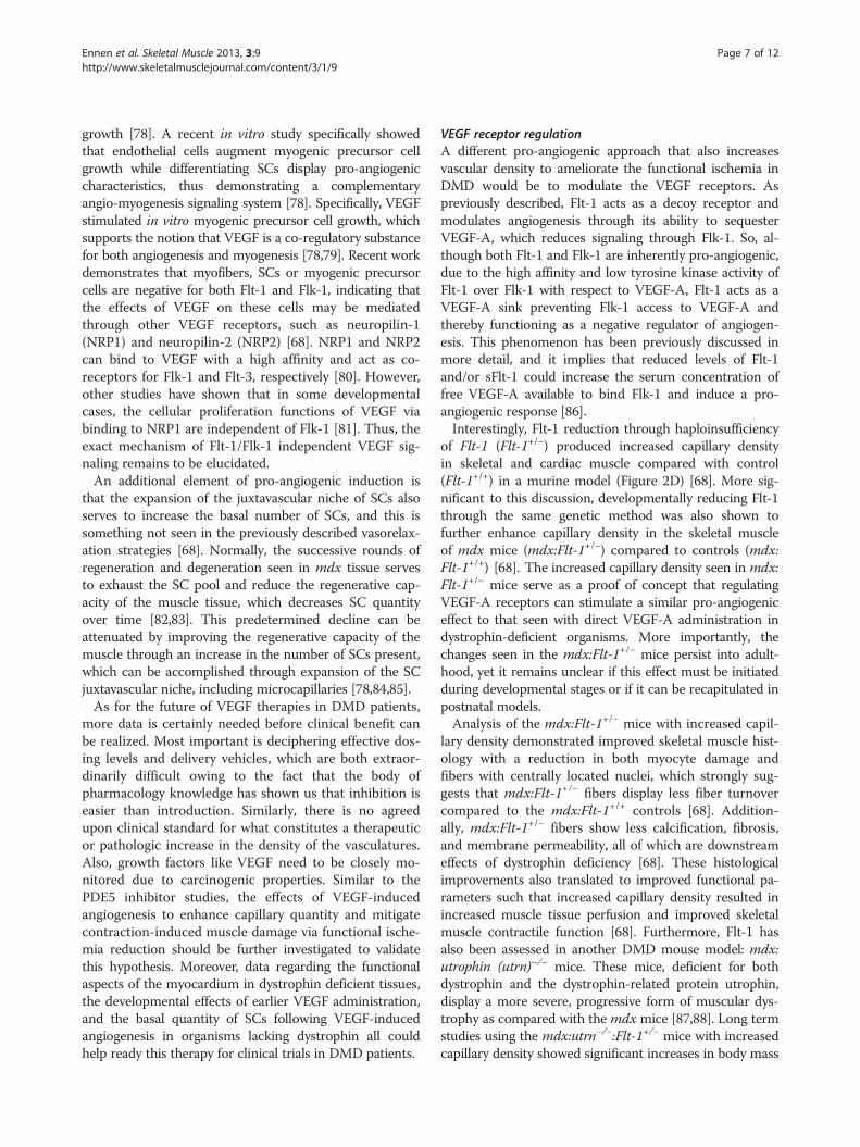
an intracellular signaling domain and thus only serves in aregulatory capacity by sequestering VEGF-A [63]. Flt-1and Flk-1 contain an extracellular VEGF-A-binding do-main and an intracellular tyrosine kinase domain, andboth show expression during the developmental stage andtissue regeneration in hemangioblasts and endothelial celllineages [63-65]. Flt-1 has a 10 times greater binding affin-ity for VEGF-A (Kd approximately 2 to 10 pM) comparedto Flk-1, but the weaker tyrosine kinase domain indicatesthat angiogenic signal transduction following VEGF-Abinding to Flt-1 is comparably weaker than the Flk-1 sig-nal (Figure 2A) [63]. As such, homozygous Flt-1 geneknockout mice die in the embryonic stage from endothe-lial cell overproduction and blood vessel disorganization(Figure 2B) [64-66]. Inversely, homozygous Flk-1 geneknockout mice die from defects in the development of or-ganized blood vessels due to lack of yolk-sac blood islandformation during embryogenesis (Figure 2C) [67]. Boththe Flt-1 and Flk-1 receptors are needed for normal devel-opment, but selective augmentation in VEGF-A concen-tration should allow for greater binding to the Flk-1receptor and induce a pro-angiogenic effect that increasescapillary density.Several studies have demonstrated that administration of
VEGF using exogenous expression mediated throughengineered myoblasts, direct systemic injections, or adeno-associated viral (AAV) vectors are capable of initiating anangiogenic signal in the myocardium and skeletal musclein both ischemic and non-ischemic conditions [69-72].However, these same studies do highlight the importanceof precisely regulating VEGF delivery quantities for futureclinical use, as overadministration has been shown to havedeleterious effects in animal models, such as hemangiomaformation [70]. Unfortunately, and to the best of ourknowledge, functional studies that assess blood flow inDMD model organisms following VEGF-induced angio-genesis have not yet been conducted. However, one studyfound that four weeks following intramuscular adminis-tration of rAAV-VEGF vectors in the bicep and tibialisanterior (TA) muscles of 4-week-old mdx mice, therAAV-VEGF-treated mdx mice showed significantlygreater forelimb strength compared to pretreatmentlevels and AAV-LacZ-treated control mdx mice [73]. Thesame study confirmed the feasibility of VEGF-mediatedangiogenic induction in mdx mice and showed greatercapillary density, particularly in the area of regeneratingfibers, as well as reduced necrotic fiber area in bicepsmuscle compared to AAV-LacZ-treated control mdxmice [73]. So, although this study did not directly assessreduction of functional ischemia via enhanced vasculardensity through VEGF-induced angiogenesis, it was ableto demonstrate similar outcomes from the PDE5 inhibi-tor studies, especially the decrease seen in necrotic fiberarea and improvement in muscle function.

Flt-1 Flk-1
Membrane
VEGF Affinity
Angiogenic
Signal Strength
Normal
Angiogenesis
Wild Type
Flt-1 Flk-1
Embryonic Lethal: Excessive
Angiogenesis
Flt-1gene KO
Flt-1 Flk-1
Flk-1gene KO
Flt-1 Flk-1
Enhanced
Angiogenesis and
Improved DMD
Phenotype
HeterozygousFlt-1gene KO
Embryonic Lethal: Signaling
Defect
sFlt-1 sFlt-1 sFlt-1 sFlt-1VEGF =
A CBD
Figure 2 Flt-1 is a decoy receptor for vascular endothelial growth factor (VEGF) pro-angiogenic signaling. (A) In the wild-type scenario,VEGF induces a pro-angiogenic signal by binding the Flt-1 or Flk-1 receptors [63]. Flt-1 has a higher binding affinity for VEGF but transmits aweaker angiogenic signal compared to Flk-1, which implies that Flt-1 acts a negative regulator of angiogenesis [63]. The soluble form of Flt-1(sFlt-1) lacks the transmembrane and intracellular signaling domains of Flt-1 and only serves a regulatory role by sequestering VEGF [63]. (B) Flt-1homozygous knockout (Flt-1−/−) mice die in the early embryonic stage from endothelial cell overproduction and blood vessel disorganization,indicating that Flt-1 is a decoy regulator for endothelial growth/differentiation [64-66]. (C) Flk-1 homozygous knockout (Flk-1−/−) mice die in theearly embryonic stage from defects in the development of organized blood vessels, indicating that Flk-1 is a positive regulator for endothelialgrowth/differentiation [67]. (D) Developmental reduction of the Flt-1 receptor through haploinsufficiency of the Flt-1 gene (Flt-1+/−) has beenshown to increase capillary density in skeletal muscle, and this same phenomenon has been demonstrated in mdx mice (mdx:Flt-1+/−) [79]. Themdx:Flt-1+/− mice also showed improved histological and functional parameters normally associated with the Duchenne muscular dystrophy(DMD) pathology [68].
Ennen et al. Skeletal Muscle 2013, 3:9 Page 6 of 12http://www.skeletalmusclejournal.com/content/3/1/9
But apart from the documented pro-angiogenic effect,VEGF delivery also has a powerful pro-myogenic effect.In normal skeletal muscle tissues, VEGF administrationinduces muscle fiber regeneration and promotes musclerecovery after ischemic and chemical damage [74]. Inin vitro studies using C2C12 myoblast cell line and pri-mary mouse myoblasts derived from cultured SCs, VEGFwas shown to promote growth and protect cells fromapoptosis [74]. Similar effects have been documentedin dystrophin deficient muscle tissues, where rAAV-VEGF-treated mdx mice showed an increase in the areaoccupied by regenerating fibers and an increased numberof activated SCs and developmental myosin-heavy chain-positive fibers in skeletal muscles [73]. In vivo transplant-ation of muscle-derived stem cells (MDSCs) engineered tooverexpress VEGF into dystrophic skeletal muscle resultsin an increase in angiogenesis and endogenous muscle re-generation along with reduction in fibrosis both two andfour weeks following transplantation [75]. Thus, the dualfunctionalities of VEGF, especially the pro-angiogenic and
pro-myogenic effects, are capable of improving both thehistological and functional parameters normally associatedwith mdx muscle pathophysiology.These data seem logical because developmentally redu-
cing angiogenesis in mdx mice through ablation of matrixmetalloproteinase-2 impairs the growth of regeneratedmyofibers and decreases VEGF expression, further com-plementing current theories about the close developmen-tal relationship between angiogenesis and myogenesis[76]. But what is the pro-myogenic mechanism of VEGFdelivery? SCs are clearly the dominant muscle-specificstem cells utilized for muscle growth, repair, and regener-ation, and the number of SCs parallels muscle capillaryquantity, largely because SCs reside in a juxtavascularniche [77,78]. In fact, most SCs maintain tight locality tocapillaries regardless of character, including quiescent SCs,proliferating SCs (myogenic precursor cells), and differen-tiating SCs (myocytes), and differentiating myogenin-positive myocytes assessed from DMD muscle biopsiesshow spatiotemporal association with new capillary
Ennen et al. Skeletal Muscle 2013, 3:9 Page 7 of 12http://www.skeletalmusclejournal.com/content/3/1/9
growth [78]. A recent in vitro study specifically showedthat endothelial cells augment myogenic precursor cellgrowth while differentiating SCs display pro-angiogeniccharacteristics, thus demonstrating a complementaryangio-myogenesis signaling system [78]. Specifically, VEGFstimulated in vitro myogenic precursor cell growth, whichsupports the notion that VEGF is a co-regulatory substancefor both angiogenesis and myogenesis [78,79]. Recent workdemonstrates that myofibers, SCs or myogenic precursorcells are negative for both Flt-1 and Flk-1, indicating thatthe effects of VEGF on these cells may be mediatedthrough other VEGF receptors, such as neuropilin-1(NRP1) and neuropilin-2 (NRP2) [68]. NRP1 and NRP2can bind to VEGF with a high affinity and act as co-receptors for Flk-1 and Flt-3, respectively [80]. However,other studies have shown that in some developmentalcases, the cellular proliferation functions of VEGF viabinding to NRP1 are independent of Flk-1 [81]. Thus, theexact mechanism of Flt-1/Flk-1 independent VEGF sig-naling remains to be elucidated.An additional element of pro-angiogenic induction is
that the expansion of the juxtavascular niche of SCs alsoserves to increase the basal number of SCs, and this issomething not seen in the previously described vasorelax-ation strategies [68]. Normally, the successive rounds ofregeneration and degeneration seen in mdx tissue servesto exhaust the SC pool and reduce the regenerative cap-acity of the muscle tissue, which decreases SC quantityover time [82,83]. This predetermined decline can beattenuated by improving the regenerative capacity of themuscle through an increase in the number of SCs present,which can be accomplished through expansion of the SCjuxtavascular niche, including microcapillaries [78,84,85].As for the future of VEGF therapies in DMD patients,
more data is certainly needed before clinical benefit canbe realized. Most important is deciphering effective dos-ing levels and delivery vehicles, which are both extraor-dinarily difficult owing to the fact that the body ofpharmacology knowledge has shown us that inhibition iseasier than introduction. Similarly, there is no agreedupon clinical standard for what constitutes a therapeuticor pathologic increase in the density of the vasculatures.Also, growth factors like VEGF need to be closely mo-nitored due to carcinogenic properties. Similar to thePDE5 inhibitor studies, the effects of VEGF-inducedangiogenesis to enhance capillary quantity and mitigatecontraction-induced muscle damage via functional ische-mia reduction should be further investigated to validatethis hypothesis. Moreover, data regarding the functionalaspects of the myocardium in dystrophin deficient tissues,the developmental effects of earlier VEGF administration,and the basal quantity of SCs following VEGF-inducedangiogenesis in organisms lacking dystrophin all couldhelp ready this therapy for clinical trials in DMD patients.
VEGF receptor regulationA different pro-angiogenic approach that also increasesvascular density to ameliorate the functional ischemia inDMD would be to modulate the VEGF receptors. Aspreviously described, Flt-1 acts as a decoy receptor andmodulates angiogenesis through its ability to sequesterVEGF-A, which reduces signaling through Flk-1. So, al-though both Flt-1 and Flk-1 are inherently pro-angiogenic,due to the high affinity and low tyrosine kinase activity ofFlt-1 over Flk-1 with respect to VEGF-A, Flt-1 acts as aVEGF-A sink preventing Flk-1 access to VEGF-A andthereby functioning as a negative regulator of angiogen-esis. This phenomenon has been previously discussed inmore detail, and it implies that reduced levels of Flt-1and/or sFlt-1 could increase the serum concentration offree VEGF-A available to bind Flk-1 and induce a pro-angiogenic response [86].Interestingly, Flt-1 reduction through haploinsufficiency
of Flt-1 (Flt-1+/−) produced increased capillary densityin skeletal and cardiac muscle compared with control(Flt-1+/+) in a murine model (Figure 2D) [68]. More sig-nificant to this discussion, developmentally reducing Flt-1through the same genetic method was also shown tofurther enhance capillary density in the skeletal muscleof mdx mice (mdx:Flt-1+/−) compared to controls (mdx:Flt-1+/+) [68]. The increased capillary density seen in mdx:Flt-1+/− mice serve as a proof of concept that regulatingVEGF-A receptors can stimulate a similar pro-angiogeniceffect to that seen with direct VEGF-A administration indystrophin-deficient organisms. More importantly, thechanges seen in the mdx:Flt-1+/− mice persist into adult-hood, yet it remains unclear if this effect must be initiatedduring developmental stages or if it can be recapitulated inpostnatal models.Analysis of the mdx:Flt-1+/− mice with increased capil-
lary density demonstrated improved skeletal muscle hist-ology with a reduction in both myocyte damage andfibers with centrally located nuclei, which strongly sug-gests that mdx:Flt-1+/− fibers display less fiber turnovercompared to the mdx:Flt-1+/+ controls [68]. Addition-ally, mdx:Flt-1+/− fibers show less calcification, fibrosis,and membrane permeability, all of which are downstreameffects of dystrophin deficiency [68]. These histologicalimprovements also translated to improved functional pa-rameters such that increased capillary density resulted inincreased muscle tissue perfusion and improved skeletalmuscle contractile function [68]. Furthermore, Flt-1 hasalso been assessed in another DMD mouse model: mdx:utrophin (utrn)−/− mice. These mice, deficient for bothdystrophin and the dystrophin-related protein utrophin,display a more severe, progressive form of muscular dys-trophy as compared with the mdx mice [87,88]. Long termstudies using the mdx:utrn−/−:Flt-1+/− mice with increasedcapillary density showed significant increases in body mass
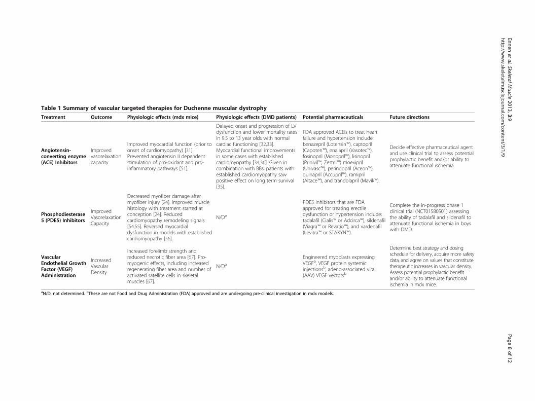
Table 1 Summary of vascular targeted therapies for Duchenne muscular dystrophy
Treatment Outcome Physiologic effects (mdx mice) Physiologic effects (DMD patients) Potential pharmaceuticals Future directions
Angiotensin-converting enzyme(ACE) Inhibitors
Improvedvasorelaxationcapacity
Improved myocardial function (prior toonset of cardiomyopathy) [31].Prevented angiotensin II dependentstimulation of pro-oxidant and pro-inflammatory pathways [51].
Delayed onset and progression of LVdysfunction and lower mortality ratesin 9.5 to 13 year olds with normalcardiac functioning [32,33].Myocardial functional improvementsin some cases with establishedcardiomyopathy [34,36]. Given incombination with BBs, patients withestablished cardiomyopathy sawpositive effect on long term survival[35].
FDA approved ACEIs to treat heartfailure and hypertension include:benazepril (Lotensin™), captopril(Capoten™), enalapril (Vasotec™),fosinopril (Monopril™), lisinopril(Prinivil™, Zestril™) moexipril(Univasc™), perindopril (Aceon™),quinapril (Accupril™), ramipril(Altace™), and trandolapril (Mavik™).
Decide effective pharmaceutical agentand use clinical trial to assess potentialprophylactic benefit and/or ability toattenuate functional ischemia.
Phosphodiesterase5 (PDE5) Inhibitors
ImprovedVasorelaxationCapacity
Decreased myofiber damage aftermyofiber injury [24]. Improved musclehistology with treatment started atconception [24]. Reducedcardiomyopathy remodeling signals[54,55]. Reversed myocardialdysfunction in models with establishedcardiomyopathy [56].
N/Da
PDE5 inhibitors that are FDAapproved for treating erectiledysfunction or hypertension include:tadalafil (Cialis™ or Adcirca™), sildenafil(Viagra™ or Revatio™), and vardenafil(Levitra™ or STAXYN™).
Complete the in-progress phase 1clinical trial (NCT01580501) assessingthe ability of tadalafil and sildenafil toattenuate functional ischemia in boyswith DMD.
VascularEndothelial GrowthFactor (VEGF)Administration
IncreasedVascularDensity
Increased forelimb strength andreduced necrotic fiber area [67]. Pro-myogenic effects, including increasedregenerating fiber area and number ofactivated satellite cells in skeletalmuscles [67].
N/Da
Engineered myoblasts expressingVEGFb, VEGF protein systemicinjectionsb, adeno-associated viral(AAV) VEGF vectorsb
Determine best strategy and dosingschedule for delivery, acquire more safetydata, and agree on values that constitutetherapeutic increases in vascular density.Assess potential prophylactic benefitand/or ability to attenuate functionalischemia in mdx mice.
aN/D, not determined. bThese are not Food and Drug Administration (FDA) approved and are undergoing pre-clinical investigation in mdx models.
Ennenet
al.SkeletalMuscle
2013,3:9Page
8of
12http://w
ww.skeletalm
usclejournal.com/content/3/1/9
Ennen et al. Skeletal Muscle 2013, 3:9 Page 9 of 12http://www.skeletalmusclejournal.com/content/3/1/9
and survival compared to the mdx:utrn−/−:Flt-1+/+ controlmice [68].Absolute mechanisms that explain the improved
phenotype and survival seen in mdx:Flt-1+/− mice andmdx:utrn−/−:Flt-1+/− mice remain to be elucidated. Butowing to pathway similarity, the explanation is probablysimilar to descriptions of VEGF-induced angiogenesis inmdx mice, namely the close developmental relationshipbetween myogenesis and angiogenesis. Increasing tissueperfusion may compensate for lack of NO-mediatedvasodilation, which would attenuate one of the proposed‘two-hits’ required for myocyte damage. Another theorybehind the progressive nature of the DMD pathology isthe SC exhaustion model [83]. This theory states thateasily damaged DMD myofibers are constantly replacedby endogenous SCs, yet the constant SC cycling leadsto rapid shortening of telomerase length and eventualexhaustion of the SC pool [82,83]. Interestingly, mdx:Flt-1+/− were shown to have developmentally increasednumbers of SCs, perhaps mediated through an expandedSC vascular niche. Thus, enhancement in the basal num-ber of SCs could mitigate the accelerated age-related de-cline seen among SCs from dystrophin-deficient muscletissue [13].Overall, Flt-1 is a novel target for pro-angiogenic ther-
apy in DMD. Greater blood perfusion alone seems tocompensate for the functional ischemic phenotype inmdx mice, but definitive studies showing attenuation offunctional ischemia through Flt-1 signal mitigation to re-duce the effects of contraction induced damage have notbeen shown. Flt-1 has also been investigated for its rolein cancer where it acts as a positive regulator of thepathological angiogenesis seen with tumor formation[89], which opposes the physiological role of Flt-1 as anegative angiogenic regulator. Thus, numerous smallmolecules have already been investigated and verified(in vivo and in vitro) that can antagonize Flt-1 bindingto VEGF, reduce angiogenesis, and prevent tumorgrowth [90]. These same small molecules could be usedto selectively block Flt-1 function and promote angio-genesis in dystrophin-deficient tissue. Still, more screen-ing studies could be needed to decipher substances thatare viable in vivo and can reduce Flt-1 function in orderto fully translate the results seen from the developmentalstudies with mdx:Flt-1+/− and mdx:utrn−/−:Flt-1+/− miceusing a pharmaceutical agent.
ConclusionsThe role of the vasculature in DMD can no longer be ig-nored in light of the mounting evidence for its role in thepathogenic process. With this new knowledge in mind andwith the dearth of current treatments, this review focusedon a variety of new therapeutic options that specifically tar-get these DMD vascular defects, namely attenuation of the
functional ischemia (see Table 1 for summary of therapies).One therapy improves systemic vasorelaxation capacityusing ACEIs with or without BBs, and this method hasshown clinical utility in both preventing and improving theadverse cardiac events normally associated with the DMDphenotype. Treatment using PDE5 inhibitors also improvessystemic vasorelaxation capacity, and preclinical evidencefrom DMD murine models demonstrates the ability ofPDE5 inhibitors to prevent skeletal and cardiac muscledamage and even reverse the functional parameters associ-ated with established cardiomyopathy. Both PDE5 andACEI therapies have a clear practical advantage as theyhave extensive clinical safety records and many of thedrugs are clinically available. There are a wide variety ofACEIs that are FDA approved to treat heart failure andhypertension, including benazepril (Lotensin™), captopril(Capoten™), enalapril (Vasotec™), fosinopril (Monopril™),lisinopril (Prinivil™, Zestril™) moexipril (Univasc™), perin-dopril (Aceon™), quinapril (Accupril™), ramipril(Altace™), and trandolapril (Mavik™). There are also sev-eral PDE5 inhibitors that received FDA approval fortreating erectile dysfunction or hypertension, includingtadalafil (Cialis™ or Adcirca™), sildenafil (Viagra™ orRevatio™), and vardenafil (Levitra™ or STAXYN™). Add-itionally, sildenafil has already been extensively studied ina pediatric population and was found to be safe for pul-monary hypertension treatment [91]. Both tadalafil andsildenafil are currently in a phase 1 clinical trial(NCT01580501) that will assess these drugs ability to at-tenuate functional ischemia in boys with DMD, and otherfuture clinical studies could address the ability of ACEIs tomitigate the same effect.Therapies that enhance the underlying vascular archi-
tecture through pro-angiogenic induction include VEGFadministration and also VEGF receptor modulation. Thepro-angiogenic therapies have shown exciting preclinicalproof of concept evidence in DMD murine models, espe-cially the expansion in the basal number of SCs mediatedthrough a larger juxtavascular SC niche and the docu-mented pro-myogenic effects. Still, the pro-angiogenicstrategies are in early stages and both methods need de-finitive means of achieving their desired result and moresafety information before clinical trial initiation. In all, thehope is that at least some or combinations of thesevascular-targeted therapies will soon have clinical utilityand provide current and future human beings living withDMD enhanced control over their own destiny.
AbbreviationsAAV: Adeno-associated virus; ACE: Angiotensin-converting enzyme;ACEI: Angiotensin-converting enzyme inhibitor; ANF: Atrial natriuretic factor;ARB: Angiotensin receptor blocker; BB: β-blocker; BMD: Becker musculardystrophy; cGMP: Cyclic guanosine monophosphate; CNS: Central nervoussystem; DAPC: Dystrophin-associated glycoprotein complex; DMD: Duchennemuscular dystrophy; EDHF: Endothelium-derived hyperpolarizing factor;LVEF: Left ventricular ejection fraction; MDSCs: Muscle-derived stem cells;

Ennen et al. Skeletal Muscle 2013, 3:9 Page 10 of 12http://www.skeletalmusclejournal.com/content/3/1/9
nNOS: Neuronal nitric oxide synthase; NO: Nitric oxide; NOS: Nitric oxidesynthase; NRP1: Neuropilin-1; NRP2: Neuropilin-2; PDE5: Phosphodiesterase 5;sGC: Soluble guanylyl cyclase; SC: Satellite cell; rAAV: Recombinant adeno-associated virus; TA: tibialis anterior; TGF- β: Transforming growth factor-β;VEGF: Vascular endothelial growth factor; VEGFR: Vascular endothelial growthfactor receptor.
Competing interestsThe authors have no financial competing interests.
Authors’ contributionsJPE completed the literature review, developed the figures, and prepared thereview. MV revised the manuscript. AA advised the literature review processand revised the manuscript. All authors read and approved the finalmanuscript.
Authors’ informationJPE is a staff researcher in the laboratory of Dr. Atsushi Asakura at theUniversity of Minnesota Stem Cell Institute.MV is an MD/PhD candidate through the Medical Scientist Training Programat the University of Minnesota Medical School.AA is an Assistant Professor of Neurology and a faculty member of the StemCell Institute in the University of Minnesota Medical School. He also belongsto the Paul & Sheila Wellstone Muscular Dystrophy Center in the Universityof Minnesota Medical School.
AcknowledgementsWe thank Dr. Lawrence Charnas and Dr. Dennis Keefe for critical reading ofthis manuscript. This work was supported by grants to AA from Grant-in-Aidof the University of Minnesota and the Muscular Dystrophy Association(MDA). The work was also supported by the NIH-T32-GM008244 grant to MV.
Author details1Stem Cell Institute, University of Minnesota Medical School, McGuireTranslational Research Facility, Room 4-220, 2001 6th Street SE, Minneapolis,MN 55455, USA. 2Paul and Shelia Wellstone Muscular Dystrophy Center,University of Minnesota Medical School, Wallin Medical Biosciences Building,2101 6th Steet SE, Minneapolis, MN 55455, USA. 3Department of Neurology,University of Minnesota Medical School, Phillips Wangensteen Building, 420Delaware Street SE, Minneapolis, MN 55455, USA. 4University of MinnesotaMedical Scientist Training Program (MD/PhD), Mayo Building, Room B-681,420 Delaware Street SE, Minneapolis, MN 55455, USA.
Received: 20 December 2012 Accepted: 25 March 2013Published: 23 April 2013
References1. Monaco AP, Neve RL, Colletti-Feener C, Bertelson CJ, Kurnit DM, Kunkel LM:
Isolation of candidate cDNAs for portions of the Duchenne musculardystrophy gene. Nature 1986, 323:646–650.
2. Hoffman EP, Kunkel LM: Dystrophin abnormalities in Duchenne/Beckermuscular dystrophy. Neuron 1989, 2:1019–1029.
3. Bushby K, Finkel R, Birnkrant DJ, Case LE, Clemens PR, Cripe L, Kaul A,Kinnett K, McDonald C, Pandya S, Poysky J, Shapiro F, Tomezsko J,Constantin C, DMD Care Considerations Working Group: Diagnosis andmanagement of Duchenne muscular dystrophy, part 1: diagnosis, andpharmacological and psychosocial management. Lancet Neurol 2010,9:77–93.
4. Ervasti JM, Campbell KP: A role for the dystrophin-glycoprotein complexas a transmembrane linker between laminin and actin. J Cell Biol 1993,122:809–823.
5. Davies KE, Nowak KJ: Molecular mechanisms of muscular dystrophies: oldand new players. Nat Rev Mol Cell Biol 2006, 7:762–773.
6. Brenman JE, Chao DS, Xia H, Aldape K, Bredt DS: Nitric oxide synthasecomplexed with dystrophin and absent from skeletal musclesarcolemma in Duchenne muscular dystrophy. Cell 1995, 82:743–752.
7. Goldspink G: Changes in muscle mass and phenotype and theexpression of autocrine and systemic growth factors by muscle inresponse to stretch and overload. J Anat 1999, 194(Pt 3):323–334.
8. Rybakova IN, Patel JR, Ervasti JM: The dystrophin complex forms amechanically strong link between the sarcolemma and costameric actin.J Cell Biol 2000, 150:1209–1214.
9. Duncan CJ: Role of intracellular calcium in promoting muscle damage:a strategy for controlling the dystrophic condition. Experientia 1978,34:1531–1535.
10. Spencer MJ, Croall DE, Tidball JG: Calpains are activated in necrotic fibersfrom mdx dystrophic mice. J Biol Chem 1995, 270:10909–10914.
11. Hopf FW, Turner PR, Steinhardt RA: Calcium misregulation and thepathogenesis of muscular dystrophy. Subcell Biochem 2007, 45:429–464.
12. Spencer MJ, Montecino-Rodriguez E, Dorshkind K, Tidball JG: Helper(CD4(+)) and cytotoxic (CD8(+)) T cells promote the pathology ofdystrophin-deficient muscle. Clin Immunol 2001, 98:235–243.
13. Jejurikar SS, Kuzon WM Jr: Satellite cell depletion in degenerative skeletalmuscle. Apoptosis 2003, 8:573–578.
14. Townsend D, Yasuda S, Metzger J: Cardiomyopathy of Duchennemuscular dystrophy: pathogenesis and prospect of membrane sealantsas a new therapeutic approach. Expert Rev Cardiovasc Ther 2007, 5:99–109.
15. Eagle M, Baudouin SV, Chandler C, Giddings DR, Bullock R, Bushby K:Survival in Duchenne muscular dystrophy: improvements in lifeexpectancy since 1967 and the impact of home nocturnal ventilation.Neuromuscul Disord 2002, 12:926–929.
16. Eagle M, Bourke J, Bullock R, Gibson M, Mehta J, Giddings D, Straub V,Bushby K: Managing Duchenne muscular dystrophy–the additive effectof spinal surgery and home nocturnal ventilation in improving survival.Neuromuscul Disord 2007, 17:470–475.
17. Rando TA: Role of nitric oxide in the pathogenesis of musculardystrophies: a "two hit" hypothesis of the cause of muscle necrosis.Microsc Res Tech 2001, 55:223–235.
18. Sander M, Chavoshan B, Harris SA, Iannaccone ST, Stull JT, Thomas GD,Victor RG: Functional muscle ischemia in neuronal nitric oxide synthase-deficient skeletal muscle of children with Duchenne muscular dystrophy.Proc Natl Acad Sci USA 2000, 97:13818–13823.
19. Loufrani L, Matrougui K, Gorny D, Duriez M, Blanc I, Levy BI, Henrion D: Flow(shear stress)-induced endothelium-dependent dilation is altered in micelacking the gene encoding for dystrophin. Circulation 2001, 103:864–870.
20. Davies PF: Flow-mediated endothelial mechanotransduction. Physiol Rev1995, 75:519–560.
21. Friebel M, Klotz KF, Ley K, Gaehtgens P, Pries AR: Flow-dependentregulation of arteriolar diameter in rat skeletal muscle in situ: role ofendothelium-derived relaxing factor and prostanoids. J Physiol 1995,483(Pt 3):715–726.
22. Ichioka S, Shibata M, Kosaki K, Sato Y, Harii K, Kamiya A: Effects of shearstress on wound-healing angiogenesis in the rabbit ear chamber. J SurgRes 1997, 72:29–35.
23. Ando J, Kamiya A: Blood flow and vascular endothelial cell function.Front Med Biol Eng 1993, 5:245–264.
24. Asai A, Sahani N, Kaneki M, Ouchi Y, Martyn JA, Yasuhara SE: Primary role offunctional ischemia, quantitative evidence for the two-hit mechanism,and phosphodiesterase-5 inhibitor therapy in mouse musculardystrophy. PLoS One 2007, 2:e806.
25. Huang PL, Dawson TM, Bredt DS, Snyder SH, Fishman MC: Targeted disruptionof the neuronal nitric oxide synthase gene. Cell 1993, 75:1273–1286.
26. Wehling-Henricks M, Jordan MC, Roos KP, Deng B, Tidball JG:Cardiomyopathy in dystrophin-deficient hearts is prevented byexpression of a neuronal nitric oxide synthase transgene in themyocardium. Hum Mol Genet 2005, 14:1921–1933.
27. Ito K, Kimura S, Ozasa S, Matsukura M, Ikezawa M, Yoshioka K, Ueno H,Suzuki M, Araki K, Yamamura K, Miwa T, Dickson G, Thomas GD, Miike T:Smooth muscle-specific dystrophin expression improves aberrantvasoregulation in mdx mice. Hum Mol Genet 2006, 15:2266–2275.
28. Garg R, Yusuf S: Overview of randomized trials of angiotensin-convertingenzyme inhibitors on mortality and morbidity in patients with heart failure.Collaborative Group on ACE Inhibitor Trials. JAMA 1995, 273:1450–1456.
29. Mant J, Al-Mohammad A, Swain S, Laramee P, Guideline Development G:Management of chronic heart failure in adults: synopsis of the NationalInstitute For Health and clinical excellence guideline. Ann Intern Med2011, 155:252–259.
30. Krause T, Lovibond K, Caulfield M, McCormack T, Williams B, GuidelineDevelopment G: Management of hypertension: summary of NICEguidance. BMJ 2011, 343:d4891.

Ennen et al. Skeletal Muscle 2013, 3:9 Page 11 of 12http://www.skeletalmusclejournal.com/content/3/1/9
31. Bauer R, Straub V, Blain A, Bushby K, MacGowan GA: Contrasting effects ofsteroids and angiotensin-converting-enzyme inhibitors in a mousemodel of dystrophin-deficient cardiomyopathy. Eur J Heart Fail 2009,11:463–471.
32. Duboc D, Meune C, Lerebours G, Devaux JY, Vaksmann G, Becane HM:Effect of perindopril on the onset and progression of left ventriculardysfunction in Duchenne muscular dystrophy. J Am Coll Cardiol 2005,45:855–857.
33. Duboc D, Meune C, Pierre B, Wahbi K, Eymard B, Toutain A, Berard C,Vaksmann G, Weber S, Becane HM: Perindopril preventive treatment onmortality in Duchenne muscular dystrophy: 10 years' follow-up. Am HeartJ 2007, 154:596–602.
34. Ramaciotti C, Heistein LC, Coursey M, Lemler MS, Eapen RS, Iannaccone ST,Scott WA: Left ventricular function and response to enalapril in patientswith duchenne muscular dystrophy during the second decade of life.Am J Cardiol 2006, 98:825–827.
35. Ogata H, Ishikawa Y, Minami R: Beneficial effects of beta-blockers andangiotensin-converting enzyme inhibitors in Duchenne musculardystrophy. J Cardiol 2009, 53:72–78.
36. Viollet L, Thrush PT, Flanigan KM, Mendell JR, Allen HD: Effects ofAngiotensin-Converting Enzyme Inhibitors and/or Beta Blockers on theCardiomyopathy in Duchenne Muscular Dystrophy. Am J Cardiol 2012,110:98–102.
37. Bish LT, Yarchoan M, Sleeper MM, Gazzara JA, Morine KJ, Acosta P, BartonER, Sweeney HL: Chronic losartan administration reduces mortality andpreserves cardiac but not skeletal muscle function in dystrophic mice.PLoS One 2011, 6:e20856.
38. Spurney CF, Sali A, Guerron AD, Iantorno M, Yu Q, Gordish-Dressman H,Rayavarapu S, van der Meulen J, Hoffman EP, Nagaraju K: Losartandecreases cardiac muscle fibrosis and improves cardiac function indystrophin-deficient mdx mice. J Cardiovasc Pharmacol Ther 2011,16:87–95.
39. Delcayre C, Swynghedauw B: Molecular mechanisms of myocardialremodeling. The role of aldosterone. J Mol Cell Cardiol 2002,34:1577–1584.
40. Lijnen P, Petrov V: Induction of cardiac fibrosis by aldosterone. J Mol CellCardiol 2000, 32:865–879.
41. Pitt B, Zannad F, Remme WJ, Cody R, Castaigne A, Perez A, Palensky J,Wittes J: The effect of spironolactone on morbidity and mortality inpatients with severe heart failure. Randomized Aldactone EvaluationStudy Investigators. N Engl J Med 1999, 341:709–717.
42. Pitt B, Remme W, Zannad F, Neaton J, Martinez F, Roniker B, Bittman R,Hurley S, Kleiman J, Gatlin M, Eplerenone post-acute myocardial infarctionheart failure efficacy and survival study I: Eplerenone, a selectivealdosterone blocker, in patients with left ventricular dysfunction aftermyocardial infarction. N Engl J Med 2003, 348:1309–1321.
43. Wynn TA: Cellular and molecular mechanisms of fibrosis. J Pathol 2008,214:199–210.
44. Martin MM, Buckenberger JA, Jiang J, Malana GE, Knoell DL, Feldman DS,Elton TS: TGF-beta1 stimulates human AT1 receptor expression in lungfibroblasts by cross talk between the Smad, p38 MAPK, JNK, and PI3Ksignaling pathways. Am J Physiol Lung Cell Mol Physiol 2007,293:L790–L799.
45. Gazzerro E, Assereto S, Bonetto A, Sotgia F, Scarfi S, Pistorio A, Bonuccelli G,Cilli M, Bruno C, Zara F, Lisanti MP, Minetti C: Therapeutic potential ofproteasome inhibition in Duchenne and Becker muscular dystrophies.Am J Pathol 2010, 176:1863–1877.
46. Russell ST, Wyke SM, Tisdale MJ: Mechanism of induction of muscleprotein degradation by angiotensin II. Cell Signal 2006, 18:1087–1096.
47. Rando TA, Disatnik MH, Yu Y, Franco A: Muscle cells from mdx mice have anincreased susceptibility to oxidative stress. Neuromuscul Disord 1998, 8:14–21.
48. Wei Y, Sowers JR, Clark SE, Li W, Ferrario CM, Stump CS: Angiotensin II-inducedskeletal muscle insulin resistance mediated by NF-kappaB activation viaNADPH oxidase. Am J Physiol Endocrinol Metab 2008, 294:E345–E351.
49. Burdi R, Rolland JF, Fraysse B, Litvinova K, Cozzoli A, Giannuzzi V, LiantonioA, Camerino GM, Sblendorio V, Capogrosso RF, Palmieri B, Andreetta F,Confalonieri P, De Benedictis L, Montagnani M, De Luca A: Multiplepathological events in exercised dystrophic mdx mice are targeted bypentoxifylline: outcome of a large array of in vivo and ex vivo tests.J Appl Physiol 2009, 106:1311–1324.
50. Shkryl VM, Martins AS, Ullrich ND, Nowycky MC, Niggli E, Shirokova N:Reciprocal amplification of ROS and Ca(2+) signals in stressed mdxdystrophic skeletal muscle fibers. Pflugers Arch 2009, 458:915–928.
51. Spurney CF, Knoblach S, Pistilli EE, Nagaraju K, Martin GR, Hoffman EP:Dystrophin-deficient cardiomyopathy in mouse: expression of Nox4 andLox are associated with fibrosis and altered functional parameters in theheart. Neuromuscul Disord 2008, 18:371–381.
52. Williams IA, Allen DG: The role of reactive oxygen species in the hearts ofdystrophin-deficient mdx mice. Am J Physiol Heart Circ Physiol 2007,293:H1969–H1977.
53. Cozzoli A, Nico B, Sblendorio VT, Capogrosso RF, Dinardo MM, Longo V,Gagliardi S, Montagnani M, De Luca A: Enalapril treatment discloses anearly role of angiotensin II in inflammation- and oxidative stress-related muscle damage in dystrophic mdx mice. Pharmacol Res 2011,64:482–492.
54. Bloom TJ: Cyclic nucleotide phosphodiesterase isozymes expressed inmouse skeletal muscle. Can J Physiol Pharmacol 2002, 80:1132–1135.
55. Lau KS, Grange RW, Chang WJ, Kamm KE, Sarelius I, Stull JT: Skeletal musclecontractions stimulate cGMP formation and attenuate vascular smoothmuscle myosin phosphorylation via nitric oxide. FEBS Lett 1998, 431:71–74.
56. Khairallah M, Khairallah R, Young ME, Dyck JR, Petrof BJ, Des Rosiers C:Metabolic and signaling alterations in dystrophin-deficient heartsprecede overt cardiomyopathy. J Mol Cell Cardiol 2007, 43:119–129.
57. Khairallah M, Khairallah RJ, Young ME, Allen BG, Gillis MA, Danialou G,Deschepper CF, Petrof BJ, Des Rosiers C: Sildenafil and cardiomyocyte-specific cGMP signaling prevent cardiomyopathic changes associatedwith dystrophin deficiency. Proc Natl Acad Sci USA 2008, 105:7028–7033.
58. Adamo CM, Dai DF, Percival JM, Minami E, Willis MS, Patrucco E, FroehnerSC, Beavo JA: Sildenafil reverses cardiac dysfunction in the mdx mousemodel of Duchenne muscular dystrophy. Proc Natl Acad Sci USA 2010,107:19079–19083.
59. Bassett DI, Currie PD: The zebrafish as a model for muscular dystrophyand congenital myopathy. Hum Mol Genet 2003, 12:R265–R270.
60. Guyon JR, Goswami J, Jun SJ, Thorne M, Howell M, Pusack T, Kawahara G,Steffen LS, Galdzicki M, Kunkel LM: Genetic isolation and characterizationof a splicing mutant of zebrafish dystrophin. Hum Mol Genet 2009,18:202–211.
61. Kawahara G, Karpf JA, Myers JA, Alexander MS, Guyon JR, Kunkel LM: Drugscreening in a zebrafish model of Duchenne muscular dystrophy. ProcNatl Acad Sci USA 2011, 108:5331–5336.
62. Ferrara N: Role of vascular endothelial growth factor in physiologic andpathologic angiogenesis: therapeutic implications. Semin Oncol 2002,29:10–14.
63. Matsumoto T, Claesson-Welsh L: VEGF receptor signal transduction. SciSTKE 2001, 2001:re21.
64. Fong GH, Rossant J, Gertsenstein M, Breitman ML: Role of the Flt-1receptor tyrosine kinase in regulating the assembly of vascularendothelium. Nature 1995, 376:66–70.
65. Fong GH, Zhang L, Bryce DM, Peng J: Increased hemangioblastcommitment, not vascular disorganization, is the primary defect in flt-1knock-out mice. Development 1999, 126:3015–3025.
66. Ferrara N, Gerber HP, LeCouter J: The biology of VEGF and its receptors.Nat Med 2003, 9:669–676.
67. Shalaby F, Rossant J, Yamaguchi TP, Gertsenstein M, Wu XF, Breitman ML,Schuh AC: Failure of blood-island formation and vasculogenesis in Flk-1-deficient mice. Nature 1995, 376:62–66.
68. Verma M, Asakura Y, Hirai H, Watanabe S, Tastad C, Fong GH, Ema M, CallJA, Lowe DA, Asakura A: Flt-1 haploinsufficiency ameliorates musculardystrophy phenotype by developmentally increased vasculature in mdxmice. Hum Mol Genet 2010, 19:4145–4159.
69. Bauters C, Asahara T, Zheng LP, Takeshita S, Bunting S, Ferrara N, Symes JF,Isner JM: Site-specific therapeutic angiogenesis after systemicadministration of vascular endothelial growth factor. J Vasc Surg 1995,21:314–324. discussion 324–315.
70. Springer ML, Chen AS, Kraft PE, Bednarski M, Blau HM: VEGF gene deliveryto muscle: potential role for vasculogenesis in adults. Mol Cell 1998,2:549–558.
71. Lee RJ, Springer ML, Blanco-Bose WE, Shaw R, Ursell PC, Blau HM: VEGFgene delivery to myocardium: deleterious effects of unregulatedexpression. Circulation 2000, 102:898–901.

Ennen et al. Skeletal Muscle 2013, 3:9 Page 12 of 12http://www.skeletalmusclejournal.com/content/3/1/9
72. Su H, Lu R, Kan YW: Adeno-associated viral vector-mediated vascularendothelial growth factor gene transfer induces neovascular formationin ischemic heart. Proc Natl Acad Sci USA 2000, 97:13801–13806.
73. Messina S, Mazzeo A, Bitto A, Aguennouz M, Migliorato A, De Pasquale MG,Minutoli L, Altavilla D, Zentilin L, Giacca M, Squadrito F, Vita G: VEGFoverexpression via adeno-associated virus gene transfer promotesskeletal muscle regeneration and enhances muscle function in mdxmice. FASEB J 2007, 21:3737–3746.
74. Arsic N, Zacchigna S, Zentilin L, Ramirez-Correa G, Pattarini L, Salvi A,Sinagra G, Giacca M: Vascular endothelial growth factor stimulatesskeletal muscle regeneration in vivo. Mol Ther 2004, 10:844–854.
75. Deasy BM, Feduska JM, Payne TR, Li Y, Ambrosio F, Huard J: Effect of VEGFon the regenerative capacity of muscle stem cells in dystrophic skeletalmuscle. Mol Ther 2009, 17:1788–1798.
76. Miyazaki D, Nakamura A, Fukushima K, Yoshida K, Takeda S, Ikeda S: Matrixmetalloproteinase-2 ablation in dystrophin-deficient mdx musclesreduces angiogenesis resulting in impaired growth of regeneratedmuscle fibers. Hum Mol Genet 2011, 20:1787–1799.
77. Dhawan J, Rando TA: Stem cells in postnatal myogenesis: molecularmechanisms of satellite cell quiescence, activation and replenishment.Trends Cell Biol 2005, 15:666–673.
78. Christov C, Chretien F, Abou-Khalil R, Bassez G, Vallet G, Authier FJ, BassagliaY, Shinin V, Tajbakhsh S, Chazaud B, Gherardi RK: Muscle satellite cells andendothelial cells: close neighbors and privileged partners. Mol Biol Cell2007, 18:1397–1409.
79. Williams RS, Annex BH: Plasticity of myocytes and capillaries: a possiblecoordinating role for VEGF. Circ Res 2004, 95:7–8.
80. Koch S: Neuropilin signalling in angiogenesis. Biochem Soc Trans 2012,40:20–25.
81. Zachary IC: How neuropilin-1 regulates receptor tyrosine kinasesignalling: the knowns and known unknowns. Biochem Soc Trans 2011,39:1583–1591.
82. Brack AS, Bildsoe H, Hughes SM: Evidence that satellite cell decrementcontributes to preferential decline in nuclear number from large fibresduring murine age-related muscle atrophy. J Cell Sci 2005, 118:4813–4821.
83. Sacco A, Mourkioti F, Tran R, Choi J, Llewellyn M, Kraft P, Shkreli M, Delp S,Pomerantz JH, Artandi SE, Blau HM: Short telomeres and stem cellexhaustion model Duchenne muscular dystrophy in mdx/mTR mice. Cell2010, 143:1059–1071.
84. Stark DA, Karvas RM, Siegel AL, Cornelison DD: Eph/ephrin interactionsmodulate muscle satellite cell motility and patterning. Development 2011,138:5279–5289.
85. Mounier R, Chretien F, Chazaud B: Blood vessels and the satellite cellniche. Curr Top Dev Biol 2011, 96:121–138.
86. Shibuya M: Vascular endothelial growth factor receptor-1 (VEGFR-1/Flt-1):a dual regulator for angiogenesis. Angiogenesis 2006, 9:225–230.
87. Deconinck AE, Rafael JA, Skinner JA, Brown SC, Potter AC, Metzinger L, Watt DJ,Dickson JG, Tinsley JM, Davies KE: Utrophin-dystrophin-deficient mice as amodel for Duchenne muscular dystrophy. Cell 1997, 90:717–727.
88. Grady RM, Teng H, Nichol MC, Cunningham JC, Wilkinson RS, Sanes JR:Skeletal and cardiac myopathies in mice lacking utrophin anddystrophin: a model for Duchenne muscular dystrophy. Cell 1997,90:729–738.
89. Hiratsuka S, Maru Y, Okada A, Seiki M, Noda T, Shibuya M, Hiratsuka S, Maru Y,Okada A, Seiki M, Noda T, Shibuya M: Involvement of Flt-1 Tyrosine Kinase(Vascular Endothelial Growth Factor Receptor-1) in PathologicalAngiogenesis. Cancer Res 2001, 61:1207–1213.
90. Bae DG, Kim TD, Li G, Yoon WH, Chae CB: Anti-flt1 peptide, a vascularendothelial growth factor receptor 1-specific hexapeptide, inhibits tumorgrowth and metastasis. Clin Cancer Res 2005, 11:2651–2661.
91. Huddleston AJ, Knoderer CA, Morris JL, Ebenroth ES: Sildenafil for thetreatment of pulmonary hypertension in pediatric patients. PediatrCardiol 2009, 30:871–882.
doi:10.1186/2044-5040-3-9Cite this article as: Ennen et al.: Vascular-targeted therapies forDuchenne muscular dystrophy. Skeletal Muscle 2013 3:9.
Submit your next manuscript to BioMed Centraland take full advantage of:
• Convenient online submission
• Thorough peer review
• No space constraints or color figure charges
• Immediate publication on acceptance
• Inclusion in PubMed, CAS, Scopus and Google Scholar
• Research which is freely available for redistribution
Submit your manuscript at www.biomedcentral.com/submit