Validity Of Cardiotocography In The Diagnosis Of Acute ...
8
ISPUB.COM The Internet Journal of Gynecology and Obstetrics Volume 17 Number 1 1 of 8 Validity Of Cardiotocography In The Diagnosis Of Acute Fetal Hypoxia In Low Resources Settings W M Aboulghar, M A Ibrahim, I S Allam, W Hosny, M Otify Citation W M Aboulghar, M A Ibrahim, I S Allam, W Hosny, M Otify. Validity Of Cardiotocography In The Diagnosis Of Acute Fetal Hypoxia In Low Resources Settings. The Internet Journal of Gynecology and Obstetrics. 2013 Volume 17 Number 1. Abstract Objective: To evaluate the validity and reliability of CTG in diagnosis of acute fetal hypoxia and to determine the influence on the rate of performing cesarean section in low resources settings. Background: Acute foetal hypoxia is one of the most serious pathological conditions in the prenatal and perinatal periods, It is routinely diagnosed by cardiotocography (CTG). However, it often gives false positive results which may be an indication for caesarean section Patients and Methods: This prospective study on 100 pregnant females, at Ain Shams University Maternity Hospital who delivered by caesarean section because of pathological or suspected pathological CTG findings suggestive of fetal hypoxia, Cord blood sample was immediately taken after birth to determine its pH , 1-min and 5-min Apgar score had been assessed by the neonatologist and need for NICU admission was recorded, the neonates were then divided into acidosis group with pH value lower than 7.2 and normal group with pH value equivalent to or more than 7.2.Results: Of the included women in this study 52% had suspicious CTG, while 48% had pathological CTG. The acidosis group comprised only 34% of their neonates, while 66% had normal cord blood pH, there was a significant positive correlation between cord blood pH and Apgar scores at 1 and 5 minutes., neonatal outcomes of Pathological and suspected pathological cardiotocographs were statistically significant regarding 1-min Apgar scores ≤ 4 and cord blood pH < 7.2, but insignificant regarding 5-min Apgar score and need for NICU admission, Also according to the results of the study; Baseline bradycardia, abnormal beat to beat variability, atypical variable decelerations, late decelerations and pathological (rather than suspicious) traces significantly increased the risk of fetal hypoxia and subsequent fetal acidosis. Conclusion: The study concluded that fetal monitoring using cardiotocography is associated with a high number of false positive results and subsequent surgical intervention was probably not necessary. Thus, using of fetal heart rate abnormalities alone as a measure of diagnosis of fetal distress during labour is a contributing factor of rising rate of cesarean sections. INTRODUCTION Acute fetal hypoxia is one of the most serious pathological conditions in the intrapartum period and is a major risk factor for significant neonatal mortality and morbidity 1.It can present as lack of oxygen, metabolic acidosis or impaired organ function as a result of cascade of events over time. 2 The fetus depends on the mother for placental exchange of oxygen and carbon dioxide. This in turn relies on adequate maternal blood gas concentrations, uterine blood supply, placental exchange and fetal gas transport. Disruption of one of these factors may lead to fetal hypoxia , and so the causes of acute fetal hypoxia and subsequent acidosis include reduced utero-placental blood flow, placental abruption or continous fetal cord compression. The consequences of acidosis depend on its severity and duration and also the state of the fetus before the insult 3 On current evidence, it is estimated that in about 10% of brain damaged infants, the cause is hypoxia during labour 4, the Pathophysiology of this hypoxic brain damage include potassium channel activation, enhanced release of excitoxic amino acids, activation of NMDA ( N-Methyl-D-aspartate) receptors, formation of free oxygen radicals and gene induction 5 In the 1970s, CEFM (Continous electoronic fetal monitoring) was introduced in the belief that it would improve the detection of fetal hypoxemia and reduce cerebral palsy and perinatal mortality, particularly in high risk pregnancies6. By the early 1990s, more than 75 percent of birth attendants had switched from intermittent auscultation to CEFM and by 2003 it was used in up to 85% of deliveries7. However, this widespread use wasn't supported by scientific evidence of efficacy in preventing
Transcript of Validity Of Cardiotocography In The Diagnosis Of Acute ...

ISPUB.COM The Internet Journal of Gynecology and ObstetricsVolume 17 Number 1
1 of 8
Validity Of Cardiotocography In The Diagnosis Of AcuteFetal Hypoxia In Low Resources SettingsW M Aboulghar, M A Ibrahim, I S Allam, W Hosny, M Otify
Citation
W M Aboulghar, M A Ibrahim, I S Allam, W Hosny, M Otify. Validity Of Cardiotocography In The Diagnosis Of AcuteFetal Hypoxia In Low Resources Settings. The Internet Journal of Gynecology and Obstetrics. 2013 Volume 17 Number 1.
Abstract
Objective: To evaluate the validity and reliability of CTG in diagnosis of acute fetal hypoxia and to determine the influence on therate of performing cesarean section in low resources settings.Background: Acute foetal hypoxia is one of the most serious pathological conditions in the prenatal and perinatal periods, It isroutinely diagnosed by cardiotocography (CTG). However, it often gives false positive results which may be an indication forcaesarean sectionPatients and Methods: This prospective study on 100 pregnant females, at Ain Shams University Maternity Hospital whodelivered by caesarean section because of pathological or suspected pathological CTG findings suggestive of fetal hypoxia,Cord blood sample was immediately taken after birth to determine its pH , 1-min and 5-min Apgar score had been assessed bythe neonatologist and need for NICU admission was recorded, the neonates were then divided into acidosis group with pH valuelower than 7.2 and normal group with pH value equivalent to or more than 7.2.Results: Of the included women in this study 52%had suspicious CTG, while 48% had pathological CTG. The acidosis group comprised only 34% of their neonates, while 66%had normal cord blood pH, there was a significant positive correlation between cord blood pH and Apgar scores at 1 and 5minutes., neonatal outcomes of Pathological and suspected pathological cardiotocographs were statistically significantregarding 1-min Apgar scores ≤ 4 and cord blood pH < 7.2, but insignificant regarding 5-min Apgar score and need for NICUadmission, Also according to the results of the study; Baseline bradycardia, abnormal beat to beat variability, atypical variabledecelerations, late decelerations and pathological (rather than suspicious) traces significantly increased the risk of fetal hypoxiaand subsequent fetal acidosis. Conclusion: The study concluded that fetal monitoring using cardiotocography is associated witha high number of false positive results and subsequent surgical intervention was probably not necessary. Thus, using of fetalheart rate abnormalities alone as a measure of diagnosis of fetal distress during labour is a contributing factor of rising rate ofcesarean sections.
INTRODUCTION
Acute fetal hypoxia is one of the most serious pathologicalconditions in the intrapartum period and is a major riskfactor for significant neonatal mortality and morbidity 1.Itcan present as lack of oxygen, metabolic acidosis orimpaired organ function as a result of cascade of events overtime. 2The fetus depends on the mother for placental exchange ofoxygen and carbon dioxide. This in turn relies on adequatematernal blood gas concentrations, uterine blood supply,placental exchange and fetal gas transport. Disruption of oneof these factors may lead to fetal hypoxia , and so the causesof acute fetal hypoxia and subsequent acidosis includereduced utero-placental blood flow, placental abruption orcontinous fetal cord compression. The consequences ofacidosis depend on its severity and duration and also the
state of the fetus before the insult 3On current evidence, it is estimated that in about 10% ofbrain damaged infants, the cause is hypoxia during labour 4,the Pathophysiology of this hypoxic brain damage includepotassium channel activation, enhanced release of excitoxicamino acids, activation of NMDA ( N-Methyl-D-aspartate)receptors, formation of free oxygen radicals and geneinduction 5In the 1970s, CEFM (Continous electoronic fetalmonitoring) was introduced in the belief that it wouldimprove the detection of fetal hypoxemia and reducecerebral palsy and perinatal mortality, particularly in highrisk pregnancies6. By the early 1990s, more than 75 percentof birth attendants had switched from intermittentauscultation to CEFM and by 2003 it was used in up to 85%of deliveries7. However, this widespread use wasn'tsupported by scientific evidence of efficacy in preventing
Validity Of Cardiotocography In The Diagnosis Of Acute Fetal Hypoxia In Low Resources Settings
2 of 8
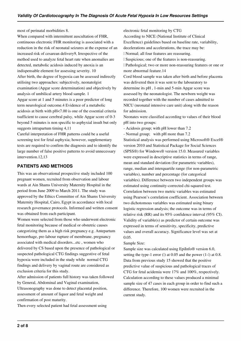
most of perinatal morbidities 8.When compared with intermittent auscultation of FHR,continuous electronic FHR monitoring is associated with areduction in the risk of neonatal seizures at the expense of anincreased risk of cesarean delivery9, Irrespective of themethod used to analyze fetal heart rate when anomalies aredetected, metabolic acidosis induced by anoxia is anindispensable element for assessing severity. 10After birth, the degree of hypoxia can be assessed indirectlyutilising two approaches: subjectively, neonatolgistexamination (Apgar score determination) and objectively byanalysis of umbilical artery blood sample. 1Apgar score at 1 and 5 minutes is a poor predictor of longterm neurological outcome.4 Evidence of a metabolicacidosis at birth with pH<7.00 is one of the essential criteriatsufficient to cause cerebral palsy, while Apgar score of 0-3beyond 5 minutes is non specific to asphyxial insult but onlysuggests intrapartum timing 4,11Careful interpretation of FHR patterns could be a usefulscreening test for fetal asphyxia; however, supplementarytests are required to confirm the diagnosis and to identify thelarge number of false positive patterns to avoid unnecessaryintervention.12,13
PATIENTS AND METHODS
This was an observational prospective study included 100pregnant women, recruited from observation and labourwards at Ain Shams University Maternity Hospital in theperiod from June 2009 to March 2011. The study wasapproved by the Ethics Committee of Ain Shams UniversityMaternity Hospital, Cairo, Egypt in accordance with localresearch governance protocols. Informed and written consentwas obtained from each participant.Women were selected from those who underwent electronicfetal monitoring because of medical or obstetric causescategorizing them as a high risk pregnancy e.g. Antepartumhemorrhage, pre-labour rupture of membrane, pregnancyassociated with medical disorders...etc , women whodelivered by CS based upon the presence of pathological orsuspected pathological CTG findings suggestive of fetalhypoxia were included in the study while normal CTGfindings and delivery by vaginal route are considered asexclusion criteria for this study.After admission of patients full history was taken followedby General, Abdominal and Vaginal examination,Ultrasonography was done to detect placental position,assessment of amount of liquor and fetal weight andconfirmation of post maturity.Then every selected patient had fetal assessment using
electronic fetal monitoring by CTGAccording to NICE (National Institute of ClinicalExcellence) guidelines based on baseline rate, variability,decelerations and accelerations, the trace may be: Normal; all four features are reassuring. Suspicious; one of the features is non-reassuring. Pathological; two or more non-reassuring features or one ormore abnormal feature.14Cord blood sample was taken after birth and before placentawas delivered then it was sent to the labouratory todetermine its pH , 1-min and 5-min Apgar score wasassessed by the neonatologist. The newborn weight wasrecorded together with the number of cases admitted toNICU (neonatal intensive care unit) along with the reasonfor admission.Neonates were classified according to values of their bloodpH into two groups:- Acidosis group; with pH lower than 7.2- Normal group; with pH more than 7.2Statistical analysis was performed using Microsoft® Excel®version 2010 and Statistical Package for Social Sciences(SPSS®) for Windows® version 15.0. Measured variableswere expressed in descriptive statistics in terms of range,mean and standard deviation (for parametric variables),range, median and interquartile range (for non-parametricvariables), number and percentage (for categoricalvariables). Difference between two independent groups wasestimated using continuity-corrected chi-squared test.Correlation between two metric variables was estimatedusing Pearson’s correlation coefficient. Association betweentwo dichotomous variables was estimated using binarylogistic regression analysis; the outcome was in terms ofrelative risk (RR) and its 95% confidence interval (95% CI).Validity of variable(s) as predictor of certain outcome wasexpressed in terms of sensitivity, specificity, predictivevalues and overall accuracy. Significance level was set at0.05.Sample Size:Sample size was calculated using EpiInfo® version 6.0,setting the type-1 error (α) at 0.05 and the power (1-β) at 0.8.Data from previous study 15 showed that the positivepredictive value of suspicious and pathological traces ofCTG for fetal acidemia were 17% and 100%, respectively.Calculation according to these values produced a minimalsample size of 47 cases in each group in order to find such adifference. Therefore, 100 women were recruited in thecurrent study.
Validity Of Cardiotocography In The Diagnosis Of Acute Fetal Hypoxia In Low Resources Settings
3 of 8
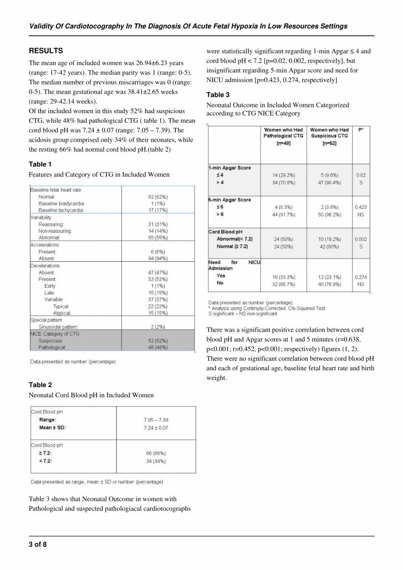
RESULTS
The mean age of included women was 26.94±6.23 years(range: 17-42 years). The median parity was 1 (range: 0-5).The median number of previous miscarriages was 0 (range:0-5). The mean gestational age was 38.41±2.65 weeks(range: 29-42.14 weeks).Of the included women in this study 52% had suspiciousCTG, while 48% had pathological CTG ( table 1). The meancord blood pH was 7.24 ± 0.07 (range: 7.05 – 7.39). Theacidosis group comprised only 34% of their neonates, whilethe resting 66% had normal cord blood pH.(table 2)
Table 1
Features and Category of CTG in Included Women
Table 2
Neonatal Cord Blood pH in Included Women
Table 3 shows that Neonatal Outcome in women withPathological and suspected pathologiacal cardiotocographs
were statistically significant regarding 1-min Apgar ≤ 4 andcord blood pH < 7.2 [p=0.02, 0.002, respectively], butinsignificant regarding 5-min Apgar score and need forNICU admission [p=0.423, 0.274, respectively]
Table 3
Neonatal Outcome in Included Women Categorizedaccording to CTG NICE Category
There was a significant positive correlation between cordblood pH and Apgar scores at 1 and 5 minutes (r=0.638,p<0.001; r=0.452, p<0.001; respectively) figures (1, 2).There were no significant correlation between cord blood pHand each of gestational age, baseline fetal heart rate and birthweight.

Validity Of Cardiotocography In The Diagnosis Of Acute Fetal Hypoxia In Low Resources Settings
4 of 8
Figure 1
Scatter-Plot showing Correlation between Cord Blood pHand 1-min Apgar Score.
Figure 2
Scatter-Plot showing Correlation between Cord Blood pHand 5-min Apgar Score
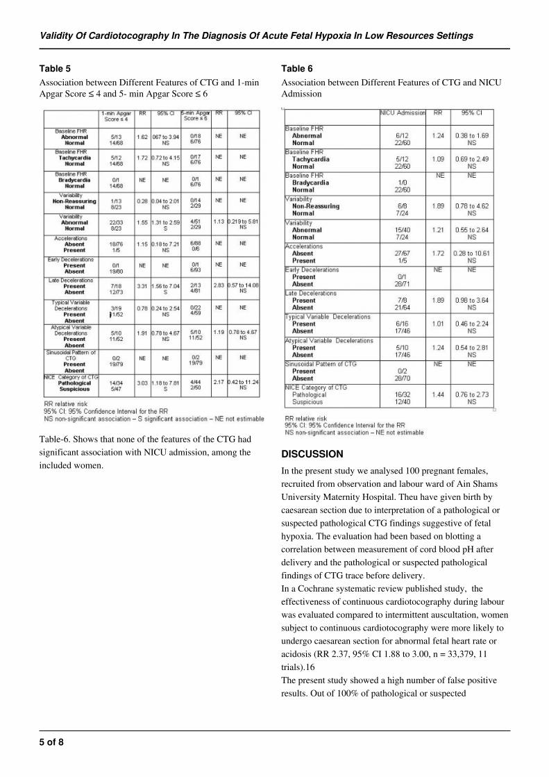
Table 4 shows Binary logistic regression analysis ofdifferent features of CTG as predictor of abnormal cordblood pH(< 7.2) Baseline bradycardia significantly increased the riskof abnormal cord blood pH almost 3-folds [RR 3.2, 95% CI(2.29 to 4.33)]., Abnormal beat-to-beat variabilitysignificantly increased the risk of abnormal cord blood pHalmost 2-folds [RR 2.35, 95% CI (1.08 to 5.09)], Latedecelerations significantly increased the risk of abnormalcord blood pH almost 7-folds [RR 7.1, 95% CI (3.86 to12.3)], Presence of atypical variable decelerationssignificantly increased the risk of abnormal cord blood pHalmost 2-folds[RR 1.9, 95% CI (1.1 to 3.27)], Pathological(rather than suspicious) CTG significantly increased the riskof abnormal cord blood pH almost 2.5-folds[RR 2.6, 95% CI(1.39 to 4.85)]., Neither of other features was significantlyassociated with abnormal cord blood pH.
Table 4
Association between Different Features of CTG andAbnormal Cord Blood pH
Table 5 shows different features of CTG as a predictor of 1-min Apgar score ≤ 4 and and 5- min Apgar Score ≤ 6,Abnormal beat-to-beat variability significantly increased therisk of having 1-min Apgar score ≤ 4 almost 1.5-folds [RR1.55; 95% CI (1.31to 2.59)]., Presence of late decelerationssignificantly increased the risk of 1-min Apgar score ≤ 4almost 3-folds [RR 3.31; 95% CI (1.56 to 7.04)].,Pathological (versus suspicious) CTG significantly increasedthe risk of 1-min Apgar score ≤ 4 almost 3-folds [RR 3.03;95% CI (1.18 to 7.81] None of other features of the CTG hadsignificant association with 1-min Apgar score ≤ 4. Whilenone of the features of the CTG had significant associationwith 5-min Apgar score ≤ 6, among the included women .
Validity Of Cardiotocography In The Diagnosis Of Acute Fetal Hypoxia In Low Resources Settings
5 of 8
Table 5
Association between Different Features of CTG and 1-minApgar Score ≤ 4 and 5- min Apgar Score ≤ 6
Table-6. Shows that none of the features of the CTG hadsignificant association with NICU admission, among theincluded women.
Table 6
Association between Different Features of CTG and NICUAdmission
DISCUSSION
In the present study we analysed 100 pregnant females,recruited from observation and labour ward of Ain ShamsUniversity Maternity Hospital. Theu have given birth bycaesarean section due to interpretation of a pathological orsuspected pathological CTG findings suggestive of fetalhypoxia. The evaluation had been based on blotting acorrelation between measurement of cord blood pH afterdelivery and the pathological or suspected pathologicalfindings of CTG trace before delivery.In a Cochrane systematic review published study, theeffectiveness of continuous cardiotocography during labourwas evaluated compared to intermittent auscultation, womensubject to continuous cardiotocography were more likely toundergo caesarean section for abnormal fetal heart rate oracidosis (RR 2.37, 95% CI 1.88 to 3.00, n = 33,379, 11trials).16The present study showed a high number of false positiveresults. Out of 100% of pathological or suspected

Validity Of Cardiotocography In The Diagnosis Of Acute Fetal Hypoxia In Low Resources Settings
6 of 8
pathological cardiotocograms, only 34 % were valid (i.e., theinfants were hypoxic after birth). The remaining 66% ofinfants, although their CTG findings were suggestive of fetalhypoxia, were born healthy and CS was probably notnecessary .The average pH value of umbilical artery blood was 7.24 ±0.07 (range 7.05 - 7.39). There was a significant positivecorrelation between cord blood pH and Apgar scores at 1and 5 minutes (r =0.638, p<0.001; r =0.452, p<0.001;respectively), There were no significant correlation betweencord blood pH and each of gestational age, baseline fetalheart rate and birth weight, These results are also supportedby that obtained by Hogan et al who performed a case-control study to evaluate how often low five-minute Apgarscores are associated with asphyxia. Cases were 183 termnewborns with five-minute Apgar scores below seven,Controls were 183 randomly selected term newborns withfive-minute Apgar scores of nine to ten. They found a strongpositive correlation between low five-minute Apgar scores,abnormal cardiotocography and cord blood acidosis. Theyconcluded that a five-minute Apgar score below four is agood proxy for asphyxia.17In the present study, Binary logistic regression analysis ofdifferent features of CTG as predictor of abnormal cordblood pH (< 7.2) was performed. Baseline bradycardiasignificantly increased the risk of abnormal cord blood pHalmost 3-folds [RR 3.2, 95% CI (2.29 to 4.33)]. reducedbeat-to-beat variability significantly increased the risk ofabnormal cord blood pH almost 2-folds [RR 2.35, 95% CI(1.08 to 5.09)]. Late decelerations significantly increasedthe risk of abnormal cord blood pH almost 7-folds [RR 7.1,95% CI (3.86 to 12.3)]. Presence of atypical variabledecelerations significantly increased the risk of abnormalcord blood pH almost 2-folds [RR 1.9, 95% CI (1.1 to3.27)]. Pathological (rather than suspicious) CTGsignificantly increased the risk of abnormal cord blood pHalmost 2.5-folds [RR 2.6, 95% CI (1.39 to 4.85)]. Neither ofother features was significantly associated with abnormalcord blood pH as well as, the present study showed thatneonatal outcomes of pathological and suspectedpathologiacal cardiotocographs were statistically significantregarding 1-min Apgar scores ≤4 and cord blood pH < 7.2[p=0.02, 0.002, respectively], but insignificant regarding 5-min Apgar score and need for NICU admission [p=0.423,0.274, respectively].The statistically significant parameters obtained by binarylogistic regression analysis of different features of CTG aspredictor of 1-min Apgar score ≤ 4 were abnormal beat-to-
beat variability [RR 1.55; 95% CI (1.31to 2.59)], presence oflate decelerations [RR 3.31; 95% CI (1.56 to 7.04)] andPathological (versus suspicious) CTG [RR 3.03; 95% CI(1.18 to 7.81] .
CONCLUSION
Fetal monitoring using cardiotocography is associated with aconsiderale false positive results and subsequent surgicalintervention that might have not been necessary. Thus, usingof fetal heart rate abnormalities alone as a measure ofdiagnosis of fetal distress during labour is a contributingfactor of increasing rate of cesarean sections.Other pathological features; Baseline bradycardia, reducedbeat to beat variability, atypical variable decelerations, latedecelerations and pathological, rather than suspicious, CTGtraces increased the risk of fetal hypoxia and subsequentfetal acidosis significantly.
References
1. Pellantova S, Roztocil A,Miklica J. "Validity of CTG inacute fetal hypoxia-neonatal status after cesarean section."Ceska Gynekol, 2000; 34-38.2. Clerici G, Luzietti R, Di Renzo G. " Monitoring ofAntepartum and intrapartum fetal hypoxemia:pathophysiological basis and available techniques. "BiolNeonate, 2001; 246-253.3. Bobrow C, Soothill P. "Causes and consequences of fetalacidosis." BMJ, 1999; F246-F249.4. Yazawa K."Neonatal encephalopathy and cerebral palsy."J Nippon Med Sch, 2005; 85-88.5. Habek D, Hodek B, Herman R. "Fetal hypoxia- etiologyand pathophyiology of hypoxic damage." Lijec vjesn, 2000;82-89.6. Goddard R. "Electronic fetal monitoring is not necessaryfor low risk labours." BMJ, 2001; 322: 1436-1437.7. Hamilton BE, Martin JA, Ventura SJ, Sutton P, MenackerF. "Births: preliminary data for 2004." National vitalstatistics, 2005; 54(2).8. Royal College Of Obstetricians and Gynecologists. "Theuse of electronic fetal monitoring." National Evidence-basedClinical guidelines , May 2001: Evidence-based clinicalguideline Number 8.9. Agrawal S, Doucette F, Gratton R, Richardson B, GagnonR. Intrapartum computerized fetal heart rate parameters andmetabolic acidosis at birth. Obstet Gynecol, 2003; 102:731-738.10. Uzan S, Berkane N, Verstraete L, Mathieu E, Breart G."Acid base balance in the fetus during labour:pathophysilogy and exploration methods." J Gynecol ObstetReprod, 2003; 1S 68-78.11. Strijbis E, Oudman I, van Essen P, Maclennan A."Cerebral palsy and the application of international criteriafor acute intrapartum hypoxia." Obstet Gynecol, 2006; 107:1357-1365.12. Larma JD, Silva AM, Holcroft CJ, Thompson RE,Donohue PK, Graham EM. "Intrapartum electronic fetalheart rate monitoring and the identification of metabolicacidosis and hypoxic-ischemic encephlopathy ." Am JObstet Gynecol, 2007; 301.e1-301.e8.13. Bahiah A, Murphy J, Sharida H. "Fetal distress in labourand cesarean section rate." Bahrain medical Bulletin, 2010;

Validity Of Cardiotocography In The Diagnosis Of Acute Fetal Hypoxia In Low Resources Settings
7 of 8
32(2):1-5.14. Baskett T, Calder A, Arulkumaran S. "Fetal surveillencein labour." In Munro Kerr's operative obstetrics eleventhedition, 4:38-39. Elsevier publishing, 2007.15. Tasnim N, Mahmud G, Akram S. "Predictive accuracy ofintrapartum cardiotocography in terms of fetal acid basestatus at birth. "J Coll Physicians Surg Pak, 2001; 19(10):632-635.
16. Alfirevic Z, Devane D, Gyte GM. "Continuouscardiotocography (CTG) as a form of electronic fetalmonitoring (EFM) for fetal assessment during labour."CochraneDatabase Syst Rev, 2006; 3:CD006066.17. Hogan L, Ingemarsson I, Thorngren-Jerneck K, HerbstA. "How often is a low 5-min Apgar score in term newbornsdue to asphyxia?" Eur J Obstet Gynecol Reprod Biol, 2007;130(2):169-75.

Validity Of Cardiotocography In The Diagnosis Of Acute Fetal Hypoxia In Low Resources Settings
8 of 8
Author Information
Wessam M Aboulghar, MDDepartment of Obstetrics and Gynecology, Faculty of Medicine, Ain Shams UniversityCairo, Egypt
Mohamed A Ibrahim, MDDepartment of Obstetrics and Gynecology, Faculty of Medicine, Ain Shams UniversityCairo, Egypt
Ihab S Allam, MDDepartment of Obstetrics and Gynecology, Faculty of Medicine, Ain Shams UniversityCairo, Egypt
Wael Hosny, MSCDepartment of Obstetrics and Gynecology, Faculty of Medicine, Ain Shams UniversityCairo, Egypt
M Otify, Dr, Msc, MRCOGKings College HospitalLondon, United [email protected]