Validation clinical classification subtypes of acute...
7
J7ournal of Neurology, Neurosurgery, and Psychiatry 1994;57: 1173-1179 Validation of a clinical classification for subtypes of acute cerebral infarction Craig S Anderson, Bruce V Taylor, Graeme J Hankey, Edward G Stewart-Wynne, Konrad D Jamrozik Abstract The validity of a clinical classification system was assessed for subtypes of cere- bral infarction for use in clinical trials of putative stroke therapies and clinical decision making in a population based stroke register (n = 536) compiled in Perth, Western Australia in 1989-90. The Perth Community Stroke Project (PCSS) used definitions and methodology similar to the Oxfordshire Community Stroke Project (OCSP) where the classification system was developed. In the PCSS, 421 cases of cerebral infarction and primary intracerebral haemorrhage (PICH), con- firmed by brain imaging or necropsy, were classified into the subtypes total anterior circulation syndrome (TACS), partial anterior circulation syndrome (PACS), lacunar syndrome (LACS), and posterior circulation syndrome (POCS). In this relatively unselected population, relying exclusively on LACS for a diagno- sis of PICH had a very low sensitivity (6%) and positive predictive value (3%). Comparison of the frequencies and out- comes (at one year after the onset of symptoms) for each subgroup of first ever cerebral infarction in the PCSS (n = 248) with the OCSP (n = 543) registers showed uniformity only for LACI. For example, there were 27% of cases of TACI in the PCSS compared with 17% in the OCSP (difference = 10%; 95% confi- dence interval (95% CI) 4% to 16%) and 15% of cases in the PCSS compared with 24% in the OCSP were POCI (difference = 9%; 95% CI 3% to 15%). Case fatalities and long term handicap across the sub- groups were not significantly different between studies, but the frequencies of recurrent stroke were significantly greater for POCI in the OCSP compared with the PCSS. Although this classifica- tion system defines subtypes of stroke with different outcomes, simple clinical measures-level of consciousness, pare- sis, disability, and incontinence at onset-are more powerful predictors of death or dependency at one year. It is concluded that simple clinical measures that reflect the severity of the neurologi- cal deficit should complement this classi- fication system in clinical trials and practice. ( Neurol Neurosurg Psychiatry 1994;57:1173-1179) Although substantial progress has been made in developing strategies for the prevention of stroke, there is still no effective treatment for the acute event; but a number of putative treatments for stroke are now undergoing evaluation in multicentre clinical trials.' A crucial issue associated with such trials is the classification of cerebral infarction because it may arise from several different pathophysio- logical processes with variable prognosis.2 Ignoring major prognostic factors in the analysis of clinical trials increases the back- ground variability of patients and may obscure differences in the efficacy of treat- ments. Despite the recognition that pathological homogeneous stroke entities need to be identi- fied and studied,3 however, the usefulness of various criteria and scoring systems for epi- demiological research, particularly clinical tri- als, is limited. Although CT differentiates accurately haemorrhage from infarction, there are differences in the quality and interpreta- tion of CT across centres, and access of patients to this investigation is not available or often delayed in many countries. Moreover, it is often impractical to performn other objective investigations such as angiography, particu- larly if the benefit of treatment is dependent on minimising the delay between the onset of symptoms and the start of treatment. The realities of the numbers of patients required to produce meaningful results from clinical trials dictate that a classification of stroke should be valid and reliable, yet simple enough to facilitate the recruitment of patients and to allow the conclusions of the trial to be communicated easily and applied widely. Bamford et al 4 considered these issues in a study that introduced a classification sys- tem for cerebral infarction based on their experience in the Oxfordshire Community Stroke Project (OCSP), a population based study of first ever stroke. They defined by simple clinical variables four subtypes of cere- bral infarction that had different patterns of case fatality, recurrence, and long term dis- ability. This classification is not only useful in clinical practice, but could increase the sensitivity of clinical trials by correcting for imbalances in study groups or improving the precision of the estimates of differences between treatments. The purpose of our study was to validate this classification system in patients registered in the Perth Community Stroke Study (PCSS), a population based study of acute cerebrovascular disease in Department of Medicine, Flinders University of South Australia, Bedford Park, South Australia 5042 C S Anderson Department of Neurology, Royal Perth Hospital, Perth, Western Australia 6000 B V Taylor G J Hankey E G Stewart-Wynne Department of Public Health, University of Western Australia, Nedlands, Western Australia 6009 K D Jamrozik Correspondence to: Dr C S Anderson. Received 20 January 1994 and in revised form 18 April 1994. Accepted 25 April 1994 1173 on 27 August 2018 by guest. Protected by copyright. http://jnnp.bmj.com/ J Neurol Neurosurg Psychiatry: first published as 10.1136/jnnp.57.10.1173 on 1 October 1994. Downloaded from
Transcript of Validation clinical classification subtypes of acute...

J7ournal ofNeurology, Neurosurgery, and Psychiatry 1994;57: 1173-1179
Validation of a clinical classification for subtypesof acute cerebral infarction
Craig S Anderson, Bruce V Taylor, Graeme J Hankey, Edward G Stewart-Wynne,Konrad D Jamrozik
AbstractThe validity of a clinical classificationsystem was assessed for subtypes of cere-bral infarction for use in clinical trials ofputative stroke therapies and clinicaldecision making in a population basedstroke register (n = 536) compiled inPerth, Western Australia in 1989-90. ThePerth Community Stroke Project (PCSS)used definitions and methodology similarto the Oxfordshire Community StrokeProject (OCSP) where the classificationsystem was developed. In the PCSS, 421cases of cerebral infarction and primaryintracerebral haemorrhage (PICH), con-firmed by brain imaging or necropsy,were classified into the subtypes totalanterior circulation syndrome (TACS),partial anterior circulation syndrome(PACS), lacunar syndrome (LACS), andposterior circulation syndrome (POCS).In this relatively unselected population,relying exclusively on LACS for a diagno-sis of PICH had a very low sensitivity(6%) and positive predictive value (3%).Comparison of the frequencies and out-comes (at one year after the onset ofsymptoms) for each subgroup of firstever cerebral infarction in the PCSS (n =248) with the OCSP (n = 543) registersshowed uniformity only for LACI. Forexample, there were 27% of cases ofTACI in the PCSS compared with 17% inthe OCSP (difference = 10%; 95% confi-dence interval (95% CI) 4% to 16%) and15% of cases in the PCSS compared with24% in the OCSP were POCI (difference= 9%; 95% CI 3% to 15%). Case fatalitiesand long term handicap across the sub-groups were not significantly differentbetween studies, but the frequencies ofrecurrent stroke were significantlygreater for POCI in the OCSP comparedwith the PCSS. Although this classifica-tion system defines subtypes of strokewith different outcomes, simple clinicalmeasures-level of consciousness, pare-sis, disability, and incontinence atonset-are more powerful predictors ofdeath or dependency at one year. It isconcluded that simple clinical measuresthat reflect the severity of the neurologi-cal deficit should complement this classi-fication system in clinical trials andpractice.
( Neurol Neurosurg Psychiatry 1994;57:1173-1179)
Although substantial progress has been madein developing strategies for the prevention ofstroke, there is still no effective treatment forthe acute event; but a number of putativetreatments for stroke are now undergoingevaluation in multicentre clinical trials.' Acrucial issue associated with such trials is theclassification of cerebral infarction because itmay arise from several different pathophysio-logical processes with variable prognosis.2Ignoring major prognostic factors in theanalysis of clinical trials increases the back-ground variability of patients and mayobscure differences in the efficacy of treat-ments.
Despite the recognition that pathologicalhomogeneous stroke entities need to be identi-fied and studied,3 however, the usefulness ofvarious criteria and scoring systems for epi-demiological research, particularly clinical tri-als, is limited. Although CT differentiatesaccurately haemorrhage from infarction, thereare differences in the quality and interpreta-tion of CT across centres, and access ofpatients to this investigation is not available oroften delayed in many countries. Moreover, itis often impractical to performn other objectiveinvestigations such as angiography, particu-larly if the benefit of treatment is dependenton minimising the delay between the onset ofsymptoms and the start of treatment.The realities of the numbers of patients
required to produce meaningful results fromclinical trials dictate that a classification ofstroke should be valid and reliable, yet simpleenough to facilitate the recruitment ofpatients and to allow the conclusions of thetrial to be communicated easily and appliedwidely. Bamford et al 4 considered these issuesin a study that introduced a classification sys-tem for cerebral infarction based on theirexperience in the Oxfordshire CommunityStroke Project (OCSP), a population basedstudy of first ever stroke. They defined bysimple clinical variables four subtypes of cere-bral infarction that had different patterns ofcase fatality, recurrence, and long term dis-ability. This classification is not only useful inclinical practice, but could increase thesensitivity of clinical trials by correcting forimbalances in study groups or improving theprecision of the estimates of differencesbetween treatments. The purpose of our studywas to validate this classification system inpatients registered in the Perth CommunityStroke Study (PCSS), a population basedstudy of acute cerebrovascular disease in
Department ofMedicine, FlindersUniversity of SouthAustralia, BedfordPark, South Australia5042C S AndersonDepartment ofNeurology, RoyalPerth Hospital, Perth,Western Australia6000B V TaylorG J HankeyE G Stewart-WynneDepartment ofPublicHealth, University ofWestern Australia,Nedlands, WesternAustralia 6009K D JamrozikCorrespondence to:Dr C S Anderson.Received 20 January 1994and in revised form18 April 1994.Accepted 25 April 1994
1173
on 27 August 2018 by guest. P
rotected by copyright.http://jnnp.bm
j.com/
J Neurol N
eurosurg Psychiatry: first published as 10.1136/jnnp.57.10.1173 on 1 O
ctober 1994. Dow
nloaded from

Anderson, Taylor, Hankey, Stewart-Wynne, J7amrozik
Perth, Western Australia.25 The PCSS usedstandard definitions, measures of outcome,and community wide methods of case ascer-tainment that were similar to those used in theOCSP.
MethodsTHE PERTH COMMUNITY STROKE STUDY(PCSS) POPULATIONThe objectives, design, and study populationof the PCSS have been described.5 In brief,the aims of the study were to determine theincidence, aetiology, and outcome of acutestroke (n = 536) in a geographically definedand representative segment (population138 708) of Perth, Western Australia, duringan 18 month period in 1989-90. The registerused a variety of overlapping sources thatincluded notifications from general practition-ers; scrutiny of attendances at, and admissionsto, all acute hospitals, rehabilitation centres,and nursing homes in and around the studyarea; coroner's reports and death certificates;and surveillance of computerised hospital dis-charge statistics that cover all inpatient sepa-rations from every hospital in the state.
EVALUATION OF PATIENTSMost patients (83%) were seen early afteronset of the event (median delay five days) bya single observer (CSA) who conducted astandardised interview and physical examina-tion. Those patients who were not personallyassessed were classified as cases on the basisof the history and neurological examinationobtained from the medical records. StandardWorld Health Organisation (WHO) criteria6were used for stroke and each event was classi-fied as being either the patient's first ever in alifetime (first ever) or recurrent event duringthe study period. Criteria were applied thatcombined clinical data with the results of CT,MRI, or necropsy so that strokes could beclassified into several "pathologically distinct"groups (in 86% of cases).2
Information obtained from each patient atbaseline included data on associated illnesses,risk factors for cardiovascular diseases, andthe patterns of disability within the immediatepremorbid period. If the patient was uncon-sious or otherwise not assessable, informationwas obtained from the patient's closest rela-tive or another reliable proxy, or hospitalrecords, or the general practitioner. Level ofconsciousness at onset was measured by theGlasgow coma scale,7 and scores 3-9 and10-14 were classified as "comatose" and"drowsy" respectively. Degree of paresis wasmeasured with the motricity index,8 whichgives a score from 0 (total plegia) to 100 (nor-mal). We defined a score of 0-50 as "severeparesis", 51-94 as "moderate paresis", and ascore of 95-100 as "normal or minimal pare-sis". Comorbid diseases of interest were diag-nosed on the basis of available informationand were considered to be present if theyoccurred at any time before the onset of theindex event. Hypertension was defined if apatient had a clinical history of hypertension,
or had two or more previously documentedsystolic blood pressures > 160 mm Hg ordiastolic blood pressures > 95 mm Hg.Coronary heart disease was defined as a his-tory of acute myocardial infarction or anginapectoris. Peripheral vascular disease (PVD)was diagnosed if the patients had a history ofintermittent claudication (WHO criteria: calfpain induced by exercise and relieved within10 minutes by rest, that was atherosclerotic inorgin). Diabetes mellitus was deemed presentif the patient gave a history of the conditionthat was confirmed in the medical records, orthere was a random blood glucose concentra-tion of > 11 mmolIl.The same investigator who made the base-
line assessments prospectively followedpatients until 12 months after their indexevent or the time of death if it occurred beforethat date. All survivors were reassessed at fourand 12 months, generally in their own homeor usual place of residence, and were classifiedas "independent" (score 0-3) or "dependent"(4-5) according to the Oxford handicapscale.9 Any patient who gave a history of arecurrent stroke during follow up wasreassessed with the same baseline assessmentschedule. Human rights and ethics commit-tees approved the study at each participatinginstitution.
CLASSIFICATION OF SUBTYPES OF STROKEFor this report, one of us (BVT) revieweddetails of the neurological examination foreach event that was recorded on the originalassessment forms while blind to the finalpathological diagnoses and outcomes. Eachstroke event was then classified into one of thefour clinical syndromes outlined in detail byBamford et al.4 The appendix includes an out-line of these specific subgroups-namely, totalanterior circulation syndrome (TACS), partialanterior circulation syndrome (PACS), lacu-nar syndrome (LACS), and posterior circula-tion syndrome (POCS). When brain imagingor necropsy had excluded primary intracere-bral haemorrhage (PICH), the subgroupswere referred to as infarcts: total anterior cir-culation infarcts (TACI), partial anterior cir-culation infarcts (PACI), lacunar infarcts(LACI), and posterior circulation infarcts(POCI).
BRAIN IMAGING AND NECROPSYConsiderable effort was made to ensure thatinformation about the nature and extent ofcerebral lesions could be correlated with thevarious clinical syndromes and outcomes. AllCT and MRI films and necropsies werereviewed by a study neuroradiologist orneuropathologist blind to clinical and otherinformation, and each cerebral region thatcontained infarction or haemorrhage wasquantified. By contrast with Bamford et al,4we did not use the Guy's Hospital strokediagnostic scale'0 to exclude PICH in thosecases where brain imaging was unavailable,and cases of "undetermined stroke" were notreclassified into one of the mentioned clinicalsyndromes. In the PCSS, infarcts associated
1174
on 27 August 2018 by guest. P
rotected by copyright.http://jnnp.bm
j.com/
J Neurol N
eurosurg Psychiatry: first published as 10.1136/jnnp.57.10.1173 on 1 O
ctober 1994. Dow
nloaded from
Validation ofa clinical classification for subtypes of acute cerebral infarction
with first ever strokes were coded into one ofthe following sites: deep (caudate, putamen,globus pallidus, thalamus, internal capsule,and deep periventricular white matter); lobar(specific cortical and subcortical locations);and brainstem (including cerebellum). Thedeep lesions were further subdivided accord-ing to size, which was estimated by multiply-ing the greatest sagittal, transverse, andcoronal measures from the brain imaging orbrain slices. These lesions were then cate-gorised as either "small" (< 8 cm3) or "large"( s 8 cm3) to differentiate lacunar infarctsfrom larger striatocapsular infarcts, whichmay have different aetiologies. If no relevantacute cerebral lesion was documented, the siteof infarction was classified as "unknown".
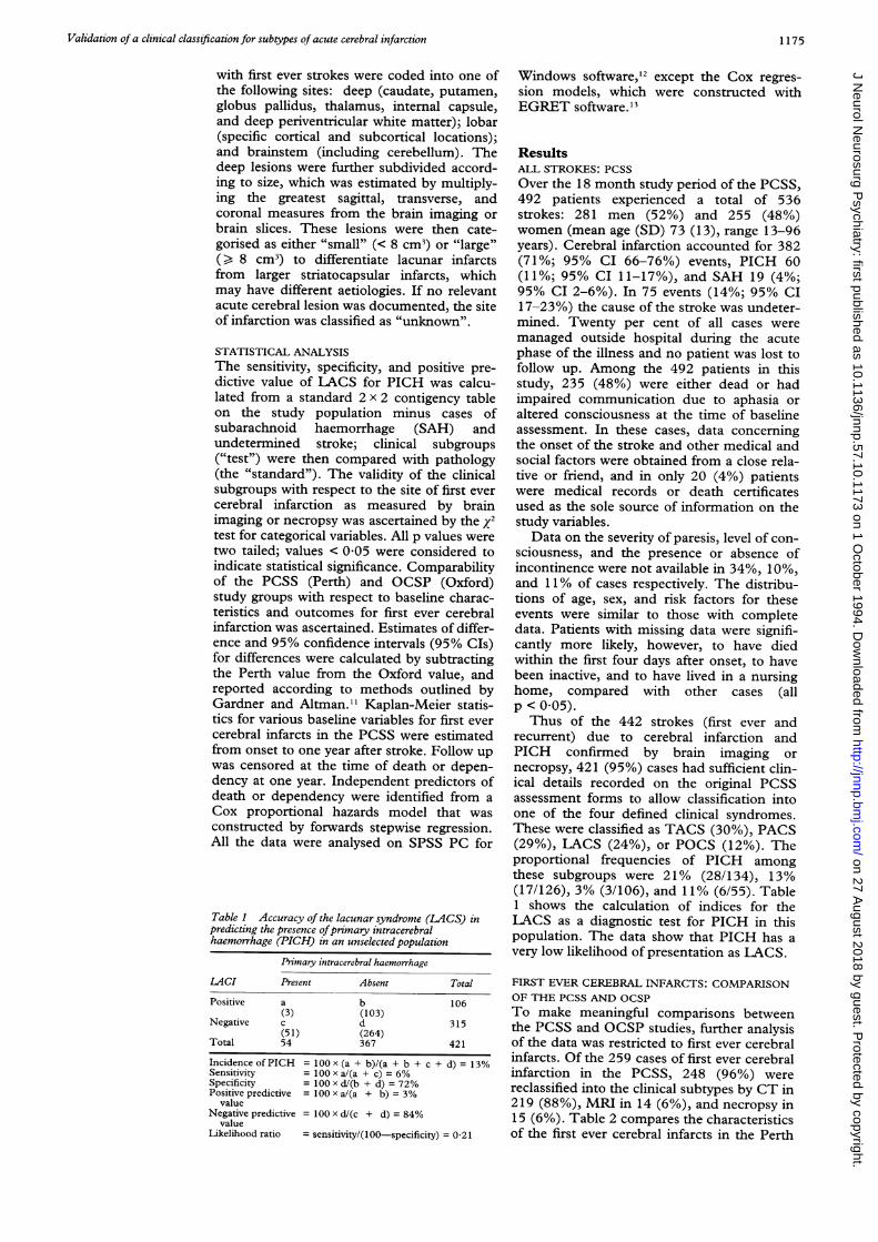
STATISTICAL ANALYSISThe sensitivity, specificity, and positive pre-dictive value of LACS for PICH was calcu-lated from a standard 2 x 2 contigency tableon the study population minus cases ofsubarachnoid haemorrhage (SAH) andundetermined stroke; clinical subgroups("test") were then compared with pathology(the "standard"). The validity of the clinicalsubgroups with respect to the site of first evercerebral infarction as measured by brainimaging or necropsy was ascertained by the X2test for categorical variables. All p values weretwo tailed; values < 0 05 were considered toindicate statistical significance. Comparabilityof the PCSS (Perth) and OCSP (Oxford)study groups with respect to baseline charac-teristics and outcomes for first ever cerebralinfarction was ascertained. Estimates of differ-ence and 95% confidence intervals (95% CIs)for differences were calculated by subtractingthe Perth value from the Oxford value, andreported according to methods outlined byGardner and Altman." Kaplan-Meier statis-tics for various baseline variables for first evercerebral infarcts in the PCSS were estimatedfrom onset to one year after stroke. Follow upwas censored at the time of death or depen-dency at one year. Independent predictors ofdeath or dependency were identified from aCox proportional hazards model that wasconstructed by forwards stepwise regression.All the data were analysed on SPSS PC for
Table 1 Accuracy of the lacunar syndrome (LACS) inpredicting the presence ofprimary intracerebralhaemorrhage (PICH) in an unselected population
Primary intracerebral haemorrhage
LACI Present Absent Total
Positive a b 106(3) (103)
Negative c d 315(51) (264)
Total 54 367 421
Incidence of PICH = 100 x (a + b)/(a + b + c + d) = 13%Sensitivity = 100 x a/(a + c) = 6%Specificity = 100 x d/(b + d) = 72%Positive predictive = 100 x a/(a + b) = 3%
valueNegative predictive = 100 x d/(c + d) = 84%
valueLikelihood ratio = sensitivity/(100-specificity) = 0-21
Windows software,'2 except the Cox regres-sion models, which were constructed withEGRET software."3
ResultsALL STROKES: PCSSOver the 18 month study period of the PCSS,492 patients experienced a total of 536strokes: 281 men (52%) and 255 (48%)women (mean age (SD) 73 (13), range 13-96years). Cerebral infarction accounted for 382(71%; 95% CI 66-76%) events, PICH 60(11%; 95% CI 11-17%), and SAH 19 (4%;95% CI 2-6%). In 75 events (14%; 95% CI17-23%) the cause of the stroke was undeter-mined. Twenty per cent of all cases weremanaged outside hospital during the acutephase of the illness and no patient was lost tofollow up. Among the 492 patients in thisstudy, 235 (48%) were either dead or hadimpaired communication due to aphasia oraltered consciousness at the time of baselineassessment. In these cases, data concerningthe onset of the stroke and other medical andsocial factors were obtained from a close rela-tive or friend, and in only 20 (4%) patientswere medical records or death certificatesused as the sole source of information on thestudy variables.
Data on the severity of paresis, level of con-sciousness, and the presence or absence ofincontinence were not available in 34%, 10%,and 11% of cases respectively. The distribu-tions of age, sex, and risk factors for theseevents were similar to those with completedata. Patients with missing data were signifi-cantly more likely, however, to have diedwithin the first four days after onset, to havebeen inactive, and to have lived in a nursinghome, compared with other cases (allp < 0 05).Thus of the 442 strokes (first ever and
recurrent) due to cerebral infarction andPICH confirmed by brain imaging ornecropsy, 421 (95%) cases had sufficient clin-ical details recorded on the original PCSSassessment forms to allow classification intoone of the four defined clinical syndromes.These were classified as TACS (30%), PACS(29%), LACS (24%), or POCS (12%). Theproportional frequencies of PICH amongthese subgroups were 21% (28/134), 13%(17/126), 3% (3/106), and 11% (6/55). Table1 shows the calculation of indices for theLACS as a diagnostic test for PICH in thispopulation. The data show that PICH has avery low likelihood of presentation as LACS.
FIRST EVER CEREBRAL INFARCTS: COMPARISONOF THE PCSS AND OCSPTo make meaningful comparisons betweenthe PCSS and OCSP studies, further analysisof the data was restricted to first ever cerebralinfarcts. Of the 259 cases of first ever cerebralinfarction in the PCSS, 248 (96%) werereclassified into the clinical subtypes by CT in219 (88%), MRI in 14 (6%), and necropsy in15 (6%). Table 2 compares the characteristicsof the first ever cerebral infarcts in the Perth
1175
on 27 August 2018 by guest. P
rotected by copyright.http://jnnp.bm
j.com/
J Neurol N
eurosurg Psychiatry: first published as 10.1136/jnnp.57.10.1173 on 1 O
ctober 1994. Dow
nloaded from

Anderson, Taylor, Hankey, Stewart- Wynne, Jramrozik
Table 2 Demographic characteristics offirst-ever cerebral infarction in the study groups
Variable Perth Oxford Difference (95% CI)
Number of events 248 543Age (mean y) 73 73Men (%) 54 50 4 (-3 to 11)Time to assessment (median days) 4 4Pathological confirmation (%) 100 81 19 (6 to 32)Independent prestroke (%) 79 85 -6 (-12 to 0)
Table 3 Comparison ofproportionalfrequencies (%) ofclinical subgroups offirst ever cerebral infarction in Perthand Oxford
Perth OxfordGroup (n = 248) (n = 543) Difference (95% CI)
TACI 27 17 10 (4 to 16)PACI 30 34 4 (-3 to 11)LACI 28 25 3 (4 to IO)POCI 15 24 -9 (-3 to -15)
TACI = Total anterior circulation infarcts; PACI = partialanterior circulation infarcts; LACI = lacunar infarcts; POCI =posterior circulation infarcts
and Oxford populations. Before the indexstroke, the patients in Perth were more likelyto be dependent than the patients in Oxford,but the age and sex distributions and time toassessment were similar. As highlighted previ-ously, the PCSS included cases of cerebralinfarction defined by CT, MRI, or necropsy,whereas 19% of cases in the OSCP had a lowprobability of PICH on the basis of their scoreon the Guy's Hospital stroke diagnosis scale.
Table 3 shows the proportional frequenciesof the clinical subtypes of first ever cerebralinfarction in the two populations. The pro-portion of PACI and LACI were similar, butPerth had a significantly greater proportion ofTACI and correspondingly fewer POCIcompared with Oxford. It was important,therefore, to determine whether these differ-ences were real and not related to misclassifi-cation. Table 4 shows that the overall andsubgroup specific case fatalities and long termdependencies did not differ significantlybetween the centres (although the 95% CIswere wide). The prognosis in terms of recur-
rent stroke for TACI and LACI was also simi-lar in the centres. A significantly higher pro-
portion of patients with PACI and POCI,however, had recurrent events in Oxford, yetthese patients seemed to be less likely toremain dependent at one year after strokecompared with Perth.
PREDICTION OF LESION LOCATIONTable 5 shows a statistically significant trendacross subgroups (p < 0O00001) indicatingthat these syndromes predict accurately thesite of cerebral infarction. This trend wouldbe even more significant if among the POCIsubgroup, the case with a deep cerebral lesion(based within the internal capsule) and threeout of the seven lobar lesions (which werelocated within an occipital lobe) were reclassi-fied into brainstem category because of thecommon major vascular supply to these terri-tories.
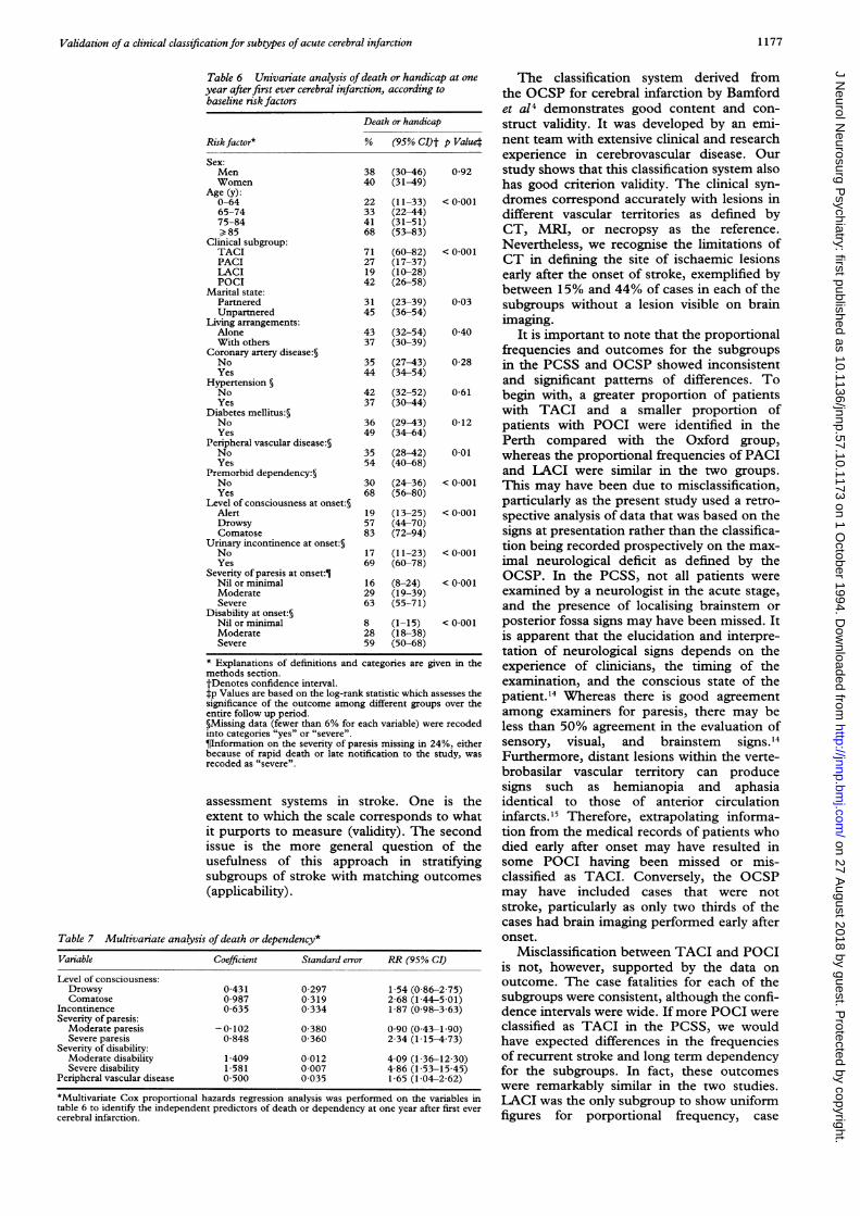
PREDICTORS OF DISABILITY FREE SURVIVALThe baseline factors found on univariateanalysis to be associated with death or depen-dency at one year in the PCSS were: age, clin-ical syndromes; marital state; premorbiddependency; level of consciousness; inconti-nence; severity of paresis; and disability (table6). In the stepwise Cox proportional hazardsmodel, one year mortality or dependency was
associated in the derivation set with beingcomatose at onset, urinary incontinence,severe paresis, and both moderate and severe
disability at onset; and evidence of peripheralvascular disease (table 7). The clinical sub-types did not enter the model as independentpredictors of outcome.
DiscussionWe wished to assess the validity of a classifica-tion system for infarction based on definedclinical syndromes that seems useful for clinicaldecision making and clinical trials. Our datahighlight two important issues relevant to thedevelopment, evaluation, and application of
Table 4 Death, recurrent stroke, and dependency over 12 months afterfirst ever cerebral infarctionCase fatality (%lo) Recurrent stroke (%) Dependency (%/6)
Difference Difference DifferenceGroup Perth Oxford (95% CI) Perth Oxford (95% CI) Perth Oxford (95% CI)
TACI 49 60 -11 (-27to 5) 4 6 -2 (-9to5) 37 36 1 (-14to 16)PACI 16 16 0 (-10 to 10) 8 17 -9 (-17 to-1) 37 29 8 (-5 to 21)LACI 10 11 -1 (-10to8) 15 9 6 (-4to 16) 29 28 1 (-12to 14)POCI 36 19 17 (0 to 34) 6 20 -14 (-24 to-4) 25 19 6 (-10 to 22)Total 26 23 3 (-4 to 10) 9 14 -5 (-10 to 0) 33 28 5 (-2 to 12)
Table 5 Comparison of the clinical syndromes with the site of cerebral infarction in the Perth Community Stroke StudyArea of cerebral infarction
Deep *
"small" "large" Lobar Brainstem UnknownGroup n(%q) n(%o) n(0/o) n(0/%) n(%O)TACI (n = 68) 9(13) 4(6) 42(62) 3(4) 10(15)PACI (n = 75) 16(8) 2(3) 23(31) 3(4) 31(41)LACI (n = 69) 30(43) 2(3) 10(14) 6(9) 21(30)POCI (n = 36) 1(3) 7(19) 12(33) 16(44)
*Deep infarcts categorised according to lesion size into "small" (volume range 0-008-7-5 cm3) and "large" (volume range 9-84 cm').X2 = 86-5, df = 12, p < 0-00001 for the comparison between groups.
1176
on 27 August 2018 by guest. P
rotected by copyright.http://jnnp.bm
j.com/
J Neurol N
eurosurg Psychiatry: first published as 10.1136/jnnp.57.10.1173 on 1 O
ctober 1994. Dow
nloaded from
Validation of a clinical classification for subtypes of acute cerebral infarction
Table 6 Univariate analysis of death or handicap at oneyear afterfirst ever cerebral infarction, according tobaseline risk factors
Death or handicap
Risk factor* % (95% CI)t p Valuet
Sex:Men 38 (30-46) 0 92Women 40 (31-49)
Age (y):0-64 22 (11-33) < 0-00165-74 33 (22-44)75-84 41 (31-51)> 85 68 (53-83)
Clinical subgroup:TACI 71 (60-82) < 0-001PACI 27 (17-37)LACI 19 (10-28)POCI 42 (26-58)
Marital state:Partnered 31 (23-39) 0-03Unpartnered 45 (36-54)
Living arrangements:Alone 43 (32-54) 0 40With others 37 (30-39)
Coronary artery disease:§No 35 (27-43) 0-28Yes 44 (34-54)
Hypertension §No 42 (32-52) 0 61Yes 37 (30-44)
Diabetes mellitus:§No 36 (29-43) 0-12Yes 49 (34-64)
Peripheral vascular disease:§No 35 (28-42) 0-01Yes 54 (40-68)
Premorbid dependency:§No 30 (24-36) < 0-001Yes 68 (56-80)
Level of consciousness at onset:§Alert 19 (13-25) < 0 001Drowsy 57 (44-70)Comatose 83 (72-94)
Urinary incontinence at onset:§No 17 (11-23) < 0-001Yes 69 (60-78)
Severity of paresis at onset:¶Nil or minimal 16 (8-24) < 0-001Moderate 29 (19-39)Severe 63 (55-71)
Disability at onset:§Nil or minimal 8 (1-15) < 0-001Moderate 28 (18-38)Severe 59 (50-68)
* Explanations of definitions and categories are given in themethods section.tDenotes confidence interval.*p Values are based on the log-rank statistic which assesses thesignificance of the outcome among different groups over theentire follow up period.§Missing data (fewer than 6% for each variable) were recodedinto categories "yes" or "severe".$Information on the severity of paresis missing in 24%, eitherbecause of rapid death or late notification to the study, wasrecoded as "severe".
assessment systems in stroke. One is theextent to which the scale corresponds to whatit purports to measure (validity). The secondissue is the more general question of theusefulness of this approach in stratifyingsubgroups of stroke with matching outcomes(applicability).
Table 7 Multivariate analysis of death or dependency*
Variable Coefficient Standard error RR (95% CI)
Level of consciousness:Drowsy 0-431 0-297 1-54 (0 86-275)Comatose 0-987 0-319 2-68 (1-445-01)
Incontinence 0-635 0-334 1 87 (0 98-3 63)Severity of paresis:
Moderate paresis -0-102 0 380 0 90 (0-43-1 90)Severe paresis 0 848 0-360 2-34 (1-154-73)
Severity of disability:Moderate disability 1 409 0-012 4 09 (1 36-12-30)Severe disability 1-581 0 007 4-86 (1-53-15 45)
Peripheral vascular disease 0 500 0 035 1-65 (1 042-62)
*Multivariate Cox proportional hazards regression analysis was performed on the variables intable 6 to identify the independent predictors of death or dependency at one year after first evercerebral infarction.
The classification system derived fromthe OCSP for cerebral infarction by Bamfordet a14 demonstrates good content and con-struct validity. It was developed by an emi-nent team with extensive clinical and researchexperience in cerebrovascular disease. Ourstudy shows that this classification system alsohas good criterion validity. The clinical syn-dromes correspond accurately with lesions indifferent vascular territories as defined byCT, MRI, or necropsy as the reference.Nevertheless, we recognise the limitations ofCT in defining the site of ischaemic lesionsearly after the onset of stroke, exemplified bybetween 15% and 44% of cases in each of thesubgroups without a lesion visible on brainimaging.
It is important to note that the proportionalfrequencies and outcomes for the subgroupsin the PCSS and OCSP showed inconsistentand significant patterns of differences. Tobegin with, a greater proportion of patientswith TACI and a smaller proportion ofpatients with POCI were identified in thePerth compared with the Oxford group,whereas the proportional frequencies of PACIand LACI were similar in the two groups.This may have been due to misclassification,particularly as the present study used a retro-spective analysis of data that was based on thesigns at presentation rather than the classifica-tion being recorded prospectively on the max-imal neurological deficit as defined by theOCSP. In the PCSS, not all patients wereexamined by a neurologist in the acute stage,and the presence of localising brainstem orposterior fossa signs may have been missed. Itis apparent that the elucidation and interpre-tation of neurological signs depends on theexperience of clinicians, the timing of theexamination, and the conscious state of thepatient.'4 Whereas there is good agreementamong examiners for paresis, there may beless than 50% agreement in the evaluation ofsensory, visual, and brainstem signs.'4Furthermore, distant lesions within the verte-brobasilar vascular territory can producesigns such as hemianopia and aphasiaidentical to those of anterior circulationinfarcts. 15 Therefore, extrapolating informa-tion from the medical records of patients whodied early after onset may have resulted insome POCI having been missed or mis-classified as TACI. Conversely, the OCSPmay have included cases that were notstroke, particularly as only two thirds of thecases had brain imaging performed early afteronset.
Misclassification between TACI and POCIis not, however, supported by the data onoutcome. The case fatalities for each of thesubgroups were consistent, although the confi-dence intervals were wide. If more POCI wereclassified as TACI in the PCSS, we wouldhave expected differences in the frequenciesof recurrent stroke and long term dependencyfor the subgroups. In fact, these outcomeswere remarkably similar in the two studies.LACI was the only subgroup to show uniformfigures for porportional frequency, case
1177
on 27 August 2018 by guest. P
rotected by copyright.http://jnnp.bm
j.com/
J Neurol N
eurosurg Psychiatry: first published as 10.1136/jnnp.57.10.1173 on 1 O
ctober 1994. Dow
nloaded from
Anderson, Taylor, Hankey, Stewart- Wynne, J7amrozik
fatality, recurrent stroke, and dependencymore than 12 months after onset.
Diagnostic accuracy is important in clinicaltrials of stroke because the various pathophys-iological entities encompassed by the term"stroke" are not necessarily related to athero-sclerosis, have different natural histories, andrequire different management. Recent studieshave shown that when there is a clear accountof symptoms and signs the clinical diagnosisof stroke can be made with great confidenceand the risk of mistaking a non-vascular lesionfor stroke is small.'6 Accurate differentiationof haemorrhage from infarction (except forSAH), however, in the acute period requiresCT. Given delays in the access of patients toCT and the necessity of early treatment toalter favourably the sequence of events aftercerebral infarction, entry into a trial of treat-ment could proceed safely if the probability ofhaemorrhage was very low and the risk oftreatment was low, and that diagnostic confir-mation could be performed at a later date.Although an infrequent stroke syndrome with arelatively good prognosis in terms of mortalityand long term disability, our findings suggestthat patients with LACS could probablyenter a clinical trial before brain imagingbecause of the low probability of underlyinghaemorrhage.
Although the OCSP classification systemdefines subgroups of stroke with different out-comes, it seems that the risk of death ordependency is best gauged from clinical vari-ables, such as the level of consciousness,severity of paresis, and disability, and thepresence of urinary incontinence at onset, thatreflect the severity of the neurological deficitrather than the presumed pathophysiologicalmechanisms. In this study, patients who werecomatose, incontinent, or severely plegic atpresentation had some two to three times therisk of death or handicap by one year afterfirst ever cerebral infarction compared withalert, continent, or non-paretic patientsrespectively. As loss of consciousness is bydefinition inconsistent with the diagnosis oflacunar infarction, the effect of these factors isconsistent with clinical findings.Many other predictors of death during the
acute phase have been identified includingvarious neurological signs such as pupillaryreaction, gaze paresis, and extensor plantarresponses, various measures of disability, andsome biochemical markers.such as blood glu-cose concentrations.'7 Some of these itemsform the basis of assessment scales such as theCanadian neurological scale'8 and theNational Institutes of Health (NIH) scale.'9Although various predictive equations usingseveral variables generate values that correlatewith outcome, no model yet examined hasbeen shown to be sufficiently accurate-thatis, to account for sufficient a proportion of thevariation in outcome-to be used as a basisfor clinical decisions about individual patientswith stroke. In the light of these difficulties,the importance of simple measures asopposed to complex models has been empha-sised.20-22
The implication from this study is that: (a)the OCSP clinical classification is a valid mea-sure of the underlying vascular territory forcerebral infarction; (b) it may allow patientspresenting with LACS to be randomised intotrials before diagnostic confirmation of infarc-tion because the likelihood of underlyinghaemorrhage is extremely low; (c) simplemeasures that reflect the severity of the neuro-logical damage, however (loss of conscious-ness, incontinence, paresis, and extent ofdisability) used in conjunction with the clini-cal classification allow the best discriminationbetween groups with different prognoses.
We are indebted to the National Health and Medical ResearchCouncil, the Australian Brain Foundation, and the RoyalPerth Hospital Medical Research Foundation who supportedthe study; to Professor B Kakulas and Associate Professor TChakera for reviewing the necropsy data and brain imagingscans respectively; and to the patients, their families, and thelocal medical staff who participated in this study.
This work was supported by grants from the NationalHealth and Medical Research Council, the Australian BrainFoundation, and the Medical Research Foundation of RoyalPerth Hospital.
AppendixDEFINITIONS FOR SUBTYPES OF CEREBRALINFARCTIONTotal anterior circulation infarcts (TACI)These were defined as acute stroke with thecombination of new higher cerebral dysfunc-tion (for example, dysphasia, dyscalculia,visuospatial disorder); homonymous visualfield deficit; and ipsilateral motor and/or sen-sory deficit of at least two areas of the face,arm, and leg. If the conscious level wasimpaired and formal testing of higher cerebralfunction or visual fields was not possible, adeficit was assumed to be present.
Partial anterior circulation infarcts (PACI)These were defined as only two of the threecomponents of the TACI, with higher cere-bral dysfunction alone, or with a motor/sen-sory deficit more restricted than thoseclassified as LACI (for example, confined tothe limb, or to face and hand, but not to thewhole arm).
Lacunar infarcts (LACI)These were defined as an acute onset of oneof the five major recognised lacunar syn-dromes: pure motor stroke, pure sensorystroke, ataxic hemiparesis, dysarthria-clumsyhand syndrome, or sensory-motor stroke.
Posterior circulation infarcts (POCI)These were defined as acute onset of focalneurological deficit that included any of thefollowing: ipsilateral cranial nerve palsy withcontralateral motor and/or sensory deficit;bilateral motor and/or sensory deficit; disor-der of conjugate eye movement; cerebellardysfunction without ispilateral long-tractdeficit (for example, ataxic hemiparesis); orisolated homonymous visual field deficit.
1 Sandercock PAG, van den Belt AGM, Lindley RI, SlatteryJ. Antithrombotic therapy in acute ischaemic stroke: anoverview of the completed randomised trials. JT NeurolNeurosurg Psychiatry 1993;56:17-25.
1178
on 27 August 2018 by guest. P
rotected by copyright.http://jnnp.bm
j.com/
J Neurol N
eurosurg Psychiatry: first published as 10.1136/jnnp.57.10.1173 on 1 O
ctober 1994. Dow
nloaded from
Validation of a clinical classification for subtypes of acute cerebral infarction
2 Anderson CS, Jamrozik KD, Burvill PW, Chakera TMH,Johnson GA, Stewart-Wynne EG. Determining the inci-dence of different subtypes of stroke: results of the PerthCommunity Stroke Study 1989-1991. Med3JAust 1993;158:85-9.
3 Mohr JP, Nicholas FT, Tatemichi TK. Classification anddiagnosis of stroke. Int Angiol 1984;3:431-9.
4 Bamford J, Sandercock P, Dennis M, Burn J, Warlow C.Classification and natural history of clinically identifiablesubtypes of cerebral infarction. Lancet 1991 ;337: 1521-6.
5 Anderson CS, Jamrozik KD, Burvill PW, Chakera TMH,Johnson GA, Stewart-Wynne EG. Ascertaining the trueincidence of stroke: experience from the PerthCommunity Stroke Study 1989-1991. MedJAust 1993;158:80-4.
6 WHO MONICA Project principal investigators. TheWorld Health Organisation MONICA Project (monitor-ing trends and determinants in cardiovascular disease)-a major international collaboration (prepared by HTunstall-Pedoe). JT Clin Epidemiol 1988;41: 105-14.
7 Teasdale G, Murray G, Parker L, Jennet B. Adding up theGlasgow coma scale. Acta Neurochir (Wein) 1979;28(suppl): 13-6.
8 Demeurisse G, Demol 0, Robaye E. Motor evaluation invascular hemiplegia. Eur Neurol 1980;19:382-9.
9 Bamford J, Sandercock P, Warlow C, Slattery P.Interobserver agreement for the assessment of handicap instroke patients. Stroke 1989;20:828.
10 Sandercock P, Allen C, Corston R, Harrison M, WarlowC. Clinical diagnosis of intracranial haemorrhage usingGuy's Hospital score. BM3r 1985;291:1675-8.
11 Gardner MJ, Altman DG. Confidence intervals ratherthan P values: estimation rather than hypothesis testing.BM3r 1986;292:746-50.
12 SPSSfor Windows V5.0.1. Chicago, Ill: SPSS Inc. 1992.13 EGRET statistical package. Seattle: Statistics and
Epidemiology Research Corporation, Seattle.Washington, 1991.
14 Lindley RI, Warlow CP, Wardlaw JM, Dennis MS,Slattery J, Sandercock PAG. Interobserver reliability of aclinical classification of acute cerebral infarction. Stroke1993;24: 1801-4.
15 Bogousslavsky J, Caplan LR. Vertebrobasilar occlusive dis-ease: review of selected aspects. Cerebrovascular Diseases1993;3: 193-205.
16 Sandercock P, Molyneux A, Warlow C. Value ofcomputed tomography on patients with stroke:Oxfordshire Community Stroke Project. BMJ 1985;290:193-7.
17 Ebrahim S. Clinical Epidemiology ofStroke. Oxford: OxfordMedical Publications, Oxford University Press, 1990:147-97.
18 C6te R, Battista RN, Wolfson C, Boucher J, Adam J. TheCanadian Neurological Scale: validation and reliabilityassessment. Neurology 1989;39:638-43.
19 Brott T, Adams HP Jr, Olinger CP, et al. Measurements ofacute cerebral infarction: a clinical examination scale.Stroke 1989;20:864-70.
20 Barer DH, Mitchell JRA. Predicting the outcome of acutestroke: do multivariate models help? Q J Med 1989;70:27-39.
21 Gladman JRF, Harwood DMJ, Barer DH. Predictingthe outcome of acute stroke: prospective evaluation offive multivariate models and comparison with simplemethods. J Neurol Neurosurg Psychiatry 1 992;55:347-51.
22 Lyden PD, Lan GT. A critical appraisal of stroke evalua-tion and rating scales. Stroke 1991;22:1345-52.
Sir Thomas Browne's observations on the pupillaryresponses to light and shade befit his stature as aFellow of the Royal College of Physicians. Both VictorHugo and Thomas Hardy confuse the dark adaptationof the retina with the more rapid pupillary reaction todark. It has been estimated that dark adaptation takesup to half an hour after a sudden transition from light torelative darkness. Thomas Mann's observations onFelix Krull are intriguing. The capacity for some peopleto dilate their pupils voluntarily is well recognised, butI have had difficulty finding any authority who believesin voluntary meiosis, other than that occurring as aresult of convergence spasm. Duke-Elder gives one ref-erence, in the German literature, to hysterical meiosis.Dickens' observations here, as almost always, areapposite. Whatever he found, Eugene Wrayburn's sur-geon gave a gloomy prognosis, confirming, not for thefirst time, the fallability of the medical profession inpredicting the outcome of head injury.
Sir Thomas Browne, 1658, The garden of CyrusAnd therefore in diffused and open aspects, men hol-low their hand above their eye, and make an artificialbrow, whereby they direct the dispersed rays of sight,and by this shade preserve a moderate light in thechamber of the eye; keeping the pupilla plump andfair, and not contracted or shrunk as in light andvagrant vision.
Victor Hugo, 1862, Les miserablesThe pupil dilates in darkness and in the end finds light,
just as the soul dilates in misfortune and in the endfinds God.
Charles Dickens, 1864-5, Our mutual friendHe appeared irresolute. He did not retain it, but laid itgently down, took a candle, looked more closely at theinjuries on the head, and at the pupils of the eyes. Thatdone, he replaced the candle and took the hand again.
Thomas Hardy, 1887, The woodlandersFor her eyes were fresh from the blaze, and here therewas no street lamp or lantern to form a kindly transi-tion between the inner glare and the outer dark ... butthe pupils of her young eyes soon expanded, and shecould see well enough for her purpose.
Thomas Mann, 1954, Confessions of Felix Krull,confidence manIt is a well-known fact that the muscles controlling thepupils of our eyes react involuntarily to the intensity ofthe light falling upon them. I decided to bring thisreaction under voluntary control. I would stand infront of my mirror, concentrating all my powers in acommand to my pupils to contract or expand . .. butlater I actually succeeded in contracting them to themerest points and then expanding them to great,round mirror-like pools.
G D PERKINRegional Neurosciences Centre,
Charing Cross Hospital,London W6 8RF, UK
NEUROLOGY IN LITERATURE
The pupil
1179
on 27 August 2018 by guest. P
rotected by copyright.http://jnnp.bm
j.com/
J Neurol N
eurosurg Psychiatry: first published as 10.1136/jnnp.57.10.1173 on 1 O
ctober 1994. Dow
nloaded from