V-L1 Virology Intro- 2013
26
Block 4 Infection and Immunity Medical Virology Behavioral Environmental Pharmacological Immuno- modulatory Surgical Innate immunity Adaptive immunity VIRUSES Bacteria Fungi Protozoa Animalia Infectious Agent Host Defenses Health Interventions
-
Upload
thescarletpimpernels -
Category
Documents
-
view
223 -
download
0
Transcript of V-L1 Virology Intro- 2013

8/12/2019 V-L1 Virology Intro- 2013
http://slidepdf.com/reader/full/v-l1-virology-intro-2013 1/26
Block 4 Infection and Immunity
Medical Virology
Behavioral
Environmental
Pharmacological
Immuno-
modulatory
Surgical
Innateimmunity
Adaptive
immunity
VIRUSES
Bacteria
Fungi
Protozoa
Animalia
Infectious
Agent
Host
Defenses
Health
Interventions

8/12/2019 V-L1 Virology Intro- 2013
http://slidepdf.com/reader/full/v-l1-virology-intro-2013 2/26
Learning Objectives for Introduction to Viruses
Students will be able to
1. define a virus.
2. define how the terms “virion” and “virus” differ.
3. identify the components of a virion.
4. list the families of viruses that infect humans.
5. describe how virus families are organized.
6. list, in order, the steps in the viral replication cycle.
7. list the kinds of infections viruses cause.

8/12/2019 V-L1 Virology Intro- 2013
http://slidepdf.com/reader/full/v-l1-virology-intro-2013 3/26
Viruses are obligate intracellular parasites
They replicate only inside living cells because they use
the cellular synthetic machinery for transcription,
translation and replication to cause the synthesis of
specialized elements called virions that serve to transfer
the viral genome, which can be either RNA or DNA, toother living cells.
Virus
• Infectious form of virus: virion
•Intracellular form of virus represented by its genome
[non-infectious form of virus]
[2 distinct stages or forms of a virus]
What is a virus?

8/12/2019 V-L1 Virology Intro- 2013
http://slidepdf.com/reader/full/v-l1-virology-intro-2013 4/26
Virion1. Genome: DNA or RNA
2. Capsid (Protein) cubic (icosahedral)
helical
complex
4. Envelope: lipid bilayer (host) with glycoprotein (virus)
external layer surrounding nucleocapsid
Virions are either enveloped or non-enveloped [naked]
Enveloped viruses are sensitive (inactivated) to
organic solvents, detergents
3. Matrix (Protein)—not in all viruseslayer between capsid and envelope

8/12/2019 V-L1 Virology Intro- 2013
http://slidepdf.com/reader/full/v-l1-virology-intro-2013 5/26
Genome
Human genome is composed of linear, double-stranded DNA with about
three billion (3x10⁹) base pairs. It is segmented and diploid.
Viral genomes can be:
• RNA or DNA
• double stranded or single stranded
• linear or circular
• continuous (non-segmented) or segmented
• haploid or diploid
1 amino acid:1 codon (3 nucleic acid bases)
Av. protein 50,000 Daltons or ~500 amino acids
Av. Gene is 1,500 nucleic acid bases (1.5 kb)
Smallest virus ~1.5 kb (2-3 genes)
Largest viruses ~1 Mb (>1,000 genes)
Smallest bacteria "free living” ~600 kb (600 genes)
Human (~20,000 genes)

8/12/2019 V-L1 Virology Intro- 2013
http://slidepdf.com/reader/full/v-l1-virology-intro-2013 6/26
Icosahedral
Helical
C a p s
i d s
Naked Enveloped

8/12/2019 V-L1 Virology Intro- 2013
http://slidepdf.com/reader/full/v-l1-virology-intro-2013 7/26
INFLUENZA A (lab strains)
INFLUENZA C
FILAMENTOUS INFLUENZA A
(most clinical strains are filamentous)

8/12/2019 V-L1 Virology Intro- 2013
http://slidepdf.com/reader/full/v-l1-virology-intro-2013 8/26
DNA Viruses
Parvoviridae
Polyomaviridae
Adenoviridae
Herpesviridae
Poxviridae
RNA Viruses
Picornaviridae
Positive RNA
Caliciviridae
Hepeviridae
FlaviviridaeTogaviridaeCoronaviridae
Negative RNA
Orthomyxoviridae
Double-Stranded RNA
segmented
Reoviridae
Use Reverse Transcription "make DNA from RNA"
Hepadnaviridae Retroviridae
Mononegavirales
Papillomaviridae
RhabdoviridaeParamyxoviridae
BunyaviridaeArenaviridae
FiloviridaeBornaviridae
Segmented
Naked virions above red line,
Enveloped virions below red line

8/12/2019 V-L1 Virology Intro- 2013
http://slidepdf.com/reader/full/v-l1-virology-intro-2013 9/26
Learning Tips for Virology
1. DNA viruses have icosahedral symmetry, except Poxviruses
that have complex symmetry.
2. Parvo-, polyoma- papilloma- and adenoviruses are naked,
herpes- and poxviruses are enveloped.
3. Positive stranded RNA viruses have icosahedral symmetry,
except coronaviruses which have helical symmetry.
4. The picorna-, hepe- and caliciviruses are naked, the flavi-, toga-and coronaviruses are enveloped.
5. All negative stranded RNA viruses are enveloped and have
helical symmetry.
6. Reoviruses have multiple capsid shells (naked).
7. Hepadna- and Retroviruses encode a reverse transcriptase,
both are enveloped and have icosahedral symmetry.
Retroviruses have an RNA genome and make DNA on the way
in (first step after uncoating). Hepadnaviruses have a DNA
genome and make DNA on the way out (last step during
assembly).

8/12/2019 V-L1 Virology Intro- 2013
http://slidepdf.com/reader/full/v-l1-virology-intro-2013 10/26

8/12/2019 V-L1 Virology Intro- 2013
http://slidepdf.com/reader/full/v-l1-virology-intro-2013 11/26
Process by which virus is taken up into cells - Two general types
•occurs at cell surface ("pH independent”)
•occurs inside-after receptor-mediated endocytosis ("pH dependent”)
at least 5 pathways for endocytosis
•clathrin-dependent pathway
•macropinocytosis
•caveolar pathway
•clathrin- and caveolin-independent pathway•phagocytosis
Actin cytoskeleton involved in all endocytotic pathways
2. Penetration

8/12/2019 V-L1 Virology Intro- 2013
http://slidepdf.com/reader/full/v-l1-virology-intro-2013 12/26
Entry [portal of entry]
In some cases it is difficult to separate penetration from uncoating,
both occur simultaneously.
3. Uncoating
Uncoating is the separation of the genome or internal nucleocapsids fromthe outer structural components of virions
"Eclipse period" results from uncoating. After uncoating, the virus is no longer
a virion, i.e., it loses its infectivity, because the particle no longer contains its VAP.

8/12/2019 V-L1 Virology Intro- 2013
http://slidepdf.com/reader/full/v-l1-virology-intro-2013 13/26
EE = early endosome
AT = actin dependent stage
ECV = endocytic carrier vesicle
or vesicular EE
ME = maturing endosome
LE = late endosome
MT = microtubule
Adsorption and Entry

8/12/2019 V-L1 Virology Intro- 2013
http://slidepdf.com/reader/full/v-l1-virology-intro-2013 14/26
Infection pathway of enveloped alphaviruses
* *
* * *
* * *
*** * **
*
**
*
*
*
*
*
*
**
**
****
**
**
**
** *
* *
*
**
*
*
*
*
** *
*
*
^ ^
^
^
H +
Receptor binding
Endocytosis
Nuc leocaps id rel ease
Fusion
* *
* * *
* * *
*** * **
*
**
*
*
*
*
*
*
**
**
****
**
**
**
** *
* *
*
**
*
*
*
*
** *
*
*
^ ^
^
^
H +
Receptor binding
Endocytosis
Nuc leocaps id rel ease
Fusion
Virus
replication
Ex: Togavirus E1
membrane-inserted
homotrimers (HTs)pH ~6.5-5.5
FUSION
Viral membrane
Cell membrane
Fusion mechanism: Membrane
bending through hairpin formation

8/12/2019 V-L1 Virology Intro- 2013
http://slidepdf.com/reader/full/v-l1-virology-intro-2013 15/26
Viruses aren't the only microorganism that are obligate, intracellular
parasites or that enter the cells through endocytosis!
• Chlamydia (bacteria) are obligate intracellular parasites with biphasic growth cycle.
• EB [elementary body] infectious, metabolically inert, extracellular form
• RB [r eticulate body] metabolically active form, conventional bacteria-like
endosome
8/12/2019 V-L1 Virology Intro- 2013
http://slidepdf.com/reader/full/v-l1-virology-intro-2013 16/26
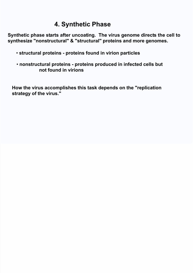
4. Synthetic Phase
Synthetic phase starts after uncoating. The virus genome directs the cell tosynthesize "nonstructural" & "structural" proteins and more genomes.
• structural proteins - proteins found in virion particles
• nonstructural proteins - proteins produced in infected cells but
not found in virions
How the virus accomplishes this task depends on the "replication
strategy of the virus."
8/12/2019 V-L1 Virology Intro- 2013
http://slidepdf.com/reader/full/v-l1-virology-intro-2013 17/26
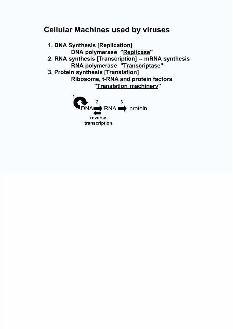
Cellular Machines used by viruses
1. DNA Synthesis [Replication]
DNA polymerase "Replicase"2. RNA synthesis [Transcription] -- mRNA synthesis
RNA polymerase "Transcriptase"3. Protein synthesis [Translation]
Ribosome, t-RNA and protein factors"Translation machinery"
DNA RNA protein reverse
transcription
121 3
8/12/2019 V-L1 Virology Intro- 2013
http://slidepdf.com/reader/full/v-l1-virology-intro-2013 18/26
• During assembly, the virions are formed by packaging the genomes
with capsid proteins; and if the virus is enveloped, wrapping the
nucleocapsid in an envelope.
• Non-enveloped viruses accumulate in the cell and are released whenthe cell dies.
• Enveloped viruses acquire their envelope by budding through a
membrane of the cell (released in same step).
5. Assembly
6. Release

8/12/2019 V-L1 Virology Intro- 2013
http://slidepdf.com/reader/full/v-l1-virology-intro-2013 19/26
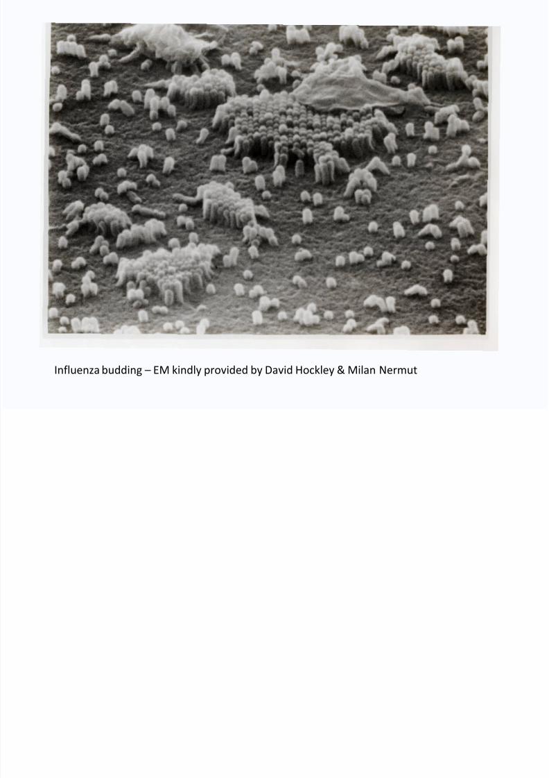
"Budding" is the process by which a virion acquires an envelope
Budding occurs at
plasma membrane
or internally (atGolgi, ER, nuclear
membranes) and is
specific to each
virus
8/12/2019 V-L1 Virology Intro- 2013
http://slidepdf.com/reader/full/v-l1-virology-intro-2013 20/26
Influenza budding – EM kindly provided by David Hockley & Milan Nermut

8/12/2019 V-L1 Virology Intro- 2013
http://slidepdf.com/reader/full/v-l1-virology-intro-2013 21/26
ONE STEP GROWTH CYCLE
V
i r i o n s
( p f u o r p l a q u
e f o r m i n g u n i t s )
106
107
108
109
1010
105
104
103
time (hours)
2 4 6 8 10 12
E C L I S P E
P E R I O D
HYPERBOLIC
GROWTH
A single infected cell can make and
release 10,000 virions within 4-6 hours.
PFU is a plaque f orming unit: one virus can infect a single cell,
spread and kill surrounding cells to cause a plaque (a clear
area of dead cells) surrounded by live cells.

8/12/2019 V-L1 Virology Intro- 2013
http://slidepdf.com/reader/full/v-l1-virology-intro-2013 22/26
Copyright ©2005 by the National Academy of Sciences
Gao, Huajian et al. (2005) Proc. Natl. Acad. Sci. USA 102, 9469-9474
Fig. 1. The life cycle of an animal virus
8/12/2019 V-L1 Virology Intro- 2013
http://slidepdf.com/reader/full/v-l1-virology-intro-2013 23/26
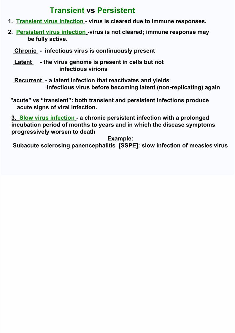
Chronic - infectious virus is continuously present
Latent - the virus genome is present in cells but not
infectious virions
Recurrent - a latent infection that reactivates and yieldsinfectious virus before becoming latent (non-replicating) again
2. Persistent virus infection -virus is not cleared; immune response may
be fully active.
1. Transient virus infection - virus is cleared due to immune responses.
"acute" vs “transient”: both transient and persistent infections produce
acute signs of viral infection.
3. Slow virus infection - a chronic persistent infection with a prolonged
incubation period of months to years and in which the disease symptomsprogressively worsen to death
Example:
Subacute sclerosing panencephalitis [SSPE]: slow infection of measles virus
Transient vs Persistent

8/12/2019 V-L1 Virology Intro- 2013
http://slidepdf.com/reader/full/v-l1-virology-intro-2013 24/26
Virus-specific antibodies:
block receptor binding (neutralizing antibodies)
bind virus and prevent uncoating (entry blocking antibodies, also neutralizing)
bind virus and remove coated virus particles via phagocytosis
may require complement to neutralize certain viruses
Virus-specific T cells:
kill virus infected cells when they recognize virus antigens on cell membranes
kill infected cells that present viral peptides using MHC I
Concepts: Virus-specific antibodies “neutralize” or remove virus particles from blood
or respiratory secretions, while virus-specific T cells eliminate virus infected
cells, preventing persistence of the virus and of disease.
How does the immune system "clear" virus?

8/12/2019 V-L1 Virology Intro- 2013
http://slidepdf.com/reader/full/v-l1-virology-intro-2013 25/26
How do viruses cause disease?
1. Virus kills cells and results in organ failure.
• Example: viral encephalitis. Patient dies less than 1 week post-infection.
2. Immune reaction to virus infection, e.g. cytotoxic T cells destroy tissues and causeorgan failure.
• Example: viral hepatitis. Patient dies more than 1 week after infection
3. Virus infection induces autoimmune disease.
• Example: type 1 diabetes
4. Congenital infection prevents or distorts organ development.
•
Example: congenital rubella5. Virus infection provokes inflammatory state.
• Example: post-infection (vaccination) encephalomyelitis
6. Virus infection induces an immunodeficient state.
• Example: HIV, AIDS, measles
“undifferentiated symptoms of viral infection”
• fever
• myalgia
• malaise
• anorexia
Symptoms occur after the host has responded immunologically
and virus often is no longer present.

8/12/2019 V-L1 Virology Intro- 2013
http://slidepdf.com/reader/full/v-l1-virology-intro-2013 26/26
Questions?