UvA-DARE (Digital Academic Repository) Vascular ... · thee cytosolic leaflet of the membrane to...
13
UvA-DARE is a service provided by the library of the University of Amsterdam (http://dare.uva.nl) UvA-DARE (Digital Academic Repository) Vascular dysfunction in preeclampsia van Wijk, M.J. Link to publication Citation for published version (APA): van Wijk, M. J. (2003). Vascular dysfunction in preeclampsia. General rights It is not permitted to download or to forward/distribute the text or part of it without the consent of the author(s) and/or copyright holder(s), other than for strictly personal, individual use, unless the work is under an open content license (like Creative Commons). Disclaimer/Complaints regulations If you believe that digital publication of certain material infringes any of your rights or (privacy) interests, please let the Library know, stating your reasons. In case of a legitimate complaint, the Library will make the material inaccessible and/or remove it from the website. Please Ask the Library: https://uba.uva.nl/en/contact, or a letter to: Library of the University of Amsterdam, Secretariat, Singel 425, 1012 WP Amsterdam, The Netherlands. You will be contacted as soon as possible. Download date: 16 Aug 2019
Transcript of UvA-DARE (Digital Academic Repository) Vascular ... · thee cytosolic leaflet of the membrane to...

UvA-DARE is a service provided by the library of the University of Amsterdam (http://dare.uva.nl)
UvA-DARE (Digital Academic Repository)
Vascular dysfunction in preeclampsia
van Wijk, M.J.
Link to publication
Citation for published version (APA):van Wijk, M. J. (2003). Vascular dysfunction in preeclampsia.
General rightsIt is not permitted to download or to forward/distribute the text or part of it without the consent of the author(s) and/or copyright holder(s),other than for strictly personal, individual use, unless the work is under an open content license (like Creative Commons).
Disclaimer/Complaints regulationsIf you believe that digital publication of certain material infringes any of your rights or (privacy) interests, please let the Library know, statingyour reasons. In case of a legitimate complaint, the Library will make the material inaccessible and/or remove it from the website. Please Askthe Library: https://uba.uva.nl/en/contact, or a letter to: Library of the University of Amsterdam, Secretariat, Singel 425, 1012 WP Amsterdam,The Netherlands. You will be contacted as soon as possible.
Download date: 16 Aug 2019

Chapterr 5
Microparticlee subpopulations are increased
inn preeclampsia: possible involvement in
vascularr dysfunction?
Marjaa J. Van Wijk, Rienk Nieuwland, Kees Boer, Joris A.M. van der Post,
Edd VanBavel, Augueste Sturk.
AmericanAmerican Journal of Obstetrics and Gynecology 2002;187:450-6.

Chapterr 5
Abstract t
Objective:: The purpose of this study was to investigate the cellular origin and numbers of circulatingg microparticles in normal pregnancy and preeclampsia. Methods:: Plasma samples from 10 women with preeclampsia, from 10 normal pregnant women, andd from 10 nonpregnant women, matched for age and gestation, were analyzed by flow cytometry. . Results:: The total number of circulating microparticles was unaltered in pregnancy and preeclampsia.. The largest portion of microparticles was derived from platelets in all groups. T-suppressorr cell microparticle numbers were decreased in normal pregnancy (P =.04). In preeclampsiaa T-suppressor, T-helper cell, and granulocyte microparticle numbers were increased (PP =.008, P =.008 and P =.03). Elastase concentration were increased in preeclampsia (P =.02) andd correlated with granulocyte microparticle numbers (P =.006). Elastase concentrations correlatedd with systolic and diastolic blood pressure (P =.001 and .003), and granulocyte microparticlee numbers correlated with systolic blood pressure (P =.05). Conclusions:: Numbers of T-cell and granulocyte microparticles are increased in preeclampsia. Whetherr these altered microparticle numbers cause vascular dysfunction in preeclampsia or are a consequencee of the disease remains to be established.
- 6 2--

Circulatingg microparticles
Introduction n
Microparticless are small membrane fragments that are released from cells during cell activationn and apoptosis. Many different cell types (such as endothelial cells, thrombocytes, leukocytes,, erythrocytes and vascular smooth muscle cells) release microparticles in vitro: the presencee of these microparticles in vivo has been demonstrated recently in various studies. Microparticlee formation is altered in several disease states. A reduced ability to generate microparticless occurs in Scott syndrome and Castaman's defect, both of which are bleeding disorderss [253]. Increased numbers of platelet microparticles have been found in patients who weree at increased risk for thromboembolic complications, such as patients with increased coagulationn activation, idiopathic thrombocytopenia, ischemic brain diseases, acute coronary syndromes,, multiple sclerosis [253], and sepsis [210,211].
Thee characteristics of microparticles seem to depend on the activation status of the cells from whichh they originate and on the mechanism of stimulation [154]. Consequently, they vary in size, structure,, antigen- and adhesion molecule-expression. The functional importance of microparticless in vivo, however, remains to be defined. Microparticles from different cell types havee different effects on vascular cells and on blood cells in vitro. Platelet microparticles induce COX-22 expression, PGI2 [157] and cytokine [199] production in endothelial cells, up-regulate adhesionn molecules on the endothelial surface [199,196], which results in monocyte adherence, causee platelet activation [157], and increase vascular smooth muscle mitogenesis and proliferationn [254]. Leukocyte microparticles also induce up-regulation of adhesion molecules on endotheliall cells and initiate the production of the cytokines IL-6 and IL-8 [198]. Endothelial cell microparticless can activate neutrophils, which results in increased neutrophil adhesion to the endotheliumm [239] and can stimulate coagulation [167]. Furthermore, microparticles that have beenn isolated from patients with acute myocardial infarction can diminish endothelium-dependentt relaxation in isolated arteries of rats [235]. Thus, microparticles act as potent pro-inflammatoryy inducers, which initiate an array of signal transduction pathways and gene expressionn profiles in endothelial cells, thereby affecting endothelial function. Finally, the surface off microparticles forms a strong procoagulant surface [184] because of the redistribution of membranee components during the formation of microparticles, in particular PS, which flips from thee cytosolic leaflet of the membrane to the extracellular leaflet. Therefore, microparticles may, in vivo,vivo, be involved in regulation of both coagulation and vascular function.
Normall pregnancy is associated with extensive anatomic and functional adaptations of the cardiovascularr system to accommodate the demands of pregnancy. In preeclampsia this adaptationn is inadequate [255]. Endothelial cells, leukocytes, and the coagulation system are all activatedd in normal pregnancy, while in preeclampsia this activation seems exaggerated (for revieww see [256]). In theory, microparticles could cause the alterations that occur in pregnancy andd preeclampsia. We therefore determined the numbers and cellular origin of circulating microparticless in pregnancy and preeclampsia. Thus far, the only microparticles that have been studiedd in pregnancy and preeclampsia are STBM, which were reported to be present in the
-63--
Chapterr 5
maternall circulation in pregnancy and to be increased in number in preeclampsia [64]. In vitro, STBMM damaged endothelial cell culture structure [65] and function [66] and activated neutrophils [76].. However, at present no information is available on the presence of microparticles from other cells. .
Methods s
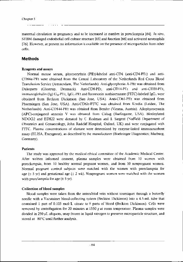
Reagentss and assays Normall mouse serum, phycoerythrin (PEt)-labeled anti-CD4 (anti-CD4-PEt) and anti-
CD66e-PEtt were obtained from the Central Laboratory of the Netherlands Red Cross Blood Transfusionn Service (Amsterdam, The Netherlands). Anti-glycophorin A-PEt was obtained from Dakopattss (Glostrup, Denmark). Anti-CD8-PEt, anti-CD14-PEt and anti-CD20-PEt, immunoglobulinn (Ig) G2b-PEt, IgGrPEt and fluorescein isothiocyanate (FITC)-labeled IgG, were obtainedd from Beckton Dickinson (San Jose, USA). Anti-CD61-PEt was obtained from Pharmingenn (San Jose, USA). Anti-CD62e-FITC was obtained from Kordia (Leiden, The Netherlands).. Anti-CD 144-PEt was obtained from Bender (Vienna, Austria). Allophycocyanin (APC)-conjugatedd annexin V was obtained from Caltag (Burlingame, USA). Biotinylated NDOG22 and ED822 were donated by C. Redman and I. Sargent (Nuffield Department of Obstetricss and Gynaecology, John Radcliff Hospital, Oxford, UK) and were conjugated with FITC.. Plasma concentrations of elastase were determined by enzyme-linked immunosorbent assayy (ELISA, Enzygnost), as described by the manufacturer (Boehringer Diagnostics; Marburg, Germany). .
Patients s Thee study was approved by the medical ethical committee of the Academic Medical Center.
Afterr written informed consent, plasma samples were obtained from 10 women with preeclampsia,, from 10 healthy normal pregnant women, and from 10 nonpregnant women. Normall pregnant control subjects were matched with the women with preeclampsia for agee ( 5 yr) and gestational age ( 2 wk). Nonpregnant women were matched with the women withh preeclampsia for age ( 5 yr).
Collectionn of blood samples Bloodd samples were taken from the antecubital vein without tourniquet through a butterfly
needlee with a Vacutainer blood-collecting system (Beckton Dickinson) into a 4.5-mL tube that containedd 1 part of 0.105 mol/L citrate to 9 parts of blood (Beckton Dickinson). Cells were removedd by centrifugation for 20 minutes at 1550 g at room temperature. Plasma samples were dividedd in 250 uL aliquots, snap frozen in liquid nitrogen to preserve microparticle structure, and storedd at -80°C until further analysis.
- 6 4--

Circulatingg microparticles
Isolationn of microparticles AA 250-uL plasma sample was thawed on ice and then centrifuged for 30 minutes at 17570 g
andd 20°C to pellet the microparticles. The plasma from which the microparticles were isolated wass not defibrinated to avoid a potential loss of platelet-derived microparticles. After centrifugation,, 225 jiL of the supernatant was removed and the microparticle-pellet and remainingg 25 uL of plasma were resuspended with 225 uL citrate-substituted PBS. After another centrifugationn period of 30 minutes at 17570 g and 20°C, again 225 uL of the supernatant was removed.. The microparticle pellet was then resuspended in 75 uL citrated PBS, of which 5 uL wass used per incubation.
Floww cytometric analysis Thee 5 uL of microparticle suspension was diluted in 35 |iL of calcium chloride (2.5 mmol/L)-
containingg PBS and 5 uX of 5000-fold prediluted microparticle-free normal mouse serum. After ann incubation period of 15 minutes at room temperature, 5 uL APC-labeled annexin V was added too all tubes plus 5 uL of one of the labeled cell-specific monoclonal antibodies or isotype-matched controll antibody (IgG,-PEt, except for anti-CD 14-PEt, which has IgG2b-PEt as a control and anti-CD62e-FITCC and NDOG2-FITC, which have IgG,-FITC as a control). The samples were thenn incubated in the dark for 15 minutes at room temperature.
AA panel of cell-specific monoclonal antibodies was used, directed against platelets (CD61), erythrocytess (glycophorin A), endothelial cells (CD62e and CD 144), T-helper (TH) cells (CD4), T-suppressorr (Ts) cells (CD8), monocytes (CD 14), B cells (CD20), granulocytes (CD66e) and syncytiotrophoblastt (NDOG2). Anti-CD62e and anti-CD 144 were used to detect endothelial cell microparticless on the basis of previous observations [210]. NDOG2 and ED822, both antibodies againstt placental alkaline phosphatase, were tested for their ability to identify syncytiotrophoblast microparticles.. Although ED822 bound to artificially prepared STBM, this antibody could not detectt STBM when the samples were diluted more than 50-fold. NDOG2 detected artificially preparedd STBM well (figure 1) also at low STBM concentrations and in plasma samples from patients.. The following final concentrations were used: anti-CD8-PEt (250 ng/mL), anti-CD 14-PEtt (250 ng/mL), anti-CD20-PEt (500 ng/mL), anti-CD62e-FITC (1.67 ug/mL), anti-glycophorinn A-PEt (200 ng/mL), anti-CD 144-PEt (500 ng/mL), IgG,-Pet (500 ng/mL), IgG2b-PEtt (500 ng/mL), IgG,-FITC (500 ng/mL) and annexin V-APC (3.33 umol/ml). For some antibodiess concentrations were not supplied by the supplier. For these antibodies the final dilutionss used were anti-CD4-PEt (1:50), anti-CD61-PE (1:100), anti-CD66e-PEt (1:100) and 1:1000 for the NDOG2-FITC antibody. All antibodies were previously validated in our laboratory onn various templates (patients with sepsis for anti-CD 14, synovial fluid from patients with arthritiss for anti-CD66e and anti-CD20, buffy coat for anti-CD4 and anti-CD8, artificially preparedd syncytiotrophoblast microparticles for NDOG2, and endothelial cell microparticles fromm IL-la-stimulated human umbilical vein endothelial cells for anti-CD62e and anti-CD144). Thee choice of antibody concentrations used in this study was based on previous experience in our laboratoryy [211,186,210].
-65--

Chapterr 5
10L L 1011 10z 10°
NDOG2-binding g \o\oH H
Figuree 1. Histogram presenting the
labelinglabeling of artificially prepared STBM
withwith NDOG2-FITC (grey area) and
withwith control IgG,-FITC (black area).
AllAll events on the right side of the
verticalvertical line are considered NDOG2-
positivepositive microparticles.
Afterr the incubation period, 200 uL of calcium-containing PBS was added to all tubes and the
microparticless were pelleted again by centrifugation for 30 minutes at 17570 g and 20°C. Finally,
2000 U.L of supernatant was removed and the microparticle pellets were resuspended in 300 uL of
calcium-containingg PBS. Samples were analyzed for one minute in a fluorescence automated cell
sorterr flow cytometer with CellQuest software (Beckton Dickinson). Both forward scatter and
sidewardd scatter were set at a logarithmic gain. Microparticles were identified on the basis of their
sizee and density, as previously described [211], and on their capacity to bind annexin V and a
celltype-specificc monoclonal antibody (the latter was used to establish the cellular origin of the
microparticles).. Annexin V measurements were corrected for autofluorescence, and binding of
cell-specificc monoclonal antibodies was corrected with identical concentrations of isotype-
matchedd control antibodies. Microparticles numbers, which were corrected for isotype control
andd autofluorescence, were calculated as the number per liter plasma with the following formula:
NN x (100/5) x (355/60) x (106/250), in which N is the number of events that stained for both
annexinn V and a cell-specific antibody.
Statisticall analysis
Dataa were analyzed in SPSS for Windows, release 10.0.7 (SPSS Inc; Chicago, USA). The
distributionn of data was tested for normality using a Kolmogorov Smirnov 2 test. The results of
thee demographic characteristics of patients were normally distributed and therefore analyzed with
aa ANOVA test with a Bonferroni post hoc test for differences between groups. Because of the
- 6 6--
Circulatingg microparticles
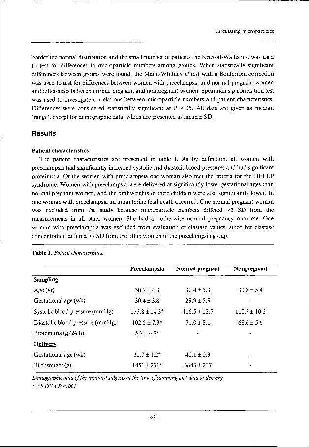
borderlinee normal distribution and the small number of patients the Kruskal-Wallis test was used
too test for differences in microparticle numbers among groups. When statistically significant
differencess between groups were found, the Mann-Whitney U test with a Bonferroni correction
wass used to test for differences between women with preeclampsia and normal pregnant women
andd differences between normal pregnant and nonpregnant women. Spearman's p correlation test
wass used to investigate correlations between microparticle numbers and patient characteristics.
Differencess were considered statistically significant at P <.05. All data are given as median
(range),, except for demographic data, which are presented as mean SD.
Results s
Patientt characteristics Thee patient characteristics are presented in table I. As by definition, all women with
preeclampsiaa had significantly increased systolic and diastolic blood pressures and had significant
proteinuria.. Of the women with preeclampsia one woman also met the criteria for the HELLP
syndrome.. Women with preeclampsia were delivered at significantly lower gestational ages than
normall pregnant women, and the birthweights of their children were also significantly lower. In
onee woman with preeclampsia an intrauterine fetal death occurred. One normal pregnant woman
wass excluded from the study because microparticle numbers differed >3 SD from the
measurementss in all other women. She had an otherwise normal pregnancy outcome. One
womann with preeclampsia was excluded from evaluation of elastase values, since her elastase
concentrationn differed >7 SD from the other women in the preeclampsia group.
Tablee I. Patient characteristics.
Preeclampsiaa Normal pregnant Nonpregnant
Sampling g
Age(yr)) 30.7 3 30.4 + 5.3 30.8 4
Gestationall age (wk) 30.4 8 29.9 9
Systolicc blood pressure (mmHg) * 116.5 12.7 110.7 10.2
Diastolicc blood pressure (mmHg) 102.5 7.3* 71.0 1 68.6 6
Proteinuriaa (g/24 h) 5.7 *
Delivery y
Gestationall age (wk) 31.7 1.2* 40.1 0.3
Birthweightt (g) 1451 * 3643 7
DemographicDemographic data of the included subjects at the time of sampling and data at delivery,
** ANOVA P <.001
-67--
Chapterr 5
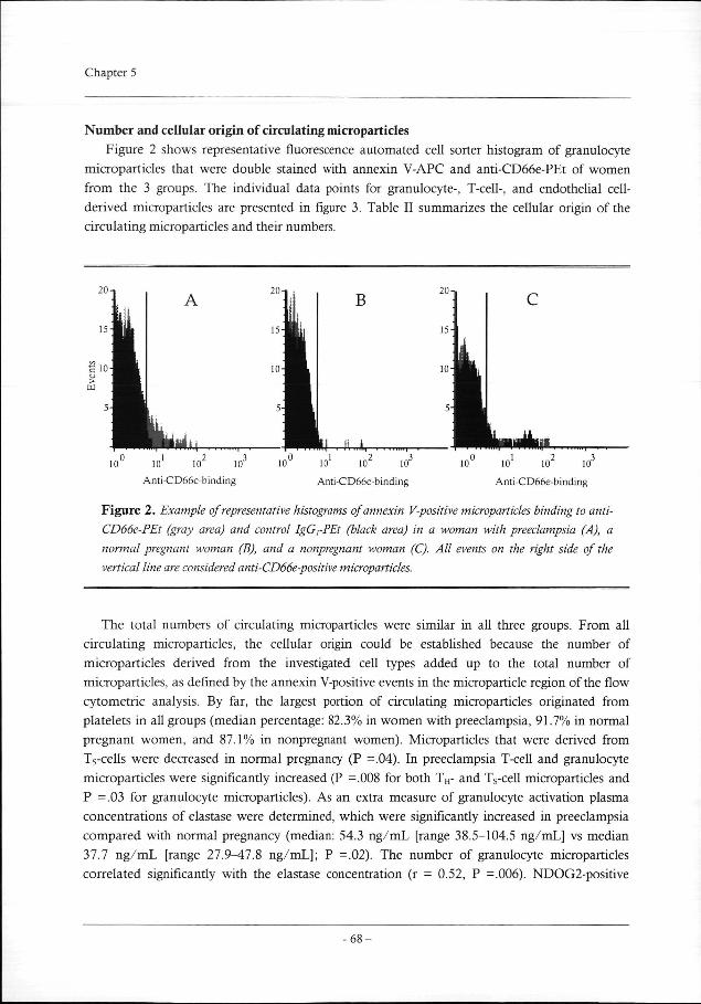
Numberr and cellular origin of circulatin g microparticles
Figuree 2 shows representative fluorescence automated cell sorter histogram of granulocyte
microparticless that were double stained with annexin V-APC and anti-CD66e-PEt of women
fromm the 3 groups. The individual data points for granulocyte-, T-cell-, and endothelial cell-
derivedd microparticles are presented in figure 3. Table II summarizes the cellular origin of the
circulatingg microparticles and their numbers.
Anti-CD66e-binding g Anti-CD66e-binding g
100 10 10 10
Anti-CD66e-binding g
Figuree 2. Example of representative histograms of annexin V-positive microparticles binding to anti-
CD66e-PEtCD66e-PEt (gray area) and control IgG,-PEt (black area) in a woman with preeclampsia (A), a
normalnormal pregnant woman (B), and a nonpregnant woman (C). All events on the right side of the
verticalvertical line are considered anti-CD66e-positive microparticles.
Thee total numbers of circulating microparticles were similar in all three groups. From all
circulatingg microparticles, the cellular origin could be established because the number of
microparticless derived from the investigated cell types added up to the total number of
microparticles,, as defined by the annexin V-positive events in the microparticle region of the flow
cytometricc analysis. By far, the largest portion of circulating microparticles originated from
plateletss in all groups (median percentage: 82.3% in women with preeclampsia, 91.7% in normal
pregnantt women, and 87.1% in nonpregnant women). Microparticles that were derived from
Ts-cellss were decreased in normal pregnancy (P =.04). In preeclampsia T-cell and granulocyte
microparticless were significantly increased (P =.008 for both TH- and Ts-cell microparticles and
PP =.03 for granulocyte microparticles). As an extra measure of granulocyte activation plasma
concentrationss of elastase were determined, which were significantly increased in preeclampsia
comparedd with normal pregnancy (median: 54.3 ng/mL [range 38.5-104.5 ng/mL] vs median
37.77 ng/mL [range 27.9^17.8 ng/mL]; P =.02). The number of granulocyte microparticles
correlatedd significantly with the elastase concentration (r = 0.52, P =.006). NDOG2-positive
- 6 8--

Circulatingg microparticles
microparticless were not only present in the circulation of normal pregnant and preeclamptic
womenn but also in the circulation of 8 of the 10 nonpregnant women, of whom five women had
neverr been pregnant.
Theree was no correlation between the total number of circulating microparticles and either
age,, systolic or diastolic blood pressure, or proteinuria. The number of granulocyte microparticles
correlatedd with systolic blood pressure (r = 0.39, P =.05) and elastase concentrations (r = 0.52, P
=.006),, which also correlated with both systolic and diastolic blood pressure (r = 0.64, P =.001
andd r =0.58, P =.003, respectively).
Tablee II . Cellular origin
Microparticl ee origin
Total l
Platelett (CD61)
Erythrocytee (GlycoA)
Granulocytee (CD66e)
Monocytee (CD 14)
BB cell (CD20)
THH cell (CD4)
Tss cell (CD8)
STBMM (NDOG2)
Endotheliall cell
(CD62e/CD144) )
andand numbers of circulating microparticles.
Preeclampsia a
22566 (994-3839)
1818(638-3561) )
234(19-520) )
799 (0-287)
00 (0-253)
5(0-479) )
299 (0-82)
155 (0-133)
422 (0-89)
233 (0-122)
Normall pregnant
1960(410-3714) )
1618(117-3450) )
1222 (0-256)
88 (0-85)
0(0-18) )
100 (0-30)
00 (0-20)
00 (0-6)
2(0-95) )
00 (0-59)
Nonpregnant t
23577 (1092-3468)
2014(405-3261) )
1500 (39-474)
77 (0-267)
00 (0-48)
33 (0-106)
13(0-258) )
12(0-101) )
844 (0-334)
11(0-75) )
p* *
.62 2
.68 8
.08 8
.05 5
.35 5
.76 6
.01 1
.008 8
.05 5
.14 4
n n
.03 3
.008 8
.008 8
.36 6
n n
.84 4
.11 1
.04 4
.08 8
DataData are presented as median number x Iff1/L plasma (range).
** differences between all groups (Kruskal-Wallis)
%% women with preeclampsia vs normal pregnant women (Mann Whitney U, with Bonferroni correction),
tt normal pregnant vs nonpregnant women (Mann Whitney U, with Bonferroni correction).
-69--

Chapterr 5
_ll 300
oo 250
S33 200
1 1 == 150 — —
100 a a CL L
§§ 50
SS 0
44 .
20 0
00 0
80 0
60 0
40 0
20 0
00 - -fr" "
B B
PEE PR NP PE E PR R NP P
350 0
300 0
xx 250
2000 -1 1 I I JJJ 150 -
100 0
50 0 i i - t --PE E PRR NP
140 0
120 0
100 0
80 0
600 \
40 0
20 0
oi i
D D
PE E PR R NP P
Figuree 3. Individual data points and medians for TH (A), Ts (B), granulocyte (C) and endothelial cell
(D)(D) microparticle numbers in women with preeclampsia (PE), normal pregnant women (PR), and
nonpregnantnonpregnant women (NP).
Comments s
Althoughh the total numbers of circulating microparticles were not significantly altered either
inn normal pregnancy or in preeclampsia, we found that numbers of microparticles of certain
subgroupss of microparticles are different in these conditions. In normal pregnancy, the number of
- 7 0 --

Circulatingg microparticles
Ts-celll microparticles was decreased as compared with the nonpregnant state, while in preeclampsiaa numbers of TH-cell, Ts-cell and granulocyte microparticles were increased compared withh normal pregnancy. The increased numbers of microparticles of these particular microparticle subgroupss in preeclampsia could be a possible mechanism for development of vascular dysfunctionn in preeclampsia and seem to reflect an altered activation status of the immune system ass well as an increased inflammatory response. The increased numbers of lymphocyte microparticless could be released into the maternal circulation from activated lymphocytes, which aree present in increased numbers in the placental tissue during preeclampsia [257]. These lymphocytee microparticles could then cause endothelial damage directly or indirectly by inducing microparticlee formation through activation of other cells, thus creating a vicious circle. Alternatively,, it is possible that cells are activated when passing through the placenta, by an unknownn factor that is released from the placenta. Microparticle formation could then be triggeredd secondarily and result in endothelial dysfunction. In this respect, it is interesting that culturee medium, after incubation with placentas from women with preeclampsia, stimulates neutrophill granulocytes to interact with the endothelium [258]. The presence of activated neutrophilss in preeclampsia has been reported in several studies (review see [256]). The increased granulocytee microparticle and elastase concentrations in this study confirm this. Both these measuress of granulocyte activation also significantly correlated with blood pressure, a measure of diseasee severity. Another mechanism that could support microparticle formation by neutrophils is theirr delayed apoptosis in preeclampsia [259] because this could result in elevated numbers of circulatingg activated neutrophils.
Microparticless derived from T-cells are increased in patients with HIV infection [188], whereas granulocytee microparticles are increased in patients with sepsis and multiple organ dysfunction syndromee [211,210]. Because altered immune and inflammatory responses are involved in these diseases,, the increased T-cell and granulocyte microparticle numbers in preeclampsia provide furtherr evidence for involvement of thee immune and inflammatory system in the pathogenesis of preeclampsiaa [256].
AA central phenomenon in the vascular dysfunction in preeclampsia is endothelial activation. Onee could therefore expect endothelial cell microparticle numbers to be increased in this disease. Thiss was not found in this study, although numbers of endothelial cell microparticles tended to be higherr in preeclampsia (figure 3D). The numbers of endothelial cell microparticles, however, correlatedd with numbers of microparticles that were derived from lymphocytes (r = 0.39, P =.04 forr TH-cells and r = 0.48, P =.008 for Ts-cells), B cells (r = 0.72, P <.001), granulocytes (r = 0.51, PP =.005) and erythrocytes (r = 0.39, P =.04), which indicates a strong relation between the activationn status of all these cell types. Two additional explanations, however, should be considered. .
First,, it is possible that not only the numbers of microparticles are important in the generation off vascular dysfunction in preeclampsia, but also their characteristics. It has been reported that microparticless that result from different stimuli show different characteristics [154]. Thus, their effectt on cells may vary as well. It is therefore possible that a subgroup of microparticles with
- 7 1--

Chapterr 5
certainn characteristics, formed after a specific stimulus (eg, hypoxia, cytokines, or free radicals), couldd result in the development of vascular dysfunction, which is observed in preeclampsia. In thiss respect it is of interest that endothelial cell microparticles, prepared in vitro by stimulation withh TNF were procoagulant [167]. The fact that these microparticles in addition exposed several adhesionn molecules leads us to the second consideration: that not only the circulating microparticlee fraction is important, but also -most likely- the fraction of microparticles that has boundd to cells. It is known that microparticles can indeed bind to cells. For example, platelet microparticless bind to neutrophils in a 150:1 ratio, which results in clustering of the neutrophils [197].. Because adhesion of microparticles to cells might induce the observed activation, the boundd fraction may prove to be important. No data are available yet on the amount of bound microparticles. .
Itt is generally accepted that the placenta plays an important role in the pathogenesis of preeclampsia.. In preeclampsia trophoblast invasion is impaired, which results in the release of an unknownn factor from the placenta into the maternal circulation that causes a generalized vascular dysfunctionn (for review see [255]). Obviously, STBM would have been an excellent candidate for thiss unknown factor, especially because increased apoptosis of trophoblast in preeclampsia has beenn reported [260]. We used two antibodies to identify STBM that have previously been used to detectt and quantify STBM in normal pregnancy and in preeclampsia. Both antibodies bound to artificiallyy prepared STBM. However, ED822 was unable to detect STBM at low concentrations. InIn addition, NDOG2 not only bound to a subpopulation of microparticles in normal pregnant womenn and women with preeclampsia, but also in 8 of the 10 nonpregnant women, 5 of whom hadd never been pregnant before. In addition, NDOG2 also bound to microparticles that had been isolatedd from some healthy men (data not shown). Therefore, NDOG2 seems to be non specific forr STBM. Thus, we were unable to identify STBM in the maternal circulation in our study. However,, because the cellular origin of virtually all microparticles could be established, the fractionn of STBM can, at most, be a modest one, in respect to the total number of circulating microparticles.. Nevertheless, we can not exclude the possibility that very low numbers of STBM circulatee in preeclampsia, which may have a high capacity to damage the vascular endothelium orr to activate neutrophils [76].
Inn conclusion, our results show increased numbers of T-cell and granulocyte microparticles in preeclampsia.. It remains to be established whether these microparticles are involved in the pathogenesiss of preeclampsia. It is important to identify the mechanisms leading to vascular dysfunctionn in preeclampsia, since this could provide us with new therapeutical options and ultimatelyy with methods to prevent the disease.
Wee thank Renée Baak-Pablo, Marianne Schaap and Krista Reijgwart for their excellent laboratoryy work.
- 7 2 --