UvA-DARE (Digital Academic Repository) Chromosome ... · semanti cc discussion is an accurate...
19
UvA-DARE is a service provided by the library of the University of Amsterdam (http://dare.uva.nl) UvA-DARE (Digital Academic Repository) Chromosome abnormalities in first-trimester pregnancy loss Goddijn, M. Link to publication Citation for published version (APA): Goddijn, M. (2003). Chromosome abnormalities in first-trimester pregnancy loss Amsterdam General rights It is not permitted to download or to forward/distribute the text or part of it without the consent of the author(s) and/or copyright holder(s), other than for strictly personal, individual use, unless the work is under an open content license (like Creative Commons). Disclaimer/Complaints regulations If you believe that digital publication of certain material infringes any of your rights or (privacy) interests, please let the Library know, stating your reasons. In case of a legitimate complaint, the Library will make the material inaccessible and/or remove it from the website. Please Ask the Library: http://uba.uva.nl/en/contact, or a letter to: Library of the University of Amsterdam, Secretariat, Singel 425, 1012 WP Amsterdam, The Netherlands. You will be contacted as soon as possible. Download date: 26 Jun 2018
Transcript of UvA-DARE (Digital Academic Repository) Chromosome ... · semanti cc discussion is an accurate...

UvA-DARE is a service provided by the library of the University of Amsterdam (http://dare.uva.nl)
UvA-DARE (Digital Academic Repository)
Chromosome abnormalities in first-trimester pregnancy loss
Goddijn, M.
Link to publication
Citation for published version (APA):Goddijn, M. (2003). Chromosome abnormalities in first-trimester pregnancy loss Amsterdam
General rightsIt is not permitted to download or to forward/distribute the text or part of it without the consent of the author(s) and/or copyright holder(s),other than for strictly personal, individual use, unless the work is under an open content license (like Creative Commons).
Disclaimer/Complaints regulationsIf you believe that digital publication of certain material infringes any of your rights or (privacy) interests, please let the Library know, statingyour reasons. In case of a legitimate complaint, the Library will make the material inaccessible and/or remove it from the website. Please Askthe Library: http://uba.uva.nl/en/contact, or a letter to: Library of the University of Amsterdam, Secretariat, Singel 425, 1012 WP Amsterdam,The Netherlands. You will be contacted as soon as possible.
Download date: 26 Jun 2018
Geneti cc aspect s of miscarriag e
MM Goddijn1 and NJ Leschot2
11 Center for Reproductive Medicine, Department of Obstetrics and Gynecology,
Academicc Medical Center, Amsterdam
22 Department of Clinical Genetics, Academic Medical Center, Amsterdam.
Baillièress Best Practice and Research. Clinical Obstetrics and Gynaecology 2000;14:855-65

Abstrac t t
Fetall chromosome abnormalities account for about 50% of first trimester pregnancy losses.
Mostt of these abnormalities are numerical abnormalities (86%) and a low percentage is
causedd by structural abnormalities (6%) or other genetic mechanisms, including chromosome
mosaicismm (8%). The recurrence risk of numerical abnormalities is low, so karyotyping of
fetall material in case of a miscarriage does not seem worthwhile in daily practice.
Halff of the structural abnormalities may be inherited from a parent carrying a balanced
chromosomee translocation orinversion. Parental carriership is found in 4-6% of the couples
withh recurrent miscarriage. In case of parental carriership of a balanced structural
chromosomee abnormality, a next pregnancy may result in a child with an unbalanced
structurall chromosome abnormality. This child can have multiple congenital malformations
and/orr a mental handicap. Prenatal diagnosis is therefore recommended.
Conventionall laboratory techniques, such as tissue culturing and karyotyping, or (semi-)
directt chromosome technique of chorionic villi, and the recently developed laboratory
techniquess such as fluorescence in situ hybridization (FISH) and comparative genomic
hybridizationn (CGH) are described successively.
Untill now, not enough evidence has been available about the role of other genetic
mechanisms,, such as single-gene abnormalities, uniparental disomy, genomic imprinting,
multifactoriall disorders and skewed X chromosome inactivation, in the occurrence of
miscarriages. .

Introductio n n
Thee prevalence of miscarriages has been estimated to be between 10 and 15% of all clinically
recognizedd pregnancies, with the majority of these occurring in the first trimester of
pregnancy.. Fetal chromosome abnormalities account for about 50% of first-trimester
pregnancyy losses. Pregnancy loss of chromosomal origin is uncommon after is weeks of
gestation1.. Therefore, this chapteris concerned mainly with first-trimester miscarriages.
Thee World Health Organization's definition of miscarriage is: the expulsion or extraction from
itss mother of an embryo orfetus weighing 500 g orless. The weight criterion corresponds
withh a gestational age of roughly 20-Z2 weeks2. Many synonyms are used for the terms
'miscarriage'' and 'recurrent miscarriage'. In this chapter we use the term 'miscarriage' to
meann spontaneous abortion or first trimester miscarriage, and the term 'recurrent
miscarriage'' to mean repeated, recurrent, multiple or habitual abortion. It is probable that the
differentt synonyms do not reflect different clinical entities, so that terms such as 'recurrent
miscarriage'' and 'habitual abortion' can be used interchangeably. More important than a
semanticc discussion is an accurate description of gestational age and number of miscarriages
inn studies concerning miscarriage or recurrent miscarriage.
Inn this chapter we present what is currently known about the laboratory techniques,
prevalencee and causes of chromosome abnormalities of miscarriages and recurrent
miscarriage.. The following mechanisms will be considered: cytogenetic abnormalities,
i.e.. numerical abnormalities, structural abnormalities and mosaicism, single-gene
abnormalitiess and other genetic mechanisms, such as multifactorial disorders and skewed
XX chromosome inactivation. Furthermore, we will discuss the clinical implications of each
geneticc mechanism.
Cytogeneti cc abnormalitie s
Cytogeneticc abnormalities can be subdivided into numerical chromosome abnormalities,
structurall chromosome abnormalities and other mechanisms, such as mosaicism.
Conventiall cytogenetic analysis of aborted fetal material depends on tissue culturing
andd karyotyping or the use of a (semi-)direct chromosome technique on chorionic villi.
Thee technique of tissue culturing is laborious and subject to problems such as external

contamination,, culture failure and selective growth of maternal cells3. A possible
disadvantagee of the (semi-)direct preparation is the discrepancy that may occur between
embryonicc cells and chorionic villi. Such a discrepancy might be due to the factthat
mosaicismm is found only in placental tissue, i.e. confined placental mosaicism. Thus, the fetal
karyotypee may not be represented correctly by the villous karyotype. The estimated
percentagee of mosaicism is 1-2% for (semi-)direct chromosome preparations in chorionic
villuss samplings (CVS)45.
Cytogeneticc analysis of fetal tissue is expensive. Formerly, it was thought that histological
featuress of miscarriages could predict karyotype, and could be a possible alternative to
karyotyping.. Examples of such histological features are: villus contour, hydropic villi,
trophoblasticc hyperplasia, trophoblastic lacunae, cisterns, inclusions, perivillous and
intervillouss fibrin deposits. So far, hydropic villi and trophoblastic lacunae showed a
significantt association with triploidy in one study6, and trophoblastic hyperplasia, cisterns
andd inclusions with triploidy in another study7. No histological features were significantly
associatedd with other chromosome abnormalities6"9. In general, histological features are
inconvenientt for predicting karyotype. The presence of a cytogenetic abnormality in
miscarriagess explains the reason for the loss10. Some authors favour routine karyotyping of
fetall material in miscarriages311. We think that this is unnecessary because, in women who
havee had only one miscarriage, the recurrence risk of another miscarriage is not, or only
slightly,, elevated (16%) when compared to the initial risk of all women (10-15%)12,13.
AA study which pooled data of 5318 miscarriages appeared in 198714. tt combined the data of
fourlargee studies1518. The overall percentage of chromosome abnormalities was reported to
bee 51%. The chromosome abnormalities were subdivided in numerical chromosome
abnormalitiess (96%), structural chromosome abnormalities (3%) and other chromosome
abnormalitiess (1%). As tissue sampling, culture technique and direct preparation of chorionic
villii have improved since then, we have pooled the data of more recent chromosome studies.
Tablee 1 gives an overview of the reported frequency of chromosome abnormalities among 11
seriess of single miscarriages (data from 4696 spontaneous miscarriages)3'19"28. These data
differr only slightly from the percentages found in the earlier studies. An overall percentage
off 4g% chromosome abnormalities was calculated, and chromosome abnormalities were
subdividedd into 86% numerical abnormalities, 6% structural chromosome abnormalities and
8%% other chromosome abnormalities. The most remarkable finding is a higher incidence of
otherr chromosome abnormalities, such as mosaicism, double and triple trisomies and
miscellaneouss chromosome abnormalities.

Itt is suggested that most of the chromosome abnormalities resultin disordered development
incompatiblee with prolonged intrauterine survival and live birth. The mechanism by which a
chromosomee abnormality could lead to regression of the conceptus is unclear.
Newerr techniques which can be used in detecting chromosome abnormalities of miscarriages,
andd which offer a possible additional role to conventional laboratory techniques are
fluorescencee in situ hybridization (FISH) and comparative genomic hybridization (CGH).
Untill now there have been only a few published studies concerning the use of these new
techniquess in relation to miscarriages. One study used FISH in pre-implantation embryos
inn patients with unexplained recurrent miscarriages, and found that 16/39 (41%) of the
pre-implantationn embryos were aneuploid29. The authors suggest that pre-implantation
diagnosiss could be a feasible method for improving the chance of a successful pregnancy.
Inn another study, the percentage of chromosome abnormalities detected by FISH in these
pre-implantationn embryos proved to be higherin the group with unexplained recurrent
miscarriagess (35/66=53%) when compared to a control group (l2/62=ig%)30.
Comparativee genomic hybridization (CGH) is a molecular-cytogenetic assay capable
off detecting chromosomal gains and losses by FISH. It provides a whole-genome screen
forr unbalanced aberrations, and can detect the origin of extra or missing chromosomal
material31.. CGH has been shown to detect chromosome abnormalities in 50% of the aborted
fetall samples, as compared with 42% abnormality displayed by culture of chorionic villi
(n=i2)32. .
Neww insights can be expected by the use of DNA microarrays. DNA microarrays consist
off short pieces of DNA, from 20 to over 1000 nucleotides in length. By using these
microarrays,, every desired DNA sequence can be stained, and microdeletion syndromes or
duplicationss can be diagnosed33. No information is yet available about the use of microarrays
inn miscarriages
NumericalNumerical abnormalities
Numericall abnormalities can be subdivided in aneuploidy (trisomiesand monosomies) and
polyploidy.. Trisomies occur most frequently (52%), followed by polyploidy (21%) and
monosomyy X (13%) (see Table 1).

|2 2
c oo c n u-i
e nn r - OO CO
mm at
58 8
cnn m —'
r*-- m ro coo ^ ro
r-jj L O <-0 i_n
E l --
- ^^ LD CO
c oo r v rsj oo m c o P-)) — —
OO cn rv
r\jj m LD L HH rvj ^-i
COO LD * T
LDD O .—
OO CO CD o fNJ
t DD c n I"* - CD ,—i
^ rr c n r ~ us **t
LDD —i cn co ^-
—— ^ E E ^" ii o C-5 o -Q
EE -5. S £ £
?? 1 1 1 33 O. OJ — uu - D -D O
55 T , H o
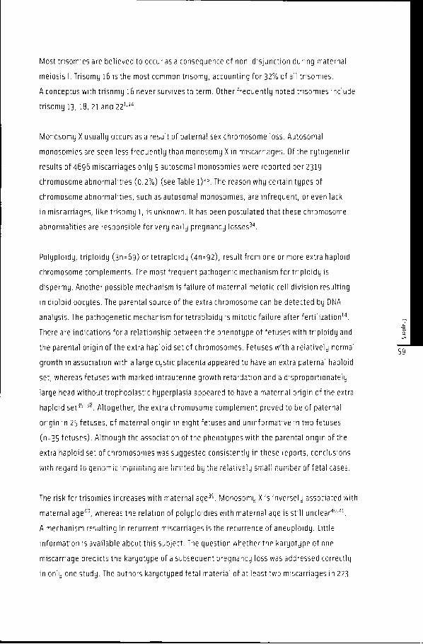
Mostt trisomies are believed to occuras a consequence of non-disjunction during maternal
meiosiss I. Trisomy 16 is the most common trisomy, accounting for 32% of all trisomies.
AA conceptus with trisomy 16 never survives to term. Other frequently noted trisomies include
trisomyy 13, 18, 21 and 22 U 4 .
Monosomyy X usually occurs as a result of paternal sex chromosome loss. Autosomal
monosomiess are seen less frequently than monosomy X in miscarriages. Of the cytogenetic
resultss of 4696 miscarriages only 5 autosomal monosomies were reported per 2319
chromosomee abnormalities (0.2%) (see Table l )2 5 . The reason why certain types of
chromosomee abnormalities, such as autosomal monosomies, are infrequent, or even lack
inn miscarriages, like trisomy 1, is unknown. It has been postulated thatthese chromosome
abnormalitiess are responsible for very early pregnancy losses34.
Polyploidy,, triploidy (3n = 6g) or tetraploidy (4n=92), result from one or more extra haploid
chromosomee complements. The most frequent pathogenic mechanism for triploidy is
dispermy.. Another possible mechanism is failure of maternal meiotic cell division resulting
inn diploid oocytes. The parental source of the extra chromosome can be detected by DNA
analysis.. The pathogenetic mechanism for tetraploidy is mitotic failure after fertilization14.
Theree are indications for a relationship between the phenotype of fetuses with triploidy and
thee parental origin of the extra haploid set of chromosomes. Fetuses with a relatively normal
growthh in association with a large cystic placenta appeared to have an extra paternal haploid
set,, whereas fetuses with marked intrauterine growth retardation and a disproportionately
largee head without trophoblastic hyperplasia appeared to have a maternal origin of the extra
haploidd set3538. Altogether, the extra chromosome complement proved to be of paternal
originn in 25 fetuses, of maternal origin in eight fetuses and uninformative in two fetuses
(n=355 fetuses). Although the association of the phenotypes with the parental origin of the
extraa haploid set of chromosomes was suggested consistently in these reports, conclusions
withh regaru to genomic imprinting are iimiteu by the reiativeiy sman number ui 1 eta 1 cases.
Thee risk fortrisomiesincreases with maternal age39. Monosomy X is inversely associated with
maternall age40, whereas the relation of polyploidies with maternal age is still unclear4041.
AA mechanism resulting in recurrent miscarriages is the recurrence of aneuploidy. Little
informationn is available about this subject. The question whetherthe karyotype of one
miscarriagee predicts the karyotype of a subsequent pregnancy loss was addressed correctly
inn only one study. The authors karyotyped fetal material of at least two miscarriages in 273

women.. The recurrence risk of a chromosome abnormality was not increased when there has
beenn a trisomy, and probably slightly increased when there has been a non-trisomic
abnormalityy (odds ratio 2.0 [95% confidence interval 1.0-4.0])42. In anotherstudy, prenatal
diagnosis,, amniocentesis or chorionic villus sampling was offered to women with recurrent
miscarriagess (without carriership) and a controlgroup. It was reported that the rate of
aneuploidd conceptions was significantly higher in women with recurrent miscarriages
(5/611 = 1.6%) when compared to controls (3/979=0.3%, P=0.02)43.
Inn our view, prenatal diagnosis is not justified when there has been a chromosome
abnormalityy in fetal material, because of the low recurrence risk of chromosome
abnormalities.. Prenatal diagnosis is also not indicated in recurrent miscarriages without
parentall carriership of a balanced abnormality.
StructuralStructural chromosome abnormalities
Structurall chromosome abnormalities can be subdivided into deletions, translocations,
inversionss and duplications, but only translocations and inversions play a role in miscarriage
andd recurrent miscarriage44'45.
Structurall chromosome abnormalities occurin 6% of chromosomally abnormal abortuses
(seee Table l ) . About half of these abnormalities may arise 'de novo' during gametogenesis,
andd the other half may be inherited from a parent carrying a 'balanced' translocation or an
inversion14. .
Thee latter has important implications and will be discussed here. In ïggo, a review was
publishedd based on 200 cytogenetic studies in 22199 couples experiencing repeated
pregnancyy loss. Overall, 5% of the couples with two or more spontaneous miscarriages
includedd a carrier individual (reciprocal and Robertsonian translocations and inversions).
Carriershipp of a balanced structural chromosome abnormalities was at least 10 times more
frequentt in couples with recurrent miscarriages when compared to the general population
(0.34%).. The distribution of chromosome abnormalities according to the number of
miscarriagess ( l , 2, >3) did not show any increase in the frequency of the inversions, sex
chromosomee aneuploidies and supernumerary chromosomes when the number of
miscarriagess increased, whereas there was a correlation between the incidence of
Robertsoniann and reciprocal translocations and the number of miscarriages (P<0.05).
Women,, rather than men, appeared to be more likely carriers of a translocation (reciprocal
orr Robertsonian), an inversion or a supernumerary chromosome ( P<0.05)4'.

Resultss of more recent parental chromosome studies report a comparable percentage of
carriership:: respectively 8% in 639 couples, 2% in 241 couples and 5% in 1743 couples46"48.
Thee overall percentage of the preceding three studies is 6% carriership in 2623 couples.
AA high incidence of cytogenetic abnormalities was found in couples with miscarriage and
aa normal child (7 and 18% respectively)49,50. The prevalence of carriership of a balanced
structurall chromosome abnormality in recurrent miscarriage is now well established.
Thee impact of carriership on fetal outcome has been studied less frequently. Only one study
wass found to report the outcome of prenatal diagnosis for pregnancies of reciprocal carriers
withh recurrent miscarriages. When the carrier was maternal 10/209 (5%) unbalanced
conceptionss were found, and when the carrier was paternal 2/139 (l%) unbalanced
conceptionss were found51.
Whenn one of the parents is a carrier of a balanced structural chromosome abnormality,
aa pregnancy can result in three types of offspring: a child with a normal chromosome pattern,
aa child with a balanced structural chromosome abnormality, or a conceptus with an
unbalancedd structural chromosome abnormality. The latter case will lead to either
aa spontaneous miscarriage or to a liveborn child with multiple congenital malformations
and/orr mental handicaps (5-10%).
Thee greater'the chromosomal unbalance', the higherthe chance of a miscarriage.
Standardd studies (blood lymphocyte karyotyping with G and Q banding) should be used as
routinee screening tests when there have been recurrent miscarriages. FISH can be considered
whenn a specific defect is suggested by routine tests52.
Untill now a genetic evaluation (parental karyotypes) is generally recommended after two
orthreee miscarriages. A cost-effectiveness study to evaluate whether karyotyping should
bee carried out after two orthree miscarriages is needed.
Whenn parental karyotyping is performed, and a translocation or inversion is found, a strong
indicationn for prenatal diagnosis in a subsequent pregnancy exists, because of the above-
mentionedd chance for a child with multiple congenital malformations and/or a mental
handicap. .
ChromosomeChromosome mosaicism
Inn mosaicism, two or more different genetic cell lines are present in an individual. Depending
onn the timing of the mutational event, i.e. prior or after the differentiation of embryonic and

chorionicc compartments, the mosaicism may be found in the placenta and embryo oronly in
onee of them. Confined placental mosaicism has been mentioned earlierasthe main source of
false-positivee results of viable pregnancies at CVS ( l-2%)4 5. Different types of mosaicism
cann play a role in fetal loss. Among them are chromosome abnormalities limited to the
placentaa with complete dichotomy between placenta and fetus53. This type of mosaicism
wass found in 2/141 abortuses (l%)54. Another type is mosaicism confined to the placenta,
withh both cell lines represented in the placenta. This type was found in respectively 10/54
abortusess (19%), while another pattern of mosaicism, confined to the embryo was found in
1/544 abortuses (2%)55.
Thee majority of mosaic miscarriages thus represent a confined placental mosaicism.
AA positive correlation between placental mosaicism, diagnosed by CVS, and fetal cell death
hass been given in one other study55. On the other hand, mosaicism can be a survival
mechanismm as well. It is described in advanced trisomy 13 and 18 gestations that have
aa normal cell line confined to the placenta. The normality of part of the placental cells
iss supposed to be a mechanism for gestational survival".
Mosaicismm originating from a trisomic zygote can result in uniparental disomy. If the cells
thatt eventually will form the fetus itself loose the extra chromosome, there is a 1:3 chance
thatt both remaining homologue chromosomes are derived from one parent only. This is
calledd uniparental disomy5859.
Forr some genes, only one of the genes (maternal or paternal allele) is expressed in certain
cells.. This phenomenon is known as genomic imprinting. If uniparental disomy occursin
chromosomess of which parts are known to be imprinted or inactivated, abnormal phenotypic
effectss are to be expected. Lethal effects have been reported in mice, but an effect on fetal
celll death, resulting in miscarriages in women, 15 still unclear59.
Singl ee gene abnormalitie s
Singlee gene disorders associated with recurrent miscarriage are myotonic dystrophy, lethal
skeletall dysplasias, such as thanatoporic dysplasia and type II osteogenesis imperfecta60"62.
Myotonicc dystrophy, an autosomal dominant disease is characterized at the molecular level,
itss gene localised at chromosome I9ql3-3. It is one of the 'trinucleotide repeat diseases'.
Thee phenomenon of anticipation which occursin myotonic dystrophy is the reason why
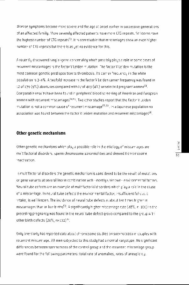
diseasee symptoms become more severe and the age at onset earlier in successive generations
off an affected family. More severely affected patients have more CTG repeats. Stillborns have
thee highest number of CTG repeats59. It is conceivable that miscarriages show an even higher
numberr of CTG repeats but there is as yet no evidence for this.
AA recently discovered single-gene abnormality which possibly plays a role in some cases of
recurrentt miscarriages is the factor V Leiden mutation. The factor V Leiden mutation is the
mostt common genetic predisposition to thrombosis. Its carrierfrequency in the white
populationn is 3-4%. A twofold increase in the factor V Leiden carrierfrequency was found in
122 of 139 (9%) abortuses compared with 1? of 403 (4%) unselected pregnant women53.
Comparablee results have been found in peripheral blood screening of American and European
womenn with recurrent miscarriages64,65. Two other studies report that the factor V Leiden
mutationn is not a common cause of recurrent miscarriage66'57. In a Japanese population no
associationn was found between the factorV Leiden mutation and recurrent miscarriages68.
Otherr geneti c mechanism s
Otherr genetic mechanisms which play a possible role in the etiology of miscarriages are
multifactoriall disorders, sperm chromosome abnormalities and skewed X chromosome
inactivation. .
Inn multifactorial disorders the genetic mechanism is considered to be the result of mutations
orr gene variants at several loci in combination with -mostly unknown- environmental factors.
Neurall tube defects are an example of multifactorial disorders which play a role in the cause
off a miscarriage. In neural tube defects the environmental factor, insufficient folic acid
intake,, is well known. The incidence of neural tube defects is about ten times higher in
miscarriagess than in live births69. A significantly higher miscarriage rate (48%, n = 100) in the
precedingg pregnancy was found in the neural tube defect group compared to the group with
otherr birth defects (20%, n = lOO)70.
Onlyy one study has reported data about chromosome studies on spermatozoa in couples with
recurrentt miscarriage. All men subjected to this study had a normal karyotype. No significant
differencess between spermatozoa of the control group and the recurrent miscarriage group
weree found for the following parameters: total rate of anomalies, rates of aneuploidy,

hypohaploidyy and hyperhaploidy and total rate of structural anomalies. However, there was
aa significant difference in the occurrence of chromosome breaks (control group: n = i0/4 i3
(2%);; recurrent miscarriage group: n=l8/308 (6%) and acentric fragments (control group:
n=lO/4l33 (2%); recurrent miscarriage group: n=25/308 (8%)) of spermatozoa71.
Anotherr genetic mechanism, possibly related to the occurrence of miscarriages, is skewed X
chromosomee inactivation. X chromosome inactivation isthe preferential use of eitherthe
paternall or maternal X chromosome. The inactivation is considered to be a random process
duringg early embryonic development. As a consequence, the maternal X chromosome is
inactivatedd approximately as often as the paternal X chromosome. Skewed X chromosome
inactivationn is defined as preferential use of one, maternal or paternal, X chromosome in
>90%% of peripheral leukocytes. Skewed X chromosome inactivation has been found more
oftenn in recurrent miscarriages when compared to controls (7/48 = 14.6% vs 1/68 = 1.5%,
P<0.01)) and (14/76=18% vs 6/111 = 5%, P<O.OOl)72'73. More evidence is needed to establish
thiss genetic mechanism.
Conclusion ss and recommendation s
Thee frequency and type of cytogenetic abnormalities in miscarriages is well established.
Afterr a single miscarriage, genetic evaluation -karyotyping of the fetal material- seems not
worthwhilee because of the low recurrence risk of another miscarriage with chromosome
abnormalitiess and the high chance of a liveborn child in a next pregnancy.
Parentall carriership is found in 4-6% of the couples with recurrent miscarriages. Although
thiss is a relatively low percentage, it is important to recognize because it can eventually
resultt in a seriously handicapped child. In case of parental carriership prenatal diagnosis is
recommendedd in the next pregnancy. Whether a couple should be karyotyped after two or
threee miscarriages needs to be evaluated by a cost-effectiveness study. Clinical
recommendationss are summarized in Figure 1.

Figuree l Clinica l recommendation s
AA singl e miscarriag e
Twoo or more miscarriage s
noo geneti c evaluatio n
parenta ll karyotypin g
prenata ll diagnosi s in the next pregnancy , in case of
carriershi pp of a balance d chromosom e abnormalit y
Acknowledgmen t t
Thee authors wish to thank F van derVeen, MD, PhD, head of the Centerfor Reproductive
Medicine,, Academic Medical Center, for critically reading the manuscript.
Reference s s
11 Bemrschke K. Current concerns in the genetics of pregnancy losses. Delaware Medical Journal
1990;62:1169-74. 1990;62:1169-74.
22 Stirrat GM. Recurrent miscarriage I: definition and epidemiology. Lancet 1990;336:673-5.
33 Eiben B, Bartels I, Bahr-Porsch 5 et al. Cytogenetic analysis of ?50 spontaneous abortions with the direct
preparationn method of chorionic villi and its implications for studying genetic causes of pregnancy
wastage.. American journal of Human Genetics 1990;47:656-63.
44 Simom G, Sirchia SM. Confined placental mosaicism. Prenatal Diagnosis 1994;14:1185-9.
55 Leschot NJ, Schuring-Blom GH, van Prooijen-Knegt AC et al. The outcome of pregnancies with confined
placentall chromosome mosaicism in cytotrophoblast cells. Prenatal Diagnosis 1996;16:705-12.
66 van Lijnschoten G, Arends JW, Leffers P et al. The value of histomorphological features of chorionic villi
inn early spontaneous abortion for the prediction of karyotype. Histopathology 1993;22:557-63.
II Lorenzato M, Visseaux-Coletto B, Lallemand A et al. Determination of reliable histological features
associatedd with early triplody using DNA image cytometry. Pathology, Research 8 Practice
1995;191:1179-85-1995;191:1179-85-
88 Genest DR, Roberts D, Boyd Tet al. fetoplacental histology as a predictor of karyotype: a controlled study
off spontaneous first trimester abortions. Human Pathology 1995;26:201-9.
99 Fukunaga M, Onda T, Endo Y et al. Is there a correlation between histology and karyotype in early
spontaneouss abortion? Internationa! Journal of Surgical Pathology 1995;2:295-300.
100 Kalousek DK. Clinical significance of morphologic and genetic examination of spontaneously aborted
embryos.. American Journal of Reproductive Immunology 1998; 39:108-19.
I II Wolf GC, Horger EO. Indications for examination of spontaneous abortion specimens: a reassessment.
AmericanAmerican Journal of Obstetrics 5 Gynecology 1995;173:1364-8.
122 Alberman E. The epidemiology of repeated abortion. In: Beard RW, Sharp F (eds) Early Pregnancy Loss:
Mechanismss and Treatment, pp 9-17. London: Springer Verlag 1988:9-17
133 Berry CW, Brambati B, Eskes TKAB et al. The Euro-Team Early Pregnancy (ETEP) protocol for recurrent
miscarriage.. Human Reproduction 1995;10:1516-20.
144 Simpson J L, Bombard A. Chromosomal abnormalities in spontaneous abortion: frequency, pathology and
geneticc counselling. In: Bennett MJ & Edmonds DK(eds) Spontaneous and recurrent abortion, Oxford:
BlackwellBlackwell Scientific Publications 1987:51 -76.
155 Kajii T, Ohama K, Niikawa N et al. Banding analysis of abnormal karyotypes in spontaneous abortion.
AmericanAmerican Journal of Human Genetics 1973;25:539.
166 Boue J, Boue A, Lazar P. Retrospective and prospective epidemiological studies of 1500 karyotyped
spontaneouss human abortions. Teratology 1975;12:11-26.
177 Hassold TJ, Chen M, Funkhouser J et al. A cytogenetic study of 1000 spontaneous abortions. Annals of
HumanHuman Genetics ig80;44:l5l-
188 Warburton D, Stein Z, Kline J et al. Chromosome abnormalities in spontaneous abortion: data from the
Meww York City Study. In: Porter IH 5 Hook EB (eds) Human Embryonic and Fetal Death. Hew York:
AcademicAcademic Press, 1980, p26l.
199 Gue rn e n 5, Be tt 10 D, Si mom G. Prevalence and distribution of chromosome abnormalities in a sample of
firstt trimester internal abortions. Human Reproduction 1987;2:735-9.
200 Minguillon C, Eiben B, Bahr-Porsch S et al. The predictive value of chorionic villus histology for
identifyingg chromosomally normal and abnormal spontaneous abortions. Human Genetics 1989;82:373-6.
211 Rehder H, Coerdt W, Eggers R et al. Is there a correlation between morphological and cytogenetic findings
inn placental tissue from early missed abortions7 Human Genetics 1989;82:377-85.

222 Ohno M, Maeda T, Matsunobu A. A cytogenetic study of spontaneous abortions with direct analysis of
chorionicc villi. Obstetrics 8 Gynecology 1991;77:394-8.
233 Strom CM, Ginsberg N, Applebaum M et al. Analyses of 95 first-trimester spontaneous abortions by
chorionicc villus sampling and karyotype. Journal of Assisted Reproduction and Genetics 1992;9:458-61.
244 Gardó 5F Bajnoczky K. Cytogenetic analysis of spontaneous abortions with direct analysis of chorionic
villi.. European Journal of Obstetrics 6 Gynecology and Reproductive Biology 1992;47:117-20.
255 DejmekJ, VojtassakJ, Malova J, Cytogenetic analysis of 1508 spontaneous abortions originating from
southh Slovakia. European Journal of Obstetrics 6 Gynecology and Reproductive Biology 1992;46:129-36.
266 Jaumaux E, Hustin J. Histological examination of first trimester spontaneous abortions: the impact of
materno-embryonicc interface features. Histopathology 1992;21:409-14-
2?? Ford JH, Wilkin HZ, Thomas P et al. A 13-year cytogenetic study of spontaneous abortion: clinical
applicationss of testing. Australian 5 New Zealand Journal of Obstetrics and Gynaecology 1996;36:314-8.
288 Brajenovic-Milic B, Petrovic 0, Krasevic M et al. Chromosomal anomalies in abnormal human pregnancies.
FetalFetal Diagnosis and Therapy 1998;13:187-91.
299 Vidal F, Giménez C, Rubio C et al. FISH preimplantation diagnosis of chromosome aneuploidy in recurrent
pregnancyy wastage. Journal of Assisted Reproduction and Genetics 1998;15:310-3-
300 Pellicer A, Rubio C, Vidal F et al. In vitro fertilization plus preimplantation genetic diagnosisin patients
withh recurrent miscarriage: an analysis of chromosome abnormalities in human preimplantation embryos.
FertilityFertility and Sterility 1999;71:1033-9-
311 Bryndorf T, Kirchhoff M, Rose H et al. Comparative genomic hybridization in clinical cytogenetics.
AmericanAmerican Journal of Human Genetics 1995:57:1211-20.
322 Daniely M, Aviram-Goldring A, Barkai G et al. Detection of chromosomal aberration in fetuses arising
fromm recurrent spontaneous abortion by comparative genomic hybridization. Human Reproduction
1998;13:805-9-1998;13:805-9-
333 Friend SH. DNA microarrays and expression profiling in clinical practice. British Medical Journal
1999;319:1306-7. 1999;319:1306-7.
344 Simpson JL. The de Watteville Memorial Lecture: Reproductive technologies and genetic advances in
obstetricss and gynecology. International Journal of Gynecology and Obstetrics 1992;38:26l -80.
355 McFadden DE, Kalousek DK. Two different phenotypes of fetuses with chromosomal triploidy: correlation
withh parental origin of the extra haploid set. American Journal of Medical Genetics 1991;38:535-8.
366 McFadden DE, Kwong LC, Yam IYL et al. Parental origin of triploidy in human fetuses: evidence for
genomicc imprinting. Human Genetics 1993;92:465-9-
377 Dietzsch E, Ramsay M, Christianson AL. Maternal origin of extra haploid set of chromosomes in third
trimesterr triploid fetuses. American Journal of Medical Genetics 1995;58:360-4-
388 Miny P, Koppers B, Dworniczak B et al. Parental origin of the extra haploid chromosome set in tnploidies
diagnosedd prenatally. American Journal of Medical Genetics 1995;57:102-6.
399 Cowchock FS, Gibas Z & Jackson LG. Chromosome errors as a cause of spontaneous abortion: the relative
importancee of maternal age and obstetric history. Fertility and Sterility 1993;59:1011-4-
400 Neuber M, Rehder H, Zuther C et al. Polyploidies in abortion material decrease with maternal age.
HumanHuman Genetics 1993;91:563-6.
411 Snijders RJM, Sebire NJ, Nicolaides KH. Maternal age and gestational age-specific risk for chromosomal
defects.. Fetal Diagnosis and Therapy 1995;10:356-67-
422 Warburton D, Kline J, Stein Z et al. Does the karyotype of a spontaneous abortion predict the karyotype of
aa subsequent abortion? Evidence from 273 women with two karyotyped spontaneous abortions. American
JournalJournal of Human Genetics 1987;41:465-83-
433 Drugan A, Koppitch FC, Williams JC et al. Prenatal genetic diagnosis following recurrent early pregnancy
loss.. Obstetrics 5 Gynecology 1990;75:8l -4.

444 Dewald GW, Michels VV. Recurrent miscarriages: cytogenetic causes and genetic counseling of affected
families.. Clinical Obstetrics and Gynecology 1986;29:865-85.
455 De Braekeleer M, Dao TN. Cytogenetic studies in couples experiencing repeated pregnancy losses.
HumanHuman Reproduction 1990;5:519-28.
466 Makino T, Tabuchi T, Nakada K et al. Chromosomal analysis in Japanese couples with repeated spontaneous
abortions.. International Journal of Fertility 1990,35:266-70.
477 Junge A, Domke N, Tolkendorf E. Zytogenetische untersuchung von penpheren Lymphozytenkulturen bei
Paarenn mit habituellen Aborten. lentral blattfur Gynakologie 1991;113:1046-58.
488 Fryns J-P, van Buggenhout G. Structural chromosome rearrangements in couples with recurrent fetal
wastage.. EuropeanJournal of Obstetrics & Gynecology 1998;81:171-6.
499 Portnoi M-F, Joye N, van den AkkerJ et al. Karyotypes of 1142 couples with recurrent abortion. Obstetrics
andand Gynaecology 1988;72:31-4.
500 Schwartz S, Palmer CG. Chromosomal findings in 164 couples with repeated spontaneous abortions:
withh special consideration to prior reproductive history. Human Genetics 1983;63:28-34.
511 Daniel A, Hook EBr Wulf G. Risks of unbalanced progeny at amniocentesis to carriers of chromosome
rearrangements:: Data from United States and Canadian laboratories. American Journal of Medical
GeneticsGenetics 1989:33:14.
522 Zhao J, Gordon PL, Wilroy RS et al. Characterization of an unbalanced de novo rearrangement by
microsatellitee polymorphism typing and by fluorescent in situ hybridization. American Journal of Medical
GeneticsGenetics 1995:56:398-402.
533 Kalousek DK. Confined placental mosaicism and intrauterine human development. Acta Medica
AuxologicaAuxologica 19Q1;23:201 -8.
544 Qumsiyeh MB. Chromosome abnormalities in the placenta and spontaneous abortions. The Journal of
Maternal-FetalMaternal-Fetal Medicine 1998;7:210-2.
555 Kalousek DK, Barrett IJ, Gartner AB. Spontaneous abortion and confined chromosomal mosaicism. Human
GeneticsGenetics 1992;88:642-6.
555 Wapner RJ, Simpson JL, Golbus MS et al. Chorionic mosaicism: association with fetal loss but not with
adversee perinatal outcome. Prenatal Diagnosis 1992; 12:347'-55-
577 Kalousek DK, Barrett IJ, McGillivray BC. Placental mosaicism and intrauterine survival of trisomies 13
andd 18. American Journal of Human Genetics 1989:44:338-43-
588 Engel E, Dawn DeLozier-Blanchet C. Uniparental disomy, isodisomy, and imprinting: probable effects
inn man and strategies for their detection. American Journal of Medical Genetics 1991;40:432-9-
599 Kotzot D. Abnormal phenotypes in uniparental disomy (UPD): fundamental aspects and a critical review
withh bibliography of UPD other than 15. American Journal of Medical Genetics 1999;82:265-74.
600 Brook JD, McCurrack ME, Harley HG et al. Molecular basis of myotonic dystrophy: Expansion of a
trinucleotidee (CTG) repeat at the 3' end of a transcript encoding a protein kinase family member. Cell
1992;69:799-808. 1992;69:799-808.
611 Byrne JLB, Ward K. Genetic factors in recurrent abortion. Clinical Obstetrics and Gynecology
1994:37:693-704. 1994:37:693-704.
622 SuthersG. Mutations, malformations and mortality. Journal of Paediatrics and Child Health 1996;32:10-5-
633 Dizon-Townson DS, Meline L, Nelson LM et al. Fetal carriers of the factor V Leiden mutation are prone to
miscarriagee and placental infarction. American Journal of Obstetrics 5 Gynecology 1997;177:402-5.
644 Grandone F, Margaglione M, Colaizzo D et al. Factor V Leiden is associated with repeated and recurrent
unexplainedd feta) losses. Thrombosis and Haemostasis 1997;77:822-4-
655 Ridker PM, Miletich JP, Buring J E et al. Factor V Leiden Mutation as a risk factor for recurrent pregnancy
)oss.)oss. Annals of Internal Medicine 1998;128:1000-3.

666 Dizon-Townson DS, Kinney S, Branch DW et al. The factor V Leiden mutation is not a common cause of
recurrentt miscarriage. Journal of Reproductive Immunology 1997;34:217-23.
6?? Coumans ABC, Huijgens PC, Jakobs C et al. Haemostatic and metabolic abnormalities in women with
unexplainedd recurrent abortion. Human Reproduction 1999,14:211-4.
688 Hashimoto K, Shizusawa Y, Shimoya K et al. The factor V Leiden mutation in Japanese couples with
recurrentt spontaneous abortion. Human Reproduction 1999;14:1872-4.
699 McFadden DE, Kalousek D. Survey of neural tube defects in spontaneous aborted embryos. American
JournalJournal of Medical Genetics 1989;32:356-8.
700 Carmi R, GoharJ, Meizner I et al. Spontaneous abortion-high risk factor for neural tube defects in
subsequentt pregnancy. American Journal of Medical Genetics 1994;51:93-7.
711 Rosenbusch B, Sterzik K. Sperm chromosomes and habitual abortion. Fertility and Sterility 1991;56:370-2.
722 Lanasa MC, Hogge WA, Hoffman EP. Sex chromosome genetics '99. The X chromosome and recurrent
spontaneouss abortion: the significance of transmanifesting carriers. American Journal of Human Genetics
1999;64:934-8. 1999;64:934-8.
733 Sangha KK, Stephenson MD, Brown CJ et al. Extremely skewed X-chromosome inactivation is increased in
womenn with recurrent spontaneous abortion. American Journal of Human Genetics 1999;65:913-7.

70 0