Use of a novel antigen expressing system to study the ... filestudy the Salmonella enterica serovar...
24
Use of a novel antigen expressing system to study the Salmonella enterica serovar Typhi protein recognition by T cells Item Type Article Authors Salerno-Goncalves, Rosangela; Tettelin, Herve; Lou, David; Steiner, Stephanie; Rezwanul, Tasmia; Guo, Qin; Picking, William D.; Nene, Vishvanath; Sztein, Marcelo B. Publication Date 2017-09-05 Abstract Salmonella enterica serovar Typhi (S. Typhi), the causative agent of the typhoid fever, is a pathogen of great public health importance. Typhoid vaccines have the potential to be cost- effective measures towards combating this disease, yet the antigen... Keywords Antigens, Bacterial; T-Lymphocytes; Typhoid-Paratyphoid Vaccines; Salmonella typhi--immunology; T-Cell Antigen Specificity Citation Salerno-Gonçalves R, Tettelin H, Lou D, et al. (2017). Use of a novel antigen expressing system to study the Salmonella enterica serovar Typhi protein recognition by T cells. PLoS Neglected Tropical Diseases, 11(9), e0005912, DOI: https://doi.org/10.1371/ journal.pntd.0005912 DOI 10.1371/journal.pntd.0005912 Publisher San Francisco: PLOS Download date 07/06/2019 21:58:33
Transcript of Use of a novel antigen expressing system to study the ... filestudy the Salmonella enterica serovar...
Use of a novel antigen expressing systemto study the Salmonella enterica serovar
Typhi protein recognition by T cells
Item Type Article
Authors Salerno-Goncalves, Rosangela; Tettelin, Herve; Lou, David;Steiner, Stephanie; Rezwanul, Tasmia; Guo, Qin; Picking, WilliamD.; Nene, Vishvanath; Sztein, Marcelo B.
Publication Date 2017-09-05
Abstract Salmonella enterica serovar Typhi (S. Typhi), the causativeagent of the typhoid fever, is a pathogen of great public healthimportance. Typhoid vaccines have the potential to be cost-effective measures towards combating this disease, yet theantigen...
Keywords Antigens, Bacterial; T-Lymphocytes; Typhoid-ParatyphoidVaccines; Salmonella typhi--immunology; T-Cell AntigenSpecificity
Citation Salerno-Gonçalves R, Tettelin H, Lou D, et al. (2017). Use of anovel antigen expressing system to study the Salmonella entericaserovar Typhi protein recognition by T cells. PLoS NeglectedTropical Diseases, 11(9), e0005912, DOI: https://doi.org/10.1371/journal.pntd.0005912
DOI 10.1371/journal.pntd.0005912
Publisher San Francisco: PLOS
Download date 07/06/2019 21:58:33

RESEARCH ARTICLE
Use of a novel antigen expressing system to
study the Salmonella enterica serovar Typhi
protein recognition by T cells
Rosangela Salerno-Goncalves1☯*, Herve Tettelin2☯, David Lou1, Stephanie Steiner2,
Tasmia Rezwanul1, Qin Guo2, William D. Picking3, Vishvanath Nene4, Marcelo B. Sztein1
1 Center for Vaccine Development (CVD), Department of Pediatrics, University of Maryland School of
Medicine, Baltimore, MD, United States of America, 2 Department of Microbiology and Immunology and
Institute for Genome Sciences (IGS), University of Maryland School of Medicine, Baltimore, MD, United
States of America, 3 Department of Pharmaceutical Chemistry, University of Kansas, Lawrence, Kansas,
United States of America, 4 International Livestock Research Institute (ILRI), Nairobi, Kenya
☯ These authors contributed equally to this work.
Abstract
Salmonella enterica serovar Typhi (S. Typhi), the causative agent of the typhoid fever, is
a pathogen of great public health importance. Typhoid vaccines have the potential to be
cost-effective measures towards combating this disease, yet the antigens triggering host
protective immune responses are largely unknown. Given the key role of cellular-mediated
immunity in S. Typhi protection, it is crucial to identify S. Typhi proteins involved in T-cell
responses. Here, cells from individuals immunized with Ty21a typhoid vaccine were col-
lected before and after immunization and used as effectors. We also used an innovative
antigen expressing system based on the infection of B-cells with recombinant Escherichia
coli (E. coli) expressing one of four S. Typhi gene products (i.e., SifA, OmpC, FliC, GroEL)
as targets. Using flow cytometry, we found that the pattern of response to specific S. Typhi
proteins was variable. Some individuals responded to all four proteins while others re-
sponded to only one or two proteins. We next evaluated whether T-cells responding to
recombinant E. coli also possess the ability to respond to purified proteins. We observed
that CD4+ cell responses, but not CD8+ cell responses, to recombinant E. coli were signifi-
cantly associated with the responses to purified proteins. Thus, our results demonstrate the
feasibility of using an E. coli expressing system to uncover the antigen specificity of T-cells
and highlight its applicability to vaccine studies. These results also emphasize the impor-
tance of selecting the stimuli appropriately when evaluating CD4+ and CD8+ cell responses.
Author summary
Salmonella enterica serovar Typhi (S. Typhi) is the causative agent of the life-threatening
typhoid fever that affects 11.9–20.6 million individuals annually in low-income and mid-
dle-income countries. The T-cells, CD4+ and CD8+ T cells, play a significant role in pro-
tection against S. Typhi infection. Yet, the antigens triggering host protective immune
PLOS Neglected Tropical Diseases | https://doi.org/10.1371/journal.pntd.0005912 September 5, 2017 1 / 22
a1111111111
a1111111111
a1111111111
a1111111111
a1111111111
OPENACCESS
Citation: Salerno-Goncalves R, Tettelin H, Lou D,
Steiner S, Rezwanul T, Guo Q, et al. (2017) Use of
a novel antigen expressing system to study the
Salmonella enterica serovar Typhi protein
recognition by T cells. PLoS Negl Trop Dis 11(9):
e0005912. https://doi.org/10.1371/journal.
pntd.0005912
Editor: Mathieu Picardeau, Institut Pasteur,
FRANCE
Received: June 20, 2017
Accepted: August 28, 2017
Published: September 5, 2017
Copyright: © 2017 Salerno-Goncalves et al. This is
an open access article distributed under the terms
of the Creative Commons Attribution License,
which permits unrestricted use, distribution, and
reproduction in any medium, provided the original
author and source are credited.
Data Availability Statement: All relevant data are
within the paper and its Supporting Information
files.
Funding: This work was supported, in part, by
National Institute of Allergy and Infectious Diseases
(NIAID), National Institutes of Health (NIH),
Department of Health and Human Services (DHHS)
federal research grants (https://www.niaid.nih.gov)
R01 AI036525, U19 AI082655 (Cooperative Center
for Human Immunology [CCHI]) and U19-
responses recognized by these cells are largely unknown. To address this shortcoming, in
this study we used an E. coli expression system methodology for identifying immunogenic
proteins of S. Typhi. We found that although the pattern of response to individual S.
Typhi proteins was variable among the typhoid vaccinees, the E. coli expressing system
uncovered the antigen specificity of T-cells, and highlight its applicability to vaccine
studies.
Introduction
Typhoid fever is caused by Salmonella enterica serovar Typhi (S. Typhi), a human-restricted
pathogen that enters the host through the gut-associated lymphoid tissue. Recent calculations
of the typhoid burden estimated that 11.9–20.6 million new cases of typhoid fever occur annu-
ally in low-income and middle-income countries with about 129,000–223,000 mortality [1–4].
Based on data provided by the World Health Organization, 90 percent of these typhoid deaths
occur in Asia, and most victims are children under five years of age [5]. Furthermore, antimi-
crobial treatment of enteric fever and asymptomatic carriers has become increasingly compli-
cated due to the emergence of multidrug-resistant strains of S. Typhi [6–8]. Thus, there has
been an increased emphasis on control measures, such as vaccination to fight S. Typhi infec-
tion [9, 10]. It has also become evident that a better understanding of the host immune
responses against S. Typhi will be required to achieve this task. Currently, two typhoid vaccines
are licensed in the USA for use in humans, the purified Vi (“virulence”) parenteral polysaccha-
ride vaccine and the oral live attenuated S. Typhi strain Ty21a vaccine. Although these vaccines
are moderately protective and able to induce herd immunity [11, 12], they also have some sig-
nificant shortcomings. Since Vi is a T-cell independent antigen, Vi vaccine does not confer
“memory,” and there are no robust data to suggest that the efficacy of Vi persists beyond three
years [11, 13, 14]. The Ty21a vaccine, which does not elicit anti-Vi antibodies, requires the
administration of three to four doses spaced at 48-hour intervals [12, 13, 15]. Moreover,
recently, Vi-protein-conjugate vaccines that consist of the S. Typhi Vi polysaccharide cova-
lently bound to a carrier protein have been developed [5, 16–19]. However, issues have been
raised about selective pressure for the development and spread of S. Typhi Vi antigen-negative
strains due to the generalized use of Vi and Vi-protein-conjugate vaccines [20, 21]. As a result,
novel approaches to typhoid vaccination are critically needed [22].
It is now widely accepted that cellular-mediated immunity (CMI) plays a significant role in
protection against S. Typhi infection [8]. These host responses rely mainly on two types of T-
cells, CD4+ and CD8+ T cells [23–26]. The presence of both CD4+ helper T-cells and classical
class Ia and non-classical HLA-E-restricted S. Typhi-specific CD8+ T cells have been observed
in individuals who recover from typhoid fever [25] or immunized with Ty21a and other atten-
uated leading typhoid vaccine candidates, including CVD 906, CVD 908, CVD 908-htrA and
CVD 909 [26–33]. Moreover, our group recently provided the first evidence that S. Typhi-spe-
cific CD8+ responses correlate with clinical outcome in humans challenged with wild-type S.
Typhi [34]. However, the antigen specificity of these T cells remains largely unknown. More-
over, most of the S. Typhi proteins described as being involved in human protection have been
derived from studies using mouse models of Salmonella infection [35, 36]. One of the reasons
for this is the inherent problems of working with humans as experimental models.
Here, we used an innovative antigen expressing system, originally developed by the Higgins
laboratory [37, 38] and based on the infection of B-cells with recombinant E. coli to evaluate
T cell responses to four S. Typhi proteins: SifA, FliC, GroEL, and OmpC (Table 1). These
S. Typhi protein recognition by T cells
PLOS Neglected Tropical Diseases | https://doi.org/10.1371/journal.pntd.0005912 September 5, 2017 2 / 22
AI109776 (Center of Excellence for Translational
Research [CETR] to MS. The content is solely the
responsibility of the authors and does not
necessarily represent the official views of the
National Institute of Allergy and Infectious
Diseases, the National Institutes of Health. The
funders had no role in study design, data collection
and analysis, decision to publish, or preparation of
the manuscript.
Competing interests: The authors have declared
that no competing interests exist.
proteins are known to confer survival properties to Salmonella and therefore might be evalu-
ated as vaccine antigens [27, 39–44]. Briefly, in this system, EBV-transformed lymphoblastoid
B-cell lines (B-LCL) were used as antigen-presenting cells (APCs). These B-LCL were infected
with E. coli expressing both S. Typhi proteins and cytoplasmic listeriolysin O (Hly). Hly is a
pore-forming hemolysin from Listeria monocytogenes, which allows leakage of E. coli antigen
from the phagolysosomal compartment into the APC cytosol, there gaining access to the MHC
class I antigen processing and presentation pathway [37, 38]. This system also allows the iden-
tification of S. Typhi-specific CD4+ T cell as the expression on E. coli also results in antigen
presentation in the context of MHC class II molecules [45]. Additionally, this approach has the
advantage of assessing T-cell responses to full-length proteins before initiating more expensive
and time-consuming procedures, such as synthesizing overlapping peptides [46]. Due to HLA
diversity in humans, host responses to subunit vaccines have a greater chance to be successful
if they encompass specific protein antigens rather than specific epitopes within those proteins
[45, 46].
By using this innovative antigen expression system, we found that the pattern of response
to individual S. Typhi proteins was variable. Some individuals responded to all four proteins
while others responded to only one or two proteins. When comparing T cells responses to
B-LCL exposed to recombinant E. coli to those to purified proteins from the same genes, we
observed that the CD4+ cell responses, but not CD8+ cell responses, to recombinant E. coliwere significantly associated with the responses to purified proteins. Thus, our results demon-
strate the feasibility of using an E. coli expressing system to uncover the antigen specificity of
T-cells, and highlight its applicability to vaccine studies. These results also emphasize the
importance of selecting the stimuli appropriately when designing experiments aimed at evalu-
ating CD4+ and CD8+ cell responses.
Results
Expression of recombinant proteins
To show the feasibility of our E. coli expressing system, we evaluate four S. Typhi proteins (i.e.,
SifA, FliC, GroEL, and OmpC) (Table 1) known to confer survival properties to Salmonellathen potentially promising as vaccine antigens [27, 39–44]. As shown in Fig 1, proper E.coliprotein expression for all four proteins, SifA, OmpC, FliC, and GroEL, as well as the Hly was
detected by Western blot.
We next evaluated the effect of the recombinant E. coli infection on B-LCL viability. Briefly,
we assessed cell viability by measuring the levels of Yevid viability staining on 2-hour-E. coliinfected B-LCLs that have been rested overnight in the presence of gentamicin. As shown in
Fig 2A, regardless of the protein being expressed, the infection did not adversely affect the
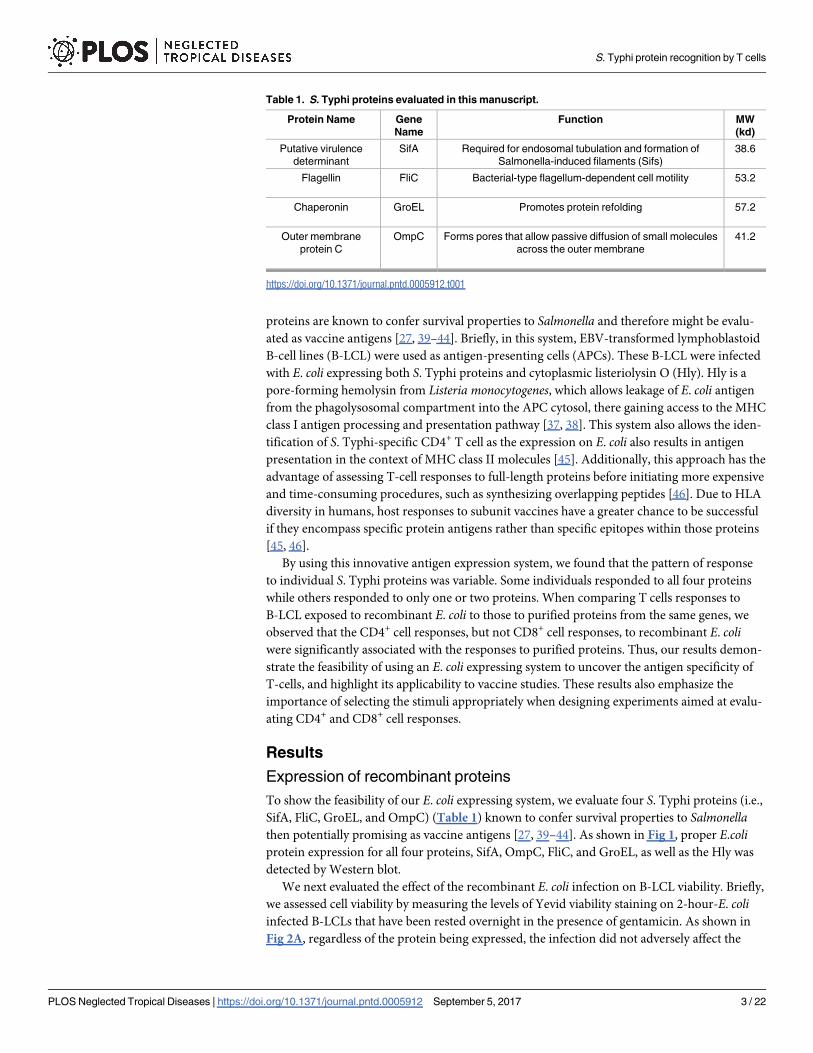
Table 1. S. Typhi proteins evaluated in this manuscript.
Protein Name Gene
Name
Function MW
(kd)
Putative virulence
determinant
SifA Required for endosomal tubulation and formation of
Salmonella-induced filaments (Sifs)
38.6
Flagellin FliC Bacterial-type flagellum-dependent cell motility 53.2
Chaperonin GroEL Promotes protein refolding 57.2
Outer membrane
protein C
OmpC Forms pores that allow passive diffusion of small molecules
across the outer membrane
41.2
https://doi.org/10.1371/journal.pntd.0005912.t001
S. Typhi protein recognition by T cells
PLOS Neglected Tropical Diseases | https://doi.org/10.1371/journal.pntd.0005912 September 5, 2017 3 / 22

viability of E. coli-infected B-LCLs. After infection, the percentage of live cells in cultures with
recombinant E. coli was comparable to control cultures with media only (uninfected). By using
the same experimental conditions as for determinations of cell viability, we also detected the
expression of bacterial antigens on B-LCLs. Similarly to the viability, regardless of the type of
protein being expressed in the recombinant E. coli, we found similar levels of E. coli-expressing
cells as assessed by surface staining with anti-E. coli antigen polyclonal antibody using flow
cytometry (Fig 2B–2D).
Hly functionality
As described above, we reasoned that Hly should promote the phagosomal escape of bacterial
antigens thereby improving MHC class I processing of S. Typhi antigens presented by B-LCLs
and hence recall immune responses from both CD4+ and CD8+ primed T cells [8, 26, 27, 29,
30, 39, 47]. To test this assumption, Hly-recombinant E. coli strain BL21, or wild type E. colistrain BL21 were used to infect B-LCL cells. Cells were infected for 2 hours using two different
multiplicity of infection (MOI, 1:30 and 1:100). After 2 hours, cells were collected, washed to
remove extracellular bacteria and cultured in the presence of gentamicin for 2 additional
hours. Thus, the ability to detect E. coli proteins in B-LCL infected cells was assessed over time
by flow cytometry (up to 120 minutes) using polyclonal anti-E. coli antibodies. As shown in
Fig 3, at all-time points evaluated, we observed higher expression of E. coli antigens on B-LCL
cells infected with the recombinant E. coli strain expressing Hly as compared to the wild-type
E. coli strain. Thus, the hly gene appears functional. These results are very significant since
based on our previous study [48], we expect to see background responses against the vector
itself (E. coli antigens). Further antigen expression might help to better discriminate T-cell
responses to S. Typhi antigens from those directed to E. coli antigens.
Identification of S. Typhi proteins that are targets of T-cells following oral
immunization with Ty21a
In order to demonstrate the feasibility of using an E. coli expression system to uncover the anti-
gen specificity of T-cells, PBMC obtained before and 42 days after immunization were exposed
Fig 1. Expression of S. Typhi proteins and lysteriolysin on recombinant E. coli. (A) Anti-HisTag antibody revealing positive
control protein Annexin3 (A3) as well as lysteriolysin (Hly) and S. Typhi gene encoded proteins (OmpC, FliC, GroEL and SifA). A
negative control with pmark (P, stop codons) clones was used instead of the protein. (B) Anti-lyteriolysin antibody revealing Hly
gene expression (red box) in all lanes except in the negative control. Expression of S. Typhi proteins and lysteriolysin on
recombinant E. coli were detected by Western blot.
https://doi.org/10.1371/journal.pntd.0005912.g001
S. Typhi protein recognition by T cells
PLOS Neglected Tropical Diseases | https://doi.org/10.1371/journal.pntd.0005912 September 5, 2017 4 / 22

to autologous B-LCL infected with recombinant E. coli expressing Hly only or co-expressing
one of the four Salmonella gene products: Hly/SifA, Hly/OmpC, Hly/FliC and Hly/GroEL.
Specifically, we studied the ability of ex-vivo PBMC from seven Ty21a-immunized volunteers
to express IL-17A, IFN-γ and TNF-α cytokines and/or CD107a and b molecules against autol-
ogous infected targets. T-cell responses (i.e., CD4+ and CD8+ T-cells) were evaluated by multi-
chromatic flow cytometry using a 10-color surface/intracellular staining panel. Unstimulated
and Staphylococcus enterotoxin B (SEB)-stimulated effector cells were used as negative and
positive controls, respectively. We observed that the pattern of response to individual S. Typhi
proteins was variable, with some individuals responding to all four proteins while others were
responding to only one or two proteins. We also observed differential CD4+ and CD8+ T cells
responses to the S. Typhi proteins (Figs 4–7). In six individuals, although the magnitude of
responses varied considerably, both CD4+ and CD8+ T cells responded to at least one protein.
In one individual, we were unable to detect CD4+ T cells responses to any of the four protein,
Fig 2. Expression of bacterial antigens on B-LCL target cells. B-LCL cells were infected with E. coli at 1:30 MOI
with one of the four recombinant E. coli expressing S. Typhi and Hly antigens: Hly/SifA (SifA), Hly/FliC (FliC), Hly/GroEL
(GroEL) and Hly/OmpC (OmpC). Uninfected B-LCLs (uninfected) and infected with recombinant E. coli expressing only
Hly antigen were used as controls. The (A) viability and the (B) percentage of the E. coli-expressing cells were assessed
by flow cytometry. Numbers correspond to the % of positive cells in the indicated quadrant in each histogram followed
by mean fluorescence intensity (MFI) of positive cells (in parenthesis). Cumulative data of (C) % and (D) mean
fluorescence intensity (MFI) were detected by using anti-E. coli antibody as described in Methods. Average of 3
independent experiments.
https://doi.org/10.1371/journal.pntd.0005912.g002
S. Typhi protein recognition by T cells
PLOS Neglected Tropical Diseases | https://doi.org/10.1371/journal.pntd.0005912 September 5, 2017 5 / 22

but CD8+ T cells responded to 3 out of 4 proteins tested (Fig 8). Representative responses
from selected volunteers are shown in Figs 4–6.
Multifunctional of CD4+ T-cells and CD8+ T- cells
Because previous results from our group have shown that multifunctional T-cells might con-
tribute to S. Typhi immunity [30, 34, 49], we then investigated the multi-functionality patterns
of CD4+ T-cells and CD8+ T-cells after exposure to infected B-LCL infected with recombinant
E. coli. We measured simultaneously four T-cell functions (i.e., expression of IL-17A, IFN-γand TNF-α cytokines, or CD107a and b molecules) by multichromatic flow cytometry using
the FCOM feature of the WinList software, which provides the % of T-cells expressing each of
the possible function combinations. Analyses of multiple function patterns (i.e., single, double,
triple or quadruple functions) revealed that, albeit different for different proteins, both CD4+
T-cells and CD8+ T-cells were multi-functional (Fig 8).
T-cell responses to exogenous proteins
An important hypothesis to evaluate is whether the T-cells from volunteers who responded to
B-LCL cultured with recombinant E. coli expressing Salmonella gene products also possess the
ability to respond to exogenous proteins. To this end, we next investigated whether there was
Fig 3. Ability of the Hly gene to improve antigen processing. B-LCL cells were infected with either E. coli
strain BL21 (BL21) or its Hly-recombinant E. coli (Hly) counterpart at 1:30 or 1:100 multiplicity of infection
(MOI). After 2 hours, the cells were washed and exposed to gentamicin for an additional 2 hours to kill and
detach extracellular bacteria. After further washings, the ability of the Hly gene to improve antigen processing
was assessed by detecting E. coli antigens at the B-cell surface over time by flow cytometry (up to 120
minutes). Cells exposed to media only were used as control (uninfected).
https://doi.org/10.1371/journal.pntd.0005912.g003
S. Typhi protein recognition by T cells
PLOS Neglected Tropical Diseases | https://doi.org/10.1371/journal.pntd.0005912 September 5, 2017 6 / 22

an association between T-cell responses after exposure to autologous B-LCL cultured with
recombinant E. coli expressing one of the four Salmonella gene products (Hly/SifA, Hly/
OmpC, Hly/FliC and Hly/GroEL) and those T-cell responses after exposure to B-LCL cultured
with individual purified recombinant proteins (SifA, OmpC, FliC, and GroEL). To this end,
B-LCLs were cultured overnight with purified SifA, OmpC, FliC or GroEL proteins, and used
as targets for T-cells. Representative responses of CD8+ and CD4+ cells from one volunteer are
shown in Figs 9 & 10 respectively. We found that the CD4+ cell responses to B-LCLs cultured
with exogenous proteins were significantly associated with the CD4+ cell responses to B-LCLs
cultured with recombinant E. coli expressing one of the four S. Typhi gene products (Fig 11;
p = 0.0111, Pearson Product Moment Correlation). However, no significant association was
observed between CD8+ cell responses to B-LCLs cultured with recombinant E. coli expressing
one of the four Salmonella gene products and CD8+ cell responses to B-LCLs cultured with
exogenous recombinant proteins (Fig 11; p = 0.0790, Pearson Product Moment Correlation).
On the other hand, CD8+ cell responses to B-LCLs cultured with exogenous proteins were
consistently higher than CD4+ cell responses (Figs 9 and 10 & S1 Table). Thus, as expected,
CD4+ and CD8+ cell responses against S. Typhi antigens depend on the nature of the stimulant
[39, 50]. These results emphasize the importance of selecting the stimuli appropriately when
designing experiments aimed at evaluating CD4+ and CD8+ cell responses cell responses [8].
Discussion
One of the characteristics that make the E. coli expression system methodology employed in
this manuscript highly relevant for identifying immunogenic proteins of S. Typhi is the use of
a translational research approach using human T cells and autologous APC to identify S.
Typhi-specific T-cell immune responses. Most published methods have relied heavily on
Fig 4. Antigen presentation of S. Typhi proteins by targets infected with recombinant E. coli. Ex vivo
PBMC from a volunteer collected before (day 0) and 42 days after immunization were co-cultured for 16–18
hrs. with autologous B-LCL targets infected at 1:30 MOI with one of the four recombinant E. coli expressing S.
Typhi/Hly (Hly/SifA (SifA), Hly/FliC (FliC), Hly/GroEL (GroEL) and Hly/OmpC (OmpC)) or only Hly (control)
proteins. After incubation, cells were stained and the ability of the PBMC to express IFN-γwas analyzed by
flow cytometry. CD4+ and CD8+ T cells were evaluated. Numbers represent the percentage of positive cells.
The data of a representative volunteer are shown.
https://doi.org/10.1371/journal.pntd.0005912.g004
S. Typhi protein recognition by T cells
PLOS Neglected Tropical Diseases | https://doi.org/10.1371/journal.pntd.0005912 September 5, 2017 7 / 22

“proof-of-concept” studies performed in mice. However, S. Typhi is a human-restricted patho-
gen and there are no good animal models that faithfully recapitulate S. Typhi infection [51]. To
partially address this shortcoming, the infection of susceptible mice with S. Typhimurium has
been used as a model for the pathogenesis of human typhoid fever [51]. Although these murine
models have provided considerable knowledge regarding host-pathogen interactions, they do
not adequately recapitulate S. Typhi infection in humans [52]. The recent availability of full
genome sequences from various Salmonella enterica serovars has uncovered many differences
in pseudo genes which can explain, at least in part, the dissimilarities observed in the immune
and other host responses to these enteric bacteria [52]. Thus, samples from human participants
exposed to the licensed Ty21a oral typhoid vaccine have the potential to provide a better charac-
terization of key Salmonella antigens involved in T cell responses than murine models.
Fig 5. CD8+ T cell responses to S. Typhi proteins presented by targets infected with recombinant E.
coli. Ex vivo PBMC from a volunteer collected 42 days after immunization were co-cultured for 16–18 hrs.
with autologous B-LCL targets infected at 1:30 MOI with one of the four recombinant E. coli expressing S.
Typhi/Hly (Hly/SifA (SifA), Hly/FliC (FliC), Hly/GroEL (GroEL) and Hly/OmpC (OmpC)) or only Hly (control)
proteins. After incubation, cells were stained and the ability of the PBMC to express one or more cytokines (IL-
17A, IFN-γ and TNF-α) and/or CD107a/b molecules was evaluated by flow cytometry. Shown are the CD8+ T
cell responses from a representative volunteer. Numbers represent the percentage of positive cells.
https://doi.org/10.1371/journal.pntd.0005912.g005
S. Typhi protein recognition by T cells
PLOS Neglected Tropical Diseases | https://doi.org/10.1371/journal.pntd.0005912 September 5, 2017 8 / 22

The main novelty of our system is that we engineered the hly gene encoding the pore-form-
ing cytoplasmic listeriolysin O (Hly) protein onto the backbone of the recombinant protein
expression plasmid pET-DEST-Hly (see methods). In contrast, Higgin’s E. coli expressing sys-
tem [37, 38] used two expression plasmids inside individual E. coli cells: one expressing Hly
and the other expressing the recombinant protein of interest. The reason for this difference
stems from our preliminary results showing that the use of two expression plasmids reduced
B-LCL infectivity (S1 Fig). We speculate that this difference is due to the fitness cost for E. colito maintain, replicate and propagate two plasmids instead of one. There may also have been a
negative effect on bacterial cell preparations resulting from growth on media containing two
selective antibiotics instead of one.
Fig 6. CD4+ T cell responses to S. Typhi proteins presented by targets infected with recombinant E.
coli. Ex vivo PBMC from a volunteer collected 42 days after immunization were co-cultured for 16–18 hrs.
with autologous B-LCL targets infected at 1:30 MOI with one of the four recombinant E. coli expressing S.
Typhi/Hly (Hly/SifA (SifA), Hly/FliC (FliC), Hly/GroEL (GroEL) and Hly/OmpC (OmpC)) or only Hly (control)
proteins. After incubation, cells were stained and the ability of the PBMC to express one or more cytokines (IL-
17A, IFN-γ and TNF-α) and/or CD107a/b molecules was evaluated by flow cytometry. Shown are the CD4+ T
cell responses from a representative volunteer. Numbers represent the percentage of positive cells.
https://doi.org/10.1371/journal.pntd.0005912.g006
S. Typhi protein recognition by T cells
PLOS Neglected Tropical Diseases | https://doi.org/10.1371/journal.pntd.0005912 September 5, 2017 9 / 22

Herein, using this methodology, we found that all the tested individuals had increased T-
cell responses over baseline (before immunization) to at least one of the four S. Typhi proteins
evaluated (i.e., SifA, OmpC, FliC, and GroEL). Moreover, multifunctional CD4+ and CD8+ T
cells that expressed two or more cytokines (IL-17A, IFN-γ and TNF-α) and/or CD107a/b mol-
ecules were detected. These results are particularly significant since we have previously dem-
onstrated that these two T-cell population might play a role in controlling Salmonella infection
[39]. These results also support previous data showing that the depletion of either CD4+ or
CD8+ T-cells had impaired recall immunity to oral challenge with the virulent S. Typhimur-
ium at different times after vaccination [53].
The reason underlying the observation that the responses to the different antigens are vari-
able among the vaccinees are unclear. However, it is reasonable to speculate that this phenom-
enon might be due to the HLA-haplotype variation between individuals. In fact, antigen
processing, together with defined MHC genes, are known to shape the individual immune
responses to a wide array of pathogens [54]. Furthermore, the differential responsiveness
among the participants supports the development of multi-component vaccines by introduc-
ing many antigenic determinants into vaccine formulations. Alternatively, these results might
encourage a renewed focus on whole-cell live attenuated preparations, especially since they
may overcome some of the inherent weaknesses associated with sub-unit vaccines such as the
need for considerable amounts of antigens and the use of adjuvants.
Fig 7. Percentage of T cell subsets specific to any S. Typhi protein. Ex vivo PBMC from 7 volunteers
collected before and 42 days after immunization were co-cultured for 16–18 hrs. with autologous B-LCL
targets infected at 1:30 MOI with one of the four recombinant E. coli expressing S. Typhi and Hly genes: Hly/
SifA (SifA), Hly/FliC (FliC), Hly/GroEL (GroEL) and Hly/OmpC (OmpC). After incubation, cells were stained
and the ability of the PBMC to express one or more cytokines (IL-17A, IFN-γ and TNF-α) and/or CD107a/b
molecules was analyzed by flow cytometry. Two T cell subset responses (i.e., CD4+ and CD8+ T cells) were
evaluated. Net responses were calculated by subtracting the T cell responses to B-LCLs infected with
recombinant E. coli expressing S. Typhi/Hly antigens from the responses to the controls (B-LCL expressing
Hly only). Increases over day 0 were calculated by subtracting the net responses of the PBMC collected 42
days after immunization from the net responses of PBMC collected before immunization. Bars represent
mean ± SE.
https://doi.org/10.1371/journal.pntd.0005912.g007
S. Typhi protein recognition by T cells
PLOS Neglected Tropical Diseases | https://doi.org/10.1371/journal.pntd.0005912 September 5, 2017 10 / 22

We also observed that the magnitude of the responses against S. Typhi SifA, OmpC, FliC
and GroEL varied among participants. Since S. Typhi GroEL has a significant degree of homol-
ogy with self-heat shock proteins in humans [27], these results provide additional information
that T-cells can discriminate between self and foreign antigens during the immune response.
Interestingly, in contrast to CD8+ cell responses, we found that the CD4+ cell responses to
B-LCLs cultured with exogenous proteins were significantly associated with the CD4+ cell
responses to B-LCLs cultured with recombinant E. coli expressing S. Typhi genes. These results
confirm and extend previous findings from our group and others that the balance of CD4+
and CD8+ cell responses against S. Typhi antigens are likely to depend on the nature of the
stimulant [8, 39, 50]. Indeed, previous work from our group has shown that CD4+ cells
respond differently to soluble antigen stimulation than CD8+ T cells [39, 50]. Future studies
will be directed to use this novel antigen discovery system to evaluate in humans the immune
responses to other S. Typhi proteins expressed in this E. coli expression system.
Since these studies were performed in immunized volunteers, it is also important to note
that the relative contributions of CD4+ and CD8+ cell responses against these S. Typhi proteins
in protection cannot be ascertained. We are directly addressing this critical issue in separate
Fig 8. Volunteer responses to S. Typhi proteins. Ex vivo PBMC from 7 immunized volunteers collected before (day 0) and 42 days after
immunization were co-cultured for 16–18 hrs. with autologous B-LCL targets infected at an 1:30 MOI with one of the four recombinant E. coli
expressing S. Typhi and Hly antigens: Hly/SifA (SifA), Hly/FliC (FliC), Hly/GroEL (GroEL) and Hly/OmpC (OmpC). After incubation, cells were
stained and the ability of the PBMC to express one or more cytokines (IL-17A, IFN-γ and TNF-α) and/or CD107a/b molecules was analyzed by flow
cytometry. Two T cell subset responses (i.e., CD4+ and CD8+ T cells) were evaluated. (A) Heat-map of the multifunctionality of CD4+ and CD8+ T
cells based on expression of cytokines and CD107a/b antigens. Percentages correspond to the net responses calculated by subtracting the T cell
responses to B-LCLs infected with recombinant E. coli expressing S. Typhi/Hly proteins from the responses to the controls (B-LCL expressing Hly
only). Volunteers were considered responders if the net responses of the PBMC collected 42 days after immunization were greater than 0.1 from the
net responses of PBMC collected before immunization. (B) Cumulative frequency of responders to any functional test.
https://doi.org/10.1371/journal.pntd.0005912.g008
S. Typhi protein recognition by T cells
PLOS Neglected Tropical Diseases | https://doi.org/10.1371/journal.pntd.0005912 September 5, 2017 11 / 22

studies in which we are evaluating whether responses to these S. Typhi proteins correlate with
protection in volunteers who have been immunized with Ty21a and subsequently challenged
with wild-type S. Typhi. Nevertheless, the results presented herein demonstrate the feasibility
of using a novel antigen discovery platform. This system could be used, for example, to system-
atically assess the specificity of T-cell immune responses against the entire proteome of a
human pathogen and generate a database of the repertoire of these human T-cell antigen spec-
ificities. This should help narrow down the proteins of interest which correlate with defined
phenotypes (e.g., responses associated with protection in human challenge studies with S.
Typhi, those directed to pathogenic determinants), ultimately leading to the identification of
candidate vaccine antigens. Finally, these results emphasize the importance of selecting the
appropriate antigens when designing experiments aimed at evaluating CD4+ and CD8+ cell
responses.
Materials and methods
Ethics statement
The human experimentation guidelines of the US Department of Health and Human Services
and those of the University of Maryland, Baltimore, were followed in the conduct of the clini-
cal research. All blood specimens were collected from volunteers who participated in the Uni-
versity of Maryland Institutional Review Board approved protocol number HP-00040022 that
authorized the collection of blood samples from healthy volunteers for the studies included in
this manuscript. Volunteers were explained the purpose and possible consequences of partici-
pating in this study and gave informed, signed consent before the blood draws. This protocol
has been conducted in accordance with the ethical standards laid down in the 1964 Declara-
tion of Helsinki and the principles of the International Conference on Harmonization Good
Clinical Practice guidelines [55].
Participants
Seven healthy adult volunteers, 20–50 (39 ± 10) years old, recruited from the Baltimore-Wash-
ington area and the University of Maryland at Baltimore campus, participated in this study.
They were immunized with four spaced doses of 2–6 x 10⁹ CFU of oral live attenuated Ty21a at
an interval of 48 hours between doses [12, 56]. Blood collection was performed before and 42
days after Ty21a immunization. Peripheral blood mononuclear cells (PBMC) were isolated from
the blood by density gradient centrifugation and cryopreserved in liquid N2 following standard
techniques [28]. These PBMC were used ex vivo as effector cells or to prepare the target cells.
Gateway cloning vectors
The entire S. Typhi strain Ty2 ORFeome was constructed as a part of the former National
Institute of Allergy and Infectious Disease (NIAID)-funded Pathogen Functional Genomics
Resource Center (PFGRC). The PFGRC provided a library of entry clones by cloning 3,381
ORFs, out of the 4,323 ORFs predicted in the Ty2 genome (78%), into the pDONR 221 vector
using methods described in Peterson et al. [57]. Entry clones can then be shuttled into a variety
of destination vectors using in vitro site-specific recombination (http://www.ncbi.nlm.nih.gov/
pubmed/11076863). For expression of the S. Typhi proteins, we engineered the pET161-DEST
destination vector (Invitrogen, Carlsbad, CA). The pET161-DEST, a commercial plasmid
from Invitrogen, is a robust E. coli destination vector that has been used for several years and
carries many desirable characteristics for this study. Gene expression is under the tight control
of the T7 promoter and lac repressor, and an optimized ribosome binding site is provided by
S. Typhi protein recognition by T cells
PLOS Neglected Tropical Diseases | https://doi.org/10.1371/journal.pntd.0005912 September 5, 2017 12 / 22

the inserted gene. The vector contains the V5 and His-tag epitopes downstream of the attRsites into which inserts from entry clones were recombined resulting in the addition of these
tags as C-terminal fusions to the S. Typhi protein. These tags were used to monitor recombi-
nant protein expression and, if necessary, for protein purification. pET161-DEST also contains
the ccdB and chloramphenicol (CmR) genes which help the recovery of correct recombination
products after the LR recombinase reaction via selection on ampicillin agar plates. We also
inserted the hly gene coding for the cytoplasmic Hly protein from L. monocytogenes into the
BglII restriction site. The hly gene was PCR-amplified with a His-tag at the 3’end of the reverse
primer, and a BgIll site was added to the forward 5’ end, and reverse 3’ end of each primer,
respectively (sense: GCGCAGATCTAGCAAGCATATAATATTGCG, anti-sense: GCGCAG
ATCTTTAGTGATGGTGATGGTGATGTTCGATTGGATTATCTAC). The resulting vector
pET-DEST-Hly was verified by Sanger sequencing. It carries a T7 promoter and the lac
Fig 9. CD8+ T cell responses to S. Typhi proteins presented by targets exposed to one of the four
recombinant S. Typhi proteins. Ex vivo PBMC from a volunteer collected 42 days after immunization were
co-cultured for 16–18 hrs. with autologous B-LCL targets exposed to 0.5ug/ml with one of the four recombinant
S. Typhi proteins: SifA, OmpC, FliC, and GroEL. Untreated B-LCL targets (media) were used as controls. After
incubation, cells were stained and the ability of the PBMC to express one or more cytokines (IL-17A, IFN-γ and
TNF-α) and/or CD107a/b molecules was evaluated by flow cytometry. Shown are the CD8+ T cell responses
from a representative volunteer. Numbers represent the percentage of positive cells.
https://doi.org/10.1371/journal.pntd.0005912.g009
S. Typhi protein recognition by T cells
PLOS Neglected Tropical Diseases | https://doi.org/10.1371/journal.pntd.0005912 September 5, 2017 13 / 22

operator preceding the gene to be cloned; the cloning site harbors attR1 and attR2 site for clon-
ing from entry clones through the LR clonase reaction (see below). The plasmid was trans-
formed into One Shot ccdB survival competent cells (Invitrogen).
Cloning of S. Typhi proteins from entry clones to pET-DEST-Hly for
protein expression
Entry clone glycerol stocks were streaked on LB agar with 50 μg/ml kanamycin and incubated
at 37˚C overnight. Single colonies were incubated overnight at 37˚C in 4ml LB broth with
50 μg/ml kanamycin. Then the entry clone plasmid was extracted using the QIA prep kit (Qia-
gen, Valencia, CA) and DNA concentration measured using a NanoDrop instrument (Thermo
Scientific, Waltham, MA). The pET-DEST-Hly glycerol stock was streaked on LB agar plates
with 100 μg/ml carbenicillin and incubated at 37˚C. After overnight incubation, single colonies
Fig 10. CD4+ T cell responses to S. Typhi proteins presented by targets exposed to one of the four
recombinant S. Typhi proteins. Ex vivo PBMC from a volunteer collected 42 days after immunization were
co-cultured for 16–18 hrs. with autologous B-LCL targets exposed to 0.5ug/ml with one of the four
recombinant S. Typhi proteins: SifA, OmpC, FliC, and GroEL. Untreated B-LCL targets (media) were used as
controls. After incubation, cells were stained and the ability of the PBMC to express one or more cytokines (IL-
17A, IFN-γ and TNF-α) and/or CD107a/b molecules was evaluated by flow cytometry. Shown are the CD4+ T
cell responses from a representative volunteer. Numbers represent the percentage of positive cells.
https://doi.org/10.1371/journal.pntd.0005912.g010
S. Typhi protein recognition by T cells
PLOS Neglected Tropical Diseases | https://doi.org/10.1371/journal.pntd.0005912 September 5, 2017 14 / 22

were isolated, incubated overnight in 50 ml LB broth with 100 μg/ml carbenicillin at 37˚C and
the destination plasmid was extracted with the HiSpeed plasmid preparation kit (Qiagen).
DNA concentration was measured by NanoDrop. We chose to evaluate four S. Typhi proteins:
SifA, FliC, GroEL, and OmpC (Table 1). Each of these four S. Typhi proteins was then trans-
ferred in vitro from the entry clone plasmid into the destination plasmid through LR clonase
reactions (Gateway LR clonase II Enzyme mix, Invitrogen) following the manufacturer’s pro-
tocol. We also cloned a short non-coding sequence called “pmark” that carried stop codons in
all six frames of translation into pET-DEST-Hly as a negative control of protein expression.
Finally, 3 μl of LR clonase reaction product was transformed into 50 μl of One Shot BL21
(DE3) competent cells (Invitrogen) by chemical transformation.
Verification of expression plasmids
Proper cloning of the target S. Typhi gene was confirmed by PCR amplification using the T7
forward and reverse primers and sequencing of the PCR products. The presence of the hlygene was verified by BgIll digestion of the plasmid.
Protein expression
A single colony from verified BL21 (DE3) expression vector clones was cultured at 37˚C over-
night in 2 ml LB broth with 100 μg/ml carbenicillin. Then 30 μl of the overnight culture was
inoculated to 3 ml fresh LB broth with 100 μg/ml carbenicillin. After ~4h of incubation at
37˚C (OD~0.6), the culture was induced for protein expression with 100 μM IPTG and incu-
bated for an additional 2 h. Bacteria were then spun down at 2,800 rpm for 10 min and the
supernatant discarded. E. coli were lysed with of 0.3 μg/ml lysozyme in Tris-HCl buffer, pH 7.5
(Thermo Scientific), then proteins were heat-denatured at 100˚C for 10 minutes and evaluated
on SDS-PAGE gels by Coomassie staining. Specificity of protein expression was confirmed by
western blot using mouse anti-His monoclonal antibodies (Sigma, St. Louis, MO), and rabbit
anti-mouse IgG conjugated to horseradish peroxidase (HRP) (Sigma) as secondary antibodies;
Fig 11. Correlation between T cell subset responses to B-LCL targets exposed to recombinant S.
Typhi proteins or infected with recombinant E. coli expressing S. Typhi proteins. Ex vivo PBMC were
analyzed as described in Figs 1 & 2. Shown are the correlation between T cell subset responses to either of
the four proteins (SifA, FliC, GroEL and OmpC) after stimulation by B-LCL targets exposed to recombinant S.
Typhi proteins or infected with recombinant E. coli expressing S. Typhi proteins. Samples are representative
of two individuals collected 42 days after vaccination. Coefficients of determination “R2” and “p” values are
shown. p values of <0.05 were considered statistically significant. Dashed lines represent 95% confidence
intervals.
https://doi.org/10.1371/journal.pntd.0005912.g011
S. Typhi protein recognition by T cells
PLOS Neglected Tropical Diseases | https://doi.org/10.1371/journal.pntd.0005912 September 5, 2017 15 / 22
as well as rabbit anti-Listeriolysin polyclonal antibodies (Abcam, Cambridge, MA, USA), and
goat anti-rabbit IgG-HRP (Millipore, Billerica, MA) as secondary antibodies. His-tagged
Annexin 3 (A3) protein (Abcam) was used as positive control for antibody detection.
Target/stimulator cells
PBMC obtained from Ty21a vaccines before immunization were used to generate autologous
Epstein-Barr Virus (EBV)-transformed B-LCLs as previously described [27, 31]. Briefly,
B-LCLs were established by using B95-8 cell line (ATCC# CRL1612) supernatants as the EBV
source. After transformation, B-LCL were maintained in culture in RPMI 1640 (Gibco, Grand
Island, New York) supplemented with 100 U/ml penicillin, 100 μg/ml streptomycin, 50 μg/ml
gentamicin, 2 mM L-glutamine, 2.5 mM sodium pyruvate, 10 mM HEPES buffer and 10%
heat-inactivated fetal bovine serum (R10) or cryopreserved until used in the experiments.
Infection of target/stimulator cells by E. coli
Target/stimulator cells were infected as previously described [27–30, 39, 47, 48] with slight
modifications. Briefly, target cells were infected by incubation in RPMI (without antibiotics) at
37˚C for 2 hours with any of the recombinant E. coli strains at 1:30 or 1:100 multiplicity of
infection (MOI). After incubation, cells were washed and incubated for an additional 2 or 16–
18 hours (overnight) in complete R10 containing gentamicin (100 μg/ml) to kill extracellular
and/or to detach cell-bound bacteria. For co-culture experiments, targets were then gamma-
irradiated (6,000 rads), surface stained with anti-CD45, a marker abundantly expressed on the
surface of hematopoietic cells [58], and used as stimulators. To confirm E. coli infection, ali-
quots of targets were surface stained with rabbit anti-E. coli antigen polyclonal antibody
(1:1000, Abcam).
Exogenous proteins
Four Salmonella purified proteins were tested: SifA, FliC, GroEL, and OmpC (Table 1). The
region encoding residues 53–450 of FliC were subcloned from S. Typhi ISP1820 into pET15b.
The plasmid was used to transform E. coli Tuner (DE3) for overexpression after being induced
with IPTG. The overexpressed protein was purified by standard immobilized metal affinity
column chromatography (IMAC) methods. OmpC was purified based on the protocol of
Kumar and Krishnaswamy [59]. The sequence encoding OmpC from S. Typhi ISP1820 was
subcloned into pET15b, which was used to transform E. coli Tuner (DE3). Overexpression
was induced using IPTG, and the OmpC was purified from inclusion bodies using IMAC after
solubilizing in buffer containing urea. Refolding was then allowed to occur in 50 mM Tris,
pH 8.5, 0.1 M NaCl, 10% (v/v) glycerol and 0.2% (v/v) polyoxyethelene-9-lauryl ether. The
sequence for GroEL was introduced into pBAD TOPO TA such that it was in-frame. The pro-
tein was overexpressed by the addition of arabinose, and the HT-GroEL purified initially by
standard IMAC, which provides good yields at high purity. Removal of contaminating pep-
tides could be achieved by adding acetone to 45% (v/v) to precipitate the GroEl which could
then be collected by centrifugation followed by solubilizing using PBS (10 mM phosphate, pH
7.4, 150 mM NaCl). The sequence encoding SifA from Salmonella Typhi ISP1820 was sub-
cloned into pET15b and used to transform E. coli c-41 (DE3). The bacteria were grown to an
absorbance of about 0.8 at 600 nm, protein expression induced with IPTG, and the cultures
immediately moved to 20˚C for overnight growth. The bacteria were then lysed and the SifA
purified by standard IMAC methods.
S. Typhi protein recognition by T cells
PLOS Neglected Tropical Diseases | https://doi.org/10.1371/journal.pntd.0005912 September 5, 2017 16 / 22

Loading of target/stimulator cells with exogenous proteins
B-LCLs were incubated overnight in 24-well plates at a density of 2 x 10⁶/ml/well in the
absence or presence of 1 μg/ml of each of the purified proteins at 37˚C in a 5% CO2 atmo-
sphere. After incubation, cells were washed and used as stimulators for T-cells.
Monoclonal antibodies for surface and intracellular staining
Cells were stained with monoclonal antibodies (mAbs) to CD69 (clone TPI-55-3) (Beckman-
Coulter, Miami, FL), CD4 (clone RPA-T4), CD8 (clone HIT8a), CD107a and b (clones H4A3
and H4B4 respectively), interferon (IFN)-γ (clone B27), tumor necrosis factor (TNF)-α (clone
MAb11) (BD Pharmingen, San Diego, CA, USA), CD14 (clone TuK4), CD19 (clone SJ25-C1),
CD45 (clone H130) (Invitrogen), interleukin (IL)-17A (clone eBio64DEC17) (eBioscience,
San Diego, CA), and CD3 (clone OKT3)(Biolegend, San Diego, CA). Antibodies conjugated to
the following fluorochromes were used in these studies: Fluorescein isothiocyanate (FITC),
PE-Cy5.5, PE-Cy7, V450, Brilliant Violet (BV)570, BV650, Energy Coupled Dye or PE-Texas-
Red conjugate (ECD), allophycocyanin (APC)-Alexa 700 and Quantum Dot (QD) 800.
Effector cells and co-culture
Ex vivo PBMC from immunized volunteers collected before and 42 days after immunization
were used as effectors as previously described [48]. Briefly, PBMC were co-cultured with autol-
ogous B-LCL cells at an effector to stimulator cell ratio of 5:1 in the presence of mAbs to
CD107a and CD107b (15 μl of each/1 x 106 cells in 500 μl of R10 medium). The CD107 a and b
antibodies were used to measure degranulation, a mechanism essential for the killing of S.
infected targets by the cytotoxic CD8+ cells [60]. PBMC cultured with uninfected target cells
or Staphylococcus enterotoxin B (SEB) (10 μg/ml, Sigma) were used as negative and positive
controls, respectively. After ~2 hours of stimulation, protein transport blockers, Monensin
(1 μg/ml, Sigma) and brefeldin-A (BFA) (2 μg/ml, Sigma), were added to the co-culture. After
overnight (16–18 hours) incubation, cells were harvested, stained with a dead-cell discrimina-
tor, yellow fluorescent viability dye (Yevid, Invitrogen)[61, 62], followed by surface staining
with mAbs against surface antigens (CD3, CD4, CD8, CD14, and CD19) and fixation and per-
meabilization with Fix & Perm cell buffers (Invitrogen, Carlsbad, CA). Cells were then stained
intracellularly for IFN-γ, TNF-α, IL-17A and CD69. Finally, cells were fixed and analyzed by
flow cytometry on an LSR-II instrument (BD Biosciences). Data were analyzed with WinList
7.0 (Verity Software House, Topsham, ME). Lymphocytes were gated based on their scatter
characteristics. Single lymphocytes were gated based on forward scatter height vs. forward
scatter area. A “dump” channel was used to eliminate dead cells (Yevid+) as well as macro-
phages/monocytes (CD14+), B lymphocytes (CD19+) and targets (CD45+) from analysis. Addi-
tional gating on CD3, CD4, and CD8 was performed to identify cytokine-producing (IFN-γ,
TNF-α and IL-17A) and CD107 expressing T cell subsets. Net responses were calculated by
subtracting the number of positive events of the experimental (Salmonella-Hly proteins) from
the negative control (Hly only). Functional responses were considered specific for S. Typhi if
the differential in the number of positive and negative events between experimental (Salmo-nella-Hly proteins) and negative control (Hly only) cultures were significantly increased
(P< 0.01) using Z-test. Volunteers were considered responders if the net responses from the
PBMC collected 42 days after immunization were greater than 0.1% from the net responses of
PBMC collected before immunization.
S. Typhi protein recognition by T cells
PLOS Neglected Tropical Diseases | https://doi.org/10.1371/journal.pntd.0005912 September 5, 2017 17 / 22

Statistical analysis
All statistical tests were performed using Prism software (version 7, GraphPad Software, La
Jolla, CA). Comparisons between two groups were carried out by Student’s t tests. Correlation
analysis was achieved by Pearson Product Moment Correlation tests. P values<0.05 were con-
sidered significant.
Supporting information
S1 Table. T-cell responses to individual S. Typhi proteins.
(TIF)
S1 Fig. Comparison between the use of one and two expression plasmids inside individual
recombinant E. coli. B-LCL cells were infected with E. coli expressing one (Hly/pmark or Hly/
Salmonella protein) or two plasmids (Hly only or Hly and S. Typhi protein) at 1:30 MOI. S.
Typhi proteins were FliC, GroEL and OmpC. Uninfected B-LCLs and B-LCLs infected with
recombinant E. coli expressing only Hly antigen were used as controls. The percentage of the
E. coli-antigen expressing B-LCLs were assessed by flow cytometry using anti-E. coli antibody
as described in Methods. Shown are the average of 3 independent experiments. p values of
<0.05 were considered statistically significant.
(TIF)
Acknowledgments
We are indebted to the volunteers who allowed us to perform this study. We also thank Mrs.
Robin Barnes and the staff from the Recruiting Section of the Center for Vaccine Development
for their help in collecting blood specimens and Dr. Haiyan Chen, and Mrs. Regina Harley
and Catherine Storrer for excellent technical assistance.
Author Contributions
Conceptualization: Rosangela Salerno-Goncalves, Herve Tettelin, William D. Picking, Vish-
vanath Nene, Marcelo B. Sztein.
Data curation: Rosangela Salerno-Goncalves, Herve Tettelin, David Lou, Stephanie Steiner,
Tasmia Rezwanul, Qin Guo, William D. Picking, Marcelo B. Sztein.
Formal analysis: Rosangela Salerno-Goncalves, Herve Tettelin, David Lou, Stephanie Steiner,
Tasmia Rezwanul, Qin Guo, William D. Picking, Marcelo B. Sztein.
Funding acquisition: Vishvanath Nene, Marcelo B. Sztein.
Investigation: Rosangela Salerno-Goncalves, Herve Tettelin, William D. Picking, Marcelo B.
Sztein.
Methodology: Rosangela Salerno-Goncalves.
Project administration: Rosangela Salerno-Goncalves.
Supervision: Rosangela Salerno-Goncalves, William D. Picking.
Validation: Rosangela Salerno-Goncalves, Herve Tettelin, William D. Picking, Marcelo B.
Sztein.
Writing – original draft: Rosangela Salerno-Goncalves, Herve Tettelin, Marcelo B. Sztein.
S. Typhi protein recognition by T cells
PLOS Neglected Tropical Diseases | https://doi.org/10.1371/journal.pntd.0005912 September 5, 2017 18 / 22
http://journals.plos.org/plosntds/article/asset?unique&id=info:doi/10.1371/journal.pntd.0005912.s001

Writing – review & editing: Rosangela Salerno-Goncalves, Herve Tettelin, David Lou, Stepha-
nie Steiner, Tasmia Rezwanul, Qin Guo, William D. Picking, Vishvanath Nene, Marcelo B.
Sztein.
References1. Mogasale V, Maskery B, Ochiai RL, Lee JS, Mogasale VV, Ramani E, et al. Burden of typhoid fever in
low-income and middle-income countries: a systematic, literature-based update with risk-factor adjust-
ment. The Lancet Global health. 2014; 2(10):e570–80. Epub 2014/10/12. https://doi.org/10.1016/
S2214-109X(14)70301-8 PMID: 25304633.
2. Mogasale V, Mogasale VV, Ramani E, Lee JS, Park JY, Lee KS, et al. Revisiting typhoid fever surveil-
lance in low and middle income countries: lessons from systematic literature review of population-
based longitudinal studies. BMC infectious diseases. 2016; 16:35. Epub 2016/01/30. https://doi.org/10.
1186/s12879-016-1351-3 PMID: 26822522; PubMed Central PMCID: PMCPMC4731936.
3. Antillon M, Warren JL, Crawford FW, Weinberger DM, Kurum E, Pak GD, et al. The burden of typhoid
fever in low- and middle-income countries: A meta-regression approach. PLoS neglected tropical dis-
eases. 2017; 11(2):e0005376. Epub 2017/02/28. https://doi.org/10.1371/journal.pntd.0005376 PMID:
28241011; PubMed Central PMCID: PMCPMC5344533.
4. Crump JA. Updating and refining estimates of typhoid fever burden for public health action. The Lancet
Global health. 2014; 2(10):e551–3. Epub 2014/10/12. https://doi.org/10.1016/S2214-109X(14)70306-7
PMID: 25304622; PubMed Central PMCID: PMCPMC4404498.
5. DeRoeck D, Jodar L, Clemens J. Putting typhoid vaccination on the global health agenda. N Engl J Med.
2007; 357(11):1069–71. Epub 2007/09/15. https://doi.org/10.1056/NEJMp078144 PMID: 17855666.
6. Connerton P, Wain J, Hien TT, Ali T, Parry C, Chinh NT, et al. Epidemic typhoid in vietnam: molecular
typing of multiple-antibiotic-resistant Salmonella enterica serotype typhi from four outbreaks. J Clin
Microbiol. 2000; 38(2):895–7. Epub 2000/02/03. PMID: 10655411; PubMed Central PMCID:
PMC86238.
7. Rowe B, Ward LR, Threlfall EJ. Multidrug-resistant Salmonella typhi: a worldwide epidemic. Clin Infect
Dis. 1997; 24 Suppl 1:S106–9. PMID: 8994789.
8. Sztein MB, Salerno-Goncalves R, McArthur MA. Complex adaptive immunity to enteric fevers in
humans: lessons learned and the path forward. Front Immunol. 2014; 5:516. Epub 2014/11/12. https://
doi.org/10.3389/fimmu.2014.00516 PMID: 25386175; PubMed Central PMCID: PMCPmc4209864.
9. Buckle GC, Walker CL, Black RE. Typhoid fever and paratyphoid fever: Systematic review to estimate
global morbidity and mortality for 2010. Journal of global health. 2012; 2(1):010401. Epub 2012/12/01.
https://doi.org/10.7189/jogh.02.010401 PMID: 23198130; PubMed Central PMCID: PMCPmc3484760.
10. Levine MM. Typhoid fever vaccines. In: Plotkin SA, Mortimer EA, editors. Vaccines. Philadelphia: W.B.
Saunders Company; 1994. p. 597–633.
11. Sur D, Ochiai RL, Bhattacharya SK, Ganguly NK, Ali M, Manna B, et al. A cluster-randomized effective-
ness trial of Vi typhoid vaccine in India. N Engl J Med. 2009; 361(4):335–44. Epub 2009/07/25. https://
doi.org/10.1056/NEJMoa0807521 PMID: 19625715.
12. Levine MM, Ferreccio C, Abrego P, Martin OS, Ortiz E, Cryz S. Duration of efficacy of Ty21a, attenu-
ated Salmonella typhi live oral vaccine. Vaccine. 1999; 17(Suppl 2):S22–7.
13. Guzman CA, Borsutzky S, Griot-Wenk M, Metcalfe IC, Pearman J, Collioud A, et al. Vaccines against
typhoid fever. Vaccine. 2006; 24(18):3804–11. https://doi.org/10.1016/j.vaccine.2005.07.111 PMID:
16278037.
14. Levine MM, Ferreccio C, Black RE, Tacket CO, Germanier R. Progress in vaccines against typhoid
fever. Rev Infect Dis. 1989; 11 Suppl 3:S552–67. Epub 1989/05/01. PMID: 2669099.
15. Ferreccio C, Levine MM, Rodriguez H, Contreras R. Comparative efficacy of two, three, or four doses of
Ty21a live oral typhoid vaccine in enteric-coated capsules: a field trial in an endemic area. The Journal
of Infectious Diseases. 1989; 159(4):766–69. PMID: 2647863
16. Chinnasami B, Mangayarkarasi V, Prema A, Sadasivam K, Davis M. Safety and immunogenicity of Sal-
monella Typhi Vi conjugate vaccine (Peda Typh) in children up to five years. International Journal of Sci-
entific and Research Publications,. 2013; 3(2):1–5.
17. Szu SC, Klugman KP, Hunt S. Re-examination of immune response and estimation of anti-Vi IgG pro-
tective threshold against typhoid fever-based on the efficacy trial of Vi conjugate in young children. Vac-
cine. 2014; 32(20):2359–63. Epub 2014/03/19. https://doi.org/10.1016/j.vaccine.2014.02.050 PMID:
24630869; PubMed Central PMCID: PMCPmc4068246.
18. van Damme P, Kafeja F, Anemona A, Basile V, Hilbert AK, De Coster I, et al. Safety, immunogenicity
and dose ranging of a new Vi-CRM(1)(9)(7) conjugate vaccine against typhoid fever: randomized
S. Typhi protein recognition by T cells
PLOS Neglected Tropical Diseases | https://doi.org/10.1371/journal.pntd.0005912 September 5, 2017 19 / 22
clinical testing in healthy adults. PLoS One. 2011; 6(9):e25398. Epub 2011/10/08. https://doi.org/10.
1371/journal.pone.0025398 PMID: 21980445; PubMed Central PMCID: PMCPmc3184126.
19. Lin FY, Ho VA, Khiem HB, Trach DD, Bay PV, Thanh TC, et al. The efficacy of a Salmonella typhi Vi
conjugate vaccine in two-to-five-year-old children. N Engl J Med. 2001; 344(17):1263–9. Epub 2001/04/
26. https://doi.org/10.1056/NEJM200104263441701 PMID: 11320385.
20. Saha MR, Ramamurthy T, Dutta P, Mitra U. Emergence of Salmonella typhi Vi antigen-negative strains
in an epidemic of multidrug-resistant typhoid fever cases in Calcutta, India. The National medical journal
of India. 2000; 13(3):164. Epub 2001/09/18. PMID: 11558122.
21. Baker S, Sarwar Y, Aziz H, Haque A, Ali A, Dougan G, et al. Detection of Vi-negative Salmonella enter-
ica serovar typhi in the peripheral blood of patients with typhoid fever in the Faisalabad region of Paki-
stan. J Clin Microbiol. 2005; 43(9):4418–25. Epub 2005/09/08. https://doi.org/10.1128/JCM.43.9.4418-
4425.2005 PMID: 16145086; PubMed Central PMCID: PMCPmc1234127.
22. Tennant SM, Levine MM. Live attenuated vaccines for invasive Salmonella infections. Vaccine. 2015;
33 Suppl 3:C36–41. Epub 2015/04/23. https://doi.org/10.1016/j.vaccine.2015.04.029 PMID: 25902362;
PubMed Central PMCID: PMCPmc4469493.
23. Salerno-Goncalves R, Sztein MB. Cell-mediated immunity and the challenges for vaccine development.
Trends in microbiology. 2006; 14(12):536–42. https://doi.org/10.1016/j.tim.2006.10.004 PMID:
17055276.
24. Lundin BS, Johansson C, Svennerholm AM. Oral immunization with a Salmonella enterica serovar
typhi vaccine induces specific circulating mucosa-homing CD4(+) and CD8(+) T cells in humans. Infect
Immun. 2002; 70(10):5622–7. Epub 2002/09/14. https://doi.org/10.1128/IAI.70.10.5622-5627.2002
PMID: 12228290; PubMed Central PMCID: PMC128315.
25. Sheikh A, Khanam F, Sayeed MA, Rahman T, Pacek M, Hu Y, et al. Interferon-gamma and proliferation
responses to Salmonella enterica Serotype Typhi proteins in patients with S. Typhi Bacteremia in
Dhaka, Bangladesh. PLoS neglected tropical diseases. 2011; 5(6):e1193. Epub 2011/06/15. https://doi.
org/10.1371/journal.pntd.0001193 PMID: 21666798; PubMed Central PMCID: PMCPmc3110156.
26. Sztein MB. Cell-mediated immunity and antibody responses elicited by attenuated Salmonella enterica
Serovar Typhi strains used as live oral vaccines in humans. Clin Infect Dis. 2007; 45 Suppl 1:S15–9.
https://doi.org/10.1086/518140 PMID: 17582562.
27. Salerno-Goncalves R, Fernandez-Vina M, Lewinsohn DM, Sztein MB. Identification of a human HLA-E-
restricted CD8+ T cell subset in volunteers immunized with Salmonella enterica serovar Typhi strain
Ty21a typhoid vaccine. J Immunol. 2004; 173(9):5852–62. PMID: 15494539
28. Salerno-Goncalves R, Pasetti MF, Sztein MB. Characterization of CD8(+) Effector T Cell Responses in
Volunteers Immunized with Salmonella enterica Serovar Typhi Strain Ty21a Typhoid Vaccine. J Immu-
nol. 2002; 169(4):2196–203. PMID: 12165550
29. Salerno-Goncalves R, Wahid R, Sztein MB. Immunization of volunteers with Salmonella enterica sero-
var Typhi strain Ty21a elicits the oligoclonal expansion of CD8+ T cells with predominant Vbeta reper-
toires. Infect Immun. 2005; 73(6):3521–30.; PubMed Central PMCID: PMC1111837. https://doi.org/10.
1128/IAI.73.6.3521-3530.2005 PMID: 15908381
30. Salerno-Goncalves R, Wahid R, Sztein MB. Ex Vivo kinetics of early and long-term multifunctional
human leukocyte antigen E-specific CD8+ cells in volunteers immunized with the Ty21a typhoid vac-
cine. Clinical and vaccine immunology: CVI. 2010; 17(9):1305–14. Epub 2010/07/28. CVI.00234-10 [pii]
https://doi.org/10.1128/CVI.00234-10 PMID: 20660136; PubMed Central PMCID: PMC2944457.
31. Sztein MB, Tanner MK, Polotsky Y, Orenstein JM, Levine MM. Cytotoxic T lymphocytes after oral immu-
nization with attenuated vaccine strains of Salmonella typhi in humans. J Immunol. 1995; 155(8):3987–
93. PMID: 7561107
32. Wahid R, Salerno-Goncalves R, Tacket CO, Levine MM, Sztein MB. Generation of specific effector and
memory T cells with gut- and secondary lymphoid tissue- homing potential by oral attenuated CVD 909
typhoid vaccine in humans. Mucosal Immunology. 2008; 1(5):389–98. PubMed Central PMCID:
PMC3215293. https://doi.org/10.1038/mi.2008.30 PMID: 19079203
33. Viret JF, Favre D, Wegmuller B, Herzog C, Que JU, Cryz SJ Jr., et al. Mucosal and systemic immune
responses in humans after primary and booster immunizations with orally administered invasive and
noninvasive live attenuated bacteria. Infect Immun. 1999; 67(7):3680–5. PMID: 10377160
34. Fresnay S, McArthur MA, Magder L, Darton TC, Jones C, Waddington CS, et al. Salmonella Typhi-spe-
cific multifunctional CD8+ T cells play a dominant role in protection from typhoid fever in humans. Jour-
nal of translational medicine. 2016; 14(1):62. Epub 2016/03/02. https://doi.org/10.1186/s12967-016-
0819-7 PMID: 26928826; PubMed Central PMCID: PMCPmc4772330.
35. Bumann D. Identification of Protective Antigens for Vaccination against Systemic Salmonellosis. Front
Immunol. 2014; 5:381. Epub 2014/08/27. https://doi.org/10.3389/fimmu.2014.00381 PMID: 25157252;
PubMed Central PMCID: PMCPmc4127814.
S. Typhi protein recognition by T cells
PLOS Neglected Tropical Diseases | https://doi.org/10.1371/journal.pntd.0005912 September 5, 2017 20 / 22

36. Barat S, Willer Y, Rizos K, Claudi B, Maze A, Schemmer AK, et al. Immunity to intracellular Salmonella
depends on surface-associated antigens. PLoS Pathog. 2012; 8(10):e1002966. Epub 2012/10/25.
https://doi.org/10.1371/journal.ppat.1002966 PMID: 23093937; PubMed Central PMCID:
PMCPmc3475680.
37. Bouwer HG, Alberti-Segui C, Montfort MJ, Berkowitz ND, Higgins DE. Directed antigen delivery as a
vaccine strategy for an intracellular bacterial pathogen. Proc Natl Acad Sci U S A. 2006; 103(13):5102–
7. Epub 2006/03/22. https://doi.org/10.1073/pnas.0509381103 PMID: 16549792; PubMed Central
PMCID: PMCPmc1458801.
38. Hu PQ, Tuma-Warrino RJ, Bryan MA, Mitchell KG, Higgins DE, Watkins SC, et al. Escherichia coli
expressing recombinant antigen and listeriolysin O stimulate class I-restricted CD8+ T cells following
uptake by human APC. J Immunol. 2004; 172(3):1595–601. Epub 2004/01/22. PMID: 14734740.
39. Salerno-Goncalves R, Wyant TL, Pasetti MF, Fernandez-Vina M, Tacket CO, Levine MM, et al. Con-
comitant Induction of CD4(+) and CD8(+) T Cell Responses in Volunteers Immunized with Salmonella
enterica Serovar Typhi Strain CVD 908-htrA. J Immunol. 2003; 170(5):2734–41. PMID: 12594304
40. Moreno-Eutimio MA, Tenorio-Calvo A, Pastelin-Palacios R, Perez-Shibayama C, Gil-Cruz C, Lopez-
Santiago R, et al. Salmonella Typhi OmpS1 and OmpS2 porins are potent protective immunogens with
adjuvant properties. Immunology. 2013; 139(4):459–71. Epub 2013/02/26. https://doi.org/10.1111/
imm.12093 PMID: 23432484; PubMed Central PMCID: PMCPmc3719063.
41. Zhao W, Moest T, Zhao Y, Guilhon AA, Buffat C, Gorvel JP, et al. The Salmonella effector protein SifA
plays a dual role in virulence. Scientific reports. 2015; 5:12979. Epub 2015/08/14. https://doi.org/10.
1038/srep12979 PMID: 26268777; PubMed Central PMCID: PMCPmc4534788.
42. Figueira R, Watson KG, Holden DW, Helaine S. Identification of salmonella pathogenicity island-2 type
III secretion system effectors involved in intramacrophage replication of S. enterica serovar typhimur-
ium: implications for rational vaccine design. mBio. 2013; 4(2):e00065. Epub 2013/04/18. https://doi.
org/10.1128/mBio.00065-13 PMID: 23592259; PubMed Central PMCID: PMCPmc3634603.
43. Salazar-Gonzalez RM, Maldonado-Bernal C, Ramirez-Cruz NE, Rios-Sarabia N, Beltran-Nava J, Cas-
tanon-Gonzalez J, et al. Induction of cellular immune response and anti-Salmonella enterica serovar
typhi bactericidal antibodies in healthy volunteers by immunization with a vaccine candidate against
typhoid fever. Immunol Lett. 2004; 93(2–3):115–22. Epub 2004/05/26. https://doi.org/10.1016/j.imlet.
2004.01.010 PMID: 15158606.
44. Carreno JM, Perez-Shibayama C, Gil-Cruz C, Lopez-Macias C, Vernazza P, Ludewig B, et al. Evolution
of Salmonella Typhi outer membrane protein-specific T and B cell responses in humans following oral
Ty21a vaccination: A randomized clinical trial. PLoS One. 2017; 12(6):e0178669. Epub 2017/06/02.
https://doi.org/10.1371/journal.pone.0178669 PMID: 28570603; PubMed Central PMCID:
PMCPmc5453566.
45. Grubaugh D, Flechtner JB, Higgins DE. Proteins as T cell antigens: methods for high-throughput identi-
fication. Vaccine. 2013; 31(37):3805–10. Epub 2013/06/29. https://doi.org/10.1016/j.vaccine.2013.06.
046 PMID: 23806245.
46. Rueckert C, Guzman CA. Vaccines: from empirical development to rational design. PLoS Pathog.
2012; 8(11):e1003001. Epub 2012/11/13. https://doi.org/10.1371/journal.ppat.1003001 PMID:
23144616; PubMed Central PMCID: PMCPmc3493475.
47. Salerno-Goncalves R, Sztein MB. Priming of Salmonella enterica serovar Typhi-specific CD8(+) T cells
by suicide dendritic cell cross-presentation in humans. PLoS ONE. 2009; 4(6):e5879. https://doi.org/10.
1371/journal.pone.0005879 PMID: 19517022; PubMed Central PMCID: PMC2691582.
48. Salerno-Goncalves R, Rezwan T, Sztein MB. B cells modulate mucosal associated invariant T cell
immune responses. Front Immunol. 2014; 4:511. Epub 2014/01/17. https://doi.org/10.3389/fimmu.
2013.00511 PMID: 24432025; PubMed Central PMCID: PMCPmc3882667.
49. McArthur MA, Sztein MB. Heterogeneity of multifunctional IL-17A producing S. Typhi-specific CD8+ T
cells in volunteers following Ty21a typhoid immunization. PLoS One. 2012; 7(6):e38408. Epub 2012/
06/09. https://doi.org/10.1371/journal.pone.0038408 PMID: 22679502; PubMed Central PMCID:
PMC3367967.
50. Wahid R, Salerno-Goncalves R, Tacket CO, Levine MM, Sztein MB. Cell-mediated immune responses
in humans after immunization with one or two doses of oral live attenuated typhoid vaccine CVD 909.
Vaccine. 2007; 25(8):1416–25. https://doi.org/10.1016/j.vaccine.2006.10.040 PMID: 17182155;
PubMed Central PMCID: PMC1840048.
51. Pasetti MF, Levine MM, Sztein MB. Animal models paving the way for clinical trials of attenuated Salmo-
nella enterica serovar Typhi live oral vaccines and live vectors. Vaccine. 2003; 21(5–6):401–18. PMID:
12531639
S. Typhi protein recognition by T cells
PLOS Neglected Tropical Diseases | https://doi.org/10.1371/journal.pntd.0005912 September 5, 2017 21 / 22

52. de Jong HK, Parry CM, van der Poll T, Wiersinga WJ. Host-pathogen interaction in invasive Salmonello-
sis. PLoS pathogens. 2012; 8(10):e1002933. https://doi.org/10.1371/journal.ppat.1002933 PMID:
23055923
53. Mastroeni P, Villarreal-Ramos B, Hormaeche CE. Role of T cells, TNF alpha and IFN gamma in recall
of immunity to oral challenge with virulent salmonellae in mice vaccinated with live attenuated aro- Sal-
monella vaccines. Microbial pathogenesis. 1992; 13(6):477–91. Epub 1992/12/01. PMID: 1363824.
54. McConnell SC, Hernandez KM, Wcisel DJ, Kettleborough RN, Stemple DL, Yoder JA, et al. Alternative
haplotypes of antigen processing genes in zebrafish diverged early in vertebrate evolution. Proc Natl
Acad Sci U S A. 2016; 113(34):E5014–23. Epub 2016/08/06. https://doi.org/10.1073/pnas.1607602113
PMID: 27493218; PubMed Central PMCID: PMCPMC5003237.
55. International Conference on Harmonisation of Technical Requirements for Registration of Pharmaceuti-
cals for Human Use (ICH) adopts Consolidated Guideline on Good Clinical Practice in the Conduct of
Clinical Trials on Medicinal Products for Human Use. International digest of health legislation. 1997; 48
(2):231–4. Epub 1997/01/01. PMID: 11656783.
56. Levine MM, Ferreccio C, Black RE, Germanier R. Large-scale field trial of Ty21a live oral typhoid vac-
cine in enteric-coated capsule formulation. Lancet. 1987; 1(8541):1049–52. PMID: 2883393.
57. P S.N., B P, T G., V P. High-Throughput Gene Cloning Using the Gateway® Technology. In: P P.F., edi-
tor. Functional Protein Microarrays in Drug Discovery: CRC Press; 2007.
58. Coren LV, Shatzer T, Ott DE. CD45 immunoaffinity depletion of vesicles from Jurkat T cells demon-
strates that exosomes contain CD45: no evidence for a distinct exosome/HIV-1 budding pathway. Ret-
rovirology. 2008; 5:64. Epub 2008/07/18. https://doi.org/10.1186/1742-4690-5-64 PMID: 18631400;
PubMed Central PMCID: PMC2490705.
59. Kumar PD, Krishnaswamy S. Overexpression, refolding, and purification of the major immunodominant
outer membrane porin OmpC from Salmonella typhi: characterization of refolded OmpC. Protein
expression and purification. 2005; 40(1):126–33. Epub 2005/02/22. https://doi.org/10.1016/j.pep.2004.
12.023 PMID: 15721780.
60. Betts MR, Brenchley JM, Price DA, De Rosa SC, Douek DC, Roederer M, et al. Sensitive and viable
identification of antigen-specific CD8+ T cells by a flow cytometric assay for degranulation. J Immunol
Methods. 2003; 281(1–2):65–78. PMID: 14580882.
61. Lamoreaux L, Roederer M, Koup R. Intracellular cytokine optimization and standard operating proce-
dure. Nature protocols. 2006; 1(3):1507–16. Epub 2007/04/05. https://doi.org/10.1038/nprot.2006.268
PMID: 17406442.
62. Booth JS, Toapanta FR, Salerno-Goncalves R, Patil S, Kader HA, Safta AM, et al. Characterization and
functional properties of gastric tissue-resident memory T cells from children, adults, and the elderly.
Front Immunol. 2014; 5:294. Epub 2014/07/06. https://doi.org/10.3389/fimmu.2014.00294 PMID:
24995010; PubMed Central PMCID: PMCPmc4062881.
S. Typhi protein recognition by T cells
PLOS Neglected Tropical Diseases | https://doi.org/10.1371/journal.pntd.0005912 September 5, 2017 22 / 22

![Pork Contaminated with Salmonella enterica Serovar …aem.asm.org/content/76/14/4601.full.pdfstudy indicates that in Germany S. enterica serovar 4,[5],12:i: strains isolated from pig,](https://static.fdocuments.in/doc/165x107/5b30ee7e7f8b9a81728b54ae/pork-contaminated-with-salmonella-enterica-serovar-aemasmorgcontent76144601fullpdfstudy.jpg)