Salmonella enterica Serovar Enteritidis (SE) Infection in Chickens
31
15 Salmonella enterica Serovar Enteritidis (SE) Infection in Chickens and Its Public-Health-Risk Control Using an SE Vaccine in Layer Flocks Hiroaki Ohta 1 and Yukiko Toyota-Hanatani 2 1 CAF Laboratories, Fukuyama, Hiroshima 2 Laboratory of Veterinary Internal Medicine, School of Veterinary Science Osaka Prefectural University, Izumisano, Osaka Japan 1. Introduction Food poisoning caused by Salmonella enterica serovar enteritidis (SE) became a major public health problem in the middle of the 1980s, and several years were required to identify that the main causative food material was chicken eggs (Altekruse S. et al. 1993, a),b) CDC 1990, Cogan TA et al., Cowden JM et al. 1989, Henzeler DJ et al. 1994, Humphrey TJ 1994, Kusunoki J et al. 1996, Lin FY et al. 1988, Shivaprad HL et al. 1990, St Louis ME et al. 1988). Since CDC had firstly-reported the main causative origin of SE food born disease being shell eggs (CDC. 1987), shell eggs as a causative food have attended (Hogue A. et al. 1997, Humphrey TJ et al. 1991, Rodrigue DC et al. 1990,). World status of SE outbreaks at around 1999 is well-reviewed in the book of “Salmonella enterica serovar enteritidis in human and animals”. (Saeed AM. Ed. 1999. Iowa State University Press). SE-contaminated chicken eggs are indistinguishable from non-contaminated eggs in appearance. As the sensory elimination of SE-contaminated chicken eggs was shown to be impossible, greater importance has been attached to the control of SE contamination in the egg production step ( a),b) CDC 1990, Okamura M et al. 2001, Rodrigue DC et al. 1990, and Stevens A et al. 1989, Thomas RD 1989). The development of live and inactivated SE vaccines has been investigated because SE contamination of chicken eggs remained even after various hygienic countermeasures were taken on layer farms, and SE vaccine administration was started in the 1990s in Western countries. However, SE vaccines have only recently been recognized as an important tool to reduce SE-contaminated chicken egg production on layer farms. Regarding efficacy evaluation of SE vaccines, the effect of live SE vaccine was understood as competitive elimination (Barrow PA et al. 1991, Hassan JO et al. 1997, Nasser TJ et al. 1994, and Parker C 2001), but many questions remained regarding inactivated SE vaccine (Davies R et al. 2003, Gast RK et al. 1992, Okamura M et al. 2007). The question concerning efficacy was whether the vaccine can eliminate SE which colonizes the digestive tract and reproductive organs even after elevating resistance in blood and parenchymal organs by SE antigen inoculation. For example, 100% elimination of gastrointestinal SE could not be achieved immediately after challenge in the chickens administrated with an inactivated SE vaccine, and also orally ingested bacteria proliferated in the gastrointestine. Moreover, concerning the www.intechopen.com
Transcript of Salmonella enterica Serovar Enteritidis (SE) Infection in Chickens

15
Salmonella enterica Serovar Enteritidis (SE) Infection in Chickens and Its Public-Health-Risk
Control Using an SE Vaccine in Layer Flocks
Hiroaki Ohta1 and Yukiko Toyota-Hanatani2 1CAF Laboratories, Fukuyama, Hiroshima
2Laboratory of Veterinary Internal Medicine, School of Veterinary Science Osaka Prefectural University, Izumisano, Osaka
Japan
1. Introduction
Food poisoning caused by Salmonella enterica serovar enteritidis (SE) became a major public health problem in the middle of the 1980s, and several years were required to identify that the main causative food material was chicken eggs (Altekruse S. et al. 1993, a),b)CDC 1990, Cogan TA et al., Cowden JM et al. 1989, Henzeler DJ et al. 1994, Humphrey TJ 1994, Kusunoki J et al. 1996, Lin FY et al. 1988, Shivaprad HL et al. 1990, St Louis ME et al. 1988). Since CDC had firstly-reported the main causative origin of SE food born disease being shell eggs (CDC. 1987), shell eggs as a causative food have attended (Hogue A. et al. 1997, Humphrey TJ et al. 1991, Rodrigue DC et al. 1990,). World status of SE outbreaks at around 1999 is well-reviewed in the book of “Salmonella enterica serovar enteritidis in human and animals”. (Saeed AM. Ed. 1999. Iowa State University Press). SE-contaminated chicken eggs are indistinguishable from non-contaminated eggs in appearance. As the sensory elimination of SE-contaminated chicken eggs was shown to be impossible, greater importance has been attached to the control of SE contamination in the egg production step (a),b)CDC 1990, Okamura M et al. 2001, Rodrigue DC et al. 1990, and Stevens A et al. 1989, Thomas RD 1989). The development of live and inactivated SE vaccines has been investigated because SE contamination of chicken eggs remained even after various hygienic countermeasures were taken on layer farms, and SE vaccine administration was started in the 1990s in Western countries. However, SE vaccines have only recently been recognized as an important tool to reduce SE-contaminated chicken egg production on layer farms.
Regarding efficacy evaluation of SE vaccines, the effect of live SE vaccine was understood as competitive elimination (Barrow PA et al. 1991, Hassan JO et al. 1997, Nasser TJ et al. 1994, and Parker C 2001), but many questions remained regarding inactivated SE vaccine (Davies R et al. 2003, Gast RK et al. 1992, Okamura M et al. 2007). The question concerning efficacy was whether the vaccine can eliminate SE which colonizes the digestive tract and reproductive organs even after elevating resistance in blood and parenchymal organs by SE antigen inoculation. For example, 100% elimination of gastrointestinal SE could not be achieved immediately after challenge in the chickens administrated with an inactivated SE vaccine, and also orally ingested bacteria proliferated in the gastrointestine. Moreover, concerning the
www.intechopen.com

Salmonella – A Diversified Superbug
280
mode of infection of pullorum disease (Salmonella enterica serovar pullorum infection) in chickens as a model (Gwatkin R. 1948, Shivaprasad HL. 2000), SE infection normally manifests no clinical symptoms and natural resistance levels rise in chickens aged 3 weeks or older, at which time inactivated SE vaccine becomes administrable, and SE colonization in the gastrointestine is very limited. Accordingly, it was considered that inactivated SE vaccine is unnecessary for chickens aged 3 weeks or older because of enhanced resistance to SE infection and the effect of inactivated SE vaccine is not useful. However, considering that the ultimate objective of inactivated SE vaccine administration to chicken flocks is to reduce SE-contaminated chicken egg production, we performed studies assuming that SE contamination of chicken eggs can be prevented by employing inactivated SE vaccine through a mechanism different from those of vaccines preventing chicken diseases, and epidemiologically clarified that inactivated SE vaccine administration in layer farms reduced the number of SE-contaminated chicken eggs to 1/260 and the isolation frequency to 1/10 as shown in Table 1 (Summary of field study on SE isolation incidence and bacterial No. in the presence and absence of SE bacterin application in four layer farms. a)Toyota-Hanatani Y et al. 2009). We also confirmed a high epidemiological risk-reducing effect of inactivated SE vaccine administration to flocks. Furthermore, we investigated the active component of inactivated SE vaccine and identified that the important activity is located at the flagellar g.m. antigen site (SEp 9) and the other components do not induce potent specific antibody production.
Incidence/Percentage Vaccination status with inactivated
SE vaccine Result
bacterial No. or Mean
(MPN/100mL) ±SE a)
Bacterial No. Yes <2 to <8 2.5±0.1**
No <2 to >1,600 674±4.1
Incidence Yes 14/571 2.45%**
No 10/40 25%
**p<0.01 a) Mean ± Standard error
Table 1. Summary of field study on SE isolation incidence and bacterial No. in the presence and absence of SE bacterin application in four layer farms. (a)Toyota-Hanatani Y 2009. et al.) This table summarizes SE detection using more than 20 kg of liquid egg in four laying houses where inactivated SE-vaccinated and -unvaccinated chickens were housed for three years. As shown in this table, SE was isolated at up to 8 MPN (Most Probable Number) per 100 ml from inactivated SE-vaccinated chickens and over 1,600 per 100 ml from some non-vaccinated flocks. The inactivated SE vaccine significantly reduced public health risks. Material and Method; Four layer farms were monitored using over 20 kg liquid eggs for 3 years according to the method described in Fig. 1. In this duration, the vaccination status with SE bacterin was mixed (Vaccinated and un-vaccinated flocks were there in each farms).
In previous studies on the mode of SE infection in flocks, the mode of infection of pullorum disease (PD) bacteria in chickens (Table 2. Mode of SE infection in chickens and chicken flocks) was referred to and investigated as a model in the SE vaccine development. However, a recent study and our survey results suggested that SE contamination/infection of chickens disseminates through mouse-mediated transmission between hen houses (Davies RH et al. 1995, Henzler DJ et al. 1992), and not by vertical infection as in PD infection model (Yamane Y et al. 2000). Unlike SE infection of mice and humans (Guiney DG
www.intechopen.com

Salmonella Enterica Serovar Enteritidis (SE) Infection in Chickens and Its Public-Health-Risk Control Using an SE Vaccine in Layer Flocks
281
et al. 1995), SE infection of individual chickens occurs as opportunistic infection excluding that immediately after hatching (a),b)Bohez L et al. 2007, and 2008, Dhillion AS et al. 2001, and Roy TP et al. 2001), and the infection is not systemic and manifests no symptoms in infected chickens. Based on previous study results reported, it is suggestive that SE ingested by a chicken evades the chicken’s immune system by changing substances expressed on its surface, which may be the essence of Salmonella infection
Mode of infection Concept of mode Examples and explanations
1. Vertical transmission
In chicks SE Infection occurred from infected parent chickens via embryonated eggs. PEQAP research data said this mode infection might be positioned at less than 5 % in all the infection cases in chicken flocks.
Infection mode of pullorum disease(PD) in chickens (PD is asymptomatic in adult chickens, but is highly lethal in chicks.) Thus, an antibody test was conducted in adult chickens to successfully eradicate positive ones.
2. Horizontal infection
Between chicken flocks (This route is more common according to the PEQAP survey)
Common route (SE may be transmitted by mice.)
Table 2. Mode of SE infection in chickens and chicken flocks. (Summarized by Ohta H)At first, many poultry farmers considered that SE was vertically transmitted. This is because both Salmonella pullorum and SE carry O9, O12, and O1 (belong to Salmonella D group and have the same antigenicity except for the presence of flagellar antigen) as bacterial antigens. Thus, many poultry farmers had tried that they could take measures by conducting a SE contamination (antibody) survey in breeding chickens to remove positive chickens. However, the PEQAP and our surveys demonstrated that SE infection was transmitted among laying flocks and that contamination was limited in growing and breeding farms. Thus, we considered that the inactivated SE vaccine might be effective.
2. Historical changes in the concept of ‘food safety’ with the recent emergence of SE food poisoning
2.1 History of occurrence of SE-contaminated chicken egg-induced SE food poisoning
Outbreaks of SE food poisoning, not previously noted, frequently occurred worldwide after 1980, mainly in Western countries, and became a social problem (Davison S et al. 2003, Stevens A et al. 1989). Before 1980, Salmonella food poisoning was mainly caused by Salmonella enterica serovar Typhimurium (ST), and so this species was mainly studied. Countermeasures against ST-induced food poisoning were taken to avoid hygiene problems of cooking facilities in many cases, and actions rarely reached the management of food material production. However, outbreaks of SE-induced food poisoning occurred in the middle of the 1980s in Western countries, and studies and study result-based countermeasures for not only ST but also SE food poisoning became necessary. The most surprising evidence with SE outbreaks for epidemiologists is that the outbreaks are
www.intechopen.com

Salmonella – A Diversified Superbug
282
sometimes caused with several number of SE not caused with numerous SE (Foley SL et al. 2008). At the beginning, contaminated food products and materials in SE food poisoning were unclear, and actions were mainly taken involving only cooking facilities, similarly to measures taken for ST. Many cooking facilities tried to eliminate ‘inappropriate food materials (food materials detectable by sensory evaluation, such as those which had started decomposition and color change)’, and complete hygiene measures were taken at cooking facilities. However, the occurrence of SE food poisoning did not stop. Around the end of the 1980s, researchers started to point out that chicken eggs were very likely to be contaminated by SE (a),b)CDC 1990, Rodrigue DC et al. 1990, Steven A et al. 1989, Thomas RD 1989). In poultry industry in United Kingdom, a poultry association consisting of eggs producers, feed suppliers, eggs traders, egg-packing sections joined together to establish an egg sanitary standard like as HACCP (Hazard analysis and critical control point) system, so called Red Lion Code. On the other hand, SE-contaminated chicken eggs were vigorously studied, and several tens of thousands of chicken eggs were individually tested for SE contamination, and the incident of SE contamination eggs originated from SE infected poultry flocks also reported to be about 10 folds increasing compared with those of ordinary shell eggs (Humphrey TJ et al. 1994). In the U.S., it has been said that several million chicken eggs were individually tested for SE. These are unbelievable numbers compared to that in the current SE test, and our studies are based on the efforts of researchers at that time, to which I express my respect.
2.2 Why did SE-contaminated chicken e ggs become a public health issue?
Since very few chicken eggs on layer farms are contaminated, reportedly, several in 10,000 eggs, SE-contaminated chicken eggs were not recognized as the cause of SE food poisoning earlier, as described above. However, many retroactive surveys suggested that the main cause was chicken egg-mediated SE contamination. On the other hand, layer farm-related test facilities performed SE tests based on the mode of PD infection as the model, and this was mainly performed for breeding flocks and chicks. Nearly 100% of samples from layer breeder farms were negative, and the isolation rate from breeding flocks and just hatched chicks was not high enough to explain the occurrence of SE food poisoning in humans. It was revealed that growing chickens and chicks are rarely contaminated with SE, which was markedly different from the retroactive survey results of food poisoning. Accordingly, chicken egg producers and chicken salmonellosis researchers believed that the frequency of SE-contaminated chicken eggs is very low, SE contamination of eggs on layer farms is not the main cause, and food poisoning occurs due to SE contamination of chicken eggs during distribution or at the consumer level due to inappropriate classic hygiene management. The detection and removal of SE-contaminated chicken eggs by employing sensory tests were considered possible at that time, and some people strongly believed that detection and removal at cooking facilities using the conventional method were possible. However, many retroactive surveys (Fris C et al. 1995, Henzler DJ et al. 1994, Kusunoki J et al. 1996) revealed that SE contamination was present in eggs that appeared normal as well as on layer farms, which led to recognizing that measures to reduce SE-contaminated chicken eggs are necessary and the safety control of food products and materials should be facilitated by producers, distributors, and consumers in unity. However, considerable time was necessary to spread the necessity of taking actions to reduce SE-contaminated chicken egg production to people related to chicken egg production.
www.intechopen.com
Salmonella Enterica Serovar Enteritidis (SE) Infection in Chickens and Its Public-Health-Risk Control Using an SE Vaccine in Layer Flocks
283
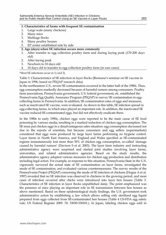
1. Characteristics of farms with frequent SE contamination
1. Large scale (many chickens) 2. Many mice 3. Multiage flocks 4. Many poultry houses 5. EP center established side by side 2. Age (days) when SE infection occurs more commonly 1. After transfer to egg collection poultry farm and during laying peak (170-200 days
old) 2. After laying peak 3. Newborn to 10 days old 4. 10 days old to transfer to egg collection poultry farm (in rare cases)
*Most SE infections occur in 1) and 2).
Table 3. Characteristics of SE infection in layer flocks (Biomune's seminar on SE vaccine in Japan in 1996, based on PEQAP research data) In the northeastern U.S., severe SE contamination occurred in the latter half of the 1980s. Thus, egg consumption markedly decreased because of harmful rumors among consumers. Poultry farm associations, Pennsylvania government, U.S. federal government, etc. established the Pennsylvania Egg Quality Assurance Program (PEQAP) to survey SE contamination in egg-collecting farms in Pennsylvania. In addition, SE contamination rates of eggs and measures, such as inactivated SE vaccine, were evaluated. As shown in this table, SE infection spread in egg-collecting farms, in which mice played an important role. In addition, the inactivated SE vaccine reduced SE-contaminated eggs, but did not effectively eradicate them.
In the 1980s to early 1990s, chicken eggs were reported to be the main cause of SE food poisoning by various media, resulting in a marked reduction of chicken egg consumption. The SE issue put chicken eggs in a disadvantageous sales situation: egg consumption decreased not due to the reports of scientists, but because consumers and egg sellers (supermarkets) considered that eggs were produced by large layer farms performing no hygiene control. Layer farms in North East America, and England and Wales specified as SE-contaminated regions instantaneously lost more than 50% of chicken egg consumption, so-called ‘damage caused by harmful rumors’ (Davison S et al. 2003). The layer farm industry and instructing administrative agency were surprised and started joint studies involving layer farms, universities, and related administrative agencies. Based on the study results, the administrative agency adopted various measures for chicken egg production and distribution including legal action. For example, in response to this situation, Pennsylvania State in the U.S. vigorously surveyed the actual state of SE contamination on layer farms, investigated the mode of SE contamination, and evaluated various countermeasures. The survey results of the Pennsylvania Project (PEQAP) concerning the mode of SE infection of chickens (Hogue A et al. 1997) revealed that no SE infection was observed in chickens in the growing period, and most cases of infection occurred after chicks were introduced into layer hen houses (Table 3. Characteristics of SE infection in layer flocks unpublished data). The point emphasized was the presence of mice playing an important role in SE transmission between hen houses as above mentioned. Based on these epidemiological study findings, the U.S. government took administrative action by establishing a law which allows selling only sterilized egg liquid prepared from eggs collected from SE-contaminated hen houses (Table 4 US-FDA egg safety rule; US Federal Register 2009. 74: 33030-330101.). In Japan, labeling chicken eggs sold in
www.intechopen.com

Salmonella – A Diversified Superbug
284
packages with a date (laid, packaged, or sell-by date) is required. Administrative actions have been taken against SE food poisoning in many countries. In which “Red Lion Code” is involved. The history of the recent emergence of SE food poisoning emphasized the necessity of analyzing the cause of food poisoning in the processes of chicken egg production through distribution and consumption (Schroeder CM et al. 2006). For analysis and the control of health damage risks of not only chicken eggs but also all food products, identification and evaluation of possible risks in each step of production, distribution, and consumption and investigation of countermeasures while considering the cost-effectiveness have been established as “ risk analysis concept” and applied to the problem of SE-contaminated chicken eggs as one case. However, no basic concept for the control of bacterial food poisoning has been established, and many epidemiological studies are still necessary.
Testing or Procedure FDA
Chicks NPIP SE Clean breeders
Pullet testing 14 to 16 weeks
Requirement for pullet + manure Egg testing of 4 sets of 1000 eggs at 2 week intervals
Layer testing 40w and 4-6 weeks after molt completion
Egg testing if manure positive 1000 eggs at 2 week intervals, 4 submissions
Diversion to pasteurization required for egg+ flocks
Yes
Return to shell market allowed Yes after a completed set of 4 submissions of 1000 eggs every 2 weeks
Egg testing after initial egg test set None if negative first set; once a month if were previously egg positive
C&D of manure or egg + houses Wet or dry cleaning
Vaccination required None
Biosecurity plan Required
Rodent Control Plan and Records Required
Fly Control Plan and Records Required
(personal information from Dr. Lozano F)
Table 4. US-FDA egg safety rule (Established by USDA, 2009)
2.3 History of SE vaccine
Live ST vaccine was used as SE control in large-scale state layer farms in Former Eastern Europe before the reunification of East and West Germany as described below. The safety and efficacy of this live ST vaccine for SE control were investigated, and several preparations are still used now.
Regarding inactivated SE vaccine, I would like to introduce the history of its first appearance in the world. The in-house vaccine system was established in the U.S. in the 1980s. In this system, farms which isolated the pathogen were allowed to use an inactivated vaccine for the infectious disease not included in highly pathogenic infectious diseases of animals, such as legal infectious diseases, and approval was granted to in-houses vaccine manufacturers. An in-house vaccine manufacturer produced a vaccine using an SE strain isolated from a layer farm,
www.intechopen.com

Salmonella Enterica Serovar Enteritidis (SE) Infection in Chickens and Its Public-Health-Risk Control Using an SE Vaccine in Layer Flocks
285
and the vaccine reduced the SE isolation frequency on the farm. This was the first preparation of inactivated SE vaccine. The world’s first state approval was granted for a vaccine which showed efficacy in the field, not prepared through establishing an evaluation method in a laboratory and then confirming the efficacy in the field. Subsequently developed inactivated SE vaccines were produced following the first inactivated SE vaccine as the standard.
3. Discussion on the usefulness of SE vaccine administration to chickens
3.1 Situation at the time of early approval of inactivated SE vaccine
The world’s first approval of inactivated SE vaccine by the administrative authority was granted to Layermune SE (Biomune Co., Kansas) in the U.S.A. in 1992. Since then, inactivated SE vaccine has been discussed with regard to not only the efficacy but also many other aspects. Discussions have mainly concerned doubt regarding the efficacy, and, secondly, vagueness of the objective of use. Generally, the objective of animal and human vaccines is the prevention of clinical problems of vaccinated animals and humans, but SE infection manifests no clinical symptoms in chickens excluding newborn chicks, causing no economic damage. For newborn chicks, there is no time for vaccination because infection occurs before inactivated SE vaccine exhibits an immunological effect. Accordingly, inactivated SE vaccine is administered to chickens developing no clinical problems, and the objective is only to reduce the public health risk (reduction of SE-infected chicken egg production). Chickens are vaccinated for a disease manifesting no clinical symptoms, but the effect of the vaccine has to be investigated in these chickens. Economically, inactivated SE vaccine has to be administered to individual chickens, requiring considerable human labor and expense for purchasing the vaccine. Since SE infection causes no direct economic damage, vaccine administration to chickens on farms requires the high-level motivation of vaccine users. The first inactivated SE vaccine was a new type of vaccine, i.e., emergence of a high-cost vaccine slightly stressful to vaccinated animals and not preventing disease in the animals white leghorn chickens (Mizumoto N et al. 2004).
We also investigated the efficacy of inactivated SE vaccine employing various challenge tests. In one of the tests, SE was orally challenged 3 or 4 weeks after inactivated SE vaccine administration, and SE was re-isolated from the gastrointestine and parenchymal organs. Concretely, 3-week-old SPF chickens were vaccinated at the normal dose and orally challenged with food poisoning-derived SE at a high bacterial count after 4 weeks (at 7 weeks of age), and the bacteria were re-isolated from the cecum after 1-7 days. SE was isolated from nearly 100% of chickens despite the vaccine having been administered. When the number of challenged bacteria was reduced to a moderate count, the number of isolated bacteria was significantly decreased in many animals in the vaccinated group, but the results were not stable. In chickens subjected to the test at 5 or 7 weeks of age, the isolation frequency after challenge (at 11 weeks of age) was markedly lower in the control non-vaccinated group. Accordingly, a large number of chickens are necessary to perform the challenge test at this age, which is not routinely possible. In this laboratory test, a significant reduction of the intestinal bacterial count was observed, but complete disappearance of the bacteria from the gastrointestine has not been confirmed within a couple of weeks.
3.2 Situation at the time of the initiation of our study
In 1990, we were informed of SE-contaminated chicken egg production on layer farms covered by our veterinary care activity. We administered bacteriostatics and organic acids
www.intechopen.com

Salmonella – A Diversified Superbug
286
on large-scale layer farms, hoping to avoid a decline in consumption, which occurred in America and England, but no effect was obtained. Thus, we performed a field epidemiological study of the efficacy of inactivated SE vaccine (Yamane Y et al. 2000). Inactivated SE vaccine was administered to flocks on a large-scale layer farm with apparent SE contamination. Eggs (500 kg) were broken in a liquid egg plant, 1,000 ml was sampled from the liquid egg batch, and 400 ml was subjected to SE isolation. The isolation rate was compared between the vaccinated and non-vaccinated groups. The results are shown in Fig. 1. (Fig. 1 Number of SE isolates and SE isolation frequency of SE-contaminated chickens and inactivated SE-vaccinated chickens in the same poultry house). Furthermore, our study confirmed horizontal infection of 4 industrial poultry farms (Table 5. SE samples monitored and their results with 4 integrated layer companies (1996-1998)). Based on those results, it was considered necessary for the positivity rate on plate agglutination with pullorum disease-diagnostic antigen to be 90% or higher in the SE-inoculated group (0% in the non-inoculated control group), while vaccination of SE-contaminated farms significantly reduced the number of bacteria isolated from liquid egg samples from 500 kg or more of eggs compared to that from non-vaccinated chickens (Table 6 Evaluation criteria for the inactivated SE vaccine (Layermune SE) in field chickens). In addition, the requirement of the number of isolated bacteria from chicken feces was set at 1 CFU or lower per 1 g in the inactivated SE vaccine-treated group. Later, similar results were obtained in the test using 20 kg of eggs (about 320 eggs). We partially demonstrated these established values epidemiologically after more than 10 years (a)Toyota-Hanatani Y et al 2009).
Vertical section for SE monitor
Sample materials Result
monitored Memo
Breeder farms (Hatchery)
1) Several swabs 2) Manure 3) Sera to detect antibody 4) Workers feces 1) Swab 2) Worker feces
No detection at all No detection
Using SE cell antigen coated ELISA
Feed mile Any protein source No detection
Growing Like as breeder No detection
Laying 1) Swabs 2) Manure 3) Dusts 4) Liquid eggs 5) Workers feces 6) Water in EP center
No detection A few positive A few positive Several positive No detection Detectable
Table 5. SE samples monitored and their results with 4 integrated layer companies (1996-1998) (Yamane Y et al. 2000. modified). Our severance studies (Yamane Y et al. 2000) summarizes the results of SE tests conducted in breeding farms, feed mills, and EP centers for three years. As shown in this table, no vertical transmission (infection from laying to adult chickens) occurred. The infection was repeated within a laying poultry houses. Materials and Methods; See Fig. 1.
www.intechopen.com

Salmonella Enterica Serovar Enteritidis (SE) Infection in Chickens and Its Public-Health-Risk Control Using an SE Vaccine in Layer Flocks
287
Case 1 Case 2
Age SE(MPN) ELISAC Age SE(MPN) ELISA
Year1 Jan. 237 ○ 0 % 181
260 ○ 252 ●( >1600) 0 %
0 %
350 ○
0 % 45 %
Jul. 20 % 349 ●(NT)
455 ●(NT) 20 % 28 %
35 %
447 ●(NT)
552 ●(39) 15 % 20 %
Year2 Jan.
35 % 25 %
25 % 10 %
25 %
40 %
Jul. 729 Replaced
881 娟
911 ▲ 35 %
218 娟
937 Replaced 245 (<2)
259 娟
Year3 Jan.
168 娟 316 娟
239 娟 386 娟
302 娟
435 娟
Fig. 1. Number of SE isolates and SE isolation frequency of SE-contaminated chickens and inactivated SE-vaccinated chickens in the same poultry house (Yamane Y et al. 2000) SE isolation in field chickens before and after inoculation of the inactivated SE vaccine (Layermune SE) (Four cases are shown in the reference. Two cases are shown here.) (Filled circles indicate SE isolation). The number indicates the number in ( ) of SE isolates. The detection rates of SE antibodies in unvaccinated chickens by ELISA coated with SE cell antigen was 0-40% for Case 1 and 0-45% for Case 2. To our experiences, the antibody positive rate of 400-500-day-old chickens inoculated with the inactivated SE vaccine (at 300-400 days after vaccination) was about 70-100%. Inaccurate administration of SE bacterin may induce the antibody positive rate of inactivated vaccine to be further decreased. Thus, vaccination and field infection cannot be distinguished at antibody level. The number and frequency of SE isolates decreased in the vaccinated group.
www.intechopen.com

Salmonella – A Diversified Superbug
288
Material &Methods: An industrial layer farm was monitored. SE isolation was done using liquid eggs samples originating from 500 kg of shell eggs. And then most probable number (MPN) per 100 m was determined. For detection of specific antibody in the sera of the flocks, an ELISA coated with SE cell antigen was used.
Test item Method Procedures Criteria
Antibody response
RPA Twenty chickens were examined at 4 weeks after vaccination.
≥90%: Markedly effective <90%~≥80%: Effective <80%: Non effective
Antibody response
ELISA Same as above Same as above
Bacterial isolation
Bacterial isolation
500 kg of eggs are collected from the vaccinated group. The eggs are broken and cultured within 48 hours after collection.
≤10MPN/100mL: Markedly effective (if materials from the unvaccinated group of the same farm showed ≥1,600 MPN/100 mL)
Table 6. Evaluation criteria for the inactivated SE vaccine (Layermune SE) in field chickens (application form for the reexamination of this formulation in Japan, provided by CAF Laboratories) The effectiveness of the formulation (Layermune SE) in Japan is evaluated based on this table. The formulation was effective in all the 12 chicken groups by an antibody test. However, SE-contaminated farms could not be surveyed by bacterial isolation.
3.3 Risk of misjudging inactivated SE v accine-treated chi ckens as SE-infected chickens
We had a problem in handling inactivated SE vaccine in our field facilities: inactivated SE vaccine-treated chickens and SE-infected chickens showed the same serological reaction (Table 7. Production of antibodies against SE bacterial antigens in inactivated SE-vaccinated and -unvaccinated chickens). Inactivated SE vaccine is generally administered at about 80 days of age. In chickens treated with a commercial inactivated SE vaccine, the anti-bacterial cell antigen-antibody positive rate determined using commercial antigen solution for the diagnosis of PD, or SE cell antigen coated ELISA reaches nearly 100% within about 120 days of age and then slowly decreases and reaches 20-60% at about 300 days of age, whereas the positive rate in SE-infected chickens is about 5-70%. We attempted to distinguish SE-infected from inactivated SE vaccine-treated chickens because eggs laid by inactivated SE vaccine-treated chickens are misjudged as those laid by SE-infected chickens, if the 2 chicken groups of SE infected and vaccinated cannot be distinguished. Thus, we investigated specific antibodies present only in chickens with ‘inactivated SE vaccine treatment’ described below (Fig. 4. Detection of specific antibodies in sera against SE cell antigen and SEp9 on oral SE administration to field white leghorn chickens) (Mizumoto N et al. 2004).
www.intechopen.com

Salmonella Enterica Serovar Enteritidis (SE) Infection in Chickens and Its Public-Health-Risk Control Using an SE Vaccine in Layer Flocks
289
Group Positive rate (references) Test methods** (References)
Inactivated SE vaccine In the laboratory In field
Vaccination At 30-40 dpv: 95-100% : ≥ 90% 300~400 days old: 70~100%
ELISA RPA
ELISA
SE infected group (Field group)
Shipping to slaughterhouse (about 700 days old): 0-15% Induced molting (400-500 days old): 0~45%
ELISA
ELISA
(Mizumoto N et al. 2004, Sunagawa H et al. 1997, Yamane Y et al. 2000)
* Age of vaccination: around 80 days old ** ELISA: Indirect method with SE cell antigen coated. RPA: rapid plate agglutination with diagnostic for pullorum disease antigen.
Table 7. Production of antibodies against SE bacterial antigens in inactivated SE-vaccinated and -unvaccinated chickens (summarized by our research group) Almost all the 3-week-old or older chickens inoculated with the inactivated SE vaccine were positive at around four weeks by both ELISA (coated with SE cell antigen) and RPA. Subsequently, the positive rate decreased at 250 days or later after inoculation. The positive rate in the ELISA coated with the g.m. antigen of SE was shown above 80% up to about 700 days old. On the other hand, SE-contaminated chickens showed the similar positive rates as those of inactivated SE-vaccinated chickens in ELISA coated with SE bacterial antigen and RPA. Generally, the positive rate of SE-contaminated chickens is lower than that of inactivated SE-vaccinated chickens. However, an antibody test cannot distinguish these 2 groups, because some SE-contaminated chickens show higher positive rate.
3.4 Active component of inactivated SE vaccine (main Fli C antigen: SEp 9)
Using sera from inactivated SE vaccine-treated and SE-infected chickens, we compared the production of antibodies against the SE cell antigen to investigate differences between the sera. A strong reaction with a 53-kDa polypeptide (Fli C) (Namba K et al. 1997) was observed in all serum samples from inactivated SE vaccine-treated chickens, but rare reaction with a specific antigen was noted in SE-infected chicken-derived serum samples (Fig. 2. Western blotting with sera from SE-infected and inactivated SE-vaccinated chickens using formalin-treated SE antigens (surface antigens)). Fli C is considered to be strongly antigenic as inactivated SE vaccine. When the SE-specific polypeptide (g.m. antigen) in Fli C (Van Asten AJ et al. 1995, and Yap LF et al. 2001) was prepared by genetic engineering and reacted with serum from inactivated SE vaccine (Layermune SE)-treated chickens, strong reactivity was noted, but SE-infected chicken-derived serum did not react with g.m. antigen. When the specific antibody reaction was investigated in sera from chickens treated with other vaccines sold in Japan (oil adjuvant vaccine 3 and aluminum hydroxide gel vaccine 1), a specific antibody reaction with g.m. antigen was noted in the serum of oil adjuvant vaccine-treated chickens (Fig. 3.
www.intechopen.com

Salmonella – A Diversified Superbug
290
Production of specific antibodies against commercial inactivated SE vaccines SE cell and SEp9 antigens). In an experiment, the inoculated chickens with SE induces antibody against SE cell antigen but not SEp 9. In field poultry flocks, inactivated SE vaccine administration was confirmed a long period persistency of specific antibody level against SEp 9 until 700 days of age (Fig. 5. Positive rates of g.m.-specific antibodies in the yolks derived from field chickens inoculated with the inactivated SE vaccine).
(a) (b)
Fig. 2. Western blotting with sera from SE-infected and inactivated SE-vaccinated chickens using formalin-treated SE antigen (surface antigen) (Nakagawa Y et al. reported by Japanese) Figure 2a shows the reactivity of sera from 3-week-old SPF chickens which received oral SE administration (C1~9, M: marker protein), examined by Western blotting (SDS-PAGE) with SE surface antigen. Fig. 2b shows Western blotting with the same antigen using sera from 3-week-old SPF chickens inoculated with the inactivated SE vaccine (Layermune SE) (at 4 weeks after inoculation) (V1~3) or from those from which SE was isolated from naturally-infected-field flocks (N1~5; 710 days old). Fig. 2a shows that light antibody response against 53 kDa (Fli C of SE) was noted in two chickens (one chicken at 2 weeks) and no band against Fli C (53 kDa polypeptide) was noted in all the nine SE-intraoral inoculated chickens. As shown this figure, one of 2 responded band at week post inoculation (wpi) was continued by 2 wpi but not by 4 wpi. Thus, the responsive antibody was considered to be IgM antibody. In another our report, a 53 kDa band was not detected in 4-week-old SPF chickens and 300-day-old field chickens, which received SE administration, but was detected in molting-induced chickens (Mizumoto N et al. 2004, Piao Z et al. 2007). Thus, the antibody against the 53 kDa polypeptide after SE inoculation is suspected no invasion into the internal organs. Fig. 2b shows strong bands against the 53 kDa polypeptides and its dimer (98 kDa) in inactivate SE-vaccinated chickens. However, in chickens from which SE could be isolated, a weak band could be detected at around 42 kDa, but no band could be detected at 53 kDa. Materials and Methods: For antigen preparation, SE was treated with formalin and centrifuged at 2000 g for 20 min. Then the supernatant was further centrifuged at 10,000g for 60 min and the precipitate dissolved in a buffered saline. The antigen was used in this analysis. The sera for SE infected chickens were prepared from the chickens inoculated with SE at the age of 3 weeks, and were weekly bled individually for this study. To the “vaccine sera”, SPF chickens were injected with Layermune SE at the age of 3 weeks and bled 4 weeks post injection. The sera were designed as vaccine sera.
www.intechopen.com

Salmonella Enterica Serovar Enteritidis (SE) Infection in Chickens and Its Public-Health-Risk Control Using an SE Vaccine in Layer Flocks
291
ケクケケ
ケクコケ
ケクサケ
ケクシケ
ケクスケ
ゲクケケ
ゲクコケ
ゲクサケ
ケ ゲ コ ゴ サ ザ シ ジ ス
Weeks post vaccinated
テLISヂ
titer ォSテギIオ
Vaccine ヂ
Vaccine ッ
Vaccine ツ
Vaccine ヅ
Nonギvaccinated
(a)
ケクケケ
ケクコケ
ケクサケ
ケクシケ
ケクスケ
ゲクケケ
ゲクコケ
ゲクサケ
ケ ゲ コ ゴ サ ザ シ ジ ス
Weeks post vaccinated
テLISヂ
titer ォSテギIIオ
(b)
Fig. 3. Production of specific antibodies SE cell (deflagellated) and SEp9 antigens (Nakagawa Y et al. Japanese report) (a; Antibody response to SE cell antigen, b; Antibody response to SEp9) Four commercial inactivated SE vaccines (Vaccine A to D) were used to inoculate five 3-week-old SPF chickens/group to examine the responsiveness to SE cell antigen and SEp9. Results shown in Fig. 3A and 3B were obtained. No response was noted in unvaccinated chickens. The inactivated SE vaccine responded to SE cell antigen in all the chickens. Notably, the antibody response of the formulation with aluminum gel used as adjuvant rapidly increased and then decreased. On the other hand, the antibody response to SEp9 was specific to each vaccine. However, this may have resulted from vaccine lot-variation. Further studies are needed to make a conclusion. Notably, there was no response to the formulation with aluminum gel used as adjuvant.
When the levels of antibodies against inactivated SE vaccine-induced SE cell antigen and flagella were compared, as shown in Table 8. (Table 8. Detection of SE-specific antibodies by
www.intechopen.com
Salmonella – A Diversified Superbug
292
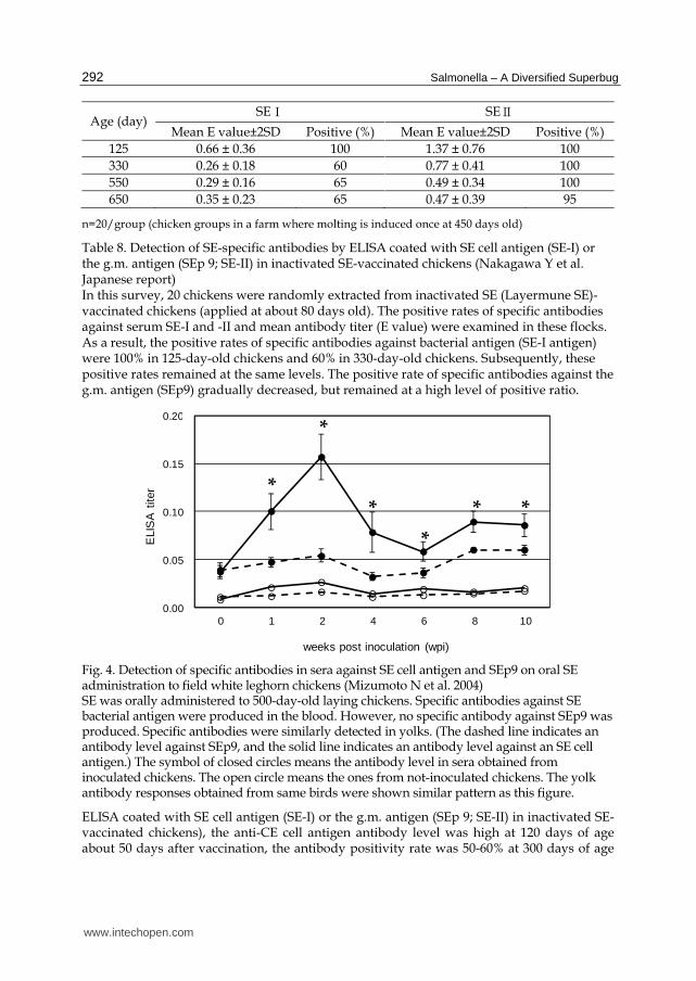
Age (day) SE⊆ SE⊇
Mean E value±2SD Positive (%) Mean E value±2SD Positive (%)
125 0.66 ± 0.36 100 1.37 ± 0.76 100
330 0.26 ± 0.18 60 0.77 ± 0.41 100
550 0.29 ± 0.16 65 0.49 ± 0.34 100
650 0.35 ± 0.23 65 0.47 ± 0.39 95
n=20/group (chicken groups in a farm where molting is induced once at 450 days old)
Table 8. Detection of SE-specific antibodies by ELISA coated with SE cell antigen (SE-I) or the g.m. antigen (SEp 9; SE-II) in inactivated SE-vaccinated chickens (Nakagawa Y et al. Japanese report) In this survey, 20 chickens were randomly extracted from inactivated SE (Layermune SE)-vaccinated chickens (applied at about 80 days old). The positive rates of specific antibodies against serum SE-I and -II and mean antibody titer (E value) were examined in these flocks. As a result, the positive rates of specific antibodies against bacterial antigen (SE-I antigen) were 100% in 125-day-old chickens and 60% in 330-day-old chickens. Subsequently, these positive rates remained at the same levels. The positive rate of specific antibodies against the g.m. antigen (SEp9) gradually decreased, but remained at a high level of positive ratio.
0.00
0.05
0.10
0.15
0.20
0 1 2 4 6 8 10
ELI
SA
tite
r
weeks post inoculation (wpi)
*
*
*
*
* *
Fig. 4. Detection of specific antibodies in sera against SE cell antigen and SEp9 on oral SE administration to field white leghorn chickens (Mizumoto N et al. 2004) SE was orally administered to 500-day-old laying chickens. Specific antibodies against SE bacterial antigen were produced in the blood. However, no specific antibody against SEp9 was produced. Specific antibodies were similarly detected in yolks. (The dashed line indicates an antibody level against SEp9, and the solid line indicates an antibody level against an SE cell antigen.) The symbol of closed circles means the antibody level in sera obtained from inoculated chickens. The open circle means the ones from not-inoculated chickens. The yolk antibody responses obtained from same birds were shown similar pattern as this figure.
ELISA coated with SE cell antigen (SE-I) or the g.m. antigen (SEp 9; SE-II) in inactivated SE-vaccinated chickens), the anti-CE cell antigen antibody level was high at 120 days of age about 50 days after vaccination, the antibody positivity rate was 50-60% at 300 days of age
www.intechopen.com

Salmonella Enterica Serovar Enteritidis (SE) Infection in Chickens and Its Public-Health-Risk Control Using an SE Vaccine in Layer Flocks
293
(220 days after vaccination), and the rate was retained thereafter. In contrast, g.m. antigen (SEp 9)-antibody level was maintained at a high level until 700 days of age (about 620 days after vaccination). An experimental inoculation with SE in SPF chickens showed similar response (Fig. 4. Detection of specific antibodies in sera against SE cell antigen and SEp9 on oral SE administration to field white leghorn chickens). This tendency of the presence of specific antibody in egg yolk was observed (date not shown).
Fig. 5. Positive rates of g.m.-specific antibodies in the yolks derived from field chickens inoculated with the inactivated SE vaccine (Publishing elsewhere by Nakagawa Y et al. ) In this study, the inactivated SE vaccine (Layermune SE) was used to inoculate about 80-day-old chickens in six farms. Ten eggs were randomly collected once in two months from 150-700-day-old chickens of each farm. Mean antibody titers (positive: ≥ 0.1 E values) against the g.m. antigen (SEp9) in yolks were determined. These chickens were giving an induced molting for about 40 days after day 450. During this period, eggs were not sampled. The determination with specific antibody to g.m. antigen was done according to the method described by Mizumoto N et al. 2004. The mean positive rate of the farms was about 88%. The positive rates were above 80% in all the farms. Thus, about 700-day-old chickens carried antibodies against SEp9. Antibodies against SEp9 markedly decreased the number of SE isolates in the gastrointestinal tract). In addition, antibodies against SEp9 inhibited SE isolation from eggs in the report. Proper vaccination prevented SE infection for a long time. Thus, specific antibodies remained in chickens inoculated with the inactivated SE vaccine, even after molting was induced once, as examined by SEp9-coated ELISA. The specific antibodies could be detected also in yolks.
3.5 Immunogenicity of SEp 9
A high specific antibody production level was noted in antibodies against a flagellar component, Fli C, in inactivated SE vaccine-treated chickens, as described above. The SE-
*
*
*
*
****
****
**
**
**
**
**
**
***
*
***
*
**
**
**
*
***
******
www.intechopen.com

Salmonella – A Diversified Superbug
294
specific region in Fli C is g.m. antigen (SEp 9), and the antigen was assumed to be effective as the antigenic site of inactivated SE vaccine (Toyota-Hanatani Y et al. 2008, and b)Toyota-Hanatani Y et al. 2009), for which we prepared SEp 9 antigen by genetic engineering and investigated the efficacy of SEp 9 vaccine. Since no international method (challenge test model) has been established for efficacy evaluation of inactivated SE vaccine, we analyzed tissue reactions at the vaccine administration site in vaccinated chickens.
Fig. 6. Histological reactions at the inoculation with f the inactivated SE vaccine or the g.m site of Fli C (b)Toyota-Hanatani H et al. 2008) We investigated a kinetic of histological reactions at the inoculation site of commercial inactivated SE vaccine or SEp 9 antigen. In the inoculation site (7a) at one week post vaccination (wpv), many histocytes were infiltrating, and hyperplastic connective issues are
a
b
a
a
b
a) b)
c) d)
e)
b
a
www.intechopen.com

Salmonella Enterica Serovar Enteritidis (SE) Infection in Chickens and Its Public-Health-Risk Control Using an SE Vaccine in Layer Flocks
295
shown (arrow a). However, tissue images, such as oil cyst, were not observed. In (7b) at 2wpv, necrosis (arrow a), surrounded by granulomatous structures (arrow b), was observed in the middle of inflammatory response. Polynuclear cells appeared in some granulomatous structures. Oil cyst was also observed. These images indicate that the antigen and oil ingredients were actively excluded from the vaccine, suggesting the establishment of specific immunity. At 4 wpv (7c), severe necrosis at 2 weeks became smaller, and the inflammatory response resolved (arrow a). In addition, peripheral lymphoid node structures (arrow b) appeared near the disappearing necrosis, suggesting active antibody production. At 6 wpv (7d), hyperplastic connective tissues also disappeared. Of the tissue reactions in the vaccination site, the characteristic responses during specific immune reaction are the emergence of polynuclear, which surrounded the granulomatous structure, and peripheral lymphoid node like structure. Thus, the inoculation site of SEp9 antigen was histologically examined at four weeks. As shown in Figure 7e, a lymphoid node like structure (arrow a) and a small number of polynuclear cells (arrow b) appeared in the SEp9 inoculation site. Thus, we concluded that SEp9 could induce specific immunity in chickens. Materials and Methods; A commercial inactivated SE vaccine was injected and weekly taken tissue sample at the injected site, and then fixed and stained as usual (HE staining, X50).
The general time course of histological changes at the inoculation site with inactivated SE vaccine (oil-adjuvant-type) is shown in Fig. 6 (Histological reactions at the inoculation with f the inactivated SE vaccine or the g.m site of Fli C); nonspecific inflammation characterized by marked monocyte infiltration was noted after 1 week, and perivascular granulomatous changes were noted at 2 weeks including the appearance of multinucleated giant cells. At 3 weeks after vaccination, lymphocyte clustering showing a lymph node-like structure, considered to be an antibody production site, was noted. These reactions then slowly disappeared. In granulomatous changes accompanied by multinucleated giant cell infiltration observed after 2 weeks, cellular reactions of delayed hypersensitivity were noted (Table10. Characteristics of histological lesions at the inoculation site in the chicken applied with commercial SE vaccine (4wpi)). The tissue reactions at the SEp 9-administered site were similar to those induced by commercial inactivated SE vaccine, confirming anti-SEp 9-specific antibody production (Table 11 Production of specific antibodies in chickens inoculated with the inactivated SE vaccine or the g.m. site of Fli C).
When SEp 9-treated and non-treated chickens were orally challenged with SE, gastrointestinal SE was significantly decreased in the SEp 9-treated group compared to that in the non-vaccinated group, and the number of isolated bacteria was decreased similarly to that in the commercial inactivated SE vaccine-treated group (Fig. 7. Challenge test in chickens inoculated with the inactivated SE vaccine or the g.m site of Fli C). Although it is not clarified why the specific immunity induced by SEp 9 injection in chickens is able to reduce SE colonization in gastrointestinal organs, we have suspected that the induced immunity may affect SE yielding lower colonization ability SE. For example, the amount of a fibrin molecular, 21 kDa polypeptide, might be reduced on surface resulting from the induced specific immunity without SE-proliferation reduction. This is because the isolation level at 1 week post challenge in Fig. 7 (Challenge test in chickens inoculated with the inactivated SE vaccine or the g.m site of Fli C) does not show different bacterial level between SEp 9 injection and non-injection groups, even though statistical difference is observed. To this point, we will attempt to further clarify the mechanisms of lower SE-colonization in SEp 9-injected birds.
www.intechopen.com

Salmonella – A Diversified Superbug
296
weeks post challenge (wpc)
Fig. 7. Challenge test in chickens inoculated with the inactivated SE vaccine or the g.m site of Fli C (Toyota-Hanatani Y et al. 2009) SEp9 induced specific immunity. Subsequently, a challenge test was conducted in SEp9-inoculated chickens. The results are shown in this figure. When buffer alone was used for inoculation, the number of SE isolates did not decrease, but remained constant. The number of SE isolates decreased in chickens, inoculated with a commercial inactivated SE vaccine or SEp9, with aging. Material and Methods; a buffered saline, SEp 9 and Layermune SE were twice-injected with mixture with an oil adjuvant, respectively. Four weeks post injection from final application, those chickens were orally challenged with SE Y 24 strain, and SE isolation was performed from intestinal samples.
Type of Bird
Type of Vaccine
Vaccination Age Route of
Administration Program Advantages
Breeders Live Killed
1 day old 7 Weeks old 12-14 Weeks of Age 18- 20 Weeks of Age
Coarse Spray Drinking Water Subcutaneous Subcutaneous
Broad Protection Selective Competitive Exclusion Strong Maternal Immunity
Layers Live Killed
1 day old 7 Weeks old 10-12 Weeks of Age
Coarse Spray Drinking Water Subcutaneous
Broad Protection Selective Competitive Exclusion Strong Immunity
Broilers Live 1 Day old Coarse Spray or Drinking Water
Coarse Spray or Drinking Water
Strong Immunity
Table 9. Recommended Salmonella vaccination programs in poultry.
www.intechopen.com

Salmonella Enterica Serovar Enteritidis (SE) Infection in Chickens and Its Public-Health-Risk Control Using an SE Vaccine in Layer Flocks
297
Indicator No.
Activation result of Characteristics in
histological observations Immunological
properties
1 Cellular immunity Granular formation (lumps) with epithelioid cells
Type 4 hypersensitivity (Pellertier M et al. 1984. Uthoaisangssok S et al. 2002)
2 Humoral immunity Perivascular accumulation with lymphocytes
Activated B-lymphocytes
3 Non-specific immunity
Hyperplastic connective tissue, infiltration of non-specific immune cells
Early or late non-specific immune reaction
Table 10. Characteristics of histological lesions at the inoculation site in the chicken applied with commercial SE vaccine (4wpi) (supplementary data by Toyota-Hanatani Y et al. 2008). This table shows three categories of reactions characteristic of tissue images on inoculation of a commercial inactivated SE vaccine: (1) cellular immunity, (2) hormonal immunity, and (3) nonspecific reaction. All the above reactions were observed in SEp9-inoculated chickens at 4 weeks. Supplementary data from other studies by Toyota-Hanatani Y et al. were also discussed in this table. Granulomatous reaction, observed at 2-4 weeks after inoculation of the inactivated SE vaccine, was considered to be the same tissue reaction as tuberculin reaction. We considered it to be cellular type IV (delayed) hypersensitive reaction. This might be a process of developing cellular immunity.
Immunizing antigen Tested chickens
Production of antibodies against g.m. (SEp 9)
Antibody positive conversion
Mean value in ELISA
g.m.(SEp 9) antigen 4 4 0.84
De-flagellated SE antigen
4 0 0.00
Buffer 4 0 0.00
Inactivated SE vaccine 4 4 0.83
Table 11. Production of specific antibodies in chickens inoculated with the inactivated SE vaccine or the g.m. site of Fli C (supplementary data by Toyota-Hanatani Y et al. 2008) This table shows specific humoral immunity induced by the g.m. antigen site (SEp9). As shown in the table, specific antibodies were produced when SEp9 was used to inoculate chickens with adjuvant. However, no specific antibodies against SEp9 were produced when SE cell antigen was used. Importantly, the g.m. antigen site has high immune induction capacity in chickens because a small amount of antigen (100 μg/bird; about 30 μg/bird of not involving GST) induces specific immunity. In another study where SEp9 in buffer was used to inoculate chickens (un-published data), specific antibodies were produced. Thus, specific immunity can be induced even without adjuvant. Materials and Method; See Fig. 7
www.intechopen.com

Salmonella – A Diversified Superbug
298
3.6 The details of attenuated live Salmonella vaccines for poultry
The first live Salmonella vaccine for poultry was a Salmonella enterica Serovar Gallinarum (SG) developed in the early 1950’s (Williams SH.et al. 1956). This attenuated SG rough strain called 9R has been used in many countries around the world for the control of fowl typhoid. However, interference with official Salmonella control and eradication programs using serological methods has limited the wider use of this attenuated strain in addition to scattered field reports of excessive attenuation and reversion to virulence. The development of paratyphoid live attenuated Salmonella vaccines is an advancement and reinforcement to the use of inactivated vaccines for Salmonella control programs in the poultry industry. These new attenuated live Salmonella vaccines elicit cell-mediated, mucosal and humoral immune responses (Gomez-Duarte. et al. 1999, Roy Curtiss R 3rd et al. 1996, Kulkarni KK et al. 2008, Ashraf S et al. 2011). In addition, new recombinant DNA technology permits the expression in Salmonella serovar strains of protective antigens from unrelated bacterial, viral or parasitic pathogens.
There are two common approaches which have been applied in the development of the new paratyphoid live Salmonella vaccines. One of them is the genetic manipulation through recombinant technology selecting virulence genes to be deleted in selected Salmonella serovars. The other approach is the manipulation of the media used for Salmonella propagation resulting in a metabolic drift mutation reducing the activity of essential enzymes and the bacterial metabolic regulatory systems resulting in slower propagation cycles under natural infection conditions and this prolonged generation time cause reduced bacterial multiplication within the host at a significant rate. Consequently, when the genetically or chemically attenuated Salmonella strain is administered to the birds, the modified bacteria lives long enough to stimulate an immune response in chickens before to be eliminated within few weeks after administration of the vaccine. Currently, two paratyphoid serovars are commercially available as live attenuated vaccines: ST and SE.
It is considered that the genetic deletion of selected virulent genes induced a more attenuated recombinant Salmonella serovar strains compared with the chemically induced metabolic mutants, which still have residual enzymatic activity and more invasivity inducing a stronger immune response.
Epidemiological markers (Specific antibiotic resistance or sensitivity patterns) are included in the development process of these live vaccines to be able to differentiate the new construct or mutant from similar wild bacterial serovars in case of a field combined infection.
The field use of these new live attenuated Salmonella vaccines has advantages and precautions to observe when administered to the chickens. The advantages of these live vaccines are: mass administration, different routes of administration (Drinking water, coarse spray), selective competitive exclusion and broader spectrum of immunity. Among the precautions to be observed are: Not compatible with antimicrobials, no water chlorination when administered in the drinking water, careful handling by the operator to protect the worker from self-infection. Different recommendations on the use of the attenuated live Salmonella vaccines may be found in the literature to obtain the best protection against field challenge in a specific environment. Short duration of immunity of the live attenuated vaccines may require 2 to 3 applications every 6 to 10 weeks to obtain a more solid protection. The combined administration of live and inactivated Salmonella vaccines provides broader and long lasting immunity, especially in breeders to transfer strong maternal immunity to the progeny. (Table 9. Recommended Salmonella vaccination programs in poultry).
www.intechopen.com
Salmonella Enterica Serovar Enteritidis (SE) Infection in Chickens and Its Public-Health-Risk Control Using an SE Vaccine in Layer Flocks
299
3.7 SE vaccine in the future
The current live and inactivated SE vaccines have advantages and disadvantages. Live vaccine is readily administrable to newborn chicks, but inactivated SE vaccine cannot be administered before 3 weeks of age. The detail potency mechanisms with live vaccine has not been clarified yet, and concerns over causing public health problems are always present: the possibility of back mutation of the vaccine production strains of SE and ST (such as reversal of pathogenicity) or mutation to a pathogenic strain cannot be completely ruled out, and, accordingly, live vaccine is not applicable for laying chickens as described above. Currently, inactivated SE vaccine is manufactured using the whole cell body containing endotoxin, which may induce stress in chickens, although this is slight.
To overcome these problems, the development of a subunit or vector vaccine comprised of active components of SE is awaited, and many researchers may have started research and development.
4. Marked usefulness of inactivated SE vaccine administration to flocks for reducing the human health risk
4.1 Reduction of SE contamination risk of chicken eggs by inactivated SE vaccine
We have surveyed the reduction of the SE contamination risk of chicken eggs by employing inactivated SE vaccine on field layer farms for a prolonged period. Herein, we report the study results.
Four-year surveys were performed on 4 field layer farms (a total of 2,300,000 chickens maintained in 37 hen-houses). Records of SE isolation from liquid eggs were analyzed. Some chickens in these layer farms were treated with inactivated SE vaccine as a trial before analysis, and all chickens were vaccinated in the 4th year of analysis.
The mean numbers of SE isolated from liquid eggs (MPN/100 mL) in the vaccinated and non-vaccinated groups were 2.5±0.1 and 674.8±162.9, respectively, and the isolation
frequencies were 2.45 and 25%, respectively, showing that the isolation frequency was reduced to 1/10 in the vaccinated group. In addition, no SE was isolated after vaccination of all chickens in the 4th year (0 of 257 samples), as described above.
It was clarified that the use of inactivated SE vaccine on layer farms significantly reduced the number of SE isolated from SE-contaminated eggs and the isolation frequency.
4.2 Risk reduction by inactivated SE vaccine on risk analysis
As described above, inactivated SE vaccine decreased the mean number of SE contaminating eggs as a food product to about 1/260 and the isolation frequency to 1/10. These occurred on SE-contaminated farms when vaccinated and non-vaccinated chickens were mixed. When these were simply compared with the number of orally ingested SE and the incidence of patients reported by the a), b)WHO and FAO-US, the incidence of SE patients in healthy subjects was estimated to be decreased to 1/100 or lower.
The 4 farms involved in our study on the reduction of SE contamination of liquid eggs by inactivated SE vaccine were large-scale farms maintaining 350,000-950,000 chickens. These were windowless farms and high-level general hygiene control was also performed.
www.intechopen.com

Salmonella – A Diversified Superbug
300
Accordingly, similar surveys should be conducted on floor feeding and loose housing layer farms, and the risk-reducing effect of SE vaccine should be investigated based on the combined results at national and community levels. In previous reports, the frequency of SE isolation from feces was reduced by about 70% in regions which applied live and
inactivated SE vaccines individually or in combination (a),b) WHO FAO-US, 2002). The accumulation of individual epidemiological surveys and studies may lead to the effective control of SE food poisoning.
5. Re-consideration of the mode of SE infection in chickens
5.1 Mode of SE infection on farms and in flocks
Many points regarding the mode of SE infection on layer farms were unclear around 1990. Layer farm veterinarians referred to the mode of infection of PD (vertical infection), considering that SE also infects in this mode, and prepared an SE detection and monitoring system. Briefly, the mode of SE infection was considered as follows: SE infects breeding chickens and the infection transmits to chicks through breeding eggs (eggs raised to chickens). Some chicks die, but latent infection occurs in survivors and these chicks grow and lay SE-contaminated eggs. Accordingly, they considered that the antibody test in breeding chickens and SE test in chicks after hatching are important, and did not attach greater importance to SE tests of grown chickens, especially laying hens. Moreover, they considered that inactivated SE vaccine is ineffective for chicks after hatching, and only bacteriostatics and analogous agents are effective. The Pennsylvania Egg Quality Assurance Project (PEQAP) of the U.S.A. actively performed field SE contamination surveys to investigate this hypothesis, and found several new facts, as described above (Davison S et al. 2003, Henzler DJ et al. 1998), Hogue A et al. 1997, Lin FY et al. 1988, Stevens A et al. 1989). The points particularly attracting attention in the PEQAP report are a very low infection frequency in newborn chicks, although contamination occurred, and the absence of SE contamination in raising houses. However, SE contamination was observed most frequently after transfer to layer hen houses over 180 days of age. Even though new episodes of SE contamination occurred thereafter (after the laying peak), the frequency was very low.
SE sensitivity of chickens is schematically presented based on the study results reported by PEQAP and our experience in Fig. 8 (Age-dependent susceptibility of chickens against SE colonization). Chicks are very sensitive to SE infection immediately after hatching, but the sensitivity rapidly decreases. No clinical symptoms develop over the growth and egg-laying periods, but the sensitivity rises around the initiation of sexual maturation (100-120 days of age). In layer hen houses, the frequency of SE contamination is high, elevating the infection risk of chickens. It is considered that most SE infection of chickens occurs after transfer to layer hen houses (around 115 days of age) over the peak laying period (around 180 days of age). The sensitivity of layer hens slightly decreases thereafter but then slowly rises with aging. SE sensitivity may be enhanced when induced molting is performed during this period, but these chickens are already infected immediately after transfer to layer hen houses. Therefore, the infection rate is not actually elevated by induced molting, although the sensitivity is high. Considering SE sensitivity and SE control of layer flocks and economic damage, chicks infected immediately after hatching may be culled because they develop clinical symptoms. The period after transfer to layer hen houses over the egg-laying peak is the most important for hygienic SE control because chickens are highly sensitive to
www.intechopen.com

Salmonella Enterica Serovar Enteritidis (SE) Infection in Chickens and Its Public-Health-Risk Control Using an SE Vaccine in Layer Flocks
301
SE but infection is unclear. The survey results of PEQAP well reflected this condition. Therefore, how hygiene control is performed during this period (after transfer to layer hen houses over the egg-laying peak) is important, and inactivated SE vaccine can be administered corresponding to this high contamination risk period.
Fig. 8. Age-dependent susceptibility of chickens against SE colonization (Ohta H et al. presented in 2nd Symposium of the Germany-Japan veterinary association 1998) The susceptibility of chickens to SE infection changed with aging. Oral SE administration killed almost 100% of chicks before feeding. However, the death rate rapidly decreased after feeding. No death was usually noted in 2-3-week-old chickens even after administering 109 FFU/bird. However, SE colonized in the intestine only for a short time. However, chickens became more susceptible to SE infection at around 100 days old when the reproductive organ developed. SE infected and colonized in the intestine for a long time in chickens of 50-100% laying age (145-180 days old). Subsequently, chickens gradually became susceptible with aging. Molting chickens were more susceptible to SE infection. More susceptible chickens were not necessarily more vulnerable to SE infection. SE infection risks became higher during the stage II (145-180 days old) in the figure, causing environmental SE contamination condition. Thus, chickens of this age group were more susceptible to SE infection. These facts were taken into consideration in the measures taken in the U.S.
5.2 SE infection of chickens
The epidemiological mode of infection of chickens is described above, but how does it occur in
individual chickens? Generally, SE is orally ingested. Regarding experimental SE infection of
chickens, Bohez et al. and other study-groups actively investigated pathogenicity in young
chickens as above mentioned, and observed that the pathogenicity manifestation mechanism
was similar to that in mice and systemic sepsis occurred and resulted in death at a high rate.
We also obtained similar results (data not shown). In contrast, pathogenicity was rarely
observed and the course was asymptomatic when grown chickens and layer hens were
infected. Weakened chickens were observed in very rare cases, but the presence of other
factors, such as stress, is generally considered for these cases, and SE infection alone is
Days of age
www.intechopen.com

Salmonella – A Diversified Superbug
302
considered to induce no morbidity. It has been considered most SE strains are not actually
pathogenic for chickens. Therefore, it is unclear what roles are played in chickens by the genes,
components, and molecules reported to exhibit pathogenicity in mice.
However, unlike other Salmonella species, such as ST, SE infection shows high tropism for
intestinal and reproductive organ epithelial cells in chickens, and the colonization rate in the
chicken intestine is high (Mizumoto N et al. 2005, Okamura M et al. 2007). Regarding
tropism, there has been no report on differences in tropism for epithelial cells of SE and
other Salmonella species in other animal species, but SE shows high species specificity for
chickens. When the oviduct surface was contacted and colonized by various Salmonella
species, the number of colonizing bacteria of SE strains was the highest and the number
decreased in the other of S. Agona, S. Typhimurium, S. Heidelberg, S. Harder, S. infantis,
and S. Montevideo. The high tropism of SE for chickens is an interesting study subject. For
example, if SE-contaminated chicken eggs serve as the main cause of SE food poisoning
resulting from the acquisition of species specificity for chickens by many currently isolated
SE strains, this property of SE will be a major epidemiological study subject, i.e., it explains
the sudden emergence of appearance of chicken egg-mediated SE food poisoning caused by
SE contaminating chicken farms in the 1980s.
6. Proposal for food safety based on the history of emergence and decline of SE food poisoning
We selected 2 topics concerning chickens and SE infection in this chapter. One was the
usefulness of inactivated SE vaccine administration to chickens to reduce the public health
risk. The other was the introduction of some of our studies on SE infection of chickens. In
the first topic, the history was described in some detail because a description of the historical
background is necessary to understand why we wanted to describe the history of the
emergence of chicken egg-induced food poisoning. In the 1980s, the production,
distribution, and consumption of food products and materials became global. Safety
standards became necessary for mass production, international distribution, and the selling
of food products and materials, with which SE food poisoning occurred and rapidly spread
in Western countries and then declined. However, this declined incidence has recently
tended to slightly re-increase in some countries, suggesting that it is time to review SE
control from the basics. Together with the history of overcoming the BSE problem of beef,
the history of emergence and control of SE food poisoning contributes to establishing the
concept of ‘risk analysis of health damage by foods’.
In the control of food poisoning before 1980, hygienic measures were mainly taken in the
steps after cooking, but analysis of SE-contaminated chicken eggs led to a new concept of
food poisoning control: SE infection of chickens should be prevented although no clinical
symptoms develop in chickens excluding chicks immediately after hatching. The problem of
food poisoning seems to widely extend over the world. This is of course due to large-scale
distribution and consumption of food products and materials, but it may also be due to
failure of inheriting food culture in countries throughout the world. Previously, sensory
elimination of problematic food products and materials was performed in each home as
‘food culture’, but this may not have been passed on in modern society in many countries.
Studies on food poisoning are required to closely investigate the safety of the globalized
www.intechopen.com

Salmonella Enterica Serovar Enteritidis (SE) Infection in Chickens and Its Public-Health-Risk Control Using an SE Vaccine in Layer Flocks
303
production, distribution, and consumption of food products and materials. These
worldwide changes in food culture are a background to the emergence and decline of SE-
contaminated chicken egg-induced food poisoning.
A large part of the text was also devoted to the usefulness of inactivated SE vaccine in this
chapter. The first vaccine approved by the US government does not completely stop SE
proliferation in the gastrointestine after SE challenge, and the bacterial count rather
increases transiently. The previous concept of vaccine for chicken diseases was the
inhibition of clinical symptoms and bacterial proliferation after challenging the pathogen,
but SE infection does not induce clinical symptoms in chickens excluding chicks
immediately after hatching. In other words, inactivated SE vaccine is administered for
asymptomatic SE infection, and stress load of vaccination gives no advantage to farms. The
use of inactivated SE vaccine was initiated in response to demands from consumers, to
which layer farms had strong resistance, and the usefulness was frequently questioned. In
this situation, we investigated the usefulness of inactivated SE vaccine.
Our study demonstrated that inactivated SE vaccine is very useful with regard to the inhibition of SE-contaminated chicken egg production, unlike conventional vaccine for chicken diseases. Although SE temporarily proliferated in the chicken gastrointestine on the SE challenge test, the production of SE-contaminated chicken eggs was markedly inhibited. Specific immunity against flagellar components plays a central role in the inhibition, and, particularly, specific immunity against g.m. antigen is assumed to play a major role. Unfortunately, the mechanism of the effect of flagellar component-specific immunity has not been clarified, and so remains to be investigated.
Reportedly, the current inactivated SE vaccine may induce stress in some cases. The development of vector vaccine with the insertion of flagellar components inducing no stress is underway, and may be realized in the near future.
Some of our study results on SE infectious disease in chickens were introduced in this chapter. SE infection of chickens may be opportunistic infection, unlike infections of mice and humans. However, SE infection of chickens was not regarded as opportunistic infection in previous studies. SE infection of chickens has been investigated employing the mode of PD infection or partially employing the mode of SE infection in mice and humans as a model, but we have been considering that it is appropriate to basically regard Salmonella-induced infectious disease as ‘opportunistic infection’ or less pathogenic ‘indigenous bacteria’. Although it causes food poisoning in humans and may result in death, it is very rarely fatal in chickens. For fatal cases, other factors may be the major cause, such as hot conditions in summer. SE infection causes no damage to chickens, but there is no doubt that SE-contaminated chicken eggs cause food poisoning in humans, although the mode of SE infection in chickens cannot be fully explained.
We attempted to describe live SE vaccine. However, I could not draw the efficacy of the live vaccine for the applied flocks in their whole the life.
Responses of SE to stimulation by chickens were confirmed as the lacy phase changed to the colonial phase when the bacteria entered the intestine, but other responses are slightly unclear. In our study, marked colonization (tropism) of the reproductive organs by SE was noted, compared to that by other Salmonella species, but enumeration of these facts will not lead to studies in the future. Thus, we selected 2 topics in this field as study subjects
www.intechopen.com

Salmonella – A Diversified Superbug
304
(working hypotheses). Various conversions occur in SE and these are important to evade the chicken’s immune system and become indigenous. We selected these conversions as one topic. The other is the colonization mechanism of SE on layer farms which should be deduced based on the history. The colonization may have resulted from the facts that flagellar antigens are not expressed in chickens and chickens do not respond to the fimbrial components. These two mechanisms have been well-known for a long time, but it may be essential to analyze the mode of SE infection of chickens. SE infection of chickens is a never-ending study area.
7. Acknowledgement
First of all, we apologize that this chapter mainly describes our field experiences, with little reference to literature. This chapter is comprised of not only studies performed mainly by us but also joint studies with many researchers, and contains fewer descriptions referring to the literature. Many contents of this chapter were those of cooperative studies performed by Osaka Prefectural University and CAF Laboratories. We would like to thank for the persons related to this publishing program, to be given a chance to write this chapter.
We express our respect to Dr. Joan D. Leonard of Biomune Co., U.S.A., who launched the world’s first inactivated SE vaccine, for her courage and fine studies, and we deeply appreciate having an opportunity to perform studies using this vaccine. We are also grateful to Dr. Fernando Lozano of Ceva/Biomune Co. for providing us with global information concerning SE vaccine.
We are grateful to the many researchers who performed cooperative studies with us, especially the following persons:
Dr. Shizunobu Igimi, National Institute of Health Sciences Japan Dr. Eiichiroh Baba, formerly of Osaka Prefectural University Dr. Kazumi Sasai, Osaka Prefectural University
8. References
Altekruse S, Koehler J, Hickman-Brenner F, Tauxe RV, Ferris K, (1993) A comparison of Salmonella enteritidis phage types from egg-associated outbreaks and implicated laying flocks. Epidemiol. Infect. 110: 17-22.
Ashraf S, Kong W, Wang S, Yang WJ, Curtiss R. (2011) Protective cellular responses elicited by vaccination with influenza nucleoprotein delivered by a live recombinant attenuated Salmonella vaccine. Vaccine. 29: 3990-4002.
Barrow PA, Lovell MA, Berchieri A. (1991) The use of two live attenuated vaccines to immunize egg-laying hens against Salmonella enteritidis phage type 4. Avian. pathol. 20: 681-692.
a)Bonhez L, Dewulf J, Ducatelle R, Pasmans F, Haesebrouck F, Van Immerseel F. (2008). The effect of oral administration of homologous HilA mutant strain on the long-term colonization and transmission of Salmonella enteritidis in brolar chickens. Vaccine. 17:372-378.
Bonhez L, Ducatelle R, Pasmans F, Haesebrouck F, Van Immerseel F. (2007) Long-term colonisation inhibition studies to protect broiler against colonization with
www.intechopen.com

Salmonella Enterica Serovar Enteritidis (SE) Infection in Chickens and Its Public-Health-Risk Control Using an SE Vaccine in Layer Flocks
305
Salmonella enteritidis, using Salmonella pathogenicity island 1 and 2 mutants. Vaccine. 22: 4235-4243.
b)Bonhez L, Gantois I, Ducatelle R, Pasmans F, Dewulf J. Haesebrouck F, Van Immerseel F.(2008) The Salmonella pathogenicity island 2 regulator ssrA promoter reproductive tract but not intestinal colonization in chickens. Vet. Microbiol. 126: 216-224.
Bumann D, Meyer TF, Gomez-Duarte OG,(1999). The attenuated Salmonella vaccine approach for the control of Helicobacter pylori-related diseases. Vaccine. 17: 1667-1673
Center for Disease Control (CDC). (1987). Update: Salmonella enteritidis infections in northeastern United State. MMWR Morb Mortal wkly Rep. 36: 204-205
a)Center for Disease control (CDC) (1990). Update: Salmonella enteritidis infections and grade A shell eggs-United States 1989. MMWR Morb Mortal Wkly Rep. 38: 877-880.
b)Center of Disease Control (CDC). (1990). Update: Salmonella enteritidis infections and shell eggs-United States,1990. MMWR Morb. Mortal. Wkly. Rep. 39: 909-912.
Cogan TA, Humphrey JM, (2003). The rise and fall of Salmonella enteritidis in the UK. J. Appl. Microbiol. 94: 114s-119s.
Cowden JM, Chisholm D, O’Manhony M, Lynch D, Mawer SL, Spain GE, Ward L, Rowe B. (1989) Two outbreaks of Salmonella enteritidis phage type 4 infection associated with the consumption of fresh shell-egg products. Epidemiol. Infect. 103: 47-52.
Curtiss R, Hassan JO. (1996). Nonrecombinant and recombinant avirulent Salmonella vaccines for poultry. Vet Immunol. Immunopathol. 54: 365-372.
Davies RH, Wray C. (1995) Mice as carriers of Salmonella enteritidis on persistently infected poultry units. Vet. Rec. 137: 337-341.
Davies R, Breslin M. (2003) Effects of vaccination and other preventive methods for Salmonella enteritidis on commercial laying chicken farms. Vet. Rec. 153: 673-677.
Davison S, Benson CE, Munro DS, Rankin SC, Ziegler AE, Eckroade RJ. (2003) The role of disinfectant resistance of Salmonella enterica serovar enteritidis recurring infections in Pennsylvania egg quality assurance program monitored flocks. Avian Dis. 47: 143-148.
Dhillion AS, Shivaprasad HL, Roy TP, Alisantosa B, Schaberg D, Bandli D, Johnson S.(2001) Pathogenicity of environmental origin Salmonellas in specific pathogen free chicks. Poul.Sci. 80: 1323-1328.
Fris C, Bos J.(1995) A retrospective case-control study of risk factors associated with Salmonella enterica subsp. enterica serovar enteritidis infections on Dutch broiler breeder farms. Avian Pathol. 24: 255-272.
Foley SL, Lynne AM. (2008) Food animal-associated Salmonella challenge: pathogenicity and antimicrobial resistance. J. Anim. Sci. Apr;86 (14 suppl): E137-187.
Gast RK, Stone HD, Holt PS, Beard CW, (1992). Evaluation of the efficacy of an oil-emulsion bacterin for protecting chickens against Salmonella enteritidis. Avian Dis. 36: 992-999.
Guard-Petter J. (1997) Induction of flagellation and a novel agar-penetrating flagellar structure in Salmonella enterica grown on solid media: possible consequences for serological identification., FEMS Microbiol Lett., 149: 173-180
Guiney DG, Fang FC, Krause M, Libby S, Buchmeier NA, Fierer J. (1995). Biology and clinical significance of virulence plasmids in Salmonella serovars. Clin Infect Dis. Oct;21 Suppl 2:S146-151
www.intechopen.com
Salmonella – A Diversified Superbug
306
Gwatkin R. 1948. Studies in Pullorum Disease: XXI Response to oral infection with Salmonella pullorum in comparable groups of turkeys and chickens.Can J Comp Med Vet Sci. 12(2):47-55.
Hassan JO, Curtiss R 3rd. (1997) Efficacy of a live avirulent Salmonella typhimurium vaccine in preventing colonization and invasion of laying hens by Salmonella typhimurium and Salmonella enteritidis. Avian. Dis. 41: 783-791.
Henzler DJ, Ebel E, Sanders J, Kradel D, Mason J, (1994) Salmonella enteritidis in eggs from commercial chicken layer flocks implicated in human outbreaks. Avian Dis. 38: 37-43.
Henzler DJ and Opitz HM. (1992). The role of mice in the epizootiology of Salmonella enteritidis infection on chicken layer farms. Avian Dis. 36: 625-631.
Henzler DJ, Kradel DC, Sischo WM. (1998). Management and environmental risk factors for Salmonella enteritidis contamination of eggs. Am. J. Vet. Res. 59: 824-829.
Hogue A, White P, Guard-Petter J, Schlosser W, Gast R, Ebel E, Farrar J, Gomez T, Madden J, Madison M, McNamara AM, Morales R. Parham D, Sparling P, Sutherlin W, Swerdlow D. (1997) Epidemiology and ontrol of eggs-associated Salmonella enteritidis in United State of America. Rev. Sci. Tech. 16: 542-553.
Humphrey TJ. (1994) Contamination of egg shell and contents with Salmonella enteritidis: a review. Int. J. Food Microbiol. 21:31-40.
Humphrey TJ, Whitehead A, Gawler Ah, Henley A, Rowe B. (1991) Number of Salmonella enteritidis in the contents of naturally contaminated hens’ eggs. Epidemiol. Infect. 106: 489-496.
Kulkarni RR, Parreira VR, Sharif S, Prescott JF. (2008) Oral immunization of broiler chickens against necrotic enteritis with an attenuated Salmonella vaccine vector expressing Clostridium perfringens antigens. Vaccine. 26: 4194-4203.
Kusunoki J, Kai A, Yanagawa Y, Takahashi M, Shingaki M, Obata H, Itho T, Ohta K, Kudoh Y, Nakamura A. (1996) Characterization of Salmonella ser. enteritidis phage type 34 isolated from food poisoning outbreaks in Tokyo by epidemiological markers. Kansenshogaku Zasshi. 70: 702-709.
Lin FY, Morris JG Jr, Trump D, Tilghman D, Wood PK, Jackman N, Israel E, Libonati JP. (1988) Investigation of an outbreak of Salmonella enteritidis gastroenteritidis associated with consumption of eggs in a restaurant chain in Maryland. Am. J. Epidemiol. 128: 839-844.
Michella SM, Slaugh BT, (2000) Producing and marketing a specialty egg. Poult. Sci. 79: 975-976.
Mizumoto N, Sasai K, Tani H, Baba E. (2005) Specific adhesion and invasion of Salmonella enteritidis in the vagina of laying hens. Vet. Microbiol. 111: 99-105.
Mizumoto N, Toyota-Hanatani Y, Sasai K, Tani H, Ekawa T, Ohta H, Baba E. (2004) Detection of specific antibodies against deflagellated Salmonella enteritidis and S. enteritidis Fli C-specific 9kDa polypeptide. Vet. Microbiol. 99: 113-120.
Namba K, Vonderviszt F. (1997) Molecular architecture of bacterial flagellum. Q. Rev. Biophys. 30:1-65.
Nassar TJ, al-Nakhli HM, al-Ogaliy Z H. (1994) Use of live and inactivated Salmonella enteritidis phage type 4 vaccines to immunise laying hens against experimental infection. Rev. Sci. Tech. 13:855-867.
Okamura M, Tachizaki H, Kubo T, Kikuchi S, Suzuki A, Takehara K, Nakamura M. (2007) Comparative evaluation of a bivalent killed Salmonella vaccine to prevent egg
www.intechopen.com

Salmonella Enterica Serovar Enteritidis (SE) Infection in Chickens and Its Public-Health-Risk Control Using an SE Vaccine in Layer Flocks
307
contamination with Salmonella enterica serovars Enteritidis, Typhimurium, and Gallinarum biovar Pullorum, using 4 different challenge models. Vaccine. 25: 4837-4844.
Okamura M, Miyamoto T, Kamijima Y, Tani H, Sasai K, Baba E. (2001) Differences in abilities to colonize reproductive organs and to contaminate eggs in intravaginally inoculated hens and in vitro adherences to vaginal explants between Salmonella enteritidis and other Salmonella serovars. Avian Dis. 45:962-971.
Piao Z, Toyota-Hanatani Y, Ohta H, Sasai K, Tani H, Baba E. (2007) Effects of Salmonella enterica subsp. enterica serovar enteritidis vaccination in layer hens subjected to S. Enteritidis challenge and various feed withdrawal regimens. Vet Microbiol. 125:111-119.
Parker C, Asokan K, Guard-Petter J. (2001) Egg contamination by Salmonella serovar enteritidis following vaccination with Delta-aroA Salmonella serovar typhimurium. FEMS. Microbiol.lett. 195 :73-78.
Pelletier M, Forget A, Bourassa D, Skamene E. (1984) Histological studies of delayed hypersensitivity reaction to tuberculin in mice. Infect. Immun. 46: 873-875.
Rodrigue DC, Tauxe RV, Rowe B. (1990) International increase in Salmonella enteritidis; a new pandemic? Epidemiol. Infect. 105: 21-27.
Roy TP, Dhillion AS, Shivaprasad HL, Schaberg DM, Bandli D, Johnson S. (2001) Pathogenicity of different serogroups of avian Salmonella in specific pathogen free chickens. Avian Dis. 45: 922-937.
Saeed AM. Ed. (1999). Salmonella enterica serovar enteritidis in human and animals. Iowa State University Press.
Schroeder CM, Latimer HK, Schlosser WD, Golden NJ, Marks HM, Coleman ME, Hogue AT, Ebel ED, Quiring NM, Kadry AR, Kause J. (2006) Overview and summary of the Food Safety and Inspection Service risk assessment for Salmonella enteritidis in shell eggs, October 2005. Foodborne Pathog Dis. 3: 403-412.
Shivaprasad HL. (2000). Fowl typhoid and pullorum disease. Rev Sci Tech. 19: 405-24. Shivaprasad HL, Timoney JF, Molares S, Lucio B, Baker RC. (1990) Pathogenesis of
Salmonella enteritidis infection in laying chickens. I. Studies on egg transmission, clinical signs, fecal shedding, and serologic responses. Avian Dis. 34: 548-557.
Stevens A, Joseph C, Bruce J, Fenton D, O’Mahony M, Cunningham D, O’Connor B, Rowe B. (1989) A large outbreak of Salmonella enteritis phage type 4 associated with eggs from overseas. Epidemiol. Infect. 103: 425-433.
St Louis ME, Morse DL, Potter ME, DeMelfi TM, Guzewich JJ, Tauxe RV, Blake PA. (1988). The emergence of grade A eggs as a major source of Salmonella enteritidis infections. New implications for the control of salmonellosis. JAMA. 259:2103-2107.
Sunagawa H, Ikeda T, Takeshi K, Takada T, Tsukamoto K, Fujii M, Kurokawa M, Watabe K, Yamane Y. Ohta H. (1997) A survey of Salmonella enteritidis in spend hens and its relation farming style in Hokkaido-Japan. Inter. J. Food Microbiol. 38: 95-102.
Thomas RD. (1989) Grade A eggs as a source of Salmonella enteritidis infections. JAMA. 261: 2064-2065.
a)Toyota-Hanatani Y, Ekawa T, Ohta H, Igimi S, Hara-kudo Y, Sasai K, Baba E. (2009) Public health assessment of Salmonella enterica serovar enteritidis inactivated-vaccine treatment in layer flocks. Appl. Environ. Microbiol . 75: 1005-1010.
www.intechopen.com

Salmonella – A Diversified Superbug
308
b)Toyota-Hanatani Y, Kyoumoto Y, Baba E, Ekawa T, Ohta H, Tani H, Sasai K (2009) Importance of subunit vaccine antigen of major Fli C antigenic site of Salmonella enteritidis II: a challenge trial. Vaccine. 27: 1680-1684.
Toyota-Hanatani Y, Inoue M, Ekawa T, Ohta H, Igimi S, Baba E. (2008) Importance of the major Fli C antigenic site of Salmonella enteritidis as a subunit vaccine antigen. Vaccine. 26: 4135-4137.
Uthaisangsook S, Day NK, Bahna SL, Good RA, Haraguchi S. (2002). Innate immunity and its role against infections. Ann. Allergy. Asthma. Immunol 88: 253-264.
Van ASTEN AJ, Zwaagstra KA, Baay MF, Kusters JG, Huis in’t Velt JH, Van der Zeijst BA. (1995) Identification of the domain which determines the g,m serotype pf the flagellin of Salmonella enteritidis. J. Bacteriol. 177:1610-1613.
Williams SH. (1956) The use of live vaccines in experimental Salmonella gallinarum infection in chickens with observation on their interference effect. J. Hyg. 54:419-432.
a)World Health Organization (WHO), Food and Agriculture Organization of the United States (FAO-US), (2002) Microbiological risk assessment series 1. Risk assessments of Salmonella in eggs and broiler chickens.
http://www.fao.org/docrep/fao/005/y4392e/y4392e00.pdf. 28-29. b)World Health Organization (WHO), Food and Agriculture Organization of the United
States (FAO-US), (2002) Microbiological risk assessment series 2. Risk assessments of Salmonella in eggs and broiler chickens. http://www.fao.org/docrep/fao/005/y4392e/y4392e00.pdf. 80-89.
Yamane Y, Leonard DJ, Kobatake R, Awamura N, Toyota Y, Ohta H, Otsuki K, Inoue T. (2000) A case study on Salmonella enteritidis (SE) origin at three egg-laying farms and its control with an S. enteritidis bacterin. Avian Dis. 44: 519-526.
Yap LF, Low S, Liu W, Loh H, Teo TP, Kwang J. (2001) Detection and screening of Salmonella enteritidis-infected chickens with recombinant flagellin. Avian Dis. 45:410-415.
www.intechopen.com

Salmonella - A Diversified SuperbugEdited by Mr. Yashwant Kumar
ISBN 978-953-307-781-9Hard cover, 576 pagesPublisher InTechPublished online 20, January, 2012Published in print edition January, 2012
InTech EuropeUniversity Campus STeP Ri Slavka Krautzeka 83/A 51000 Rijeka, Croatia Phone: +385 (51) 770 447 Fax: +385 (51) 686 166www.intechopen.com
InTech ChinaUnit 405, Office Block, Hotel Equatorial Shanghai No.65, Yan An Road (West), Shanghai, 200040, China
Phone: +86-21-62489820 Fax: +86-21-62489821
Salmonella is an extremely diversified genus, infecting a range of hosts, and comprised of two species:enterica and bongori. This group is made up of 2579 serovars, making it versatile and fascinating forresearchers drawing their attention towards different properties of this microorganism. Salmonella relateddiseases are a major problem in developed and developing countries resulting in economic losses, as well asproblems of zoonoses and food borne illness. Moreover, the emergence of an ever increasing problem ofantimicrobial resistance in salmonella makes it prudent to unveil different mechanisms involved. This book isthe outcome of a collaboration between various researchers from all over the world. The recent advancementsin the field of salmonella research are compiled and presented.
How to referenceIn order to correctly reference this scholarly work, feel free to copy and paste the following:
Hiroaki Ohta and Yukiko Toyota-Hanatani (2012). Salmonella enterica Serovar Enteritidis (SE) Infection inChickens and Its Public-Health-Risk Control Using an SE Vaccine in Layer Flocks, Salmonella - A DiversifiedSuperbug, Mr. Yashwant Kumar (Ed.), ISBN: 978-953-307-781-9, InTech, Available from:http://www.intechopen.com/books/salmonella-a-diversified-superbug/salmonella-enterica-serovar-enteritidis-se-infection-in-chickens-and-its-public-health-risk-control-