Urine Bench - KSU Facultyfac.ksu.edu.sa/sites/default/files/4-_urine_bench.pdf · Urine specimen...
37
Urine Bench Laboratory Examination of Urine
Transcript of Urine Bench - KSU Facultyfac.ksu.edu.sa/sites/default/files/4-_urine_bench.pdf · Urine specimen...

Urine Bench Laboratory Examination of Urine
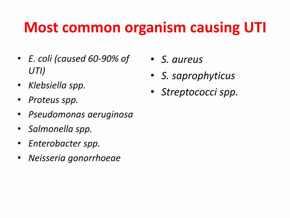
Most common organism causing UTI
• E. coli (caused 60-90% of
UTI)
• Klebsiella spp.
• Proteus spp.
• Pseudomonas aeruginosa
• Salmonella spp.
• Enterobacter spp.
• Neisseria gonorrhoeae
• S. aureus
• S. saprophyticus
• Streptococci spp.
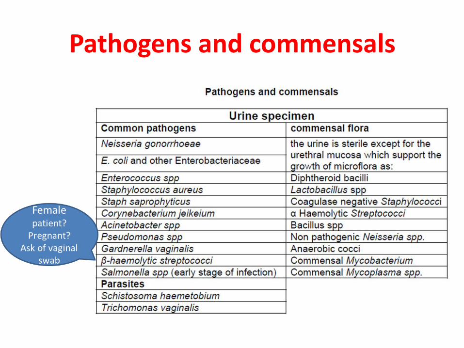
Pathogens and commensals
Urine specimen
• Normally urine is a sterile body fluid
• The bladder and urinary tract are sterile, the urethra may contain commensals
• Presence of bacteria in urine called bacteriuria
• Clean-catch midstream urine specimens that have more than 100,000 colonies of bacteria per mL of urine may be indicative of infection
• UTI occur more frequently in women than men due to the shortness of the female urethra
Types of Urine Specimens
1. First ori i g prefera ly Midstrea lea at h spe i e M“U .
2. Catheterized specimen.
3. Suprapubic aspiration.
4. Pediatric specimen.
Collection of Urine Specimen
• The first urine passed by the patient at the beginning of the
day should be sent for examination. This specimen is the most
concentrated and therefore the most suitable for lab
examination.
• 10-20 ml specimen is needed.
• Explain to the patient the need to collect the urine with as
little o ta i atio as possi le, i.e. a lea - at h spe i e . • Wash hands thoroughly before beginning the collection. Clean
the area very well with water and soap.
• Label the container with the date, name and number of the
patient, and the time of collection.
Transport of Urine Specimen
• Urine specimens should be delivered to the
laboratory immediately with a request form.
• When immediate delivery is not possible,
refrigerate the urine at 4–6 °C.
• When a delay in delivery of more than 2 hours
(maximum 48 hrs) is anticipated, add boric
acid preservative to the urine (no need to
refrigerate).
Deterioration of Urine Specimen The following changes occur when unpreserved urine is left at room
temperature:
Any bacteria in the urine will multiply so that the bacterial count will be unreliable.
When the organisms are urease-producing, the ammonia released will increase the pH of the specimen which will result in the destruction of cells and casts. Bacteria will also break down any glucose which may be present.
When white cells, red cells, and casts are present, these will begin to lyze especially in a concentrated specimen.
The concentration of protein in the urine will be altered.
When bilirubin is present this may be oxidized to biliverdin which will not be detected. Likewise, urobilinogen will not be detected because it will be oxidized to urobilin.
Lab Examination of Urine
• Urine Analysis:
Macroscopic examination
Biochemical Analysis
Microscopic examination
• Urine Culture
• Biochemical Reactions
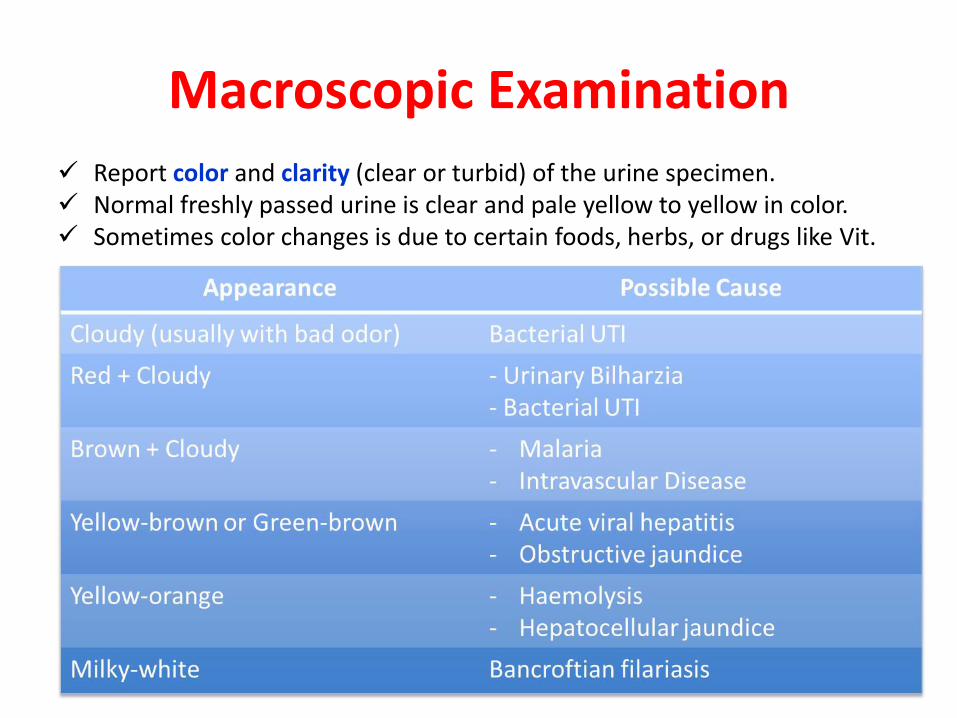
Macroscopic Examination
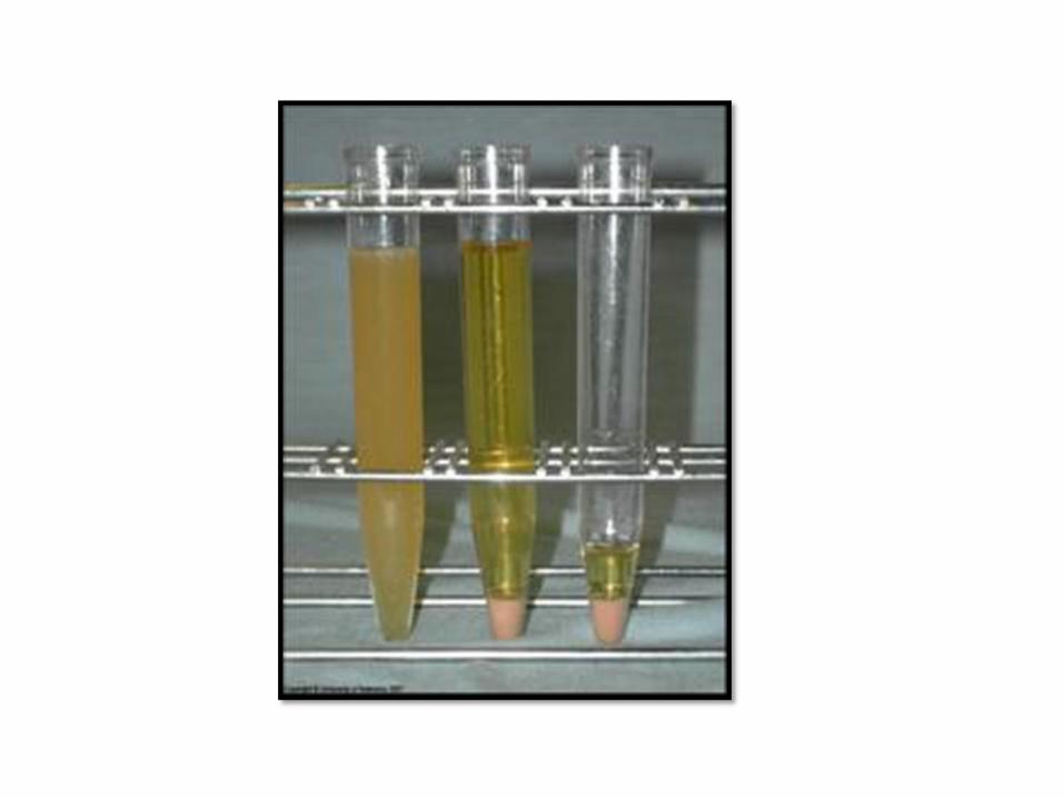
Report color and clarity (clear or turbid) of the urine specimen.
Normal freshly passed urine is clear and pale yellow to yellow in color.
Sometimes color changes is due to certain foods, herbs, or drugs like Vit.
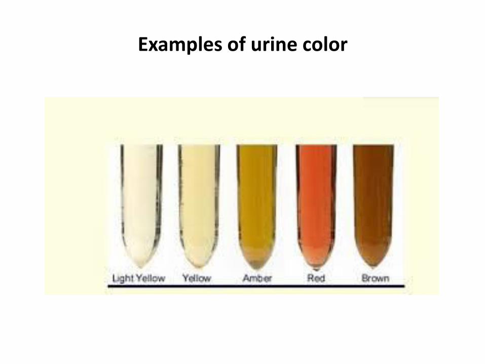
Examples of urine color
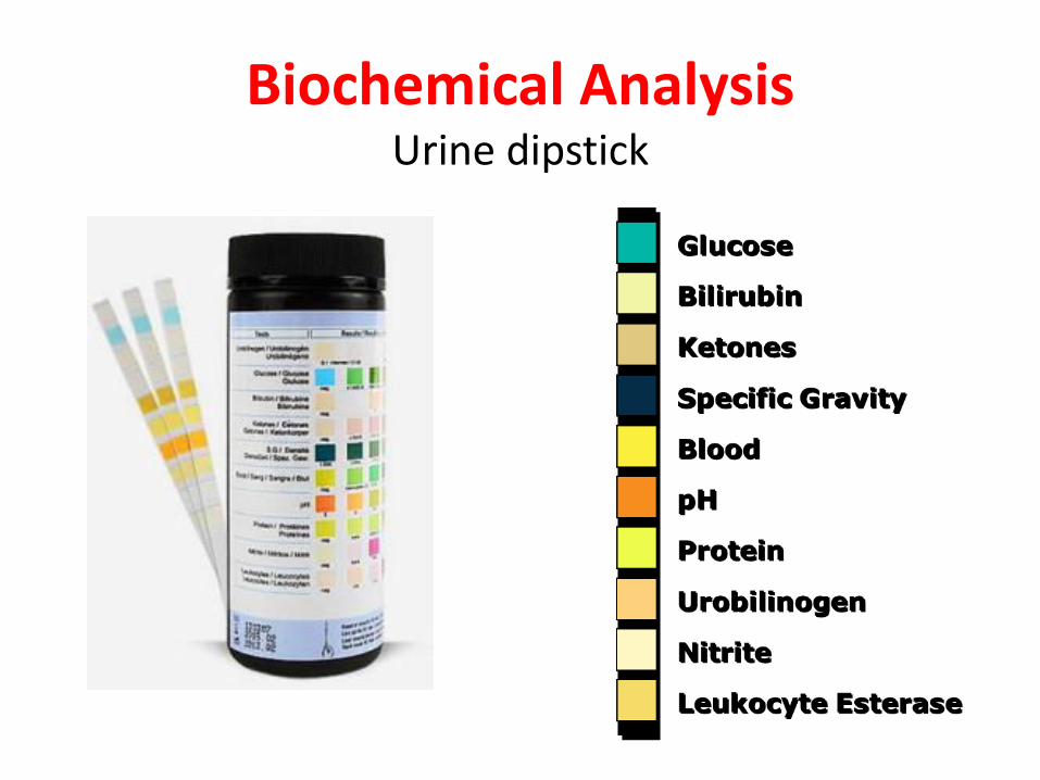
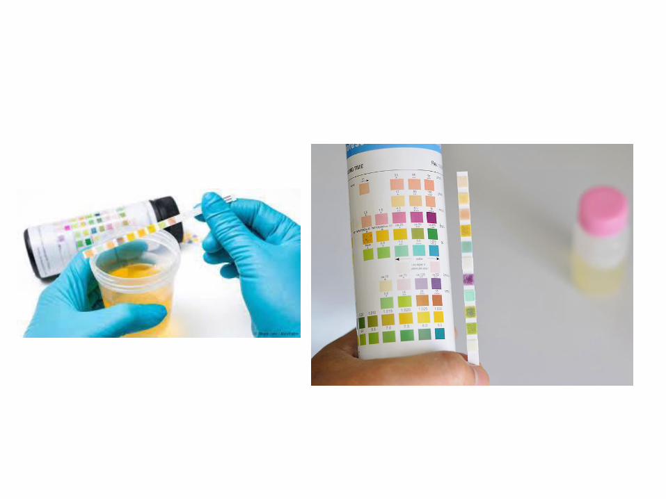
Biochemical Analysis Urine dipstick
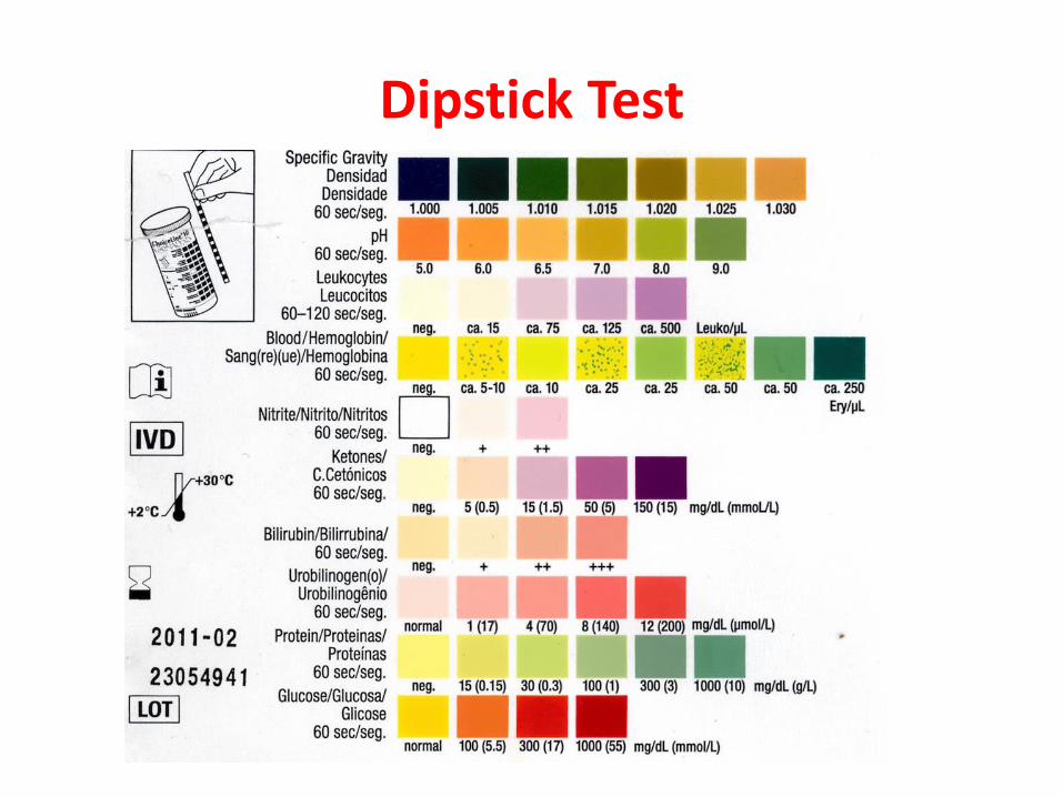
Dipstick Test
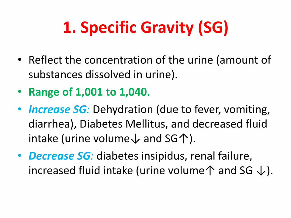
1. Specific Gravity (SG)
• Reflect the concentration of the urine (amount of
substances dissolved in urine).
• Range of 1,001 to 1,040.
• Increase SG: Dehydration (due to fever, vomiting,
diarrhea), Diabetes Mellitus, and decreased fluid
i take uri e volu e↓ a d “G↑ . • Decrease SG: diabetes insipidus, renal failure,
increased fluid intake uri e volu e↑ a d “G ↓ .
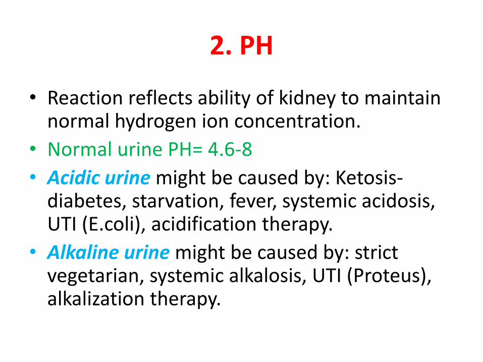
2. PH
• Reaction reflects ability of kidney to maintain normal hydrogen ion concentration.
• Normal urine PH= 4.6-8
• Acidic urine might be caused by: Ketosis-diabetes, starvation, fever, systemic acidosis, UTI (E.coli), acidification therapy.
• Alkaline urine might be caused by: strict vegetarian, systemic alkalosis, UTI (Proteus), alkalization therapy.
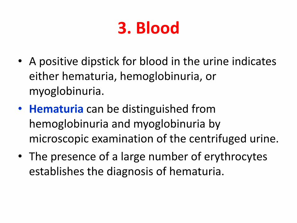
3. Blood
• A positive dipstick for blood in the urine indicates
either hematuria, hemoglobinuria, or
myoglobinuria.
• Hematuria can be distinguished from
hemoglobinuria and myoglobinuria by
microscopic examination of the centrifuged urine.
• The presence of a large number of erythrocytes
establishes the diagnosis of hematuria.
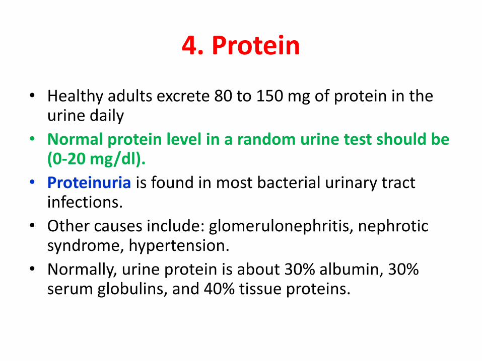
4. Protein
• Healthy adults excrete 80 to 150 mg of protein in the urine daily
• Normal protein level in a random urine test should be (0-20 mg/dl).
• Proteinuria is found in most bacterial urinary tract infections.
• Other causes include: glomerulonephritis, nephrotic syndrome, hypertension.
• Normally, urine protein is about 30% albumin, 30% serum globulins, and 40% tissue proteins.
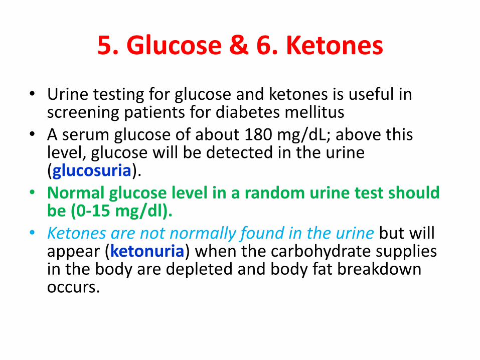
5. Glucose & 6. Ketones
• Urine testing for glucose and ketones is useful in screening patients for diabetes mellitus
• A serum glucose of about 180 mg/dL; above this level, glucose will be detected in the urine (glucosuria).
• Normal glucose level in a random urine test should be (0-15 mg/dl).
• Ketones are not normally found in the urine but will appear (ketonuria) when the carbohydrate supplies in the body are depleted and body fat breakdown occurs.
7. Nitrite • Nitrites are not normally found in the urine, and its presence is
strongly suggestive of bacteriuria.
• Many species of gram-negative bacteria can convert nitrate (normally present in urine) to nitrite, e.g. E. coli, Proteus species, and Klebsiella species if the organisms are present in the urine in sufficient concentration. When first morning urine is tested, about 80–90% of UTI caused by nitrate-reducing pathogens can be detected.
• The test is negative when the infection is caused by pathogens that do not reduce nitrate such as Enterococcus faecalis, Pseudomonas species, Staphylococcus species and Candida organisms, or when as previously mentioned the bacteria are too few in the urine.
• Occasionally the nitrite test is negative because nitrate is lacking in the urine due to the person being on a diet lacking in vegetables.
8. Leukocyte Esterase (LE)
• This enzyme is specific for polymorphonuclear neutrophils (pus
cells). It detects the enzyme from both active and lyzed WBCs.
• LE testing is an alternative method of detecting pyuria when:
1. It is not possible to examine fresh urine microscopically for
WBCs.
2. When the urine is not fresh and likely to contain mostly lyzed
WBCs.
• False negative strip test results can occur when the urine
contains boric acid or excessive amounts of protein (500 mg/100
ml) or glucose (2 g/100 ml).
• The major cause of false-positive leukocyte esterase tests is
specimen contamination.
9. Bilirubin & 10. Urobilinogen
• Normal urine contains no bilirubin and only very
small amounts of urobilinogen.
• The normal level of bilirubin is 0-1.2 mg/dl.
• There are many causes of bilirubin in urine:
1) Blockage of bile ducts due to gall stones
2) Infection
3) Decreased conjugation.
• Presence of bilirubin and urobilinogen in urine
usually indicate of liver diseases (ex: liver hepatitis,
cirrhosis)
Microscopic Examination
A- Wet preparation to look for:
1. Cells: - WBCs
- RBCs
- Epithelial cells
2. Bacteria
3. Yeast
4. Parasites
5. Casts
6. Crystals
A normal urine microscopy contains few epithelial cells, occasional RBC’s, few crystals.

How to make Wet Preparation?
1. Aseptically transfer about 10 ml of well mixed urine to a
labelled conical tube.
2. Centrifuge at 500–1000 rpm for 5 minutes. Pour the
supernatant fluid by completely inverting the tube.
3. Remix the sediment by tapping the bottom of the tube.
4. Transfer one drop of the well-mixed sediment to a slide and
cover with cover glass.
5. Examine the preparation microscopically using the 10 and 40
objective.
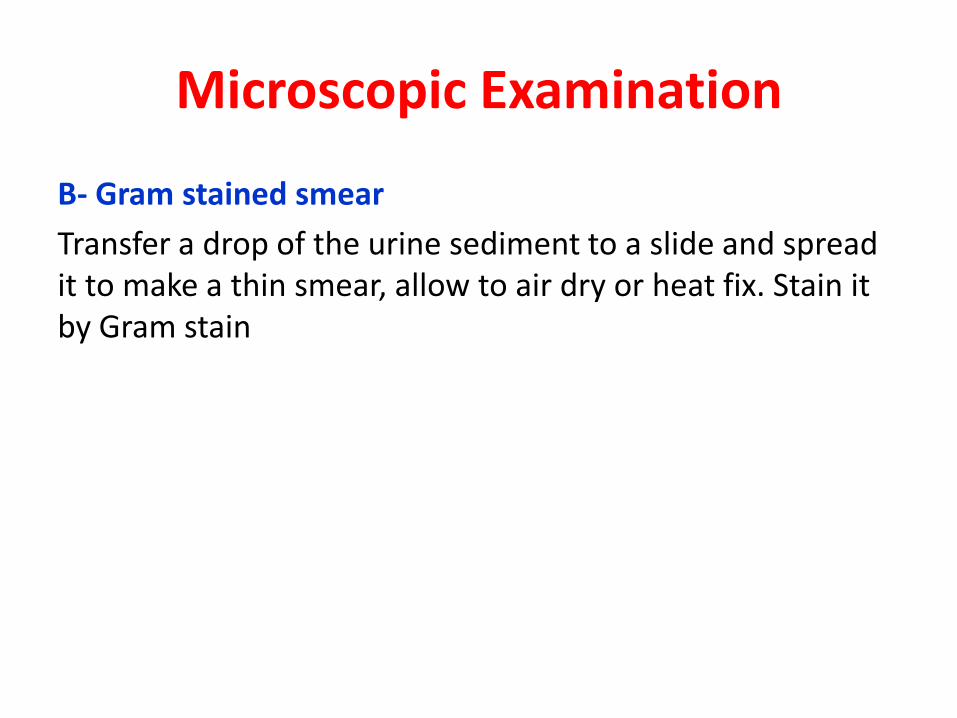
Microscopic Examination
B- Gram stained smear
Transfer a drop of the urine sediment to a slide and spread
it to make a thin smear, allow to air dry or heat fix. Stain it
by Gram stain

Urine culture
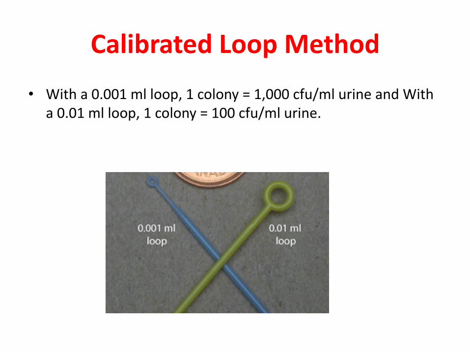
Calibrated Loop Method
• With a 0.001 ml loop, 1 colony = 1,000 cfu/ml urine and With
a 0.01 ml loop, 1 colony = 100 cfu/ml urine.
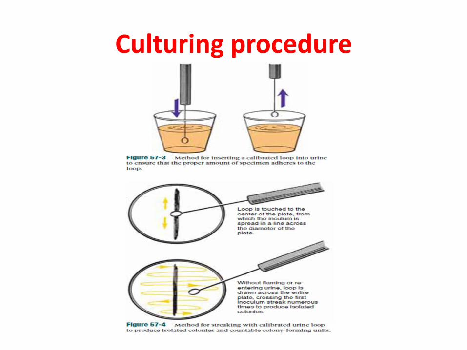
Culturing procedure
Reporting Bacterial Numbers
• Not significant: <104 organisms/ml.
• Doubtful significance: 104–105/ml (suggest repeat
specimen)
• Significant bacteriuria: >105/ml perform full ID and
susceptibility testing.
Culture the specimen
Media used:
• CLED Agar
• MacConkey Agar
• Blood Agar
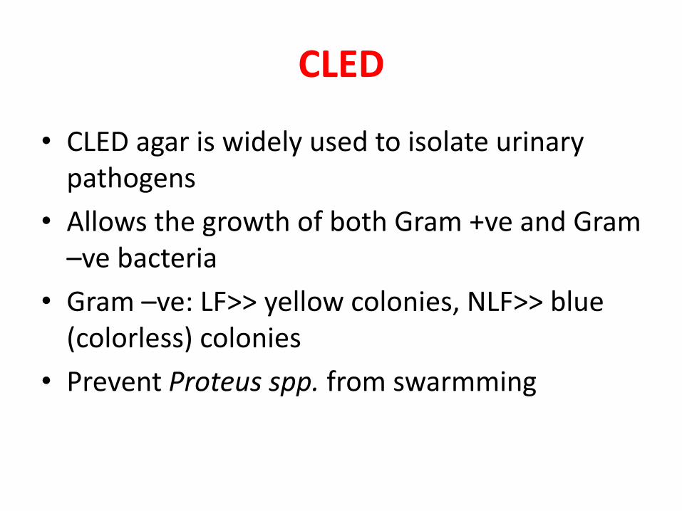
CLED
• CLED agar is widely used to isolate urinary
pathogens
• Allows the growth of both Gram +ve and Gram
–ve bacteria
• Gram –ve: LF>> yellow colonies, NLF>> blue
(colorless) colonies
• Prevent Proteus spp. from swarmming
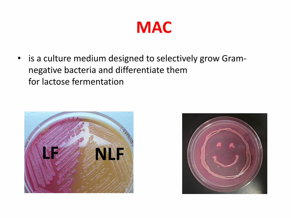
MAC
• is a culture medium designed to selectively grow Gram-
negative bacteria and differentiate them
for lactose fermentation
LF NLF
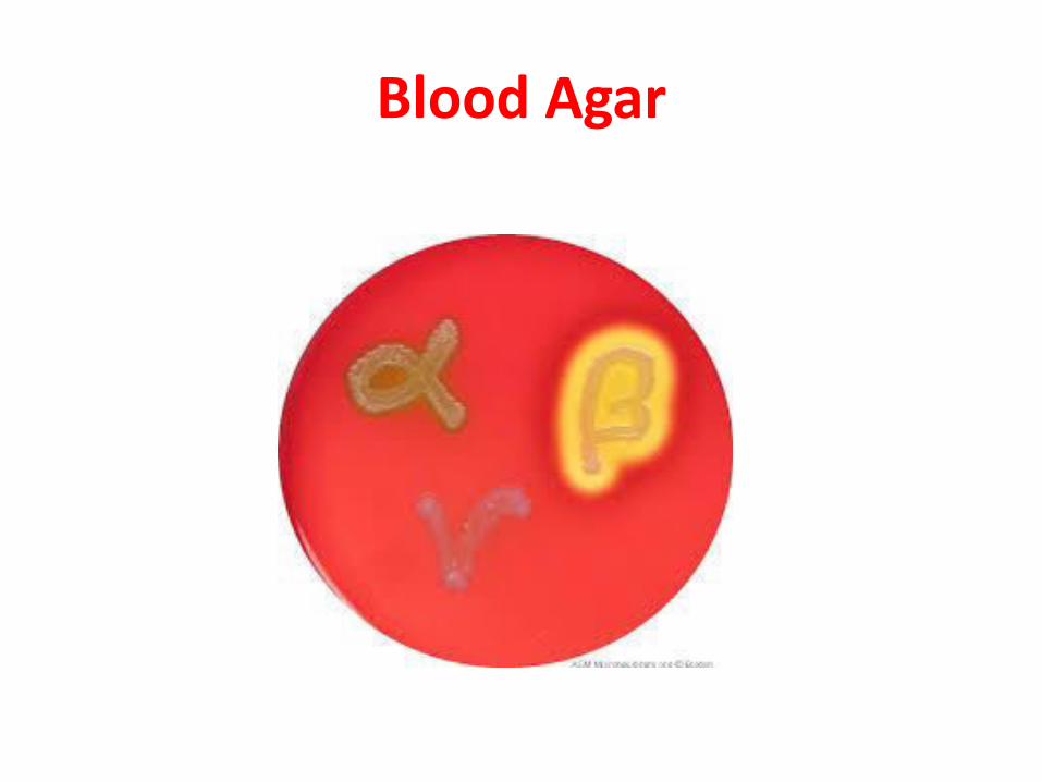
Blood Agar
Specimen Result
Turn around time:
• Wet mount results should be available 1 hour after
specimen receipt.
• Isolation of a possible pathogen can be expected
after 2-3 days.
• Negative culture will be reported out 1-2 days after
the receipt of the specimen.
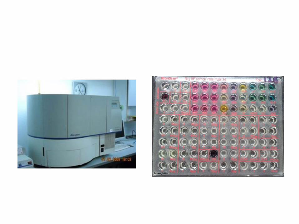
Automated Microbiology Method
• Microscan: for Id of bacteria
Antibiotic sensitivity
• Ready Kit
• Make suspension
• Fill wells: Red for G +ve & Blue for G-ve
• Run the machine & get the report.