University of Groningen Marfan syndrome and related connective … · pulmonary artery, including...
11
University of Groningen Marfan syndrome and related connective tissue disorders Aalberts, Jan IMPORTANT NOTE: You are advised to consult the publisher's version (publisher's PDF) if you wish to cite from it. Please check the document version below. Document Version Publisher's PDF, also known as Version of record Publication date: 2014 Link to publication in University of Groningen/UMCG research database Citation for published version (APA): Aalberts, J. (2014). Marfan syndrome and related connective tissue disorders: Cardiological and genetic aspects. [S.l.]: s.n. Copyright Other than for strictly personal use, it is not permitted to download or to forward/distribute the text or part of it without the consent of the author(s) and/or copyright holder(s), unless the work is under an open content license (like Creative Commons). Take-down policy If you believe that this document breaches copyright please contact us providing details, and we will remove access to the work immediately and investigate your claim. Downloaded from the University of Groningen/UMCG research database (Pure): http://www.rug.nl/research/portal. For technical reasons the number of authors shown on this cover page is limited to 10 maximum. Download date: 09-03-2019
Transcript of University of Groningen Marfan syndrome and related connective … · pulmonary artery, including...

University of Groningen
Marfan syndrome and related connective tissue disordersAalberts, Jan
IMPORTANT NOTE: You are advised to consult the publisher's version (publisher's PDF) if you wish to cite fromit. Please check the document version below.
Document VersionPublisher's PDF, also known as Version of record
Publication date:2014
Link to publication in University of Groningen/UMCG research database
Citation for published version (APA):Aalberts, J. (2014). Marfan syndrome and related connective tissue disorders: Cardiological and geneticaspects. [S.l.]: s.n.
CopyrightOther than for strictly personal use, it is not permitted to download or to forward/distribute the text or part of it without the consent of theauthor(s) and/or copyright holder(s), unless the work is under an open content license (like Creative Commons).
Take-down policyIf you believe that this document breaches copyright please contact us providing details, and we will remove access to the work immediatelyand investigate your claim.
Downloaded from the University of Groningen/UMCG research database (Pure): http://www.rug.nl/research/portal. For technical reasons thenumber of authors shown on this cover page is limited to 10 maximum.
Download date: 09-03-2019
33
CHA
PTER3
the many faces of aggressive aortic pathology: Loeys-dietz syndrome
Jan J.J. AalbertsMaarten P. van den Berg
Jorieke E.H. BergmanGideon J. du Marchie Sarvaas
Jan G. PostHans van Unen
Gerard PalsPiet W. Boonstra
J. Peter van Tintelen
Neth Heart J. 2008;16:299-304

34
Chapter 3
ABstrACt
Background
Loeys-Dietz syndrome (LDS) is a new disorder of connective tissue which shares overlapping
features with Marfan syndrome (MFS) and the vascular type of Ehlers-Danlos syndrome, including
aortic root dilatation and skin abnormalities. It is clinically classified into types I and II. LDS type I can
be recognized by craniofacial characteristics, e.g. hypertelorism, bifid uvula or cleft palate, whereas
these are absent in LDS type II. It is important to recognize LDS because its vascular pathology is
aggressive. We describe nine LDS patients from four families, relate their features to published cases,
and discuss important aspects of the diagnosis and management of LDS in order to make clinicians
aware of this new syndrome.
Results
Characteristics found in the majority of these LDS patients were aortic root dilatation, cleft palate
and/or a bifid/abnormal uvula.
Conclusion
Because aortic dissection and rupture in LDS tend to occur at a young age or at aortic root diameters
not considered at risk in MFS, and because the vascular pathology can be seen throughout the
entire arterial tree, patients should be carefully followed up and aggressive surgical treatment is
mandatory. Clinicians must therefore be aware of LDS as a cause of aggressive aortic pathology and
that its distinguishing features can sometimes be easily recognized.
35
The many faces of aggressive aortic pathology: Loeys-Dietz syndrome
3
introduCtion
Aortic dissection and rupture can be associated with generalized connective tissue disorders,
especially when these catastrophic events occur at a young age. The vascular type of Ehlers-Danlos
syndrome (EDS) and Marfan syndrome (MFS) are the most prevalent connective tissue disorders
in these patients.1,2 Recently, however, a new connective tissue disorder, Loeys-Dietz syndrome
(LDS), has been described, which is also accompanied by these severe aortic complications.3 LDS
is an autosomal dominantly inherited disorder of connective tissue caused by mutations in the
transforming growth factor beta (TGF-β) receptor 1 or 2 genes.3,4 The exact prevalence is unknown
but given the fact that LDS has only recently been discovered, many cases might not have been
diagnosed yet. The first type of this syndrome (LDS type I) has many overlapping features with
Marfan syndrome (MFS), including aortic root dilatation, arachnodactyly (long slender fingers),
dolichostenomelia (thin body habitus and long extremities), pectus deformity and joint laxity,
whereas LDS type II has features that overlap with the vascular type of EDS. (see table 1 for an
overview of differentiating characteristics of the syndromes)3,4 Vascular pathology in LDS, however,
is more aggressive than in MFS. 3-5 It is therefore of uttermost importance to recognize this disorder.
This is facilitated by distinctive, frequently occurring and easily recognizable characteristics of LDS
(see table 1).3,4
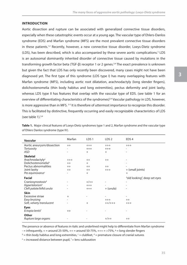
vascular Marfan LDS 1 LDS 2 EDS 4
Aortic aneurysm/dissection ++ +++ +++ +++Tortuosity - +++ +++ -ASD - + + -skeletalArachnodactylya +++ ++ ++ -Dolichostenomeliab ++ + -Pectus abnormalities ++ ++ ++ -Joint laxity ++ ++ +++ + (small joints)Pes equinovarusc - + +
facial “old looking”, deep set eyesCraniosynostosisd - +/++ - -Hypertelorisme - +++ - -Cleft palate/bifid uvula - +++ + (uvula) -skinExcessive striae + - - -Easy bruising - +++ ++Soft, velvety translucent - + ++/+++ +++eyesEctopia lentisf ++ - - -otherRupture large organs - - +/++ ++
The presence or absence of features in italic and underlined might help to differentiate from Marfan syndrome- = infrequently, + = around 25-50%, ++ = around 50-75%, +++ = >75%, a = long slender fingersb = thin body habitus and long extremities, c = clubfeet, d = premature closure of cranial sutures e = increased distance between pupil, f = lens subluxation
table 1. Major clinical features of Loeys-Dietz syndromes type 1 and 2, Marfan syndrome and the vascular type of Ehlers Danlos syndrome (type IV).
36
Chapter 3
In order to publicize these features, we here describe 9 patients, from 4 families, who presented at
our department and in whom we genetically confirmed LDS. In addition, we discuss some important
aspects concerning the diagnosis and management of LDS.
PAtients And Methods
Patients
All patients and families were referred to our Marfan outpatients’ clinic for evaluating a possible
diagnosis of MFS or other connective tissue disorder. Subjects were characterized both clinically
and genetically, and the diagnosis of LDS was confirmed by a mutation in the TGF-β receptor 1 or 2
gene in 7/9 patients. In one family the results of DNA analysis are still pending.
Mutation analysis
DNA analysis of the TGFBR1 and TGFBR2 genes was performed by direct DNA sequencing of genomic
DNA, using the BIG Dye version 3 dye terminator kit (Applied Biosystems, Torrence, CA, USA). Primer
sequences are available on request.
resuLts
Clinical and genetic evaluation
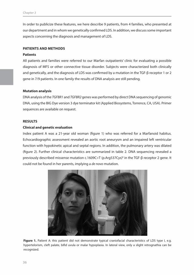
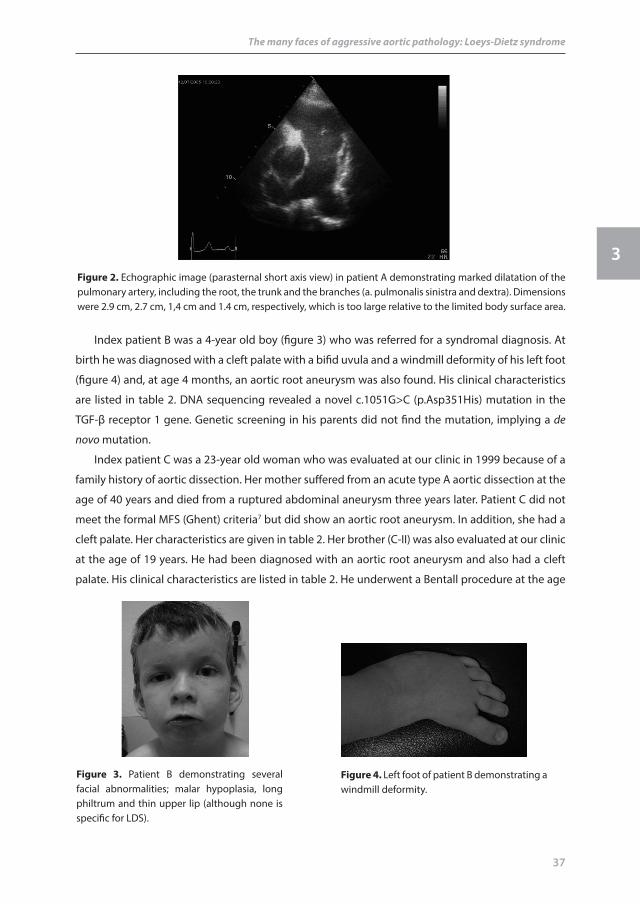
Index patient A was a 21-year old woman (figure 1) who was referred for a Marfanoid habitus.
Echocardiographic assessment revealed an aortic root aneurysm and an impaired left ventricular
function with hypokinetic apical and septal regions. In addition, the pulmonary artery was dilated
(figure 2). Further clinical characteristics are summarized in table 2. DNA sequencing revealed a
previously described missense mutation c.1609C>T (p.Arg537Cys)6 in the TGF-β receptor 2 gene. It
could not be found in her parents, implying a de novo mutation.
figure 1. Patient A: this patient did not demonstrate typical craniofacial characteristics of LDS type I, e.g. hypertelorism, cleft palate, bifid uvula or malar hypoplasia. In lateral view, only a slight retrognathia can be recognized.
37
The many faces of aggressive aortic pathology: Loeys-Dietz syndrome
3
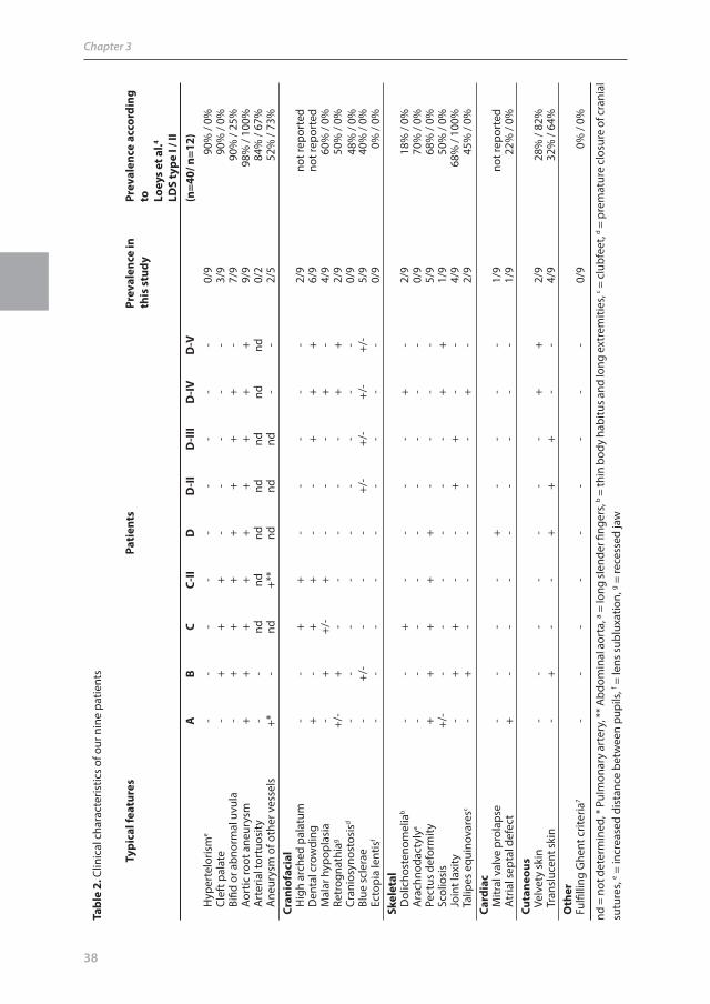
Index patient B was a 4-year old boy (figure 3) who was referred for a syndromal diagnosis. At
birth he was diagnosed with a cleft palate with a bifid uvula and a windmill deformity of his left foot
(figure 4) and, at age 4 months, an aortic root aneurysm was also found. His clinical characteristics
are listed in table 2. DNA sequencing revealed a novel c.1051G>C (p.Asp351His) mutation in the
TGF-β receptor 1 gene. Genetic screening in his parents did not find the mutation, implying a de
novo mutation.
Index patient C was a 23-year old woman who was evaluated at our clinic in 1999 because of a
family history of aortic dissection. Her mother suffered from an acute type A aortic dissection at the
age of 40 years and died from a ruptured abdominal aneurysm three years later. Patient C did not
meet the formal MFS (Ghent) criteria7 but did show an aortic root aneurysm. In addition, she had a
cleft palate. Her characteristics are given in table 2. Her brother (C-II) was also evaluated at our clinic
at the age of 19 years. He had been diagnosed with an aortic root aneurysm and also had a cleft
palate. His clinical characteristics are listed in table 2. He underwent a Bentall procedure at the age
figure 2. Echographic image (parasternal short axis view) in patient A demonstrating marked dilatation of the pulmonary artery, including the root, the trunk and the branches (a. pulmonalis sinistra and dextra). Dimensions were 2.9 cm, 2.7 cm, 1,4 cm and 1.4 cm, respectively, which is too large relative to the limited body surface area.
figure 3. Patient B demonstrating several facial abnormalities; malar hypoplasia, long philtrum and thin upper lip (although none is specific for LDS).
figure 4. Left foot of patient B demonstrating a windmill deformity.
38
Chapter 3
typi
cal f
eatu
res
Pati
ents
Prev
alen
ce in
th
is s
tudy
Prev
alen
ce a
ccor
ding
to
Lo
eys
et a
l.4
Lds
type
i / i
iA
BC
C-ii
dd
-iid
-iii
d-iv
d-v
(n=4
0/ n
=12)
H
yper
telo
rism
e -
--
--
--
--
0/9
90%
/ 0%
Cl
eft p
alat
e-
++
+-
--
--
3/9
90%
/ 0%
Bi
fid o
r abn
orm
al u
vula
-+
++
++
++
-7/
990
% /
25%
Ao
rtic
root
ane
urys
m+
++
++
++
++
9/9
98%
/ 10
0%
Art
eria
l tor
tuos
ity
--
ndnd
ndnd
ndnd
nd0/
284
% /
67%
A
neur
ysm
of o
ther
ves
sels
+*
-nd
+**
ndnd
nd-
-2/
552
% /
73%
Cran
iofa
cial
H
igh
arch
ed p
alat
um-
-+
+-
--
--
2/9
not r
epor
ted
D
enta
l cro
wdi
ng+
-+
+-
-+
++
6/9
not r
epor
ted
M
alar
hyp
opla
sia
-+
+/-
+-
--
+-
4/9
60%
/ 0%
Re
trog
nath
iag
+/-
+-
--
--
++
2/9
50%
/ 0%
48%
/ 0%
Cr
anio
syno
stos
isd
--
--
--
--
-0/
9
Blue
scl
erae
-+/
--
--
+/-
+/-
+/-
+/-
5/9
40%
/ 0%
Ec
topi
a le
ntis
f -
--
--
--
--
0/9
0% /
0%sk
elet
al
D
olic
host
enom
elia
b -
-+
--
--
+-
2/9
18%
/ 0%
A
rach
noda
ctyl
ya -
--
--
--
--
0/9
70%
/ 0%
Pect
us d
efor
mity
++
++
+-
--
-5/
968
% /
0%
Scol
iosi
s+/
--
--
--
-+
+1/
950
% /
0%
Join
t lax
ity-
++
--
++
--
4/9
68%
/ 10
0% T
alip
es e
quin
ovar
esc
-+
--
--
-+
-2/
945
% /
0%Ca
rdia
c
Mitr
al v
alve
pro
laps
e-
--
-+
--
--
1/9
not r
epor
ted
A
tria
l sep
tal d
efec
t+
--
--
--
--
1/9
22%
/ 0%
Cuta
neou
s V
elve
ty s
kin
--
--
--
-+
+2/
928
% /
82%
Tra
nslu
cent
ski
n-
+-
-+
++
--
4/9
32%
/ 64
%o
ther
Fulfi
lling
Ghe
nt c
riter
ia7
--
--
--
--
-0/
90%
/ 0%
nd =
not
det
erm
ined
, * P
ulm
onar
y ar
tery
, ** A
bdom
inal
aor
ta, a
= lo
ng sl
ende
r fing
ers,
b = th
in b
ody
habi
tus a
nd lo
ng e
xtre
miti
es, c =
clu
bfee
t, d =
pre
mat
ure
clos
ure
of c
rani
al
sutu
res,
e = in
crea
sed
dist
ance
bet
wee
n pu
pils
, f = le
ns s
ublu
xatio
n, g =
rece
ssed
jaw
tabl
e 2.
Clin
ical
cha
ract
eris
tics
of o
ur n
ine
patie
nts
39
The many faces of aggressive aortic pathology: Loeys-Dietz syndrome
3
of 22 years because of an aortic root aneurysm of 51 mm and experienced a myocardial infarction
shortly after surgery due to an unknown cause. A computed tomography (CT) scan of the coronary
arteries did not reveal any explanation for the myocardial infarction but did show a rapid tapering
of the right coronary artery and the left anterior descending coronary artery. At the age of 24 years
he died suddenly during physical activity. DNA sequencing is pending.
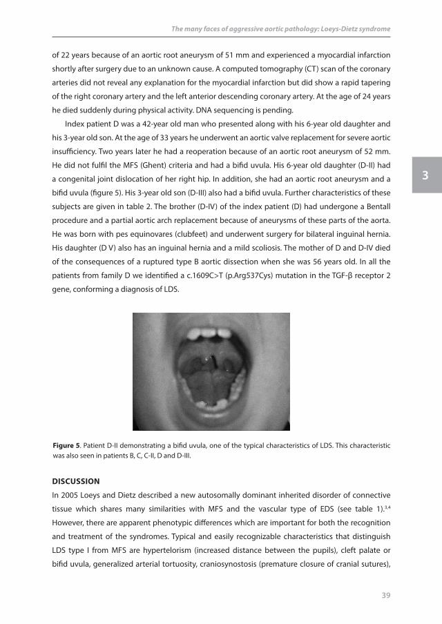
Index patient D was a 42-year old man who presented along with his 6-year old daughter and
his 3-year old son. At the age of 33 years he underwent an aortic valve replacement for severe aortic
insufficiency. Two years later he had a reoperation because of an aortic root aneurysm of 52 mm.
He did not fulfil the MFS (Ghent) criteria and had a bifid uvula. His 6-year old daughter (D-II) had
a congenital joint dislocation of her right hip. In addition, she had an aortic root aneurysm and a
bifid uvula (figure 5). His 3-year old son (D-III) also had a bifid uvula. Further characteristics of these
subjects are given in table 2. The brother (D-IV) of the index patient (D) had undergone a Bentall
procedure and a partial aortic arch replacement because of aneurysms of these parts of the aorta.
He was born with pes equinovares (clubfeet) and underwent surgery for bilateral inguinal hernia.
His daughter (D V) also has an inguinal hernia and a mild scoliosis. The mother of D and D-IV died
of the consequences of a ruptured type B aortic dissection when she was 56 years old. In all the
patients from family D we identified a c.1609C>T (p.Arg537Cys) mutation in the TGF-β receptor 2
gene, conforming a diagnosis of LDS.
figure 5. Patient D-II demonstrating a bifid uvula, one of the typical characteristics of LDS. This characteristic was also seen in patients B, C, C-II, D and D-III.
disCussion
In 2005 Loeys and Dietz described a new autosomally dominant inherited disorder of connective
tissue which shares many similarities with MFS and the vascular type of EDS (see table 1).3,4
However, there are apparent phenotypic differences which are important for both the recognition
and treatment of the syndromes. Typical and easily recognizable characteristics that distinguish
LDS type I from MFS are hypertelorism (increased distance between the pupils), cleft palate or
bifid uvula, generalized arterial tortuosity, craniosynostosis (premature closure of cranial sutures),
40
Chapter 3
patent ductus arteriosus and atrial septal defect, whereas LDS type II patients demonstrate skin
abnormalities (easy bruising, translucent velvety skin), joint laxity and rupture of visceral organs
(see also table 1 and 2).3,4 Ectopia lentis (lens subluxation), which can be found in 40-56% of patients
with MFS, as well as other ocular manifestations are absent in LDS.3,4,8 However LDS is clinically
highly variable, as demonstrated by our nine patients with LDS.9 This variability may hamper proper
classification in the two subtypes of the syndrome. Note for example the differences between
patients A and B (table 2, figures 1, 2 and 3). Remarkably, none of our patients demonstrated
hypertelorism (increased distance between pupils) or craniosynostosis (premature closure of cranial
sutures), which are allegedly encountered in 90% and 50% of LDS type I patients, respectively.4 In
addition, arterial tortuosity, although it has not yet been systematically evaluated in all our patients,
was not found in our group. This example underscores the wide clinical variability and the need for
a critical evaluation of all patients presenting with one of the symptoms of the syndrome.
Patients with LDS type I tend to require earlier cardiovascular surgery and have a shorter life
expectancy than those with LDS type II.4 However, it should be kept in mind that all LDS patients
show aggressive vascular disease and thus all require careful follow-up and management; we will
discuss this later. Patients suspected of having LDS should undergo a physical examination, paying
special attention to the typical characteristics of LDS, echo cardio graphic assessment and genetic
testing for TGF-β receptor 1 and 2 gene mutations.
An altered signalling of the TGF-β cytokine family, which plays an important role in cell
proliferation and differentiation, apoptosis and extracellular matrix formation, underlies both LDS
and MFS.10,11 The altered TGF-β signalling causes disarrayed elastic fibres and loss of elastin content
in the aortic media, predisposing to dilatation and dissection of the aortic wall.3 In MFS an abnormal
fibrillin protein causes an increased activity of TGF-β and this leads to the typical phenotypic
characteristics of the disease.11 However, in LDS, disruption of the normal TGF-β signalling takes
place at the level of the TGF-β receptors.3 Mutations in either the TGF-β receptor 1 or 2 genes have
been identified in one-third and two-thirds of LDS patients, respectively.3,4
As TGF-β molecules also play an important role in cardiovascular embryogenesis, including
guidance of the formation of the ventricular myocardium, mutations in the TGF-β receptors could
well have an effect on ventricular function and might underlie the impaired left ventricular function
with hypokinetic apical and septal regions in patient A, a feature which has not been described in
patients with LDS.12
The recognition of LDS is especially important from a management point of view. Cardiovascular
disease seems to be more aggressive and widespread in LDS than in MFS. Recently Williams et
al.5 reported aortic rupture and dissection in young patients and in patients with aortic root
diameters not considered at risk in MFS (< 4.5 cm), in addition they reported high rates of repeat
surgical interventions. Furthermore, aneurysms were not confined to the aortic root but occurred
throughout the entire arterial tree.5,13
The above observations have important clinical consequences for patients with LDS. Firstly,

41
The many faces of aggressive aortic pathology: Loeys-Dietz syndrome
3
table 3. Indications for surgical intervention in Loeys-Dietz syndrome*
Adultsa. Aortic root > 4.0 cm or expanding rapidly (> 0.5 cm/year)b. Descending thoracic aorta > 5.0 cm or expanding rapidly (> 0.5 cm/year)c. Abdominal aorta > 4.0 cm or expanding rapidly (> 0.5 cm/year)d. Rapid expansion of peripheral aneurysms
Children a. 1. Severe craniofacial features: aortic root z-score > 3.0 or expanding rapidly (> 0.5 cm/year)
2. Mild craniofacial features: aortic root z-score > 4.0 or expanding rapidly (> 0.5 cm/year)b. Effort should be made to delay surgery until the annulus reaches 1.8 cm, allowing placement of a
valve-sparing graft of sufficient size to accommodate growthc. Large size or rapid expansion of the descending aorta or other vessels
*derived from Williams et al.5
after establishing the diagnosis, regular repeated echocardiographic assessment should be
performed, paying particular attention to the aortic root diameter and the growth of this diameter.
In addition, patients should undergo MR angiography from head to pelvis to discover other possible
aneurysms.5 Secondly, when there is an aortic root aneurysm present, beta-blockade therapy
should be initiated to reduce further dilatation. Alternatively, LDS patients might benefit more from
losartan, an angiotensin II receptor antagonist, as this drug antagonizes TGF-β and has proven to be
effective in mouse models of MFS.14 Thirdly, surgical intervention should probably be considered
earlier than in MFS. Current recommendations for adult and paediatric patients, as proposed by
Williams et al., are summarized in table 3.5 However, it should be noted that these recommendations
were not empirically established. Since little is known about the natural history of the syndrome,
these are the only recommendations currently available. Earlier intervention should be considered
based upon the family history or an assessment of risk versus benefit for an individual patient.
Although vascular disease is aggressive in LDS, patients do not have friable vascular tissue,
unlike the vascular type of EDS, and they appear to tolerate surgery well in an elective setting.5,15
After surgical intervention, follow-up should be continued and regular imaging of the entire arterial
tree should be performed so that new arterial pathology can be recognized and treated in good
time.
ConCLusion
We have described nine LDS patients and discussed important aspects concerning the diagnosis
and management of the disease. Every cardiologist and cardiothoracic surgeon, particularly those
specializing in aortic pathology, should be aware of this new syndrome. Young patients presenting
with aortic dissection, aortic root aneurysm, or aneurysms elsewhere should be evaluated for LDS.
A simple physical examination – checking for hypertelorism, cleft palate or bifid uvula and skin
abnormalities – can provide important clues to the presence of the syndrome. Given the potentially
aggressive nature of LDS, careful follow-up and early surgical intervention are mandatory.
42
Chapter 3
ACknowLedGMents
We thank Jackie Senior for reading the final version of this paper.
referenCes 1 Nienaber CA, Eagle KA. Aortic dissection: New Frontiers in diagnosis and management Part I: From Etiology
to Diagnostic Strategies. Circulation 2003; 108: 628-635.
2 Januzzi JL, Isselbacher EM, Fattori R, Cooper JV, Smith DE, Fang J, et al. Characterizing the young patient with aortic dissection: results from the International Registry of Aortic Dissection (IRAD). J Am Coll Cardiol 2004; 43: 665-669.
3 Loeys BL, Chen J, Neptune ER, Judge DP, Podowski M, Holm T, et al. A syndrome of altered cardiovascular, craniofacial, neurocognitive and skeletal development caused by mutations in TGFBR1 or TGFBR2. Nat Genet 2005; 37: 275-281.
4 Loeys BL, Schwarze U, Holm T, Callewaert BL, Thomas GH, Pannu H, et al. Aneurysm syndromes caused by mutations in the TGF-beta receptor. N Engl J Med 2006: 355: 788-798.
5 Williams JA, Loeys BL, Nwakanma LU, Dietz HC, Spevak PJ, Patel ND, et al. Early surgical experience with Loeys-Dietz: A new syndrome of aggressive thoracic aortic aneurysm disease. Ann Thorac Surg 2007; 83: S757-S763.
6 Mizuguchi T, Collod-Beroud G, Akiyama T, Abifadel M, Harada N, Morisaki T, et al. Heterozygous TGFBR2 mutations in Marfan syndrome. Nat Genet 2004; 36: 855-860
7 De Paepe A, Devereux RB, Dietz HC, Hennekam RCM, Pyeritz RE. Revised diagnostic criteria for the Marfan syndrome. Am J Med Genet 1996 ; 62: 417-426
8 Dean JCS. Marfan syndrome: clinical diagnosis and management. Eur J Hum Genet 2007; 15: 724-33.
9 Akutsu K, Morisaki H, Takeshita S, Sakamoto S, Tamori Y, Yoshimuta T, et al. Phenotypic heterogeneity of Marfan-like connective tissue disorders associated with mutations in the transforming growth factor-beta genes. Circ J 2007; 71: 1305-9.
10 Annes JP, Munger JS, Rifkin DB. Making sense of latent TGF-beta activation. J Cell Sci 2003; 116: 217-224.
11 Mizuguchi T, Matsumoto N. Recent progress in genetics of Marfan syndrome and Marfan-associated disorders. J Hum Genet 2007; 52: 1-12.
12 Azhar M, Schultz JEJ, Grupp I, Dorn GW, Meneton P, Molin DGM, et al. Transforming growth beta in cardiovascular development and function. Cytokine Growth Factor Rev 2003; 14: 391-407.
13 LeMaire SA, Pannu H, Tran-Fadulu V, Carter SA, Coselli JS, Milewicz DM. Severe aortic and arterial aneurysms associated with a TGFBR2 mutation. Nat Clin Pract Cardiovasc Med 2007; 4: 167-171.
14 Habashi JP, Judge DP, Holm TM, Cohn RD, Loeys BL, Cooper TK et al. Losartan, an ATI antagonist, prevents aortic aneurysm in a mouse model of Marfan syndrome. Science 2006; 312: 117-121.
15 Oderich GS, Pannenton JM, Bower TC, Lindor NM, Cherry KJ, Noel AA, et al. The spectrum, management and clinical outcome of Ehlers-Danlos syndrome type IV: a 30-year experience. J Vasc Surg 2005; 42: 98-106.