Universities of Leeds, Sheffield and York ...eprints.whiterose.ac.uk/78555/1/Full_Field.pdf ·...
8
promoting access to White Rose research papers White Rose Research Online [email protected] Universities of Leeds, Sheffield and York http://eprints.whiterose.ac.uk/ This is a copy of the final published version of a paper published via gold open access in PLoS One. This open access article is distributed under the terms of the Creative Commons Attribution Licence (http://creativecommons.org/licenses/by/3.0), which permits unrestricted use, distribution, and reproduction in any medium, provided the original work is properly cited. White Rose Research Online URL for this paper: http://eprints.whiterose.ac.uk/78555 Published paper Field, K, George, RM, Fearne, B, Quick, WP and Davey, MP (2013) Best of both worlds: Simultaneous high-light and shade-tolerance adaptations within individual leaves of the living stone Lithops aucampiae. PLoS ONE. Doi: 10.1371/journal.pone.0075671
Transcript of Universities of Leeds, Sheffield and York ...eprints.whiterose.ac.uk/78555/1/Full_Field.pdf ·...

promoting access to White Rose research papers
White Rose Research Online [email protected]
Universities of Leeds, Sheffield and York http://eprints.whiterose.ac.uk/
This is a copy of the final published version of a paper published via gold open access in PLoS One.
This open access article is distributed under the terms of the Creative Commons Attribution Licence (http://creativecommons.org/licenses/by/3.0), which permits unrestricted use, distribution, and reproduction in any medium, provided the original work is properly cited. White Rose Research Online URL for this paper: http://eprints.whiterose.ac.uk/78555
Published paper
Field, K, George, RM, Fearne, B, Quick, WP and Davey, MP (2013) Best of both worlds: Simultaneous high-light and shade-tolerance adaptations within individual leaves of the living stone Lithops aucampiae. PLoS ONE. Doi: 10.1371/journal.pone.0075671

Best of Both Worlds: Simultaneous High-Light andShade-Tolerance Adaptations within Individual Leaves ofthe Living Stone Lithops aucampiaeKatie J. Field1, Rachel George1, Brian Fearn2, W. Paul Quick1, Matthew P. Davey3*
1Animal and Plant Sciences, Western Bank, University of Sheffield, Sheffield, United Kingdom, 2Abbey Brook Cactus Nursery, Matlock, Derbyshire, United Kingdom,
3Department of Plant Sciences, Downing Street, University of Cambridge, Cambridge, United Kingdom
Abstract
‘‘Living stones’’ (Lithops spp.) display some of the most extreme morphological and physiological adaptations in the plantkingdom to tolerate the xeric environments in which they grow. The physiological mechanisms that optimise thephotosynthetic processes of Lithops spp. while minimising transpirational water loss in both above- and below-groundtissues remain unclear. Our experiments have shown unique simultaneous high-light and shade-tolerant adaptations withinindividual leaves of Lithops aucampiae. Leaf windows on the upper surfaces of the plant allow sunlight to penetrate tophotosynthetic tissues within while sunlight-blocking flavonoid accumulation limits incoming solar radiation and aidsscreening of harmful UV radiation. Increased concentration of chlorophyll a and greater chlorophyll a:b in above-groundregions of leaves enable maximum photosynthetic use of incoming light, while inverted conical epidermal cells, increasedchlorophyll b, and reduced chlorophyll a:b ensure maximum absorption and use of low light levels within the below-groundregion of the leaf. High NPQ capacity affords physiological flexibility under variable natural light conditions. Our findingsdemonstrate unprecedented physiological flexibility in a xerophyte and further our understanding of plant responses andadaptations to extreme environments.
Citation: Field KJ, George R, Fearn B, Quick WP, Davey MP (2013) Best of Both Worlds: Simultaneous High-Light and Shade-Tolerance Adaptations withinIndividual Leaves of the Living Stone Lithops aucampiae. PLoS ONE 8(10): e75671. doi:10.1371/journal.pone.0075671
Editor: Douglas Andrew Campbell, Mount Allison University, Canada
Received April 25, 2013; Accepted August 16, 2013; Published October 23, 2013
Copyright: � 2013 Field et al. This is an open-access article distributed under the terms of the Creative Commons Attribution License, which permitsunrestricted use, distribution, and reproduction in any medium, provided the original author and source are credited.
Funding: This research was funded by the Natural Environment Research Council – Post-Genomics and Proteomics programme (NE/C507837/1) who financedMPD and the chlorophyll fluorescence imager equipment. The funders had no role in study design, data collection and analysis, decision to publish, orpreparation of the manuscript.
Competing Interests: BF is the owner of the Abbey Brook Cactus Nursery, Derbyshire, United Kingdom (www.abbeybrookcacti.com). This does not alter theauthors’ adherence to all the PLOS ONE policies on sharing data and materials.
* E-mail: [email protected]
Introduction
Xerophytes often display unusual morphological and physio-
logical characteristics to tolerate the challenging abiotic conditions
of their native environments. Few traits are more extreme than
those that have been selected for in South African ‘‘living stones’’
(Lithops spp.) where the majority of their biomass, including much
of their photosynthetic tissue, is underground [1]. Subterranean
autotrophy seems at first counterintuitive, however, the stresses
imposed by the xeric environment in which Lithops inhabit [2] can
be mitigated by the cooler and more stable conditions in the soil.
Despite this, the precise mechanisms by which Lithops maximises
subterranean photosynthesis while minimising transpirational
water loss remain unknown.
Lithops is a member of the Aizoaceae, sub family Ruschiodiae
[2]. The plant body consists of a very short stem with a pair of
fused, succulent leaves (Fig. 1a i), resulting in low surface area to
volume ratio. This allows Lithops to store substantial quantities of
water per unit of exposed plant surface, enabling it to survive
prolonged periods of drought [3]. The epidermis of many arid-
dwelling plants is specialised for water conservation by reducing
transpirational water loss [4,5], optimising photosynthetic rate and
regulating energy budgets [3]. This is taken to the extreme in
Lithops; the top surface, or ‘‘face’’, possesses ‘‘windows’’ of
translucent epidermis [6] allowing light penetration to photosyn-
thetic tissues deep within the subterranean leaf (Fig. 1(a) i-iii). Until
recently it was thought the function of these windows was to
enhance below-ground photosynthesis by allowing increased solar
radiation to penetrate photosynthetic tissues, however it is now
known that the presence of large epidermal windows can actually
inhibit photosynthesis as a result of increased internal leaf
temperatures through greater penetration of solar radiation [7].
An additional epidermal adaption of members of the Aizoaceae is
the formation of enlarged, specialised epidermal cells. These occur
throughout the plant kingdom, sometimes functioning as storage
for various secondary metabolites and crystallised minerals [8,9].
In common with other members of the Mesembryanthemaceae,
Lithops employs a mode of photosynthesis known as Crassulacean
Acid Metabolism (CAM) [5]. CAM reduces transpirational water
loss while optimising photosynthetic rate by opening stomata for
CO2assimilation only at nightwhen temperatures are cooler and the
drop in water vapour concentration from leaf–to–air is minimal [3].
By fixing atmospheric CO2 into malate via oxaloacetate, CAM
plants are able to store carbon at night for use in the Calvin cycle in
daylight hours allowing stomata to be closed, thus preventing
desiccation through excess transpiration [3].
Xerophytes are unavoidably over-exposed to solar irradiance,
resulting in excess absorption of light energy beyond that which
PLOS ONE | www.plosone.org 1 October 2013 | Volume 8 | Issue 10 | e75671

can be utilized effectively for photosynthesis [10]. In order to offset
the inevitable excess of absorbed energy, plants utilise non-
photochemical mechanisms that quench singlet-excited chloro-
phylls and dissipate the excess excitation energy as heat [11], a
process known as non-photochemical quenching (NPQ). In plants
with above-ground photosynthetic organs, the ability to dissipate
inescapable excess solar radiation through NPQ is essential in
maintaining optimal rates of photosynthesis while affording the
plant protection against oxidative damage. The degree or
significance of NPQ in plants with partially subterranean
photosynthetic tissues is currently not well described in the
literature.
In this study we examine the functional significance of
physiological and morphological adaptations in the yellow-
flowering, medium sized Lithops aucampiae (Fig. 1a) in relation to
its photosynthetic processes using a unique combination of
ecophysiological and metabolomics approaches. Specifically, we
test the hypotheses that there is spatial heterogeneity in the
photosynthetic adaptations of individual L. aucampiae leaves to
both high light and shading; that regional differences exist in
epidermal adaptations (windows, epidermal cells, pigmentation)
and that these, combined with specialised regional physiologies
(NPQ, photosynthetic pigmentation), enhance photosynthesis
while minimising transpirational water loss under variable
external light conditions. Our results provide novel insights into
the specialised morphology and biochemistry of L. aucampiae,
revealing unprecedented physiological flexibility in a xerophyte.
Methods
Lithops aucampiae were selected from Abbey Brook Lithops UK
national collection (Derbyshire, UK) and transferred to a
controlled environment chamber (SGC2352/FM chamber, Sa-
nyo-Gallenkamp, Japan), maintained under a day/night regime
designed to mimic optimal growth conditions experienced in
autumn/winter within their natural habitat. These conditions
were 12/12 hour, 20/15uC, 550 mmol m2 s21 irradiance, 50%
RH, 440 ppm CO2. 60 nm leaf sections were viewed with an
Olympus BX51 microscope (Olympus, Essex, UK) under white
and UV fluorescence (UV excitation: 380–385 nm; emission:
420 nm) and with a cross-polarising light microscope (Swift,
Basingstoke, UK) to visualise mineral deposition within epidermal
cells/cell walls.
Tissue samples from the face and both the above- and below-
ground regions of each leaf edge (3 tissue samples per region per
plant, 3 plants; n=3) surface (Fig. 1) were frozen in liquid nitrogen
and homogenized. Owing to the morphology of L. aucampiae, the
leaf edges comprise greater surface area and experience greater
exposure to abiotic stresses than either the abaxial or adaxial leaf
surfaces; therefore tissue sampling was conducted using these
areas. Pigments were extracted using standard protocols (see
Methods S1) and absorbance measured at 665 nm, 649 nm and
470 nm using a spectrophotometer (Lambda 40 UV/VIS, Perkin
Elmer, Massachusetts, USA). Concentrations of chlorophyll a, b
and carotenoids were calculated as in [12]. Steady-state quantum
yield (FPSII) and NPQ of entire leaves (n=36) were measured
through chlorophyll fluorescence imaging (Technologica LTD,
Colchester, UK). Actinic light levels of 100 and 500 mmol m22 s21
were applied prior to a further photosynthesis-saturating
3000 mmol m22 s21 PPFD pulse of 200 ms in duration in order
to obtain measures of the operational efficiency of photosystem II
(FPSII) and the levels of light dissipated as heat (NPQ). Epidermal
tissue samples (3 leaf samples per plant, 3 individual plants; n=3)
from all leaf regions were analysed by HPLC for pigmentation. Bi-
Figure 1. Anatomy of Lithops aucampiae A i). Un-earthed plant, lateral view; ii) Plant face and iii) vertical section through leaf transect. Bar= 10 mm B longitudinal leaf transect showing epidermis under crossed-polarised light (i, ii, iii), UV (iv, v, vi) and white light (vii, viii, ix). Scale bar= 50 mm.doi:10.1371/journal.pone.0075671.g001
Ecophysiology of Lithops aucampiae
PLOS ONE | www.plosone.org 2 October 2013 | Volume 8 | Issue 10 | e75671
phasic metabolite extracts were prepared directly from frozen
plant tissue (see SI). Both phases were analysed by HPLC (Hewlett
Packard Series 1090 liquid chromatography system), full protocols
are in SI.
Metabolite fingerprinting of plants sampled at 05:30 (pre-dawn)
and three at 17:30 (pre-dusk) was carried out using Direct
Injection Mass Spectrometry (SI) (3 samples per plant, 3 individual
plants; n=3) as in [13]. Metabolite profiles were compared by
unsupervised Principal Component Analysis (PCA) using Simca-
P+V12.0 (Umetrics, Sweden). Further details are available in SI.
Treatment effects were analysed using ANOVA (Minitab v13,
Minitab Inc., Pennsylvania, USA) following assumptions of a
general linear model factorial design with post-hoc tests where
appropriate (see SI).
Results
Epidermal anatomy and pigmentationLongitudinal leaf sections viewed under both white and UV
light show the epidermis of L.aucampiae consists of tightly packed
cells (Fig. 1b). The transect images show the face and above-
ground region of the plant to be covered in large, flattened cells
while the below-ground region is made up of clearly defined
inverted conical shaped epidermal cells (Fig. 1(b)iv-ix). When
viewed under cross-polarised light, the conical below-ground
epidermal cells showed internal microcrystalline deposits around
or within cell walls with light inference properties corresponding to
that of calcium oxalate [14] (Fig. 1(b)iii), previously observed in
foliar tissues of other plant species [15,16]. No reflectance of cross-
polarised light was observed in any of the other sections
(Fig. (b)i,ii).
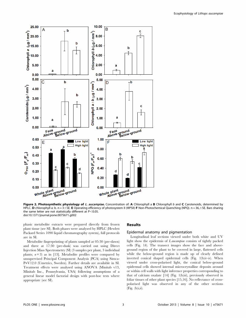
Figure 2. Photosynthetic physiology of L. aucampiae. Concentration of: A Chlorophyll a B Chlorophyll b and C Carotenoids, determined byHPLC. D chlorophyll a: b. n = 36SE. E Operating efficiency of photosystem II (FPSII) F Non-Photochemical Quenching (NPQ). n = 36,6SE. Bars sharingthe same letter are not statistically different at P,0.05.doi:10.1371/journal.pone.0075671.g002
Ecophysiology of Lithops aucampiae
PLOS ONE | www.plosone.org 3 October 2013 | Volume 8 | Issue 10 | e75671

Chlorophyll a was the most abundant pigment in each region of
L. aucampiae, followed by chlorophyll b, and carotenoid pigments
being of lowest concentration (Fig. 2a-c). The face contained the
lowest abundance of pigments in any of the regions measured.
There was no significant difference in the concentration of
carotenoids between the above- and below-ground regions
(Fig. 2c).The chlorophyll b concentration of the below ground
section was also significantly lower in the above-ground section.
Chlorophyll a: chlorophyll b was significantly lower in the below-
ground region than in the above-ground region (Fig. 2d).
Photosynthetic activity in L. aucampiaeOptimised studies using plants acclimated to actinic light levels
of 500 mmol m22 s21 and 100 mmol m22 s21 revealed that all
regions of the plants exposed to the high light treatment had lower
mean FPSII values compared to those grown at lower actinic light
(Fig. 2e; F(1,215) = 289.11, P,0.001). FPSII was significantly greater
in the above-ground region than the below-ground region at each
light level; however the difference between light levels was less
marked in the below-ground region than in the above-ground
region (Fig. 2e).
The opposite trend was true for NPQ with the high actinic light
treatment producing the greatest mean NPQ values in all regions
compared to the lower actinic treatment (F(1,215) = 143.65,
P,0.001). The highest mean NPQ value was observed in the
below-ground region of the plant (Fig. 2f).
When plants were exposed to increasing actinic light levels,
FPSII decreased dramatically in the above-ground region (Fig. 2e).
FPSII also decreased in the below-ground region, although these
values remained consistently lower than the values for the above-
ground region at every actinic level measured (Fig. 2e). NPQ
values had the opposite trend, as values increased with increasing
actinic light levels (Fig. 2f).
Metabolite profilingMetabolites were identified as photosynthetic pigments, being
either chlorophylls (eluted after 19 min, 20 min and 21 min;
Table 1, Fig. 3) or carotenoids (eluted after 8 min, 15 min and
22 min; Table 1, Fig. 3). UV-absorbing metabolites, likely to be
anthocyanins, were identified in the above-ground region along
with other non-photosynthetic pigments such as xanthone in the
face (Table 1, Fig. 3).
Metabolite fingerprinting of whole plants revealed clear
metabolic differences between pre-dawn and pre-dusk samples.
Figure 3. UV absorbance traces for metabolite identification.UV traces showing absorbance spectra and putative ID of metabolitesobtained through HPLC-PDAD.doi:10.1371/journal.pone.0075671.g003
Table 1. Metabolite identification by HPLC.
Plant section Solvent phase Retention time (min) Putative compound PDA UV lmax (nm)
Whole plant Organic 8 Neoxanthin 440, 465
Whole plant Organic 15 a-carotene 445, 475
Whole plant Organic 19 Chlorophyll b 345, 460
Whole plant Organic 20 Chlorophyll a 385, 415, 430
Whole plant Organic 21 Chlorophyll 410, 505
Whole plant Organic 22 b-carotene 455, 485
Face Aqueous 1 Xanthone 285, 330, 390
Face Aqueous 2 Vitamin K1- like compound 275, 315, 370
Above-ground Aqueous 2 Flavonoid 275, 305, 380
Above-ground Aqueous 3 Flavonoid 270, 320
doi:10.1371/journal.pone.0075671.t001
Ecophysiology of Lithops aucampiae
PLOS ONE | www.plosone.org 4 October 2013 | Volume 8 | Issue 10 | e75671

Principal Component Analysis (PCA) of the molecular ions
detected within the aqueous phase showed distinct separation of
the pre-dawn and pre-dusk samples along PC1 (Methods S1).
Malate (133 m/z) was identified as a highly discriminatory
compound between treatments and was found in greater
abundance (530855.6 TIC) in pre-dawn samples compared to
pre-dusk (289211.1 TIC) alongside many key tricarboxylic acid
(TCA) cycle components (Table 2). This analysis also showed that
carbohydrates, including hexose sugars, were of lower abundance
in pre-dawn samples compared to those taken pre-dusk (Table 2).
Discussion
Our research has demonstrated for the first time that L.
aucampiae exhibits simultaneous high-light and shade-specific
functional adaptations simultaneously within individual leaf
structures. We hypothesise that this maintains optimal photosyn-
thesis under the xeric conditions experienced in its native desert
habitat.
Our anatomical observations of L. aucampiae using white-light
and UV microscopy show L. aucampiae leaf structures to possess a
highly varied epidermal structure, in common with others within
the genera [17]. Cells on the face and above-ground regions of L.
aucampiae leaves are large and flat, while those covering the below-
ground foliar region are conical (Fig. 1b). Epidermal cells such as
those in our observations are thought to have important functions
in water storage, ion and pigment accumulation, light guiding and
photosynthetic capacity [18–21]. It is possible the cone-shaped
epidermal cells that cover the below-ground regions of the plant
leaves serve to minimize loss of light and to maximise below-
ground internal reflectance.
Calcium oxalate druses are often found within epidermal cells
similar in structure to those we observed in Lithops in a diverse
array of plant species including Dieffenbachia seguine [9], Aesculus
hippocastanum [22] and, notably, more than 80 Conophytum species
[23], themselves being close relatives of Lithops. Previous studies on
the function of similar foliar calcium oxalate formations show
them to be translucent and to possess light scattering properties
[16]. These properties suggest the presence of microcrystalline
deposits within the below-ground epidermis of L. aucampiae leaves
(Fig. 1(b)iii) may be associated with photosynthesis by scattering
light within the below-ground region of the leaves, thus enriching
the lower tissues with photons [16]. By increasing internal
reflectance of incoming light from the above-ground plant
mesophyll to the less illuminated below-ground photosynthetic
tissues, we hypothesise that the presence of conical epidermal cells
with polarised-light inference properties provide critical evidence
of shade-specific adaptations present within the below-ground
portions of L. aucampiae leaves with below-ground microcrystalline
deposits in Lithops being analogous in function to the crystals
frequently observed embedded within the epidermis and/or
mesophyll of many shade-tolerant plant species. Cone-shaped
epidermal cells were not observed on the epidermis and polarised
light was not reflected in the above-ground region of the leaves
(Figs. 1(b)i,ii,vii,viii).
Further evidence of the shade-tolerance capabilities of the
below-ground region of L. aucampiae leaves lie in the increased
concentration of chlorophyll b and reduced chlorophyll a:b ratio of
that region (Fig. 2a-d). Chlorophyll a occurs in all photosynthetic
eukaryotes and is essential for commencement of photosynthesis.
Chlorophyll b is not directly involved in photosynthetic processes
but is important for broadening the wavelengths of light that can
be absorbed by the organism and transferring ‘extra’ captured
photons to chlorophyll a for use in photosynthesis [24]. In
possessing a low chlorophyll a:b ratio, a plant is effectively able to
capture more photons across a broader range of wavelengths – an
adaptation particularly important for plants growing in shaded
environments. The above ground region appears to contain a
marginally higher concentration of chlorophyll a and has a greater
a:b ratio than the below ground portion of the leaves, indicating
greater photosynthetic capacity at higher light levels (Figs. 2a, d).
This is confirmed by there being greater FPSII (photosynthetic
rate) at both high and low light levels in the above-ground region
of the leaf compared to the below-ground portion. The lower FPSII
and greater NPQ in the lower parts of the leaf suggest that the
effective ‘cost’ of increased chlorophyll b and reduced a:b is borne
through lower operational photosynthetic efficiency and greater
dissipation of energy through NPQ (Figs. 2e & f).
These differences in regional photosynthetic function within the
leaf structure may be enhanced by variation in light-blocking
pigmentation. Pigment analysis identified numerous non-photo-
Table 2. Putative metabolites in L. aucampiae sampled pre-dawn and pre-dusk (n= 3). *P,0.05, Student’s T-test.
Mean molecularmass (Da) SE
Class ofcompound Pre-dawn TIC Pre-dusk TIC Fold change Sig.
Oxaloacetate 132.0 0.004 Organic acid 11669.8 5614.4 2.08 *
Citrate 192.0 0.006 Organic acid 328133.3 166400.0 1.97 *
Uracil 112.0 0.004 Nucleobase 244377.8 122427.8 1.99 *
Fumarate 116.0 0.004 Organic acid 466200.0 241944.4 1.93 *
Pyruvic acid 88.0 0.003 Organic acid 27568.9 52361.1 1.90 *
Malate 134.0 0.004 Organic acid 530855.6 289211.1 1.84 *
Succinate 118.0 0.004 Organic acid 10527.7 5702.0 1.84 *
cis-Aconitate 174.0 0.006 Organic acid 33801.1 19471.1 1.76 *
2-Oxoglutarate 146.0 0.005 Organic acid 945.9 1429.0 1.51 *
Phosphoenolpyruvate 167.9 0.005 Organic acid 2806.8 1870.3 1.50 *
Oxalosuccinic acid 190.0 0.006 Organic acid 1152.1 993.0 1.16 *
Hexose 342.1 0.011 Monosaccharide 17172.2 14943.3 1.15 *
Phenylpyruvate 164.0 0.005 Organic acid 55703.3 89098.9 0.63 *
doi:10.1371/journal.pone.0075671.t002
Ecophysiology of Lithops aucampiae
PLOS ONE | www.plosone.org 5 October 2013 | Volume 8 | Issue 10 | e75671

synthetic pigments contained in face and above-ground plant
tissues. Flavonoids, of which several were identified to be present
in the above-ground region (Table 1, Fig. 3), are known to filter
UV radiation, and have been found to accumulate in the
epidermal cells of other Mesembryanthemum species (Vogt et al.
1999). Unlike plants of the Caryophyllales, extracts obtained from
L. aucampiae did not contain any compounds with UV absorbance
within the range characteristic for betalain compounds. Flavonoid
compounds, such as those detected here in L. aucampiae, generally
accumulate in peripheral tissues exposed to high irradiance [25],
therefore we might expect to see fewer of these compounds in the
below-ground region. The brown pigmentation resulting from
high accumulation of flavonoids is visibly reduced in below-ground
leaf tissue compared to the above-ground leaf tissues (Fig. 1).
Flavonoid pigments preferentially absorb green and blue light but
reflect red wavelengths [26], thereby reducing the light energy
available for photosynthesis, resulting in the lower NPQ values
observed within the above-ground leaf tissues compared to the
below-ground. This photo-protective biochemical mechanism is
likely to be highly beneficial to wild-growing L. aucampiae which
experience extremely high daily irradiance and would otherwise
be highly susceptible to severe photo-damage and inhibition. The
pigmentation of L. aucampiae is likely to also serve to protect it from
herbivory by small mammals through camouflage.
Our metabolomic analysis confirmed previous findings that L.
aucampiae, in common with other Lithops species [5], utilises CAM
photosynthesis. PCA analysis demonstrated that L. aucampiae plants
sampled pre-dawn had a very different metabolic profile to those
sampled pre-dusk (SI). Many of the most highly discriminatory
compounds identified through this analysis were organic acids, one
of which was malate. The majority of these organic acids were in
far greater abundance within plant tissues in the pre-dawn
sampled plants when compared to those sampled pre-dusk
(Table 2). Organic acids, malate in particular, are vital compo-
nents of the CAM photosynthetic pathway, being produced
overnight through CO2 fixation and stored in plant cell vacuoles
until sunrise and the re-commencement of photosynthesis [24].
Plants that operate using CAM close their stomata during the day
to reduce water loss through transpiration, inevitably inhibiting
CO2 assimilation for use in photosynthesis. CAM plants, are able
to mobilise and breakdown the vacuolar acids, such as those in
Table 2, releasing carbon which is then utilised in the Calvin-
Benson cycle [24]. Plants with these adaptations have relatively
high tissue concentrations of organic acids (1.84 fold increase in
the case of malate observed here, Table 2) when sampled pre-
dawn compared to those sampled pre-dusk.
The simultaneous high-light, shade-tolerance strategies we have
revealed within individual leaves of the xerophyte L. aucampiae are
previously unreported in the plant kingdom and are, to our
knowledge, unique to the Aizoaceae. We have shown the leaves of
L. aucampiae are uniquely adapted to optimise photosynthesis under
very different environments within the same structure: extreme
high-irradiance in the AG region and shade conditions BG, and
that these regions have sufficient physiological flexibility to
respond to variable light conditions in a way that affords
maximum protection against photo-inhibition and oxidative
damage while maintaining optimal photosynthetic rates. This
demonstrates unprecedented physiological flexibility in a xero-
phyte and is a step forward in our understanding of plant
responses and adaptations to extreme environments.
Supporting Information
Figure S1 Principal component analysis (PCA) scorescatter plot (a) of the metabolic fingerprinting data(direct injection mass spectrometry m/z values) ob-tained from Lithops aucampiae sampled at pre-duskand pre-dawn. The percent of the variation explained by each
component is given. (b) molecular mass ions (m/z) differing the
most between pre-dusk and pre-dawn samples. The molecular
weight of malate is highlighted.
(PDF)
Methods S1
(DOCX)
Acknowledgments
We thank Jennifer Morris for her help and expertise in cross-polarised light
microscopy; Duncan Cameron, Janice Lake, Lyla Taylor, Kate Allinson,
Joe Quirk and the anonymous reviewers for helpful comments on our
manuscript.
Author Contributions
Conceived and designed the experiments: KJF RG MPD. Performed the
experiments: RG MPD KJF. Analyzed the data: KJF MPD. Contributed
reagents/materials/analysis tools: BF PQ. Wrote the paper: KJF MPD.
References
1. Ellis AG, Weis AE (2006) Coexistence and differentiation of ‘flowering stones’:
the role of local adaptation to soil microenvironment. J Ecol 94: 322–335.
2. Kellner A, Ritz CM, Schlittenhardt P, Hellwig FH (2011) Genetic differentiation
in the genus Lithops L. (Ruschioideae, Aizoaceae) reveals a high level of
convergent evolution and reflects geographic distribution. Plant Biol 13: 368–
380.
3. Gibson (1998) Photosynthetic organs of desert plants. BioScience 48: 911–920.
4. Raven PH, Evert RF, Eichhorn SE (1999) Biology of Plants: W.H. Freeman and
Company Worth Publishers
5. Von Willert DJ, Eller BM, Werger MJA, Brinckmann E, Ihlenfeld H-D (1992)
Life Strategies of Succulents in Deserts with special reference to the Namib
desert: Cambridge University Press.
6. Egbert KJ, Martin CE (2002) The influence of leaf windows on the utilization
and absorption of radiant energy in seven desert succulents. Photosynthetica 40:
35–39.
7. Martin CE, Brandmeyer EA, Ross RD (2013) Ecophysiological function of leaf
‘windows’ in Lithops species – ‘Living Stones’ that grow underground. Plant Biol
15: 243–247.
8. Foster AS (1956) Plant idioblasts: Remarkable examples of cell specialization.
Protoplasma 46 184–193.
9. Cote GG (2009) Diversity and distribution of idioblasts producing calcium
oxalate crystals in Dieffenbachia seguine (Araceae). American Journal of Botany
96: 1245–1254.
10. Demmig-Adams B, Adams WW (1992) Photoprotection and other responses of
plants to high light stress. Annu Rev Plant Physiol Plant Molec Biol 43: 599–626.
11. Muller P, Li XP, Niyogi KK (2001) Non-Photochemical Quenching. A
Response to Excess Light Energy. Plant Physiology 125: 1558–1566.
12. Wellburn AR (1994) The spectral determination of chlorophyll-A and
chlorophyll-B, as well as total carotenoids, using various solvents with
spectrophotometers of different resolution. J Plant Physiol 144: 307–313.
13. Davey MP, Burrell MM, Woodward FI, Quick WP (2008) Population-specific
metabolic phenotypes of Arabidopsis lyrata ssp petraea. New Phytol 177: 380–
388.
14. Nesse WD (2000) Introduction to Mineralogy: Oxford University Press.
15. Kuo-Huang LL, Ku MSB, Franceschi VR (2007) Correlations between calcium
oxalate crystals and photosynthetic activities in palisade cells of shade-adapted
Peperomia glabella. Bot Stud 48: 155–164.
16. Gal A, Brumfeld V, Weiner S, Addadi L, Oron D (2012) Certain Biominerals in
Leaves Function as Light Scatterers. Adv Mater 24: OP77–OP83.
17. Fearn B (1977) Epidermal structure, stomatal distribution and water-loss in
Lithops marmorate. Cactus and Succulent Journal of Great Britain 39: 68–70.
18. Ihlenfeldt HD, Juergens N (1982) Epidermis Types with Reduced Bladder
Idioblasts in Mesembryanthemaceae. Mitteilungen aus dem Institut fuer
Allgemeine Botanik Hamburg 18: 103–116.
19. Tanner V, Eller BM (1986) Epidermis Structure and Its Significance for the
Optical-Properties of Leaves of the Mesembryanthemaceae. J Plant Physiol 125:
285–294.
Ecophysiology of Lithops aucampiae
PLOS ONE | www.plosone.org 6 October 2013 | Volume 8 | Issue 10 | e75671

20. Vogt T, Ibdah M, Schmidt J, Wray V, Nimtz M, et al. (1999) Light-induced
betacyanin and flavonol accumulation in bladder cells of Mesembryanthemum
crystallinum. Phytochemistry 52: 583–592.
21. Whitney HM, Bennett KMV, Dorling M, Sandbach L, Prince D, et al. Why do
so many petals have conical epidermal cells? Ann Bot.
22. Weryszko-Chmielewska E, Haratym W (2011) Changes in leaf of common horse
chestnut (Aesculus hippocastanum L.) colonised by the horse-chestnut lead miner
(Cameraria ochridella). Acta Agrobotanica 64: 11–21.
23. Opel MR (2005) Leaf anatomy of Conophytum N.E.Br. (Aizoaceae). Haseltonia:
27–52.24. Raven P, Evert R, Eichhorn S (1999) Biology of plants: WH Freeman and
Company.
25. Steyn WJ, Wand SJE, Holcroft DM, Jacobs G (2002) Anthocyanins in vegetativetissues: a proposed unified function in photoprotection. New Phytol 155: 349–
361.26. Chalker-Scott L (1999) Environmental Significance of Anthocyanins in Plant
Stress Responses. Photochemistry and Photobiology 70: 1–9.
Ecophysiology of Lithops aucampiae
PLOS ONE | www.plosone.org 7 October 2013 | Volume 8 | Issue 10 | e75671