UNIVERSITI PUTRA MALAYSIA LUNG INJURIES INDUCED BY...
25
UNIVERSITI PUTRA MALAYSIA LUNG INJURIES INDUCED BY INDEN0(1,2,3-CD)PYRENE IN RATS ON GARLIC SUPPLEMENTATION SANAZ MOVASSAGH FPV 2007 7
Transcript of UNIVERSITI PUTRA MALAYSIA LUNG INJURIES INDUCED BY...

UNIVERSITI PUTRA MALAYSIA
LUNG INJURIES INDUCED BY INDEN0(1,2,3-CD)PYRENE IN RATS ON GARLIC SUPPLEMENTATION
SANAZ MOVASSAGH
FPV 2007 7

LUNG INJURIES INDUCED BY INDEN0(1,2,3-CD)PYRENE IN RATS ON GARLIC SUPPLEMENTATION
BY
SANAZ MOVASSAGH
Thesis Submitted to the School of Graduate Studies, Universiti Putra Malaysia, in Fulfilment of the Requirements for the Degree of Doctor of Philosophy
April 2007

Abstract of thesis presented to the Senate of Universiti Putra Malaysia in fulfilment of the requirement for the degree of Doctor of Philosophy
LUNG INJURIES INDUCED BY INDEN0(1,2,3-CD)PYRENE IN RATS ON GARLIC SUPPLEMENTATION
SANAZ MOVASSAGH
April 2007
Chairman: Associate Professor Noordin Mohamed Mustapha, PhD
Faculty : Veterinary Medicine
The impact of air pollution on health and economic well-being of nation is an important
worldwide issue. Indeno[l,2,3-cdlpyrene (IP) is a particulate matter amongst
environmental pollutants found in the Malaysian haze episodes and was claimed to
induce deleterious effects on humans or animals. However, such claims have never been
scientifically substantiated. In manifesting the noxious effects of haze and in view of
developing strategies and bringing about a remission of such effects in humans, the
symptoms of both acute exposure and chronic response to IP were studied on the
pulmonary system of rats.
The acute exposure studies were conducted to evaluate the histopathological and
ultrastructural changes and detection of apoptotic cells in the lung of rats following
treatment with IP with or without garlic supplementation. Furthermore, the
immunological responses and elastolytic enzymes activities were also determined. The
IP-treated rats received 13 ng (6.5 ~ 1 ) of IP that was instilled intra-tracheally without
garlic supplementation. Rats from the garlic (G) group were fed the basal ration
containing garlic incorporated at the rate of 80 mglkg body-weight/rat/day while those
from the G+IP group fed on garlic diet for two weeks before instillation with IP. All
animals were sacrificed at 8, 16, 24 and 32 hours (hrs) post-instillation (pi.) and
according to the respective interval design. Histopathological alterations were studied on
haematoxylin-eosin (H&E) stained sections and ultrastructural changes revealed by
transmission electron microscope (TEM). Apoptosis assessment was made on terminal
desxynucleotidyl transferase-mediated dUTP nick-end labeling (TUNEL) analysis and
caspase 3 colorimetric assay. The broncho-alveolar lavage (BAL) assays, ELISA
method and alveolar macrophages (AM0) activities assay were used to detect the
elastolytic enzymes activities and lung defense.
Histopathological, ultrastructural, TUNEL and caspase 3 assays findings showed
apoptosis which advances with time in the pneumocytes and bronchial epithelium of all
IP-treated rats euthanised at the respective time. In addition, inflammation, necrosis and
epithelisation were also observed in the IP group. Likewise, the IP group had the highest
elastase-like and neutrophil elastase specific activities but the lowest IgA and IgG levels,
AM0 phagocytosis, and intracellular killing activities of AM0 in the BAL.
Supplementation with garlic was able to minimize the deleterious effect of IP.
In the chronic response studies, in addition to the other parameters measured in the acute
study, an assessment on the level of glutathione S-transferase (GST) was also made.
Similarly, there was an additional treatment group that was instilled with IP once at the
beginning of the study and fed with garlic during the whole period of experiment (IP+G)
to assess the effect of consumption of garlic during the response to IP.

The microscopic and ultrastructure studies revealed evidence of apoptosis,
inflammation, necrosis and epithelisation in the lung of rats treated with IP. In addition,
rats treated with IP had the lowest levels of IgA, IgG, alveolar macrophages activities
and GST specific activity in the lung. Furthermore, there was a dreadful effect on
elastolytic enzymes activities present in the IP-treated rats.
In contrast, the IP-treated groups fed with garlic showed significant improvement
towards normal histology of the lung or trachea. However, it was found that regularly
consumption of garlic during the response to IP have a better result than a prophylactic
regime before response.
In conclusion, IP poses as an environmental hazard to the lungs of rats triggering
deleterious changes either due to short exposure or long term response and garlic has a
great potential in alleviating these adverse effects.

Abstrak tesis yang dikemukakan kepada Senat Universiti Putra Malaysia sebagai memenuhi keperluan untuk ijazah Doktor Falsafah
KECEDERAAN PARU-PARU TERARUH INDENO(l,2,3-CD)PYRENE PADA TIKUS YANG MENERIMA TAMBAHAN BAWANG PUTIH
Oleh
SANAZ MOVASSAGH
April 2007
Pengerusi: Profesor Madya Noordin Mohamed Mustapha, PhD
Fakulti : Perubatan Veterinar
Kesan pencemaran udara pada kesihatan manusia dan status ekonomi negara menjadi
salah satu isu penting di seluruh dunia. Indeno[l,2,3-capyrene (IP) adalah antara bahan
partikel dari pencemar alam sekitar yang ditemui semasa episod jerebu yang melanda
Malaysia dan dipercayai boleh mengakibatkan kesan yang merbahaya kepada manusia
dan haiwan. Walaubagaimanapun, dakwaan tersebut tidak pernah dibuktikan secara
saintifik. Untuk menunjukkan kesan berbahaya jerebu dan untuk menyelidik strategi
bagi mengurangkan kesan tersebut pada manusia, simptom-simptom pendedahan akut
dan kronik terhadap IP dikaji ke atas sistem pulmonari tikus.
Kajian mengenai pendedahan akut telah dijalankan untuk menilai perubahan
histopatologi dan ultrastruktur dan pengenalpastian sel-sel apoptosis pada paru-paru
tikus setelah diberi rawatan dengan IP. Selain daripada itu, tindakbalas imunologi dan
aktiviti enzim elastolitik telah ditentukan. Tikus dari kumpulan rawatan IP telah diberi
13 ng (6.5 p1) IP secara instilasi intra-trakea tanpa suplementasi bawang putih. Tikus
dari kumpulan bawang putih (G) diberi makanan komersial dengan penambahan bawang

kadar yang bersamaan dengan pengambilan sebanyak 80 mgkg berat badan/tikus/sehari
manakala tikus dari kumpulan G+IP telah diberi makan diet yang mengandungi bawang
putih selama dua minggu sebelum pemberian IP. Kesemua tikus tersebut telah dibunuh
pada 8, 16, 24 dan 32 jam (hrs) pasca-pemberian ( p i ) dan mengikut rekabentuk kajian
masing-masing. Perubahan secara histopatologikal telah dikaji pada hirisan tisu yang
telah diwarnakan dengan haematoxylin-eosin (H&E) manakala perubahan ultrastruktur
dikaji dengan menggunakan elektron mikroskop transmisi (TEM). Penilaian apoptosis
telah dibuat berdasarkan analisis pelabelan potongan hujung TUNEL dan penilaian
caspase 3 kalorimetrik. Penilaian cecair bronko-alveolus (BAL), kaedah ELISA dan
aktiviti makrofaj alveolus telah dilakukan untuk mengkaji enzim elastolitik dan
pertahanan peparu.
Penemuan histopatologi, ultrastruktur TUNEL dan penilaian caspase 3 menunjukkan
tahap apoptosis yang berbeza pada pneumosit and epitelium bronkiol pada semua tikus
yang diberi rawatan IP yang dibunuh mengikut jangkamasa yang telah ditetapkan pasca
pemberian. Di samping itu, inflamasi, nekrosis dan epitelialisasi telah diperhatikan
dalam kumpulan IP. Disamping ini, kumpulan IP telah menunjukkan aktiviti spesifik
elastase yang tertinggi dan aktiviti spesifik elastase neutrofil tetapi tahap aktiviti yang
rendah bagi IgA dan IgG, fagositosis A M 0 dan pemusnahan intrasel aktiviti A M 0
dalam BAL. Supplementasi dengan bawang putih telah meminimumkon kesan
pemusnahan IP.
Dari kajian pendedahan kronik, perubahan pada peparu kumpulan IP selama tiga bulan
telah dinilai melalui tahap enzim penanda, glutathion S-transferase (GST). Begitu juga
vii

bagi kumpulan yang diberi IP pada permulaan kajian dan diberi makan bawang putih
sepanjang tempoh kajian dijalankan (IP+G) untuk menilai kesan pengambilan bawang
putih sepanjang pendedahan terhadap IP.
Dalam kajian mikroskopik dan ultrastruktur menunjukkan bukti apoptosis, inflamasi,
nekrosis dan epitelialisasi di dalam peparu tikus yang dirawat dengan IP. Selain daripada
itu, tikus yang diberi IP mempunyai tahap IgA, IgG dan aktiviti makrofaj alveolus serta
aktiviti spesifik khas GST dalam peparu yang paling rendah. Tambahan lagi, terdapat
kesan yang dahsyat pada aktiviti enzim elastolisis dalam kumpulan IP.
Sebaliknya, kumpulan yang menerima rawatan IP yang diberi makan bawang putih
menunjukkan keadaan yang lebih baik dalam semua perubahan histologi pada peparu
dan trakea. Oleh yang demikian, pengambilan bawang putih yang kerap semasa
pendedahan terhadap 1P menunjukkan keputusan yang lebih baik berbanding yang
menerima pemakanan kurang sebelum pendedahan.
Kesimpulannya, kehadiran IP dalam alam sekitar adalah merbahaya kepada paru-paru
tikus yang bertindak sebagai organ target di mana ia mencetuskan perubahan yang
berbahaya untuk pendedahan jangkamasa pendek mahupun panjang dan didapati
bawang putih mempunyai potensi yang amat baik dalam mengurangkan kesan
merbahaya tersebut.
... V l l l

ACKNOWLEDGEMENTS
The highest praise and gratefulness to ALLAH, who shines my soul towards truth and
faith, and fills my mind with knowledge and wisdom. To him do I entrust myself, to him
be in grace, and with him be in success, immunity and comfort.
It was such a great honor to work under Associate Professor Dr. Noordin Mohamed
Mustapha supervision who guided me throughout this research with his patience,
continuing guidance and support by giving greater latitude of freedom in conducting this
study as well as completion of this thesis. I never can repay him back all his favour.
I would like to extend my gratitude and thanks to my supervisory committee members,
Professor Dr. Mohd Zamri Saad and Assoc. Professor Dr. Hassan Hj. Mohd Daud for
their contribution and advices during this research. I also gratefully acknowledge
Professor Dato' Dr. Sheikh Omar Abdul Rahman for his concern, encouragement and
his valuable time to guide me toward completion of my study.
My sincere thanks are due to Ministry of Science, Technology and Innovation (MOSTI)
for the financial support through IRPA grant for this research. My deep appreciations to
all the staff of the Faculty of Veterinary Medicine and Institute of Bioscience for their
cooperation and facilities throughout this work.
I highly appreciate all my colleagues, friends, family and husband for their support and
kindness, especially to Dr. Mazlina binti Mazlan for her taintless helps during this study.

I certify that an Examination Committee has met on 25'h April 2007 to conduct the final examination of Sanaz Movassagh on her Doctor of Philosophy thesis entitled "Lung Injuries Induced by Indeno(l,2,3-Cd) Pyrene in Rats on Garlic Supplementation" in accordance with Universiti Pertanian Malaysia (Higher Degree) Act 1980 and Universiti Pertanian Malaysia (Higher Degree) Regulations 1981. The Committee recommends that the candidate be awarded the relevant degree. Members of the Examination Committee are as follows:
Md Zuki Abu Bakar , PhD Associate Professor Faculty of Veterinary Medicine Universiti Putra Malaysia (Chairman)
Dato' Sheikh Omar Abdul Rahman, PhD Professor Faculty of Veterinary Medicine Universiti Putra Malaysia (Internal Examiner)
Jasni Sabri, PhD Associate Professor Faculty of Veterinary Medicine Universiti Putra Malaysia (Internal Examiner)
Ian A Silver, PhD Professor School of Veterinary Science Bristol University (External Examiner)
HD. GHAZALI, PhD
School of ~raduate Studies Universiti Putra Malaysia
Date: 21 JUNE 2007

This thesis submitted to the Senate of Universiti Putra Malaysia and has been accepted as fulfilment of the requirement for the degree of Doctor of Philosophy. The members of the Supervisory Committee are as follows:
Noordin Mohamed Mustapha, PhD Associate Professor Faculty of Veterinary Medicine Universi ti Putra Malaysia (Chairman)
Mohd Zamri Saad, PhD Professor Faculty of Veterinary Medicine Universi ti Putra Malaysia (Member)
Hassan Hj Mohd Daud, PhD Associate Professor Faculty of Veterinary Medicine Universiti Putra Malaysia (Member)
AINI IDERIS, PhD Professor1 Dean School of Graduate Studies Universiti Putra Malaysia
Date: 17 JULY 2007

DECLARATION
I hereby declare that the thesis is based on my original work except for quotations and citations which have been duly acknowledged. I also declare that it has not been previously or concurrently submitted for any other degree at UPM or other institutions.
7 SANAZ MOVASSAGH
xii
Date: 10 JUNE 2007

TABLE OF CONTENTS Page
DEDICATION ABSTRACT ABSTRAK ACKNOWLEDGEMENTS APPROVAL DECLARATION LIST OF TABLES LIST OF FIGURES LIST OF ABBREVIATIONS
CHAPTER
GENERAL INTRODUCTION Air Pollution and Haze The Impacts of Haze on Human Health Haze Episodes in Malaysia Objectives of the Study
REVIEW OF LITERATURE Introduction Chemical Carcinogens Polycyclic aromatic hydrocarbons Indeno[ l,2,3-cdJpyrene
Metabolism Toxicity Indeno[1,2,3-cdJpyrene as a Carcinogen Pathology of Cancer Induced by Indeno[l,2,3-cclJpyrene
Target Oragn Toxicity Lung The pulmonary Defense during Air pollution Mechanisms of Cell Death
Cancer Prevention and Treatment Garlic (Allium sativum) Constituents and Beneficial Effects Anti-carcinogenic and Anti-tumour Potentials Toxicity
GENERAL MATERIALS AND METHODS Animals and Management Inoculum Garlic Experimental Design Pathology
. . 11 ... 111
vi ix X
xii xvi xviii xxiii
. . . X l l l

Histopathology In Situ Detection of Apoptotic Cells Ultrastructural Study
Lavage Fluid Assays and Immunology Broncho-alveolar Lavage P A L ) Measurment of Elastase-like Activity Measurment of Neutrophil Elastase Activity Total Protein Measurment Enzyme-Linked Immunosorbent Assay (ELISA) Assay of Alveolar Macrophages Activity
Estimation of GST Activities in Lung and Liver Tissues Preparation of Cytosol Enzyme Assay Protein Determination
Caspase 3 Colorimetric Assay Statistical Analysis
EFFECT O F GARLIC ON GLUTATHIONE S- TRANSFERASE Introduction Materials and Methods Results Discussion
ACUTE LUNG EXPOSURE TO INDEN0[1,2,3-cd]PYRENE IN RATS ON GARLIC SUPPLEMENTATION Introduction Materials and Methods Results
Clinical Signs Gross Pathological Findings Histopathological Findings In Situ Detection of Apoptotic Cells Ultrastructural Findings Elastase-Like and Neutrophil Elastase Specific Activities Level of IgA in the Lung Lavage Fluid Level of IgG in the Lung Lavage Fluid Level of IgG in Serum Alveolar Macrophages Activities Caspase 3 Specific Activity
Discussion
CHRONIC LUNG RESPONSE T O INDEN0[1,2,3- cdjPYRENE IN RATS ON GARLIC SUPPLEMENTATION 97 Introduction 97 Materials and Methods 99 Results 100
xiv

VII
Clinical Signs Gross Pathological Findings Histopathological Findings In Situ Detection of Apoptotic Cells Ultrastructural Findings Elastase-Like and Neutrophil Elastase Specific Activities Level of IgA in the Lung Lavage Fluid Level of IgG in the Lung Lavage Fluid Level of IgG in Serum Alveolar Macrophages Activities Glutathione S-transferase (GST) Specific Activity Caspase 3 Specific Activity
Discussion
GENERAL DISCUSSION AND CONCLUSIONS
REFERENCES APPENDICES BIODATA OF THE AUTHOR

LIST OF TABLES
Table
1
2
Page
13 Some important characters of indeno[l,2,3-cdJpyrene
The GST specific activity in the lung of rats at necropsy (pmollminlmg protein; Mean + SE)
The GST specific activity in the liver of rats at necropsy (pmol/min/mg protein; Mean + SE)
The experimental design of acute exposure of indeno[l,2,3- cdlpyrene in rats
Percentage of neutrophils in the lungs of rats during acute exposure (mean + SE)
Percentage of alveolar macrophages in the lungs of rats during acute exposure (mean + SE)
Percentage of necrosis in the lungs of rats during acute exposure (mean + SE)
8 Percentage of apoptosis in the lungs of rats during acute exposure (mean + SE)
Percentage of epithelisation in the lungs of rats during acute exposure (mean + SE)
Percentage of TUNEL-positive cells in the lungs of rats during acute exposure (mean + SE)
The elastase-like and neutrophil elastase specific activities in the lung lavage of rats during acute exposure (unitslmg protein; Mean + SE)
The levels of IgA and IgG in the lung lavage fluid of rats during acute exposure (OD; Mean + SE) 79
The levels of IgG in the serum of rats during acute exposure (OD; Mean + SE)
The alveolar macrophage (AM@) activities in the lung lavage fluid of rats during acute exposure (Mean + SE)
The experimental design of chronic response to IP in rats
xvi

16 The lesion scores in the lungs of rats at necropsy (%; mean * SE)
17 Percentage of apoptosis in the lungs of rats at necropsy (mean * SE) 109
18 Percentage of TUNEL-positive cells in the lungs of rats at necropsy (mean SE) 111
19 The elastase-like and neutrophil elastase specific activities in the lung lavage of rats at necropsy (unitslmg protein; Mean A SE) 118
20 The levels of IgA and IgG in the lung lavage fluid and IgG in serum of rats at necropsy (OD; Mean k SE)
21 The alveolar macrophage (AM@) activities in the lung lavage fluid of rats at necropsy (Mean * SE) 121
xvii
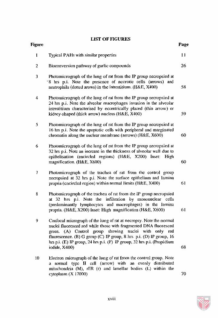
LIST OF FIGURES Figure
1 Typical PAHs with similar properties
2 Bioconversion pathway of garlic compounds
3 Photomicrograph of the lung of rat from the IP group necropsied at -8 hrs p-i. Note the presence of necrotic cells (arrows) and neutrophils (dotted arows) in the intrestitium. (H&E, X400)
Page
1 I
26
4 Photomicrograph of the lung of rat from the IP group necropsied at 24 hrs p.i. Note the alveolar macrophages invasion in the alveolar intrestitium characterised by eccentrically placed (thin arrow) or kidney-shaped (thick arrow) nucleus (H&E, X400) 59
Photomicrograph of the lung of rat from the IP group necropsied at 16 hrs p.i. Note the apoptotic cells with peripheral and marginated chromatin along the nuclear membrane (arrows) (H&E, X600) 60
Photomicrograph of the lung of rat from the IP group necropsied at 32 hrs p.i. Note an increase in the thickness of alveolar wall due to epithelisation (encircled regions) (H&E, X200) Inset: High magnification. (H&E, X600) 60
Photomicrograph of the trachea of rat from the control group necropsied at 32 hrs p.i. Note the surface epithelium and lamina propria (encircled region) within normal limits (H&E, X400) 6 1
Photomicrograph of the trachea of rat from the IP group necropsied at 32 hrs pi. Note the infiltration by mononuclear cells (predominantly lymphocytes and macrophages) in the lamina propria. (H&E, X200) Inset: High magnification (H&E, X600)
Confocal micrograph of the lung of rat at necropsy. Note the normal nuclei fluoresced red while those with fragmented DNA fluoresced green. (A) Control group showing nuclei with only red fluorescence. (B) G group (C) IP group, 8 hrs p.i. (D) IP group, 16 hrs p.i. (E) IP group, 24 hrs p.i. (F) IP group, 32 hrs p.i. (Propidium iodide, X400)
Electron micrograph of the lung of rat from the control group. Note a normal type I1 cell (arrow) with an evenly distributed mitochondria (M), rER (r) and lamellar bodies (L) within the cytoplasm (X 17000) 70
xviii

11 Electron micrograph of the lung of rat from the IP group killed 16 hours p.i. Note the apoptotic type I1 pneumocyte (arrow) depicting nuclear condensation and margination of chromatin along the nuclear membrane (X 18000)
12 Electron micrograph of the lung of rat from the IP group killed 32 hours p.i. Note the necrotic type I1 pneumocyte (arrow). The nucleus is compact (N) and lamellar bodies contain flocculent material (L). Note the congestion of sub-epithelial capillaries (C) (X 10000) 7 1
13 Electron micrograph of the lung of rat from the IP group killed 32 hours p.i. Note the hyperplasia of type I1 pneumocytes on the basement membrane (X 10000) 72
14 Electron micrograph of the lung of rat from the control group. Note thin interalveolar septa (arrow), flat pneumocyte (arrow head) (X 8000)
15 Electron micrograph of the lung of rat from the IP group killed 32 hours p.i. Note the sick lymphocyte with membrane-bound intra- nuclear inclusion (arrow) presented in the nuclei (X 10000) 7 3
16 Electron micrograph of the lung of rat from IP group killed 32 hours p i . Note the activated neutrophil with cytoplasmic abnormal dense granules (X 10000)
17 Electron micrograph of the lung of rat from the IP group killed 24 hours p.i. Note the pulmonary macrophage with dense cytoplasmic granules inside the alveolar lumen (X 10000) 74
18 Electron micrograph of the lung of rat from the IP group killed 32 hours p.i. Note the secretory plasma cells in the interstitium (arrows) with dilated and fragmented cisternae of rER (X 9000)
Linear relationship between elastase-like specific activity and time in the lung lavage fluid of rats for each treatment during acute exposure 7 7
Linear relationship between neutrophil elastase specific activity and time in the lung lavage fluid of rats for each treatment during acute exposure
Linear relationship between IgG level and time in the lung lavage fluid of rats for each treatment during acute exposure 8 0
xix

Linear relationship between phagocytosis percentage and time in the lung lavage fluid of rats for each treatment during acute exposure
Linear relationship between intracellular killing (%) and time in the lung lavage fluid of rats for each treatment during acute exposure
Photomicrograph of AM0 in the BAL fluid of rat from the IP group necropsied at 32 hrs p.i. Note the A M 0 (arrow) with cytoplasmic phagocytosed bacteria (Acridine orange and Crystal violet, X1000)
The caspase 3 specific activity in the lung of rats with different treatments during acute exposure (pmol pNA/min/mg protein; Mean + SE)
Relationship of caspase 3 specific activity and time (linear regression) in the lung of rats for each treatment during acute exposure
Colorimetric detection of caspase 3 activity based on the cleavage of chromophore pNA from the substrate
Photograph of the lung from the IP group necropsied at 90 days p.i. Note the presence of pinpoint whitish nodule (arrow) on the right apical lobe 100
Photograph of the lung from the IP group necropsied at 90 days p.i. Note the haemorrhagic foci on the left side (encircled region)
Photomicrograph of the lung of rat from the IP group necropsied at 90 days p.i. Note hyperplasia of the BALT, with extension of lymphoid tissue towards the epithelial lining of the bronchus (H&E, X200) 1 02
Photomicrograph of the lung of rat from the IP group necropsied at 90 days p.i. Note the invasion of neutrophils (thick arrow) and macrophages (thin arrow) within the interstitium of the alveolar wall (H&E, X400) 102
Photomicrograph of the lung of rat from the IP group necropsied at 90 days p.i. Note the presence of solid macrophages in the alveoli (arrow) (H&E, X400)
Photomicrograph of the lung of rat from the IP group necropsied at 90 days p.i. Note the clusters of admixture of macrophages and lymphocytes (arrows) (H&E, X400) 103

Photomicrograph of the lung of rat from the IP group necropsied at 90 days p.i. Note the presence of fibrinous exudates (arrows) in the alveoli (H&E, X400)
Photomicrograph of the lung of rat from the IP group necropsied at 90 days p.i. Note the necrotic cells (arrows) (H&E, X400) 1 04
Photomicrograph of the lung of rat from the IP group necropsied at 90 days p i . Note an increase in the thickness of alveolar wall due to epithelisation (encircled regions) (H&E, X200) Inset: High magnification. (H&E, X600)
Photomicrograph of the lung of rat from the IP group necropsied at 90 days p.i. Note marked thickening of the epithelium with the presence of large bi-nucleated cells (arrows) and the pulmonary blood vessel with thick muscular wall (arrowhead) (H&E, X400)
Photomicrograph of the lung of rat from the IP group necropsied at 90 days p.i. Note an abnormal enlargement of alveolar space due to emphysema (arrows). (H&E, X200) Inset: Presence of neutrophils and macrophages in the intresttitium (H&E, X400)
Photomicrograph of the trachea of rat necropsied at 90 days p.i. (A) Control group, with normal epithelium and lamina propria (dotted circle). (H&E, X600) (B) IP group, with infiltration of mononuclear cells in lamina propria (arrow). (H&E, X400) (C) Denudation of ciliated epithelium (arrows). (H&E, X400) (D) Marked hyperplasia of the surface epithelial cells and loss of cilia (arrow) (H&E, X400)
Confocal micrograph of the lung of rat necropsied at 90 days p.i. Note the normal nuclei fluoresced red while those with fragmented DNA fluoresced green. (A) IP+G group (B) G+IP group (C) IP group (D) G group (Propidium iodide, X400)
Electron micrograph of the lung of rat from the IP group necropsied at 90 days p.i. Note the hyperplastic pneumocytes type I1 on the basement membrane (arrows) (X 9000)
Electron micrograph of the lung of rat from the IP group necropsied at 90 days p.i. Note a degenerated granular pneumocyte with remarkable proliferation of sER (s), swelling of mitochondria with the presence of opaque particle (arrow) and dilatation of lamellar bodies (L). Also note the disruption of lamellar material (X 15000)
xxi

Electron micrograph of the lung of rat from the IP group necropsied at 90 days p.i. Note a degenerated granular pneumocyte with membrane-bound intra-nuclear inclusion (arrow) presented in nuclei (X 12000)
Electron micrograph of the lung of rat from the IP group necropsied at 90 days p.i. Note secretory plasma cells contain extensive rough endoplasmic reticulum (arrows) and globulin-laden granules (arrow heads) inside the cytoplasm (X 9000) 114
Electron micrograph of the lung of rat from the IP group necropsied at 90 days p.i. Note the eosinophil infiltration in the interstitial tissue with intraccytoplasmic granules (arrow) (X 9000) 115
Electron micrograph of the lung of rat from the IP group necropsied at 90 days p.i. Note the endothelial macrophage migrating into the alveolar lumen (arrow) (X 10000) 115
Electron micrograph of the lung of rat from the IP group necropsied at 90 days p i . Note the fibrin deposition in the alveolar lumen (arrow) (X 10000) 116
Electron micrograph of the lung of rat from the IP group necropsied at 90 days p.i. Note the collagen fiber production in the interstitium (arrow) (X 9000) 116
Electron micrograph of the trachea of rat from the IP group necropsied at 90 days p i . Note the swelling of the epithelial cells. The nucleus scattered within the vacuolated cytoplasm (arrows) (X 9000) 117
The GST specific activity in the lung of rats at necropsy (pmol/min/mg protein; Mean + SE) 122
The GST specific activity in the liver of rats at necropsy (pmol/min/mg protein; Mean + SE)
The caspase 3 specific activity in the lung of rats with different treatments at necropsy (pmol pNAlmin/mg protein; Mean + SE) 123
xxii

LIST OF ABBREVIATIONS
Ahr
AM0
ANOVA
BAL
BALT
BSA
COPD
CRD
cv
DADS
DAS
DATS
ELISA
G
GSH
GST
aryl hydrocarbon receptor
alveolar macrophage
analysis of variance
broncho-alveolar lavage
bronchial associated lymphoid tissue
bovine serum albumin
chronic obstructive pulmonary disease
completely randomized design
crystal violet
diallyl disulphide
diallyl sulphide
diallyl trisulphide
enzyme-linked immunosorbent assay
garlic
glutathione
glutathione S-transferase
haematoxylin-eosin
hours
Tukey's studentized range
immunoglobulin A
immunoglobulin G
indeno[l,2,3-cdlpyrene
xxiii
OD
PAHs
PBS
p.i.
PM
PMNs
P A
RBC
rER
rTDT
SAC
SAMC
SAS
S.C.
SE
sER
SLAPN
TAMS
TEM
TNFa
optic density
polycyclic aromatic hydrocarbons
phosphate buffered saline
post instillation
particulate matter
polymorphonuclear leukocytes
pnitroaniline
red blood cell
rough endoplasmic reticulum
recombinant terminal deoxynucleotidyl transferase
S-all yl cysteine
S-ally1 mercaptocysteine
statistical analysis system
subcutaneous
standard error
smooth endoplasmic reticulum
N-Succinyl-Ala-Ala-Ala-p-nitroanilide
tumour associated macrophages
transmission electron microscopy
tumor necrosis factor alpha
Tricaprylin
terminal deoxynucleotidyl transferase-mediated dUTP nick-end labeling
working reagent
xxiv