UNIVERSITA' DEGLI STUDI DI PADOVA -...
176
UNIVERSITÀ DEGLI STUDI DI PADOVA Centro di Ricerca Interdipartimentale per le Biotecnologie Innovative CRIBI SCUOLA DI DOTTORATO DI RICERCA IN BIOCHIMICA E BIOTECNOLOGIE INDIRIZZO DI BIOTECNOLOGIE XXI CICLO -SYNUCLEIN AND POLYUNSATURATED FATTY ACIDS MOLECULAR CHARACTERIZATION OF THE INTERACTION AND IMPLICATION IN PROTEIN AGGREGATION Direttore della scuola: Chiar.mo Prof. Giuseppe ZANOTTI Supervisore: Dr.ssa Patrizia POLVERINO DE LAURETO Dottoranda: Giorgia DE FRANCESCHI A. A. 2008/2009
Transcript of UNIVERSITA' DEGLI STUDI DI PADOVA -...

UNIVERSITÀ DEGLI STUDI DI PADOVA
Centro di Ricerca Interdipartimentale per le Biotecnologie Innovative
CRIBI
SCUOLA DI DOTTORATO DI RICERCA IN BIOCHIMICA E BIOTECNOLOGIE
INDIRIZZO DI BIOTECNOLOGIE
XXI CICLO
-SYNUCLEIN AND POLYUNSATURATED FATTY ACIDS
MOLECULAR CHARACTERIZATION OF THE INTERACTION AND IMPLICATION IN PROTEIN AGGREGATION
Direttore della scuola: Chiar.mo Prof. Giuseppe ZANOTTI
Supervisore: Dr.ssa Patrizia POLVERINO DE LAURETO
Dottoranda: Giorgia DE FRANCESCHI
A. A. 2008/2009


Alla mia piccola, grande famiglia


THESIS CONTENTS
Abbreviations pag. 3 Riassunto 5 Summary 9 Chapter 1 Protein aggregation and amyloidosis
1.1. Protein misfolding diseases 15 1.2. Amyloid diseases and amyloid fibril structure 17 1.3. Mechanisms of amyloid fibril formation 21 1.4. References 24
Chapter 2 Role of electrostatic interactions in the formation and stabilization of fibrillar aggregates
2.1 Introduction 27 2.1.1. Net charge as a determinant in protein fibrillogenesis 2.1.2. ApoMb1-29, a peptide model
2.2. Effect of pH in apoMb1-29 fibril formation and identification of the most amyloidogenic region 32
2.2.1. Materials and Methods 2.2.2. Results 2.2.3. Discussion and Conclusions
2.3. Electrostatic interaction as determinant in apoMb1-29 fibrils disaggregation 44 2.4. References 45
(Published paper- Picotti et al.,2007) Chapter 3 Parkinson’s disease and -synuclein
3.1. Parkinson’s disease: neuropathological hallmarks 49 3.2. Pathogenesis of Parkinson’s disease 53
3.2.1. Pathogenic mutations in PD pathogenesis 3.2.2. Intersecting pathways in PD pathogenesis
3.3. -Synuclein 61 3.3.1. Structure and conformational properties of -synuclein 3.3.2. Physiological role of -synuclein 3.3.3. Aggregation properties of -synuclein
3.4. Lipids and -synuclein 67 3.4.1. -Synuclein and membrane properties 3.4.2. Structure of membrane-bound -synuclein 3.4.3. -Synuclein fibrillogenesis and membranes 3.4.4. -Synuclein and fatty acids
3.5. References 74
1

Chapter 4 Insights into the molecular interaction between -synuclein and fatty acids
4.1. Introduction 87 4.1.1. Nature of the interaction between -synuclein and fatty acids 4.1.2. Docosahexaenoic acid: a polyunsaturated fatty acid 4.1.3. Aim of the study
4.2. Materials and Methods 92 4.3. Results 99
4.3.1. Characterization of the -synuclein-DHA complex Conformational analysis of -syn in the presence of fatty acids NMR structural characterization Proteolytic mapping of the complex between -syn and DHA
4.3.2. -Synuclein affects the aggregative properties of DHA Critical aggregate concentration Morphological analysis and sizing of DHA aggregates
4.3.3. Role of different regions of -synuclein in the interaction with DHA Conformational analysis Proteolytic mapping Effects on DHA aggregates
4.3.4. Aggregation process of -synuclein in the presence of DHA 4.4. Discussion and Conclusion 133 4.5. References 140
Appendix Main analytical techniques used 147
I. Circular Dichroism II. Fluorescence III. Proteolysis IV. Mass Spectrometry V. Transmission Electron Microscopy VI. Dynamic Light Scattering VII. References
2

ABBREVIATIONS
ApoMb apomyoglobin ApoMb1-29 N-terminal region of apomyoglobin, spanning the first 29 residues CD circular dichroism CAC critical aggregates concentration CMC critical micelle concentration DA dopaminergic Da Dalton DHA 4,7,10,13,16,19-docosahexaenoic acid [22:6(3)] DLS dynamic light scattering TEM transmission electron microscopy ESI electrospray ionization E:S enzyme to substrate ratio FA fatty acid GdnHCl guanidine hydrochloride HPLC high-performance liquid chromatography LB Lewy body Mb myoglobin MM molecular mass MRW mean residue weight MS mass spectrometry MW molecular weight NAC non-amyloid component NMR nuclear magnetic resonance OA oleic acid OD optical density PA palmitic acid PBS phosphate buffer saline PD Parkinson's disease PUFA polyunsaturated fatty acid RP reverse-phase RT retention time SDS sodium dodecyl sulphate SDS-PAGE polyacrylamide gel electrophoresis in presence of SDS SUV small unilamellar vesicles -Syn -synuclein [] mean residue ellipticity TFA trifluoroacetic acid ThT thioflavin T Tris tris(hydroxymethyl)aminomethane UV ultraviolet w/w weight/weight
3

4
AMINO ACID ABBREVIATIONS
Alanine Ala A
Arginine Arg R
Asparagine Asn N
Aspartic acid Asp D
Cysteine Cys C
Glutamic acid Glu E
Glutamine Gln Q
Glycine Gly G
Histidine His H
Isoleucine Ile I
Leucine Leu L
Lysine Lys K
Methionine Met M
Phenylalanine Phe F
Proline Pro P
Serine Ser S
Threonine Thr T
Tryptophan Trp W
Tyrosine Tyr Y
Valine Val V

Summary
RIASSUNTO
Il progetto della mia Tesi di dottorato riguarda il problema del folding di proteine
ed il loro misfolding, in linea con la ricerca condotta nel laboratorio di Chimica delle
Proteine dove è stato principalmente svolto lo studio. La ricerca svolta può essere divisa
in due parti. Durante il primo anno di dottorato è stato studiato l’effetto del pH nella
fibrillogenesi di proteine, mediante l’analisi delle caratteristiche di un peptide modello.
Nel secondo e terzo anno di dottorato, è stata analizzato il complesso formato da -
sinucleina umana ed acidi grassi e le implicazioni di questa interazione nel processo di
aggregazione della proteina. Di conseguenza, la Tesi è composta da una prima parte
riguardante lo studio delle proprietà di aggregazione del peptide apoMb1-29 (Capitolo 1, 2)
e di una seconda parte dedicata alla caratterizzazione dell’interazione di -sinucleina con
acidi grassi (Capitolo 3, 4).
Molte malattie umane, definite anche misfolding diseases, derivano da una non
corretta strutturazione delle proteine coinvolte. Un numero sempre maggiore di malattie,
come il morbo di Alzheimer e di Parkinson, è correlato al fenomeno dell’aggregazione
proteica e all’accumulo anomalo di depositi proteici in diversi tessuti e organi. Questi
depositi patologici sono formati da aggregati proteici fibrillari, chiamati fibrille amiloidi.
L’amiloide è un polimero proteico non-covalente, stabilizzato da struttura di tipo beta, in
cui i diversi -strands sono lateralmente associati e formano aggregati fibrillari. Poiché
anche proteine e peptidi non direttamente coinvolti in patologie sono in grado di formare
fibrille amiloidi in appropriate condizioni, si ritiene che la capacità di formare fibrille sia
una proprietà generica delle backbone polipeptidico (Chiti and Dobson, 2006).
Comunque, la tendenza ad aggregare e la stabilità delle fibrille dipende dalla sequenza
aminoacidica, quindi determinanti intrinseci, come la carica netta, l’idrofobicità, la
presenza di residui aromatici e la propensione a formare struttura beta, hanno un ruolo
determinante nell’amiloidogenicità di una catena polipeptidica (Pawar et al., 2005).
Per comprendere l’importanza della carica netta di una proteina nel suo processo
di aggregazione e per analizzare gli effetti dell’interazione elettrostatica nella stabilità
delle risultanti fibrille, le proprietà di aggregazione di un peptide, corrispondente al
frammento 1-29 di apomioglobina da cuore di cavallo (apoMb1-29), sono state studiate in
differenti condizioni di pH. Questo peptide forma velocemente fibrille amiloidi a pH
acidi. Il processo a pH 2.0 segue un meccanismo di crescita nucleazione-dipendente,
come determinato dall’analisi fluorimetrica mediante Tioflavina T (ThT). Osservazioni
5
Summary
mediante microscopia elettronica (TEM) confermano la presenza di fibrille e misure di
dicroismo circolare (CD) indicano l’acquisizione di un alto contenuto di struttura
secondaria di tipo beta. Mediante l’uso di peptidi derivanti dalla proteolisi di apoMb1-29, è
stata poi identificata la regione 7-16 come la più amiloidogenica, infatti, ha un alto grado
di idrofobicità, propensione a formare -sheet e bassa carica netta. In conclusione, la
modulazione della carica netta dei peptidi analizzati, derivante da un cambiamento del
pH, è il fattore che primariamente regola formazione di aggregati fibrillari. Inoltre, è stato
dimostrato che interazioni di tipo elettrostatico hanno un ruolo determinante anche nel
stabilizzare la struttura beta di fibrille mature. Infatti, ThT, TEM e CD hanno evidenziato
una veloce e completa disaggregazione delle fibrille, se il pH della sospensione viene
portato a valori più basici.
Nella seconda parte del mio progetto di dottorato, ho studiato i dettagli molecolari
che regolano l’interazione tra -sinucleina (-syn) e acidi grassi, analizzando sia le
caratteristiche conformazionali della proteina acquisite in presenza dell’acido grasso, sia
lo stato fisico dello stesso lipide. Inoltre, è stato studiato il processo di aggregazione di -
syn mediato da acidi grassi, allo scopo di comprendere l’implicazione dei lipidi nella
formazione amiloide in vivo.
α-Sinucleina è una proteina solubile di 140 aminoacidi, natively unfolded con
funzione sconosciuta. Essa è altamente espressa nel sistema nervoso centrale ed è
abbondante nei terminali presinaptici dei neuroni. Questa proteina è caratterizzata dalla
presenza di sette ripetizioni imperfette di sequenza aminoacidica (KTKEGV) nella
regione N-terminale, da una regione idrofobica centrale (NAC, non-amyloid component)
e da una coda C-terminale che presenta numerosi residui acidi. La sovraespressione di α-
syn e mutazioni nel suo gene sono associati a forme precoci della sindrome di Parkinson.
Inoltre, -syn è il componente principale dei corpi di Lewy, accumuli citoplasmatici
caratteristici del morbo di Parkinson (Spillantini et al., 1998). Il meccanismo con cui un
cambiamento nella struttura e nell’espressione della proteina possa portare allo sviluppo
della malattia non è ancora stato chiarito. Nonostante l’evidenza di un ruolo chiave nella
patogenesi, ci sono ancora poche informazioni sulla funzione fisiologica di -syn a livello
neuronale.
Tra le varie ipotesi, la funzione di -syn è stata associata anche ad acidi grassi. -
Syn sembra essere in grado di interagire con acidi grassi insaturi e polinsaturi, ma non è
ancora chiaro se l’interazione coinvolga molecole libere (Sharon et al., 2001), o stati
6

Summary
aggregati (micelle, vescicole, oil droplets) di acidi grassi (Broersen et al., 2006; Lücke et
al., 2006). Questa interazione modula anche l’oligomerizzazione della proteina. Infatti,
studi in vitro hanno evidenziato come -Syn formi multimeri in seguito all’esposizione a
vescicole formate da lipidi contenenti PUFA (Perrin et al., 2001). Inoltre, in linee cellulari
neuronali trattate con PUFA è stato descritto un aumento della formazione di oligomeri di
-syn. Queste strutture potrebbero precedere la formazione di aggregati associati alla
neurodegenerazione (Sharon et al., 2003).
In questo lavoro di tesi è stato effettuato in primo luogo uno studio sistematico
sulle transizioni conformazionali di -syn in presenza di diversi acidi grassi. Dato che il
numero di insaturazioni e la lunghezza della catena acilica hanno un importante effetto
sullo stato aggregativo dell’acido grasso (monomero, micella, vescicola o oil droplet) e di
conseguenza anche nell’interazione con la proteina, l’analisi è stata condotta usando acidi
grassi con diverse caratteristiche: acido palmitico (saturo), acido oleico (monoinsaturo) e
acido docosaesaenoico (DHA, polinsaturo). Quest’ultimo è un acido grasso omega-3
abbondante a livello delle membrane neuronali. E’ stato osservato che in aree del cervello
di pazienti affetti da morbo di Parkinson contenenti inclusioni di -syn, si registra un
aumento nel livello di DHA. Gli effetti degli acidi grassi sulla struttura di -syn sono stati
analizzati mediante CD e mapping proteolitico. La proteina è unfolded in assenza degli
acidi grassi e in presenza di acido palmitico, mentre in seguito al legame con acido oleico
e DHA, acquisisce una conformazione -elicoidale mediante una semplice transizione a
due stadi. In presenza di DHA, -syn è abbastanza resistente alla proteolisi con proteinasi
K e tripsina e il segmento 70-90 contenuto nella regione NAC è maggiormente
suscettibile all’attacco proteolitico rispetto alla regione N-terminale. Probabilmente,
questo segmento è flessibile ed sufficientemente esposto all’azione proteolitica,
nonostante l’analisi CD dimostri la presenza di struttura -elica e gli esperimenti NMR
indichino che solo 40 residui del C-terminale risultano essere destrutturati e mobili in
presenza di DHA.
Successivamente, abbiamo osservato che -syn altera il processo di auto-
associazione di DHA. Lo stato fisico del lipide in presenza di -syn è stato analizzato
mediante misure di torbidità, dynamic light scattering (DLS), TEM e studi di
fluorescenza utilizzando il pirene come sonda. DHA forma oil droplets polidisperse a pH
neutro (Namani et al., 2007). -Syn disgrega questi aggregati lipidici, favorendo una
diversa forma di auto associazione di DHA. In presenza di -syn sono necessarie
7

Summary
concentrazioni minori di acido grasso per ottenere questa specie, caratterizzata da forma
più regolare, diametro inferiore e volume idrofobico ridotto.
Forme tronche di -syn corrispondenti a diverse parti della catena polipeptidica
(syn1-99, syn1-52, syn57-102, syn108-140) sono state utilizzate per ulteriori studi sul
ruolo che ciascuna regione ha nell’interazione con il lipide. Analisi CD evidenziano come
le sequenze ripetute svolgano un’importante funzione nella transizione ad -elica e, di
conseguenza, nell’interazione con DHA. Invece, la regione C-terminale sembra modulare
la porzione di proteina che si colloca nel compartimento lipidico. Questi peptidi sono stati
utilizzati anche nello studio delle proprietà aggregative di DHA. Ad eccezione di syn108-
140, tutti gli altri peptidi alterano il processo di auto-associazione di DHA. Possiamo,
quindi, ipotizzare che la regione N-terminale svolge un ruolo cruciale anche nel regolare
il processo aggregativo di DHA.
Infine, in questo lavoro di tesi si discute l’abilità del DHA, e probabilmente di altri
acidi grassi polinsaturi, di indurre la formazione di oligomeri e fibrille di -syn (Perrin et
al., 2001; Sharon et al., 2003; Broersen et al., 2006). Gli effetti molecolari di DHA
sull’aggregazione di -syn sono stati analizzati mediante CD, elettroforesi su gel nativo,
ThT e TEM. La presenza di DHA in un rapporto molare [DHA]/[-syn] di 10, promuove
l’aggregazione e la formazione di fibrille della proteina. Al contrario, condizioni saturanti
di DHA inducono la formazione di sole specie oligomeriche. DHA esercita un effetto
diretto sulla struttura proteica, stabilizzandone una conformazione amiloidogenica, e crea
un ambiente che promuove l’aggregazione proteica.
8

Summary
SUMMARY
The project of my PhD Thesis focuses on the general problem of the protein
folding and misfolding in line with the research conducted in the laboratory of Protein
Chemistry at CRIBI, where the work was mainly conducted.
My research activity can be divided in two parts. In the first year of the PhD
course I studied the effect of pH in protein fibrillogenesis using a peptide model. During
the second and the third years, my research was focused into the molecular interaction
between -synuclein and fatty acids and its implications in -synuclein aggregation.
Thus, this PhD Thesis is composed of a minor part concerning the analysis of the
aggregative properties of the peptide model apoMb1-29 (Chapter 1 and 2) and of a major
part dealing with the characterization of the interaction of -synuclein and fatty acids
(Chapter 3 and 4).
Several human diseases, defined also misfolding disease, result from the failure of
protein folding of the involved proteins. An increasing number of human diseases, such as
Alzheimer’s and Parkinson’s diseases (PD), have been linked to protein aggregation and
the aberrant accumulation of protein deposits in different tissues and organs. These
pathological deposits are characterized by the presence of highly organized fibrillar
aggregates called amyloid fibrils. Amyloid is a non-covalent polymer of extended,
intermolecularly hydrogen bonded -sheets that laterally self-assemble to yield twisted
fibers. Since amyloid fibrils are formed from disease-associated as well as from disease
unrelated proteins and peptides under appropriate conditions, there is the belief that the
ability to form fibrils is a generic property of the polypeptide chain (Chiti and Dobson,
2006). However, the propensity to aggregate and the stability of the mature fibrils
depends on the amino acid sequence, so intrinsic determinants, such as net charge,
hydrophobicity, the presence of aromatic residues and -sheet propensity, have important
roles in amyloidogenicity of polypeptides (Pawar et al., 2005).
In order to investigate the role of the net charge in the aggregation process of
unfolded proteins and to analyze the importance of electrostatic interaction in the stability
of the resulting fibrils, the aggregation properties of a peptide model derived from the N-
terminal region of apomyoglobin were analyzed under different pH conditions. The N-
terminal fragment 1-29 of horse heart apomyoglobin (apoMb1-29) is highly prone to form
amyloid-like fibrils at low pH. Fibrillogenesis at pH 2.0 occurs following a nucleation-
9

Summary
dependent growth mechanism, as evidenced by the thioflavin T (ThT) assay.
Transmission electron microscopy (TEM) confirms the presence of regular amyloid-like
fibrils and far-UV circular dichroism (CD) spectra indicate the acquisition of a high
content of β-sheet structure. Using peptides deriving from the proteolysis of apoMb1–29,
we identified the region 7-16 as the most amyloidogenic, indeed it contains in terms of
hydrophobicity, -sheet propensity and low net charge, all the determinants that favor the
aggregation. In conclusion, the modulation of the net charge of apoMb1-29 and its sub-
fragments by change of pH is of utmost importance for fibril formation. Moreover, we
demonstrated that the electrostatic interaction, in apoMb1-29 system, is the force that
primarily stabilizes the -sheet structure of the mature fibrils. Indeed, ThT assay, TEM
and CD highlight fast and complete disaggregation of the fibrils, if the pH of a suspension
of mature fibrils is increased to neutral values.
In the second part of my PhD project, I investigated the molecular details that
regulate the interaction between-synuclein (-syn) and fatty acids (FAs), analyzing the
conformational features of the protein bound to FAs and the physical state of the lipids.
Moreover, the aggregation process FA-mediated was analyzed in order provides insights
into the implication of lipids in amyloid formation in vivo.
Human -syn is a 140 amino acid natively unfolded protein of still unknown
function. It is highly expressed in the central nervous system and enriched in the
presynaptic nerve terminals. -Syn is characterized by 7 repetitive amino acid sequences
(KTKEGV) in the N-terminal portion, by a central hydrophobic region (non-amyloid
component, NAC) and by acidic stretches in the C-terminal tail. Mutations or
overexpression of the human -syn gene cause early-onset autosomal dominant
Parkinson’s disease (PD). -Syn is the major component of Lewy bodies, the cytoplasmic
proteinaceous aggregates pathognomonic for PD (Spillantini et al., 1998). The mechanism
by which an abnormality in structure or expression of -syn causes PD has not been
elucidated. Despite the evidence for a key role of -syn in the onset of PD, there is very
little information about its physiological function in the brain.
Among several hypotheses, the role of -syn is also associated to FAs. -Syn
seems to interact with unsatured and polyunsatured fatty acids (PUFAs), but it is not
known if this interaction involves free FA molecules (Sharon et al., 2001), or aggregate
states of FAs (micelles, vesicles, oil droplets) (Broersen et al., 2006; Lücke et al., 2006).
Furthermore, this interaction promotes the oligomerization of -syn. -Syn forms
10

Summary
multimers in vitro upon exposure to vesicles containing certain PUFA acyl groups and
this process occurs at physiological concentration (Perrin et al., 2001). Moreover, since
exposure of neuronal cell lines to PUFA increases -syn oligomer levels, the in vivo
interaction of -syn with PUFAs seems to promote the formation of soluble oligomers
that precede the aggregates associated with neurodegeneration (Sharon et al., 2003).
First, a systematic study on the conformational transitions of -syn in the presence
of several fatty acids was conducted. Since the number of unsaturations and the length of
the acyl chain have been shown to deeply affect the aggregate state of fatty acid
(monomer, micelle, vesicle and oil droplet) and consequently, the interaction with the
protein, the analysis was conducted using several fatty acids: palmitic acid (saturated),
oleic acid (unsaturated), and docosahexaenoic acid (DHA, polyunsaturated). In particular,
the last one is an essential omega-3 fatty acid, abundant in brain. DHA levels have been
shown to be elevated in those brains areas containing -syn inclusions in PD patients
(Sharon et al., 2003). The FAs effects on -syn structure were analyzed by far-UV
circular dichroism and by proteolytic mapping. The protein is unfolded in the absence of
FAs or in the presence of palmitic acid. Instead, upon binding to oleic acid (OA) and
DHA, -syn acquires -helical conformation in a simple two-state transition. In the
presence of DHA, -syn is quite resistant to proteolysis by proteinase K and trypsin. We
reported that the segment 70-90 in the NAC region is more susceptible to proteolytic
attack than the N-terminal region. Probably, This region is flexible and sufficiently
protruded to be protease-sensitive even if the analysis of CD spectra in the far-UV
demonstrates that this region has an -helix conformation and the NMR experiment
indicates that only the C-terminal ~ 40 residues continue to be unfolded and mobile in the
presence of DHA.
Furthermore, we observed that -syn strongly affects the self-association process of
DHA. The physical state of the lipid in the presence of the protein was analyzed by
turbidity measurements, dynamic light scattering (DLS), pyrene fluorescence analysis and
transmission electron microscopy (TEM). At pH 7.4, DHA assembles in oil droplets with
a large size distribution (Namani et al., 2007). -Syn disrupts these lipid aggregates,
stabilizing a new product of DHA self-assembly. These species are formed at lower
concentrations range and they have a regular shape, a smaller diameter and a reduced
hydrophobic volume. Truncated forms of -syn corresponding to different parts of its
polypeptide chain (syn1-99, syn1-52, syn57-102, and syn108-140) were also used to
11

Summary
extend the knowledge on the role of different protein regions in the interaction with the
lipid. CD data suggest that there is an important role of the repeats in the -helix
transition and thereby in the interaction with DHA. The C-terminal region, at variance,
seems to modulate the portion of -syn buried into the lipid compartment. Moreover, with
the exception of syn 108-140, all the polypeptides affect the self-assembly process of
DHA. We can hypothesize that the N-terminal region of -syn has a crucial role even in
the regulation of DHA aggregation process.
Finally, a general consideration concerns the ability of DHA and probably of other
long chain PUFAs to induce oligomerization and fibrillation of -syn (Perrin et al., 2001;
Sharon et al., 2003; Broersen et al., 2006). The molecular effect of DHA on aggregation
process of -syn was analyzed by CD, native gel electrophoresis, Thioflavin T assay and
TEM observation. The presence of DHA, in a molar ratio [DHA]/[-syn] of 10, promotes
aggregation and fibrils formation of -syn. On the contrary, in the presence of saturating
conditions of DHA, only oligomeric species are formed. DHA exerts a direct effect on
protein structure, stabilizing an amyloidogenic conformation and generates an
environment that can promote protein aggregation.
Sharon R., Goldberg M.S., Bar-Josef I., Betensky R.A., Shen J., Selkoe D.J. (2001). Alpha-Synuclein occurs in lipid-rich high molecular weight complexes, binds fatty acids, and shows homology to the fatty acid-binding proteins. Proc. Natl. Acad. Sci. U S A. 98(16), 91109115.
Broersen K., van den Brink D., Fraser G., Goedert M., Davletov B. (2006). Alpha-
synuclein adopts an alpha-helical conformation in the presence of polyunsaturated fatty acids to hinder micelle formation. Biochem. 45(51), 1561015616.
Chiti, F. and Dobson, C. M. (2006). Protein misfolding, functional amyloid, and
human disease. Annu. Rev. Biochem., 75, 333366. Lücke C., Gantz D. L., Klimtchuk E., Hamilton J. A. (2006). Interactions between
fatty acids and alpha-synuclein. J. Lipid Res. 47, 17141724. Namani T., Ishikawa T., Morigaki K., Walde P. (2007). Vesicles from
docosahexaenoic acid. Colloids and Surfaces B: Biointerfaces. 54, 118123. Pawar, A. P., DuBay, K. F., Zurdo, J., Chiti, F., Vendruscolo, M. & Dobson, C. M.
(2005). Prediction of “aggregation-prone” and “aggregation-susceptible” regions in proteins associated with neurodegenerative disease. J. Mol. Biol. 350, 379392.
12

Summary
Perrin R.J., Woods W.S., Clayton D.F., George J.M. (2001). Exposure to long chain polyunsaturated fatty acids triggers rapid multimerization of synucleins. J. Biol. Chem. 276(45), 4195841962.
Sharon R., Bar-Joseph I., Frosch M.P., Walsh D.M., Hamilton J.A., Selkoe D.J.
(2003). The formation of highly soluble oligomers of alpha-synuclein is regulated by fatty acids and enhanced in Parkinson's disease. Neuron. 37(4), 583595.
Spillantini, M. G., Crowther, R. A., Jakes, R., Hasegawa, M. & Goedert, M.
(1998). alpha-Synuclein in filamentous inclusions of Lewy bodies from Parkinson's disease and dementia with Lewy bodies. Proc. Natl. Acad. Sci. U S A, 95, 6469–6473.
13

Summary
14
Chapter 1
Chapter 1
Protein aggregation and amyloidosis
1.1 PROTEIN MISFOLDING DISEASES
A broad range of human diseases arises from the failure of a specific peptide or
protein to adopt, or remain in, its native functional conformational state (Chiti and
Dobson, 2006). These pathological conditions are generally referred to protein misfolding
diseases. They include pathological states in which impairment in the folding efficiency
of a given protein results in a reduction in the quantity of the protein that is available to
play its normal role. This reduction can arise as the result of a posttranslational process,
such as an increased probability of degradation via the quality control system of the
endoplasmic reticulum, as occurs in cystic fibrosis (Amaral, 2004), or the improper
trafficking of a protein, as seen in early-onset emphysema (Lomas & Carrel, 2002). The
largest group of misfolding diseases, however, is associated with the conversion of
specific peptides or proteins from their soluble functional states into highly organized
fibrillar aggregates (Table 1.1). These structures are generally described as amyloid fibrils
or plaques when they accumulate outside the cell. The term “intracellular inclusions” has
been suggested as more appropriate when fibrils morphologically and structurally related
to extracellular amyloid form inside the cell (Westermark et al., 2005).
Current interest in studying amyloid fibrils arises from their involvement in
different fields. First, they play a crucial role in disorders such as Alzheimer’s and
Parkinson’s diseases. Second, since it has been proved that all polypeptide chains form
this kind of fibrils under appropriate conditions, the understanding of why and how this
process happen has become central problem in protein knowledge. Last, the ordered
ultrastructure characterizing amyloid fibrils may be thought as a basis for nanomaterials
with possible technological applications (Cherny & Gazit, 2008). However, despite the
ability of most proteins to form amyloid fibrils, very little is known about their structures
and the factors that govern their formation.
15

Chapter 1
Table 1.1 Human diseases associated with formation of extracellular amyloid deposits or intracellular inclusions with amyloid-like characteristics (Chiti & Dobson, 2006)
Disease Aggregating protein or peptide
Native structure of protein or peptide
Neurodegenerative diseases Alzheimer’s disease Amyloid peptide Natively unfolded Spongiform encephalopathies Prion protein or fragments thereof Natively unfolded (residues 1-120) and -
helical (residues 121-230) Parkinson’s disease -synuclein Natively unfolded Dementia with Lewy bodies -synuclein Natively unfolded Frontotemporal dementia with Parkinsonism
Tau Natively unfolded
Amyotrophic lateral sclerosis Superoxide dismutase I All-, Ig like Huntington’s disease Huntingtin with polyQ expansion Largely natively unfolded Spinocerebellar ataxia Ataxins with polyQ expansion All-, AXH domain (residues 562-694);
the rest are unknown Spinocerebellar ataxia 17 TATA box-binding protein with polyQ
expansion +, TBP like (residues 159-339); unknown (residues 1-158)
Spinal and bulbar muscular atrophy Androgen receptor with polyQ expansion All-, (residues 669-919); the rest are unknown
Hereditary dentatorubral-pallidoluysian atrophy
Atrophin-1 with polyQ expansion Unknown
Familial British dementia ABri Natively unfolded Familial Danish dementia ADan Natively unfolded
Nonneuropathic systemic amyloidosis AL amyloidosis Immunoglobulin light chains or fragments All-, Ig like AA amyloidosis Fragments of serum amyloid A protein All-, unknown fold Familial Mediterranean fever Fragments of serum amyloid A protein All-, unknown fold Senile systemic amyloidosis Wild-type transthyretin All-, prealbumin like Familial amyloidotic polyneuropathy Mutants of transthyretin All-, prealbumin like Hemodialysis-related amyloidosis 2-microglobulin All-, Ig like ApoAI amyloidosis N-terminal fragments of apolipoprotein AI Natively unfolded ApoAII amyloidosis N-terminal fragments of apolipoprotein
AII Unknown
ApoAIV amyloidosis N-terminal fragments of apolipoprotein AIV
Unknown
Finnish hereditary amyloidosis Fragments of gelsolin mutants Natively unfolded Lysozyme amyloidosis Mutants of lysozyme +, lysozyme fold Fibrinogen amyloidosis Variants of fibrinogen -chain Unknown Icelandic hereditary cerebral amyloid angiopathy
Mutant of cystatin C +cystatin like
Nonneuropathic localized diseases Type II diabetes Amylin,also called islet amyloid
poltpeptide (IAPP) Natively unfolded
Medullary carcinoma of the tyroid Calcitonin Natively unfolded Atrial amyloidosis Atrial natriuretic factor Natively unfolded Hereditary cerebral haemorrhage with amyloidosis
Mutants of amyloid peptide Natively unfolded
Pituitary prolactinoma Prolactin All-, 4-helical cytokines Injection-localized amyloidosis Insulin All-, insulin like Aortic medial amyloidosis Medin Unknown Hereditary lattice corneal dystrophy Mainly C-terminal fragments of kerato-
epithelin Unknown
Corneal amyloidosis associated with trichiasis
Lactoferrin , periplasmic-binding protein like II
Cataract -Crystallins All-, -crystallin like Calcifying epithelial odontogenic tumors Unknown Unknown Pulmonary alveolar proteinosis Lung surfactant protein C Unknown Inclusion-body myositis Amyloid peptide Natively unfolded Cutaneous lichen amyloidosis Keratins Unknown
16

Chapter 1
1.2 AMYLOID DISEASES AND AMYLOID FIBRIL STRUCRURE
A number of human diseases, including amyloidosis and many other
neurodegenerative diseases, originate from the deposition of stable, ordered, filamentous
protein aggregates, commonly referred to as amyloid fibrils. In each of these pathological
states, a specific protein or protein fragment changes from its natural soluble form into
insoluble fibrils, which accumulate in a variety of organs and tissues (Kelly, 1998;
Dobson, 1999; Rochet & Lansbury, 2000). In Table 1.1 it is reported a list of diseases that
are associated with the formation of extracellular amyloid fibrils or intracellular
inclusions. The proteins primarily involved in these diseases are unrelated in terms of
sequence or structure. Prior to fibrillation, amyloidogenic polypeptides may be rich in
beta-sheet, alpha-helix, beta-helix, or contain both alpha-helix and beta-sheets (Fig.1.1
A). They may be globular proteins with rigid 3D-structure or belong to the class of
natively unfolded (or intrinsically unstructured) proteins (Wright and Dyson, 1999;
Uversky, 2002). Despite these differences, the fibrils from different pathologies display
many common properties (Fig. 1.1 B, C). They have similar morphologies when observed
with atomic force microscopy (AFM) or transmission electron microscopy (TEM).
Typically mature fibrils consist of two to six unbranched protofilaments, 2-5 nm in
diameter, associated laterally or twisted together to form fibrils with 7-13 nm diameter
(Fändrich, 2007). Amyloid fibrils have a core cross-beta-sheet structure: within individual
protofilaments a -sheet structure is present, characterized by -strand running parallel to
each other and perpendicular to the axis of the fibrils. This repeated structure has a
distinct X-ray diffraction pattern. The meridional reflection at 4.7-4.8 Å results from the
interstrand repeats (the inter beta-strand distance), and the 10-11 Å equatorial reflection
arises from intersheet packing (Fig. 1.1 C). Finally, the fibrils have the ability to bind
specific dyes such as Thioflavin T (ThT) and Congo Red. However, it is difficult to
obtain high-resolution structures of amyloid fibrils as these species are insoluble and non-
crystalline. Recent advances in cryo-electron microscopy (cryoEM), solid state NMR
(ssNMR), electron paramagnetic resonance (EPR) and X-ray crystallography are now
provide detailed information on the fibril architecture both about the local structural order
and about the overall fibril morphology, including the packing of protofilaments.
Recently, for -synuclein, the protein involved in Parkinson’s disease, it was proposed a
structural model for its mature amyloid fibrils by using the data obtained by ssNMR,
hydrogen-deuterium exchange by NMR and high resolution cryoEM (Vilar et al., 2008).
17

Chapter 1
Mer
idio
nal d
irect
ion
Equatorial direction
Fib
er a
xis
A
C
B
Fig. 1.1 (A) Representative structures of proteins involved in disease-related amyloid fibril formation (reprinted from Jahn & Radford, 2008). (B) Fibrils have a long and unbranched morphology. (C) Fibrils show a cross- structure. This scheme represents the typical appearance of fibrils when analyzed by X-ray fibre diffraction (reprinted from Serpell, 2000)
18

Chapter 1
The proposed fold (Fig.1.2) comprises a five-layered 1–loop–2–loop–3–loop–
4–loop–5 -sandwich, which, when incorporated into a protofilament of a fibril,
generates five layers of parallel, in-register -sheets. In the straight fibril type, two
protofilaments align with each other to form a fibril, which can align again itself. In the
twisted fibrils two protofilaments twist around each other, and such a twisted filament
twists again with another one.
Fig. 1.2 -Syn fibrils structure consistent with the experimental data obtained by ssNMR, hydrogen-deuterium exchange by NMR and high resolution cryoEM. The proposed fold of a monomeric -syn within a protofilament is shown in Center. The incorporation of a protofilament into the straight (Left) and twisted (Right) fibril type is indicated by a schematic drawing (reprinted from Vilar et al., 2008).
Amyloid fibrils have been formed in vitro from disease-associated as well as from
disease-unrelated proteins and peptides (Table 1.2). Moreover, there is an increasing
belief that the ability to form fibrils is a generic property of the polypeptide chain, i.e.
many proteins, perhaps all, are potentially able to form amyloid fibrils under appropriate
conditions (Dobson, 1999, 2001; Fändrich et al., 2001).
19

Chapter 1
Table 1.2 Non-disease-related amyloidogenic proteins and peptides
Protein (peptide) Type of structure Protein (peptide) Type of structure Betabellins 15D and 16D
-Sandwich Prothymosin a Natively unfolded
Cytochrome c552 -Helical Myoglobin -Helical Methionine aminopeptidase -Helical Muscle acylphosphatase /Phosphoglycerate kinase / Hen egg white lysozyme + Hen egg white lysozyme, -domain
-Sheet Acidic fibriblast growth factor -Barrel
PI3-SH3 domain -Barrel OspA protein, BH9–10 peptide -Turn -Lactoglobulin -Sheet (predominantly) De novo ata peptide -Helix-turn-a-helix Monellin / Lung surfactant protein C -Helix Immunoglobulin light chain LEN
-Sheet -Lactalbumin +
HypF, N-terminal domain / VL domain of mouse antibody F11
-Sheet
Human complement receptor 1, 18– 34 fragment
Unfolded Apolipoprotein C-II Natively unfolded
Human stefin B / Cold shock protein A -Barrel GAGA factor
Natively unfolded Protein G, B1 Ig-binding domain
Four-stranded -sheet with a flanking -helix
Yeast prion Ure2p - Helical/unfolded Cold shock protein B, 1 –22 fragment
Unfolded
Herpes simplex virus glycoprotein B fragment
-Structural De novo proteins from combinatorial library
-Structural
The fiber protein of adenovirus, 355–396 peptide from shaft
Fibrillar Soluble homopolypeptides: poly-L-lysine poly-L-glutamic acid poly-L-threonine
Unordered
20

Chapter 1
1.3 MECHANISMS OF AMYLOID FIBRIL FORMATION
Amyloid fibrils are formed in a nucleation-dependent manner, in which the protein
monomer is converted into a fibrillar structure via a transiently populated aggregation
nucleus (Harper & Lansbury, 1997). After the rate limiting step of nucleus formation,
aggregates growth proceeds rapidly by addition of monomers or other assembly
competent species. The first stage is thermodynamically unfavorable; on the contrary the
elongation phase is highly favored. Indeed, in order to generate long-range interactions in
such structures, a critical number of molecules must be present such that the favorably
enthalpic terms, associated with their regular stacking, can mostly offset the
accompanying loss of configurational entropy.
Recently, numerous studies have identified and characterized several intermediate
structures populated during fibril formation, including soluble oligomers and protofibrils
(spherical or annular particles and worm-like fibrils) (Lashuel et al., 2002). These
prefibrillar species have been shown to be the most toxic for the cells (Bucciantini et al.,
2002). However in some cases, oligomeric species have been suggested to be the direct
precursors of mature fibrils, whilst in other cases, an off-pathway role has been proposed
(Jahn & Radford, 2008; Necula et al., 2007).
To explain the molecular basis of amyloid fibril formation, it is important to
elucidate which are the conformational states that can be the starting point for the
aggregation. For globular proteins, it has been proposed that fibrillation can occur when
the rigid native structure of a protein is destabilized, favoring partial unfolding and
culminating in the formation of a partially unfolded conformation or intermediate (Rochet
and Lansbury, 2000; Fink, 1998; Dobson, 2001). In fact, since all fibrils, independently of
the original structure of the amyloidogenic protein, have a common cross-beta-structure,
considerable protein conformation rearrangements have to occur to form fibrils. Such
changes cannot take place in the typical tightly packed native protein conformation, due
to the constraints of the tertiary structure. Thus, formation of non-native partially
unfolded conformation is required. Presumably, such a partially unfolded conformation
enables specific intermolecular interactions, including electrostatic attraction, hydrogen
bonding and hydrophobic contacts, which are necessary for oligomerization and
fibrillation (Fink, 1998). This model, however, cannot directly apply to intrinsically
unstructured (natively unfolded) proteins, as they are devoid of secondary structure.
Indeed, the primary step of their fibrillogenesis has been shown to be the stabilization of a
partially folded conformation. Partial folding rather than unfolding occurs in natively
21

Chapter 1
unfolded proteins (Uversky et al., 2001; Uversky et al., 2004). Although the
“conformational change hypothesis” can account for the aggregation properties of many
proteins, both globular and intrinsically disordered proteins, recent observations suggest
that the aggregation of some globular proteins could occur via formation of native-like
oligomers (Plakoutsi et al, 2005). For example, formation of amyloid-like fibrils of
insulin at low pH is preceded by an oligomerization step in which a native-like -helical
content is retained, while -sheet rich aggregates form only later on (Bouchard et al.,
2000).
In conclusion, the different features of the aggregation processes, described in this
section, reveal that polypeptide chains can adopt a multitude of conformational states and
interconvert between them on a wide range of timescales. The network of equilibria,
which link some of the most important of such states is schematically illustrated in Fig.
1.3 (Chiti and Dobson, 2006). Following biosynthesis on a ribosome, a polypeptide chain
is initially unfolded. It can then populate a wide distribution of conformations, each of
which contains little persistent structure, as in the case of natively unfolded proteins, or
fold to a unique compact structure, often through one or more partly folded intermediates.
In such a conformational state, the protein can remain as a monomer or associate to form
oligomers or higher aggregates, some of which are functional with characteristics far
from those of amyloid structures, such as in actin, myosin, and microtubules. Finally, the
vast majority of proteins will be degraded, usually under very carefully controlled
conditions as part of normal biochemical processes, with their amino acids often being
recycled.
However, fully or partially unfolded ensembles on the pathways to their functional
states (or generated as a result of stress, chemical modification, or genetic mutation) are
particularly vulnerable to aggregation. Furthermore, peptides and proteins that are
natively unfolded, as well as fragments of proteins generated by proteolysis and unable to
fold in the absence of the remainder of the polypeptide chain, can also aggregate under
some circumstances, for examples, if their concentrations become elevated. Some of the
initial amorphous aggregates simply dissociate again, but others may reorganize to form
oligomers and protofibrils that precede the amyloid fibril formation. The structured
polypeptide aggregates can then sometimes grow into mature fibrils by further self-
association or through the repetitive addition of monomers. Also proteins that adopt a
folded structure under physiological conditions could aggregate. This latter type of
22

Chapter 1
proteins can either unfold, fully or partially, and aggregate through the mechanism
described above or they could oligomerize prior to such a substantial conformational
change, forming native-like prefibrillar assembly.
Fig. 1.3 Different conformational states can be populated by a polypeptide chain (reprinted from Chiti & Dobson, 2006). All of these different conformational states and their interconversions are carefully regulated in the biological environment, by using machinery such as molecular chaperones, degradatory systems, and quality control processes. Conformational diseases, in particular amyloid diseases, will occur when such regulatory systems fail. The several pathways that can result in amyloid fibrils formation are reported in the bottom of the figure.
23

Chapter 1
1.4 REFERENCES
Amaral, M. D. (2004). CFTR and chaperones: processing and degradation. J Mol Neurosci., 23, 4148.
Bouchard, M., Zurdo, J., Nettleton, E. J., Dobson, C. M., Robinson, C. V. (2000).
Formation of insulin amyloid fibrils followed by FTIR simultaneously with CD and electron microscopy. Protein Sci., 9, 19601967.
Bucciantini, M., Giannoni, E., Chiti, F., Baroni, F., Formigli, L., Zurdo, J., Taddei,
N., Ramponi, G., Dobson, C. M., Stefani, M. (2002). Inherent toxicity of aggregates implies a common mechanism for protein misfolding diseases. Nature, 416, 507511.
Chiti, F. and Dobson, C. M. (2006). Protein misfolding, functional amyloid, and
human disease. Annu. Rev. Biochem., 75, 333366. Cherny, I., Gazit, E. (2008). Amyloids: not only pathological agents but also
ordered nanomaterials. Angew. Chem. Int. Ed. Engl., 47, 40624069. Dobson, C. M. (1999). Protein misfolding, evolution and disease. Trends
Biochem. Sci., 24, 329332. Dobson, C. M. (2001). Protein folding and its links with human disease. Biochem.
Soc. Symp. 126. Dobson, C. M. (2001). The structural basis of protein folding and its links with
human disease. Philos. Trans. R. Soc. Lond., B Biol. Sci., 356, 133145. Fändrich, M. (2007). On the structural definition of amyloid fibrils and other
polypeptude aggregates. Cell. Mol. Life Sci., 64, 20662078. Fändrich, M., Fletcher, M. A., Dobson, C. M. (2001). Amyloid fibrils from muscle
myoglobin. Nature, 410, 165166. Fink, A. L. (1998). Protein aggregation: folding aggregates, inclusion bodies and
amyloid. Fold. Des., 3, 915.
Harper, J. D., Lansbury, P. T. Jr. (1997). Models of amyloid seeding in Alzheimer's disease and scrapie: mechanistic truths and physiological consequences of the time-dependent solubility of amyloid proteins. Annu. Rev. Biochem., 66, 385407.
Jahn, T. R., Radford, S. E. (2008). Folding versus aggregation: polypeptide
conformations on competing pathways. Arch. Biochem. Biophys., 469, 100117. Kelly, J. W. (1998). The alternative conformations of amyloidogenic proteins and
their multi-step assembly pathways. Curr. Opin. Struct. Biol.,8, 101106. Lashuel, H. A., Hartley, D., Petre, B. M., Walz, T., Lansbury, P. T. Jr. (2002).
Neurodegenerative disease: amyloid pores from pathogenic mutations. Nature, 418, 291.
24

Chapter 1
Lomas, D. A. and Carrell, R. W. (2002). Serpinopathies and the conformational dementias. Nat. Rev. Genet., 3, 759768.
Necula, M., Kayed, R., Milton, S., Glabe, C. G. (2007). Small molecule inhibitors
of aggregation indicate that amyloid beta oligomerization and fibrillization pathways are independent and distinct. J. Biol. Chem., 282, 1031110324.
Plakoutsi, G., Bemporad, F., Calamai, M., Taddei, N., Dobson, C. M., Chiti, F.
(2005). Evidence for a mechanism of amyloid formation involving molecular reorganisation within native-like precursor aggregates. J. Mol. Biol., 351, 910922.
Rochet, J. C. and Lansbury, P. T. Jr. (2000). Amyloid fibrillogenesis: themes and
variations. Curr. Opin. Struck. Biol., 10, 6068. Serpell, L. C. (2000). Alzheimer's amyloid fibrils: structure and assembly.
Biochim Biophys Acta., 1502, 1630. Uversky, V. N. (2002). Natively unfolded proteins: a point where biology waits
for physics. Protein Sci., 11, 739756. Uversky, V. N., Fink, A. L. (2004). Conformational constraints for amyloid
fibrillation: the importance of being unfolded. Biochim Biophys Acta., 1698, 131153. Uversky, V. N., Li, J., Fink, A. L. (2001). Evidence for a partially folded
intermediate in alpha-synuclein fibril formation. J. Biol. Chem., 276, 1073710744. Vilar, M., Chou, H. T., Lührs, T., Maji, S. K., RiekLoher, D., Verel, R.,
Manning, G., Stahlberg, H., Riek, R. (2008) The fold of alpha-synuclein fibrils. Proc. Natl. Acad. Sci. U S A, 105, 86378642.
Westermark, P., Benson, M. D., Buxbaum, J. N., Cohen, A. S., Frangione, B., Ikeda, S., Masters, C. L., Merlini, G., Saraiva, M. J., Sipe, J. D. (2005). Amyloid: toward terminology clarification. Amyloid., 12, 14.
Wright, P. E. and Dyson, H. J. (1999). Intrinsically unstructured proteins:
reassessing the protein structure-function paradigm. J. Mol. Biol., 293, 321331.
25

Chapter 1
26

Chapter 2
Chapter 2
Role of the electrostatic interactions in the formation and
stabilization of fibrillar aggregates
2.1 INTRODUCTION
2.1.1 Net charge as a determinant in protein fibrillogenesis
A wide range of human diseases is associated with the conversion of specific
peptides or proteins from their soluble state into highly organized aggregates known as
amyloid fibrils (Stefani et al., 2003). It has been demonstrated that under appropriate
conditions many, if not all, polypeptide chains can form amyloid-like fibrils (Guijarro et
al., 1998), but it is clear that they show different propensities. Therefore, an
understanding of the parameters that modulate the aggregation propensity of a
polypeptide chain and the mechanism of fibrils formation is fundamental to gain insight
into the pathogenesis of protein diseases and for a general knowledge of the process of
amyloid formation.
A determinant factor in the aggregation process of an unfolded polypeptide chain
is the hydrophobicity of its side chains. Amino acid substitutions within regions of the
sequence, crucial for the aggregative behavior of the whole sequence, can reduce the
aggregation propensity when they decrease the hydrophobicity at the site of mutation
(Otzen et al., 2000; Calamai et al., 2003; Chiti et al., 2006). A low propensity to form α-
helical structure and a high propensity to form β-sheet structure are also likely to be
important factors for amyloid formation. Indeed, it has been shown that several proteins
forming amyloid structures under physiological conditions present an -helix in a
segment that has a high propensity to form a -strand according to secondary structure
predictions (Kallberg et al., 2001; Villegas et al., 2000). It was also suggested that the
presence of aromatic residues, particularly phenylalanine and tyrosine, promotes amyloid
formation and stabilizes the resulting amyloid fibrils by a -stacking interaction (Gazit,
2002; Porat et al., 2004).
27

Chapter 2
Another property likely to be a key factor in protein aggregation is the charge,
since a high net charge either globally or locally may hinder the protein self-association.
Numerous observations indicate that the electrostatic interactions play an important role
in the formation and stabilization of fibrillar aggregates. Ribonuclease Sa, for example,
forms ThT-positive and -sheet containing aggregates in the presence of denaturing
concentrations of trifluoroethanol (Schmittschmitt and Scholtz, 2003). The minimum
solubility of the trifluoroethanol-denatured protein was observed at a pH value
corresponding to the isoionic point (pI) of ribonuclease Sa, when the protein has a net
charge of zero. Two variants of ribonuclease Sa, having respectively 3 and 5 solvent-
exposed acidic residues replaced by lysine (3K and 5K RNase Sa), displayed different
profiles of protein solubility versus pH, but in both cases the pH of minimum solubility
equaled the pI. This result indicates that amyloid aggregation of ribonuclease Sa is most
favored under conditions in which the net charge of the protein molecules is equal to zero
and the electrostatic repulsion is minimized. Along the same lines, the effects of single
amino acid substitutions were investigated on the propensity of human muscle
acylphosphatase, partially unfolded in trifluoroethanol, to aggregate. Mutations
decreasing the positive net charge of the protein resulted in an accelerated formation of -
sheet containing aggregates able to bind CR and ThT, whereas mutations increasing the
net charge resulted in the opposite effect (Chiti et al., 2002). Further indications of the
importance of the charge in protein aggregation derive from observations on -synuclein.
At neutral pH -synuclein has a high negative net charge due to the presence of several
acidic residues in the C-terminal region. Deletions of the C-terminal segment cause fibril
formation to be accelerated, with the acceleration being proportional to the extent of the
truncation (Hoyer et al. 2004). Acceleration of fibril formation by full-length -synuclein
at neutral pH can also be achieved by polyamines having a high positive compensatory
charge, such as spermine, or small cations such as Na+ or Mg2+ (Hoyer et al., 2004). The
relevance of electrostatic interactions in amyloid fibril formation is not limited to the
chemistry of the process, as it clearly has implications for the pathogenesis of diseases
arising from amyloid aggregation. It was shown that many mutations associated with
familial forms of protein deposition diseases are pathogenic because they decrease the net
charge of the protein forming the deposits in each specific case (Chiti et al., 2002).
The aim of this study is to investigate further the role of the net charge in the
aggregation process of unfolded proteins and to analyze the importance of electrostatic
28

Chapter 2
interaction in the stability of the resulting fibrils. For this purpose a peptide model derived
from the N-terminal region of apomyoglobin, spanning the first 29 residues (ApoMb1-29)
was used (Fig. 2.1).
2.1.2 ApoMb1-29, a peptide model
In this study, the peptide apoMb1-29 is a suitable experimental model as it is
unstructured and does not form a folded conformation in any condition investigated.
Moreover it has a high ratio of acidic versus basic residues (Fig. 2.1), so it has a high
negative net charge at neutral pH and a low net charge at acidic pH. This peptide was
obtained by limited proteolysis of horse hearth apomyoglobin by pepsin. Previous studies
on the independent propensity to aggregate of segments corresponding to different
regions of the polypeptide chain of apoMb had demonstrated that the N-terminal region is
highly prone to aggregate.
Myoglobin (Mb) is a full -helical protein of 153 residues. The three-dimensional
structure of sperm-whale myoglobin was solved in 1958 by Kendrew and co-workers and
it was shown that this protein has a relatively simple and compact globular structure. The
polypeptide chain of Mb is organized in 8 -helical segments (designated helix A to H).
Amino acid residues packed into the interior of the molecule have predominantly
hydrophobic character, while those exposed on the surface are generally hydrophilic, thus
making the molecule readily soluble in water, and making it a perfect model of globular
proteins structure. The heme group is inserted in a hydrophobic cleft of the protein, in
close contact with helices E and F (Kendrew et al., 1958; Jennings & Wright, 1993;
Nishimura et al., 2002). Mb is an oxygen carrier protein, mainly found within striated and
cardiac myocites. Mb was proposed to act as an oxygen storage protein, releasing the
ligand to mitochondria upon periods of oxygen deprivation or intense metabolism, to
support oxidative energy production. It was demonstrated that apoMb, even if it is not
involved in any amyloid disorder, can aggregate and forms fibrillar structures under
strong denaturant conditions (Fändrich et al., 2001). Moreover, the aggregation process
can be selectively modulated changing the grade of unfolding of the polypeptide chain
(Fändrich et al., 2003). In the same work, it was shown that the segment corresponding to
G helix in the polypeptide chain of native Mb could be relevant in the → transition of
the molecule during aggregation. Other studies demonstrated that the substitution of Trp
residues in position 7 and 14 by Phe induces apoMb aggregation in vitro under
29

Chapter 2
physiological conditions (Sirangelo et al., 2002). Moreover, early pre-fibrillar aggregates
from mutated apoMb show cytotoxic activity on fibroblast cells (Sirangelo et al., 2004),
in analogy with other amyloid-forming proteins (Bucciantini et al., 2002). W7FW14F
apoMb fibrils elongation, but not their toxicity, can be inhibited by antibiotics such as
tetracycline (Malmo et al., 2005). In conclusion, since Mb is a completely -helix
protein, it should be substantially destabilized in order to aggregate. However, Yoon &
Welsh (2005) identified by an algorithm the regions 27-33 and 66-72 of the polypeptide
chain of apoMb as “hidden regions” with the strongest propensity to form -strand. These
authors indicate also regions 10-19, 101-108 and 111-118, as potential amyloidogenic.
More recently, prediction of aggregation profiles of globular proteins identified in Mb
four region with a high intrinsic aggregation propensity (9-12, 31-33, 65-70 ad 108-118)
(Tartaglia et al, 2008). However these regions that are the most amyloidogenic are not
those with a low structural protection in the native state. This result provides an example
of how native states of proteins have evolved to avoid aggregation.
30

Chapter 2
Fig. 2.1 (A) Three dimensional structure of horse heart apo-myoglobin taken from the Brookhaven Protein Data Bank (PDB file 1DWR), utilizing the program WebLab (Molecular Simulations Inc.). The model was obtained by Chu and coworkers from the X-ray diffraction analysis (Chu et al., 2000). Chain segment apoMb1-29 is colored in green. (B) Schematic representation of the apoMb1-29 fragment resulting from proteolysis of apomyoglobin (apoMb). In the sequence of apoMb1-29 the acidic residues are underlined and the basic are bold.
1 29
GLSDGEWQQVLNVWGKVEADIAGHGQEVL
1
Pepsin
153
31

Chapter 2
2.2 EFFECT OF PH IN APOMB1-29 FIBRIL FORMATION AND
IDENTIFICATION OF THE MOST AMYLOIDOGENIC REGION
2.2.1 Materials and Methods
Materials
Horse heart myoglobin, porcine pepsin, trypsin from bovine pancreas, pepstatin A
and thioflavin-T (ThT) were purchased from the Sigma Chemical Company (St. Luis,
MO), whereas V8-protease (endoproteinase Glu-C) from Staphylococcus aureus,
acetonitrile and triluoroacetic acid (TFA) from Fluka (Buchs, Switzerland). All other
chemicals were of analytical reagent grade and were obtained from Sigma or Fluka.
Apomyoglobin (apoMb) was obtained from holomyoglobin by removal of heme by the
acetone extraction procedure (Ascoli et al., 1981). The possible contamination of
apomyoglobin by heme was assessed checking the absorbance spectrum in the UV-vis
region.
Preparation and purification of apomyoglobin fragments
Peptide apoMb1-29 was produced by limited proteolysis of apoMb by pepsin
(Fruton, 1970). Digestion was conducted at 25°C by incubating apoMb (2 mg/ml) in 0.01
M HCl, pH 2.0, at an E/S ratio of 1:100 (by weight). The reaction was stopped after 90
sec adding pepstatin A (1mg/ml) at an E/I ratio of 1:5 (by weight). The fragment was
purified by hydrophobic interaction chromatography (HIC) using a ResourceTM Phe
column (Amersham Bioscience, Uppsala, Sweden), eluted with a step gradient of 0.01 M
HCl, 10 mM Tris-HCl and 6M GndHCl at a flow rate of 1 ml/min. The effluent from the
column was monitored at 280 nm. Further purification was obtained by RP-HPLC, using
an Eclipse XDB-C8 column (4.6 150 mm) (Zorbax Columns, USA) eluted with a linear
gradient of acetonitrile, containing 0.1 % (v/v) TFA, from 5% to 40% in 5 min and from
40% and 60% in 25 min at a flow rate of 0.8 ml/min. Sub-fragments 1-16, 17-29, 7-18
and 7-16 were produced by proteolysis of peptide apoMb1-29, in particular the
complementary fragments 1-16 and 17-29 were prepared by trypsin digestion (Young,
1961) (E/S ratio of 1:100), whereas fragments 7-16 and 7-18 by proteolysis with trypsin
and V8-protease (for both, E/S ratio of 1:50). Proteolysis were conducted at 37°C by
incubating fragment 1-29 (0.2-0.3 mg/ml), in 10 mM Tris-HCl buffer, pH 8.0-8.5 for 24
hours with the proteases. Fragments were purified by RP-HPLC using an Eclipse XDB-
32

Chapter 2
C8 column (4.6 150 mm) (Zorbax Columns, USA) eluted with a linear gradient of
acetonitrile, containing 0.1% (v/v) TFA from 5% to 60% in 30 min at a flow rate of 0.8
ml/min. The effluent was monitored by recording the absorbance at 226 nm. The identity
of all fragments was assessed by mass-spectrometry. Electrospray ionization (ESI) mass
spectrometry (MS) analysis of protein material was carried out on a Q-Tof Micro
spectrometer (Micromass, Manchester, UK).
Aggregation and amyloid fibrils formation
Aggregation and fibrils formation were carried out incubating lyophilized
fragments in 10 mM Tris-HCl at 37°C, adjusting the pH value with 1M HCl or 1M Tris,
in order to follow the aggregation process under several pH conditions. The peptide
solutions were clarified with 0.22 µm Millex-GV filter (Millipore Corporation, Bedford,
MA, USA) to eliminate soluble oligomers that could affect the aggregation process. For
all fragments a peptide concentration of 0.2 mg/ml was used.
Spectroscopic measurements
CD spectra were recorded at 25°C with a Jasco J-710 spectropolarimeter (Tokyo,
Japan) equipped with a thermostatically controlled cell holder. The instrument was
calibrated with d-(+)10-camphorsulfonic acid. Quartz cells with a 1-mm and 10-mm path-
length were used for measurements in the far-UV and near-UV region, respectively. The
mean residue ellipticity [] (deg cm2dmol-1) was calculated using the formula [] =
(obs/10)(MRW/lc), where obs is the observed ellipticity in deg, MWR the mean residue
molecular weight, l the path-length in cm and c the protein concentration in g/ml. Protein
and fragment concentrations were evaluated from absorption measurements at 280 nm on
a double-beam Lambda-25 spectrophotometer (Perkin Elmer, USA). Extinction
coefficients at 280 nm were calculated according to Gill & von Hippel (1989). The
concentration of fragment 17-29 was calculated by monitoring sample absorbance at 205
nm (Scopes, 1974). CD measurements were performed in 10 mM Tris-HCl buffer, pH
2.0, using a protein concentration of 0.2 mg/ml.
Thioflavin T fluorescence assays
Thioflavin-T (ThT) binding assay was performed according to LeVine (1993). A
freshly prepared 25 μM ThT solution in 25 mM sodium phosphate (pH 6.0) was added to
33

Chapter 2
protein samples from suspension containing aggregates (120 μl for a peptide
concentration of 0.2 mg/ml) to reach a final volume of 500 l. Fluorescence emission
measurements were conducted at 25°C on a Perkin-Elmer model LS-50B
spectrofluorimeter, utilizing a 10 mm path-length cuvette. The excitation wavelength was
fixed at 440 nm and ThT fluorescence emission recorded at 485 nm. ThT fluorescence
measurements of the fragments were plotted as a function of time and fitted to a sigmoid
curve described by equation y = a/(1+exp(-(x-x0)/b)), where y is the fluorescence
intensity and x0 is the time to reach 50% of maximal fluorescence (TFmax/2). In some case,
the ThT intensities have been normalized in percent data.
Transmission electron microscopy (TEM)
A drop of the peptide samples (0.2 mg/ml) was placed on a Butvar-coated copper
grid (400-square mesh) (TAAB, Berks, UK) and dried. The samples were then negatively
stained with a drop of 1% (w/v) uranyl acetate solution and observed with a Tecnai G2 12
Twin transmission electron microscope (FEI Company, Hillsboro, OR), operating at an
excitation voltage of 100 kV.
34

Chapter 2
2.2.2 Results
Aggregation and fibrils formation of apoMb1-29
Aggregation and fibrils formation of apoMb1-29 were investigated under different
pH conditions by ThT binding assay (LeVine et al., 1993), circular dichroism (CD) and
transmission electron microscopy (TEM). In Fig. 2A the fluorescence emission values at
485 nm, after excitation at 440 nm, of a solution of ThT in the presence of apoMb1-29,
incubated at 37°C for 6 and 24 hours under different pH, are reported. A significant
increase of ThT fluorescence intensity is observed at acidic pH. Indeed, as shown in Fig.
2.2 B, aggregation process of fragment apoMb1-29 at pH 2.0 follows a nucleation-
dependent growth mechanism, as described by a sigmoid curve. This process is
characterized by a very short lag time, suggesting that the formation of the nuclei, on
which fibrils growth proceeds, is very rapid. This phase is followed by a growth phase
that reaches a plateau after 30 hours of incubation. The morphology of the aggregates,
formed at different pH values, is shown in Fig. 2.2 C. Electron micrographs confirm that
in acidic environment apoMb1-29 forms fibrils. In particular, these fibrils are long and
unbranched, with a diameter between 8-12 nm. Some fibrils show a twisted morphology,
in which two filaments associate together to form a larger fibril. Amyloid-like fibrils were
observed also at pH 5.0, but they are shorter and with a diameter between 5-7.5 nm.
Samples incubated at pH 7.0 or at pH 9.0 do not form regular fibrils in 6 hours (Fig. 2.2
C, bottom), on the contrary TEM reveals the presence of amorphous aggregates.
The conformation analysis of apoMb1-29 at different stages of aggregation under
acidic conditions was carried out by using CD measurements (Fig. 2.3). In Fig. 2.3 A the
far UV spectra acquired at pH 2.0 are reported. ApoMb1-29 at pH 2.0 shows a spectrum
typical of a random coil. During aggregation, the peptide undergoes a large
conformational rearrangement and a band at 217 nm characteristic of -structure appears
after 6 hours of incubation. The negative band at 200 nm, indicative of a random
structure, becomes less intense, while the band at 217 nm becomes gradually more
pronounced upon more prolonged incubation. After 24 hours, the CD signal at 200 nm is
absent and the spectrum is dominated by the band at 217 nm, evidencing a high content in
-structure. Fig. 2.3 B shows the evolution of the morphology of the fibrils of apoMb1-29
at pH 2.0 monitored by TEM. A TEM image, taken after 3 hours of incubation at pH 2.0,
shows the presence of small amount of ordered fibrils with a diameter of 8-12 nm. After
18 hours of incubation the diameter is always ranging from 8 to 12 nm, but the length
35

Chapter 2
increases as well as the twisting between the fibrils (Fig. 2.3, middle). TEM pictures
taken after 96 hours indicate that there is no further change in the morphology of the
aggregates, but only the amount of ordered fibrils increases (Fig. 2.3 B, right). In
conclusion, at pH 2.0 apoMb1-29 forms fibrillar aggregates those are morphologically,
structurally and tinctorially indistinguishable from classical amyloid fibrils forming under
pathological conditions.
36

Chapter 2
Time (h)0 20 40 60 80 100 120
ThT
Flu
ores
cenc
e a
t 48
5 nm
(a.
u.)
0
100
200
300
400
pH2 3 4 5 6 7 8 9
Fig. 2.2 (A) Fluorescence emission of ThT in the presence of fragment apoMb1-29 (0.2 mg/ml) incubated for 6 or 24 h at 37°C at different pH values (2.0-9.0). (B) Time-course analysis of the aggregation process of apoMb1-29 peptide at pH 2.0 and 37°C by ThT fluorescence. (C) Electron micrographs of apoMb1-29 samples after 6 h of incubation at 37°C under different pH conditions. The scale bar represents 200 nm.
ThT
Flu
ores
cenc
e at
485
nm
(a.
u.)
0
40
80
120
160
200
240
280
24 h
6 h
A B
C
pH 2.0 pH 5.0 200 nm 200 nm
200 nm 200 nm pH 7.0 pH 9.0
37

Chapter 2
Wavelength (nm)
Fig. 2.3 (A) Evolution of the secondary structure of apoMb1-29 fragment (0.2 mg/ml) monitored by far-UV during aggregation at pH 2.0 and 37°C. (B) Time evolution of apoMb1-29 fibrils morphology at pH 2.0 and 37°C followed by TEM. The scale bar represents 200 nm
200 210 220 230 240 250
-10
-5
0
5
24
A
h
7 h
5.30 h
0 h3 h
[]
x 10
-3 (
degc
m2d
mol
-1)
B 3 h 18 h 96 h
200 nm 200 nm 200 nm
38

Chapter 2
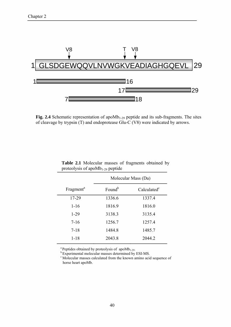
Dissection of apoMb1-29 and identification of the most amyloidogenic region
ApoMb1-29 peptide was subjected to proteolysis by using two proteases with
different specificity and fragments of various lengths were obtained (Fig. 2.4). Taking
advantage of the unique Lys residue in position 16 in the sequence, a proteolysis by
trypsin was carried out, producing the complementary fragments 1-16 and 17-29.
Furthermore, the presence of few Glu residues (position 6, 18 and 27) suggested the
possibility to produce some peptides from apoMb1-29 by using V8-protease, that in Tris-
buffer, pH 7.5 selectively cleaves only Glu-X peptide bonds (Drapeau, 1977). In order to
obtain also the decapeptide 7-16, a proteolysis, using both the proteases at the same time,
was performed. However, the low solubility in aqueous buffer of this peptide makes very
difficult its re-suspension after lyophilisation, even in acidic conditions. For this reason,
the species 7-18, produced from the same reaction, was isolated and purified. All peptide
species, purified by RP-HPLC, was characterized by mass spectrometry in order to assess
their chemical composition (Table 2.1). The secondary structure of these peptides was
analyzed by CD spectroscopy in the far UV range (data not shown); all the peptides show
to be essentially random in solution at pH 2.0 and insoluble under neutral conditions,
while, being the spectrum of 7-18 masked by the strong signal due to the two tryptophan
residues, we can not evaluate if this peptide is unstructured or in -conformation. The
amyloidogenic properties and the kinetic of aggregation of fragments 1-29, 1-16, 17-29
and 7-18 were compared. As assessed by ThT assay (Fig. 2.5 A), fragments 1-16 and 7-
18 aggregate very quickly, while the fluorescence emission in presence of fragment 17-29
does not change at all, also after long incubation time. In the case of fragments 1-16 and
7-18, there is a very short or nor lag time, indicating that their propensity to aggregate is
very high, more than apoMb1-29 and for both the peptides, the fluorescence emission of
ThT reaches a plateau within 10 h of incubation. Electron micrographs taken from the
solution of fragments apoMb1-29, 1-16 and 7-18, after incubation for 18, 20 and 29 h
respectively, are reported in Fig. 2.5 B. The TEM picture of 1-16 shows the presence of a
large network of fibrils, with a diameter of 10-14 nm. The aggregates from peptide 7-18
exhibit the typical feature of amyloid fibrils but are straighter and shorter than those
formed by apoMb1-29 and 1-16. They are approximately 20-22 nm in width and appear to
be composed of filaments laterally associated. They show a distribution of various lengths
with some evidence of twisted ribbons morphology.
39
Chapter 2
Molecular Mass (Da)
Fragmenta Foundb Calculatedc
17-29
1-16
1-29
7-16
7-18
1-18
1336.6
1816.9
3138.3
1256.7
1484.8
2043.8
1337.4
1816.0
3135.4
1257.4
1485.7
2044.2
GGLSDGEWQQVLNVWGKVEADIAGHGQEVL LSDGEWQQVLNVWGKVEADIAGHGQEVL GLSDGEWQQVLNVWGKVEADIAGHGQEVL
1 16
2917
7 18
1 29
T V8 V8
Fig. 2.4 Schematic representation of apoMb1-29 peptide and its sub-fragments. The sites of cleavage by trypsin (T) and endoprotease Glu-C (V8) were indicated by arrows.
Table 2.1 Molecular masses of fragments obtained by proteolysis of apoMb1-29 peptide
a Peptides obtained by proteolysis of apoMb1-29. b Experimental molecular masses determined by ESI-MS. c Molecular masses calculated from the known amino acid sequence of
horse heart apoMb.
40

Chapter 2
Time (h)
Fig. 2.5 (A) Time course analysis of aggregation of fragments apoMb1-29, 1-16, 7-18 and 17-29 at pH 2.0, 37°C. (B) TEM pictures of peptide samples after 18 h (1-29), 20 h (1-16), and 29 h (7-18) of incubation at 37°C in 10 mM Tris-HCl pH 2.0. The scale bar represents 200 nm.
0 10 20 30 40 50 60 70 80
ThT
Flu
ores
cenc
e at
485
nm
(%
)
0
20
40
60
80
100 A
1-29
1-16
7-18
17-29
B
1-29 200 nm
200 nm1-16
200 nm7-18
41

Chapter 2
2.2.3 Discussion and Conclusions ApoMb is able to form amyloid fibrils only under strong denaturing conditions
(Fändrich et al., 2001; Fändrich et al., 2003) or under more physiological conditions
substituting the Trp residues 7 and 14 with Phe (W7FW14F apoMb) (Sirangelo et al.,
2002). Here, we demonstrated that the N-terminal region of apoMb is highly prone to
aggregate, indeed peptide ApoMb1-29 and its sub-fragments 1-16 and 7-18 readily form
amyloid-like fibrils at pH 2.0. In the folded protein, the region comprised between amino
acid residues 3 and 18 is involved in the formation of helix A, which, together with
helices G and H, constitutes the main structural core of the folding pathway of apoMb
(Reymond et al., 1997; Loh et al., 1995). Of note, the W7FW14F amino acid substitution
in apoMb promotes misfolding and fibril formation of the protein even at neutral pH, as
the mutation destabilizes the folding core of the protein, despite maintaining the aromatic
character of the N-terminal region of apoMb (Sirangelo et al., 2002). At pH 2.0, apoMb is
unfolded and the N-terminal region of the protein is exposed to the solvent, but the
protein does not form fibrillar aggregates. The high degree of protonation of apoMb at pH
2.0 hinders intermolecular aggregation, due to electrostatic repulsion between positively
charged apoMb monomers. ApoMb, therefore, despite the presence in its polypeptide
chain of a region highly prone to aggregate, is protected from misfolding and loss of
function by a compact three-dimensional structure that masks potentially amyloidogenic
stretches of amino acids. The peptides studied here are isolated from protein and are
unfolded in solution, so their intrinsic propensity to form amyloid fibril can be
emphasized. The aggregation tendency can be evaluated in terms of net charge, -sheet
propensity, hydrophobicity and presence of aromatic residues (Chiti et al., 2003; Pawar et
al., 2005). In apoMb1-29, the net charge of the peptide changes upon acidification. The
theoretical value of net charge of apoMb1-29, calculated on the base of the average pKa of
its amino acid residues, is + 2.6 at pH 2.0 and -2.6 at pH 5.0. However at pH 2.0 the
presence of Cl- anions (derived from the HCl used for the acidification) attenuates the
electrostatic repulsive forces, resulting in an increase of the hydrophobic character of the
peptide (Aggeli et al., 2003; Hoyer et al., 2004). On the other hand, this shielding effect
does not occur at pH 5.0, slowing down the aggregation process. Under neutral and
alkaline conditions the net charge is ~-4, value considered not suitable for aggregation
(Calamai et al., 2003). Regarding the -sheet propensity, secondary structure prediction
analysis reveals that the region 9-16 shows the highest -sheet propensity and a -helical
structure is not favored (Chou & Fasman, 1978; Garnier et al., 1996). The segment 22-29
42

Chapter 2
on the contrary shows a preferentiality for random conformation. Taking together, these
results suggest that there is an intrinsic strong tendency to form -sheet that can lead to
the easy association of the peptides corresponding to the N-terminal part of apoMb. The
process is fastened at pH 2.0, because the hydrophobic collapse, important for the first
step of protein aggregation (Meersman and Dobson, 2006), is promoted by the charge
shielding effect (Goers et al., 2002; Hoyer et al., 2004). The complementary peptides 1-16
and 17-29 have the same net charge of +2 at pH 2.0, but only the fragment 1-16 quickly
aggregates, while 17-29 is stable in acid solution as monomer. Several algorithms (Pawar
et al., 2005; DuBay et al., 2004; Tartaglia et al., 2008) agree that N-terminal region of
apoMb, spanning approximately 8-16, is one of the regions with high intrinsic
aggregation propensity. In conclusion, we have clarified that in this system the
electrostatic interactions have the main role in the aggregation process. So peptides, as
apoMb1-29, are an important research tool for analyzed the intrinsic determinants that
regulate the molecular self-assembly in fibrillogenesis.
43

Chapter 2
2.3 ELECTROSTATIC INTERACTION AS DETERMINANT IN APOMB1-29
FIBRIL DISAGGREGATION
Since the aggregation process of apoMb1-29 is highly regulated by pH and
therefore by net charge, the effect of a change of net charge on the stability to dissociation
of mature fibrils was studied. Investigations into the stability of amyloid fibrils in terms
of stabilizing interactions can help to rationalize hypothesized kinetic and structural
models (Meersman and Dobson, 2006). Moreover, elucidation of the factors that
determine disaggregation may represent an additional approach to develop therapeutic
strategies for the treatment of amyloid disease based on the conversion of misfolded
proteins to their native states or on the clearance of amyloid deposits (Calamai et al.,
2005; Calamai et al., 2008).
The mature apoMb1-29 fibrils formed at pH 2.0 go into a fast and complete
disaggregation, if the pH of the suspension is increased at 8.3. The variation of pH causes
a large morphological alteration of the fibrils, as observed by TEM, Moreover, ThT assay
and CD show a significant reversion of ordered aggregates into more immature specie,
including the unstructured monomer. The increase of net charge of apoMb1-29 caused by
the change of pH back to neutral values promotes disaggregation. However, the amyloid-
like fibrils form again if the sample is acidified.
In conclusion, the electrostatic interaction, in apoMb1-29 system, is the force that
primarily stabilized the -sheet structure of the mature fibrils. However, other factors,
such as hydrophobicity and the presence of two aromatic residues have an important role
in the intrinsic aggregation propensity of the polypeptide.
The work attached hereby shows all the results relative to these studies and
contains a more complete discussion. In this publication are reported also proteolysis
experiments of the isolated fibrils. In brief, apoMb1-29 fibrils are resistant to the
proteolytic degradation by pepsin at pH 2.0, while the disaggregate fibrils are easily
proteolyzed by V8 protease and trypsin at pH 8.3. However, in the last case, in the
proteolytic mixture fibrillar aggregates can be isolated. The pellet is mostly constituted by
fragments 7-16 and 7-18. These peptides, as described in 2.2.1, have an high tendency to
aggregate and this property is much more highlighted at pH 8.3, since they have a net
charge value of +1 and 0, respectively.
Published paper (Picotti et al., 2007, J.M B., 367, 12371245)
44

Chapter 2
2.4 REFERENCES
Aggeli, A., Bell, M., Carrick, L. M., Fishwick, C. W., Harding, R., Mawer, P. J., Radford, S. E., Strong, A. E , Boden, N. (2003). pH as a trigger of peptide beta-sheet self-assembly and reversible switching between nematic and isotropic phases. J. Am. Chem. Soc., 125, 96199628.
Ascoli, F., Fanelli, M. R., Antonini, E. (1981). Preparation and properties of
apohemoglobin and reconstituted hemoglobins. Methods Enzymol., 76, 7287. Calamai, M., Canale, C., Relini, A., Stefani, M., Chiti, F., Dobson, C. M. (2005).
Reversal of protein aggregation provides evidence for multiple aggregated states. J. Mol. Biol., 346, 603616.
Calamai, M., Taddei, N., Stefani, M., Ramponi, G., Chiti, F. (2003). Relative
influence of hydrophobicity and net charge in the aggregation of two homologous proteins. Biochemistry 42, 1507815083.
Calamai, M., Tartaglia, G. G., Vendruscolo, M., Chiti, F., Dobson, C.M. (2008).
Mutational Analysis of the Aggregation-Prone and Disaggregation-Prone Regions of Acylphosphatase. J. Mol. Biol., epub ahead of print.
Chiti, F., Calamai, M., Taddei, N., Stefani, M., Ramponi, G., Dobson, C. M.
(2002). Studies of the aggregation of mutant proteins in vitro provide insights into the genetics of amyloid diseases. Proc. Natl. Acad. Sci. USA, 99, 1641916426.
Chiti, F., Dobson, C. M. (2006). Protein misfolding, functional amyloid and
human disease. Annu.Rev.Biochem., 75, 33366. Chiti, F., Stefani, M., Taddei, N., Ramponi, G., Dobson, C. M. (2003).
Rationalization of the effects of mutations on peptide and protein aggregation rates. Nature, 424, 805808.
Chou, P. Y. & Fasman, G. D. (1978). Prediction of the secondary structure of
proteins from their amino acid sequence. Adv. Enzymol. Relat. Areas Mol. Biol., 47, 45148.
Chu, K., Vojtchovsky, J., McMahon, B. H., Sweet, R. M., Berendzen, J.,
Schlichting, I. (2000). Structure of a ligand-binding intermediate in wild-type carbonmonoxy myoglobin. Nature, 403: 921923.
Drapeau, G. R. (1977). Cleavage at glutamic acid with staphylococcal protease.
Methods Enzymol., 47, 189191. DyBay, K. F., Pawar, A. P., Chiti, F., Zurdo, J., Dobson, C. M. & Vendruscolo,
M. (2004). Prediction of the absolute aggregation rates of amyloidogenic polipeptide chains. J. Mol. Biol. 341, 13171326.
Fändrich, M., Flecther, M. A., Dobson, C. M. (2001). Amyloid fibrils from
muscle myoglobin. Nature, 410, 165166.
45
Chapter 2
Fändrich, M., Forge, V., Buder, K., Kittler, M., Dobson, C. M., Diekmann, S. (2003). Myoglobin forms amyloid fibrils by association of unfolded polypeptide segments. Proc. Natl. Acad. Sci. USA, 100, 1546315468.
Fruton, J. S. (1970) The specificity and mechanism of pepsin action. Adv.
Enzymol. Relat. Areas Mol. Biol., 33, 401443.
Garnier, J., Gibrat, J-F., Robson, B. (1996). GOR secondary structure prediction method version IV. Meth. Enzymol. 266, 540553.
Gazit, E. (2002). A possible role for -stacking in the self-assembly of amyloid
fibrils. FASEB J., 16, 7783. Gill, S. C. & von Hippel, P. H. (1989). Calculation of protein extinction
coefficients from amino acid sequence data. Anal. Biochem., 182, 319326. Guijarro, J. I., Sunde, M, Jones, J. A., Campbell, I. D., Dobson, C. M. (1998).
Proc. Natl. Acad. Sci. USA, 95, 42244228. Hoyer, W., Cherny, D., Subramaniam, V, Jovin, T. M. (2004) Impact of the acidic
C-terminal region comprising amino acids 109-140 on -synuclein aggregation in vitro. Biochemistry 43, 1623316242.
Jennings, P. A. and Wright, P. E. (1993). Formation of a molten globule
intermediate early in the kinetic folding pathway of apomyoglobin. Science, 262, 892896.
Kallberg, Y., Gustafsson, M., Persson, B., Thyberg, J., Johansson, J. (2001).
Prediction of amyloid fibril-forming proteins. J. Biol. Chem., 276, 1294512950. Kendrew, J. C., Bodo, G., Dintzis, H. M., Parrish, R. G., Wyckoff, H. & Phillips,
D. C. (1958). A three-dimensional model of the myoglobin molecule obtained by x-ray analysis. Nature 181, 662666 .
Le Vine, H. (1993). Thioflavin T interaction with synthetic Alzheimer’s disease beta-amyloid peptides: Detection of amyloid aggregation in solution. Protein Sci., 2, 404410.
Loh, S. N., Kay, M. S., Baldwin, R. L. (1995). Structure and stability of a second
molten globule intermediate in the apomyoglobin folding pathway. Proc. Natl. Acad. Sci USA, 92, 54465450.
Malmo, C., Vilasi, S., Iannuzi, C., Tacchi, S., Cametti, C., Irace, G. and Sirangelo,
I. (2006) Tetracycline inhibits W7FW14F apomyoglobin fibril extension and keeps the amyloid protein in a pre-fibrillar, highly cytotoxic state. The FASEB J, 20, 346347.
Meersman, F. and Dobson, C. M. (2006). Probing the pressure-temperature
stability of amyloid fibrils provides new insights into their molecular properties. Biochim. Biophys Acta., 1764, 452460.
46
Chapter 2
Nishimura, C., Dyson, H. J., Wright, P. E. (2002) The apomyoglobin folding pathway revisited: structural heterogeneity in the kinetic burst phase intermediate. J. Mol. Biol., 322, 483-489.
Otzen, D. E., Kristensen, O., Oliveberg, M. (2000). Designed protein tetramer
zipped together with a hydrophobic Alzheimer homology: a structural clue to amyloid assembly. Proc. Natl. Acad. Sci. USA, 97, 99079912.
Pawar, A. P., DuBay, K. F., Zurdo, J., Chiti, F., Vendruscolo, M. & Dobson, C.
M. (2005). Prediction of “aggregation-prone” and “aggregation-susceptible” regions in proteins associated with neurodegenerative disease. J. Mol. Biol. 350, 379392.
Porat, Y., Mazor, Y., Efrat, S., Gazit, E. (2004). Inhibition of islet amyloid
polypeptide fibril formation: A potential role for heteroaromatic interactions . Biochemistry, 43, 1445414462.
Schmittschmitt, J. P., Scholtz, J. M. (2003). The role of protein stability,
solubility, and net charge in amyloid fibril formation. Protein Sci., 12, 23742378. Scopes R. K. (1974). Measurement of protein by spectraphotometry at 205 nm.
Anal. Biochem., 59, 277282. Sirangelo, I., Malmo, C., Casillo, M., Mezzogiorno, A., Papa, M., Irace, G.
(2002). Tryptophanyl Substitutions in Apomyoglobin Determine Protein Aggregation and Amyloid-like Fibril Formation at Physiological pH. J. Biol. Chem., 277, 4588745891.
Sirangelo, I., Malmo, C., Iannuzzi, C., Mezzogiorno, A., Bianco, M. R., Papa, M.,
Irace, G. (2004) Fibrillogenesis and Cytotoxic Activity of the Amyloid-forming Apomyoglobin Mutant W7FW14F. J. Biol. Chem., 279, 1318313189.
Stefani, M., Dobson, C. M. (2003). Protein aggregation and aggregate toxicity:
new insights into protein folding, misfolding diseases and biological evolution. J. Mol. Med., 81, 678699.
Tartaglia, G. G., Pawar, A. P., Campioni, S., Dobson, C. M., Chiti, F. &
Vedruscolo, M. (2008). Prediction of Aggregation-Prone Regions in Structured Proteins. J. Mol. Biol. 380, 425436.
Villegas, V., Zurdo, J., Filimonov, V. V., Avilés, F. X., Dobson, C. M., Serrano,
L. (2000). Protein engineering as a strategy to avoid formation of amyloid fibrils. Protein Sci., 9,17001708.
Yoon, S. and Welsh, W. J. (2004) Detecting hidden sequence propensity for
amyloid fibril formation. Protein Sci., 13, 21492160. Young, J. D. and Carpenter, F. H. (1961). Isolation and characterization of
products formed by the action of trypsin on insulin. J. Biol. Chem., 236, 743748.
47

Chapter 2
48

doi:10.1016/j.jmb.2007.01.072 J. Mol. Biol. (2007) 367, 1237–1245
COMMUNICATION
Amyloid Fibril Formation and Disaggregation ofFragment 1-29 of Apomyoglobin: Insights into theEffect of pH on Protein Fibrillogenesis
Paola Picotti1, Giorgia De Franceschi1, Erica Frare1, Barbara Spolaore1
Marcello Zambonin1, Fabrizio Chiti2, Patrizia Polverino de Laureto1⁎and Angelo Fontana1⁎
1CRIBI Biotechnology Centre,University of Padua,Viale G. Colombo 3,35121 Padua, Italy2Department of BiochemicalSciences, University of Florence,Viale G. Morgagni 50,50134 Florence, ItalyAbbreviations used: apoMb, apomN-terminal fragment 1–29 of horse henzyme to inhibitor ratio; E/S, enzyESI-MS, electrospray ionization-masGdnHCl, guanidine hydrochloride;interaction chromatography; RP, revmean residue ellipticity; TEM, transmicroscopy; TFA, trifluoroacetic acidE-mail addresses of the correspon
[email protected]@unipd.it
0022-2836/$ - see front matter © 2007 E
The N-terminal fragment 1–29 of horse heart apomyoglobin (apoMb1–29) ishighly prone to form amyloid-like fibrils at low pH. Fibrillogenesis at pH 2.0occurs following a nucleation-dependent growth mechanism, as evidencedby the thioflavin T (ThT) assay. Transmission electron microscopy (TEM)confirms the presence of regular amyloid-like fibrils and far-UV circulardichroism (CD) spectra indicate the acquisition of a high content of β-sheetstructure. ThT assay, TEM and CD highlight fast and complete disaggrega-tion of the fibrils, if the pH of a suspension of mature fibrils is increased to8.3. It is of interest that amyloid-like fibrils form again if the pH of thesolution is brought back to 2.0. While apoMb1–29 fibrils obtained at pH 2.0are resistant to proteolysis by pepsin, the disaggregated fibrils are easilycleaved at pH 8.3 by trypsin and V8 protease, and some of the resultingfragments aggregate very quickly in the proteolysis mixture, formingamyloid-like fibrils. We show that the increase of amyloidogenicity ofapoMb1–29 following acidification or proteolysis at pH 8.3 can be attributedto the decrease of the peptide net charge following these alterations. Theresults observed here for apoMb1–29 provide an experimental basis forexplaining the effect of charge and pH on amyloid fibril formation by bothunfolded and folded protein systems.
© 2007 Elsevier Ltd. All rights reserved.
Keywords: protein aggregation; amyloid; protein fragments; electronmicroscopy; circular dichroism
*Corresponding authorsIntroduction
Polypeptide chains appear to have a general ten-dency to convert, under specific experimental con-ditions, from their soluble states into well-organizedaggregates that have structural properties similar to
yoglobin; apoMb1–29,eart apoMb; E/I,me to substrate ratio;s spectrometry;HIC, hydrophobicerse-phase; [θ]MRW,mission electron; ThT, thioflavin T.ding authors:t;
lsevier Ltd. All rights reserve
those of the extracellular amyloid fibrils associatedwith a number of diseases, such as the amyloidosesand Alzheimer's disease, or of the intracellularinclusions that form in a number of neuropathicconditions, including Parkinson's disease or fronto-temporal dementia.1–10 These aggregates are char-acterized by a fibrillar morphology, as shown bytransmission electron microscopy (TEM) or atomicforce microscopy, an extended cross-β structure, asrevealed by X-ray fibre diffraction data, and a highaffinity for specific dyes such as Congo red andthioflavin T (ThT).3,11–13 The generic potential ofpolypeptide chains to generate these fibrillar aggre-gates is relevant for a number of reasons. Firstly,fibril formation represents an essential feature ofthe behavior of polypeptide chains that needs tobe fully understood for a thorough characterizationof the dynamics and conformational changes of
d.

Figure 1. (a) Amino acid sequence of the peptidefragment spanning residues 1–29 of horse hearth apo-myoglobin (apoMb1–29).
56 (b) Far-UV CD spectra ofapoMb1–29 at pH 7.5 and 2.0. ApoMb1-29 was obtainedby limited proteolysis of apoMb (2 mg/ml) by pepsinat 25 °C, in 0.01 M HCl (pH 2.0), at an E/S ratio of 1:100(w/w). The reaction was stopped after 90 s by addingpepstatin A (1 mg/ml) at an E/I ratio of 1:5 (w/w). TheapoMb1–29 fragment contained in the proteolysis mixtureat pH 2.0 was purified by hydrophobic interactionchromatography (HIC) using a Resource™ Phe column(Amersham Bioscience, Uppsala, Sweden), equilibratedand eluted at a flow-rate of 1 ml/min initially with 0.01 MHCl (pH 2.0) and then with 10 mM Tris–HCl (pH 7.5). Theeffluent from the column was monitored by measuringthe absorbance at 280 nm. Essentially homogeneousapoMb1–29 fragment was eluted from the HIC columnwith Tris–HCl (pH 7.5) as a single chromatographic peak.The CD spectrum at pH 7.5 was obtained from a sample ofthe fragment just recovered from the HIC column and thatat pH 2.0 was recorded immediately after pH adjustment.The same CD spectra were obtained at pH 7.5 and 8.3. Far-UV CD spectra were recorded at 25 °C with a Jasco J-710spectropolarimeter (Tokyo, Japan) using quartz cells witha 1 mm path-length. Fragment concentration was eval-uated from absorption measurements at 280 nm utilizing adouble-beam Lambda-25 spectrophotometer (PerkinElmer, Norwalk, CT, USA). Extinction coefficients at280 nm were calculated as described.57 CD measurementswere performed at a peptide concentration of about0.2 mg/ml. The results are reported in terms of meanresidue ellipticity [θ]MRW.
1238 Dissolution of Amyloid Fibrils
proteins.6,8,9 Secondly, formation of amyloid fibrils,or intracellular inclusions with amyloid-like char-acteristics, is associated with more than 40 patholo-gical conditions in humans, all having distinct andwell-described clinical profiles.10 Finally, such fibril-lar species can serve biological functions in livingorganisms,10 the most fascinating of them being theability of amyloid-like fibrils to serve as transmis-sible genetic traits distinct from DNA genes.14
Therefore, investigating the mechanism of amyloidfibril formationwill hopefully shed light on a processthat represents an essential feature of the chemistryof proteins, has a central role in human pathologyand constitutes an important aspect of the biology ofliving organisms.Numerous observations indicate that electrostatic
interactions play a key role in the formation andstabilization of fibrillar aggregates. For example,ribonuclease Sa easily forms ThT-positive and β-sheet-containing aggregates in the presence ofdenaturing conditions of trifluoroethanol at a pHvalue corresponding to its isoelectric point (pI) andthus when the protein has a net charge of zero.15
Similarly, mutations decreasing the positive netcharge of human muscle acylphosphatase, partiallyunfolded in trifluoroethanol, resulted in an acceler-ated formation of β-sheet-containing aggregatesable to bind ThT, whereas mutations increasingthe net charge resulted in the opposite effect.16
Further indications of the importance of charge inprotein aggregation come from observations onα-synuclein. This “natively unfolded” protein17–23
at neutral pH has a high negative net charge due tothe presence of several acidic residues in the C-terminal region. Deletions of the C-terminal segmentcauses fibril formation to be accelerated, with theacceleration being proportional to the extent of thetruncation.21 Acceleration of fibril formation by full-length α-synuclein at neutral pH can be achieved bypolyamines having a high positive compensatorycharge, such as spermine and other polycations,22,24or small cations such as Na+, Mg2+ or Ca2+.21,25,26
We note here that many mutations associated withfamilial forms of protein deposition diseases arepathogenic because they decrease the net charge ofthe protein forming the deposits.10,16 Overall, theseresults indicate that amyloid aggregation is mostfavored under conditions in which the net charge ofprotein molecules is diminished and, consequently,the electrostatic repulsion between them is mini-mized. However, the inverse correlation betweenamyloidogenicity of natively or chemically unfoldedproteins and net charge apparently contrasts withthe increased tendency of some globular proteins toform amyloid-like fibrils following acidification (pH∼2.0), when the net charge of the protein is generallyhighly positive.27–33
In order to investigate further the importance ofnet charge on protein aggregation, we propose tostudy the aggregation and disaggregation processesof a fragment of horse heart apomyoglobin (apoMb)spanning residues 1–29 (apoMb1–29) (see Figure1(a)). This peptide appears to be a suitable experi-
mental model as: (i) it is unstructured and does notform a folded conformation in any of the conditionsinvestigated; (ii) it forms fibrillar aggregates thatare morphologically, structurally and tinctorially in-

1239Dissolution of Amyloid Fibrils
distinguishable from those forming under patholo-gical conditions; and (iii) it has a high ratio of acidicversus basic residues. The latter property is particularimportant, as this system has a high negative netcharge at neutral pH and a low net charge at acidicpH. We show that formation of amyloid-like fibrilsby apoMb1–29 occurs readily at pH 2.0, whereas atpH values close to neutrality the peptide remainssoluble and even its fibrils, pre-formed at low pH,disaggregate.
Effect of pH on the formation of amyloid fibrilsby apoMb1–29
ApoMb1–29 was prepared by limited proteolysis ofapoMb with pepsin at pH 2.0, following our studiesaimed at unraveling molecular features of the partlyfolded states of apoMb34 under acidic solvent con-ditions using the limited proteolysis approach.35–37
The fragment was purified from the proteolysis mix-ture by hydrophobic interaction chromatography(HIC) (see the legend to Figure 1). The apoMb1–29fragment was eluted from the HIC-column withTris–HCl buffer (pH 7.5) and used immediately foradditional experiments. We have found that lyophi-lization causes significant aggregation of the frag-ment and was therefore avoided. A sample of thepeptide in Tris–HCl buffer (pH 7.5) was shown bygel-filtration chromatography on a Superdex-75column (not shown) to be largely monomeric.The far-UV circular dichroism (CD) spectrum of
apoMb1–29 is typical of a fully unfolded polypeptide,since it is characterized by a pronounced minimumat ∼200 nm,38 at pH 7.5 or 2.0 (Figure 1(b)). Thelargely unfolded structure of horse apoMb1–29 atboth acidic and neutral pH contrasts with the β-sheet structure at pH 5.8 reported for the analogous1–36 fragment of sperm whale apoMb.39 Since thislast fragment was prepared by recombinant meth-ods, purified by chromatography and lyophilized, itcould be well that the far-UV CD spectra of thesperm whale peptide were taken on an aggregatedmaterial resulting from the lyophilization step.39
Alternatively, the different behavior of the twopeptides may reside in the amino acid sequencedifferences or from the fact that the extra sevenresidues at the C terminus can stabilize β-sheetstructure in sperm whale apoMb1–36 that is other-wise unstable in horse apoMb1–29.To induce fibril formation, the apoMb1–29 frag-
ment has been incubated at 37 °C for five days at aconcentration of 0.2 mg/ml and pH 2.0. The analysisof the aggregated peptide sample by TEM shows thepresence of long, unbranched fibrils with a diameterof 9.0(±1.2) nm, characteristic of amyloid structures(Figure 2(a)). Some larger structures appear to bemade by two twisted fibrils, with an overalldiameter of 16.1(±0.8) nm. The sample produces amarked intensity gain of ThT fluorescence, support-ing further the presence of ordered amyloid-likestructures that selectively bind the dye (Figure 3(a),filled circles).40 The far-UV CD spectrum of thispeptide sample (Figure 3(b), sample a) displays the
minimum at ∼218 nm characteristic of β-sheetsecondary structure, whereas the monomeric pep-tide appears to be largely unfolded (see Figure 1(b)).In order to verify the stability of the amyloid-like
aggregates to pH changes, the pH of the fibrilsample was adjusted to 8.3 by adding a minimalvolume of a 1 M Tris. The sample was analyzedimmediately by CD, TEM and ThT-binding assay toevaluate the effect induced by the pH change on thestructural and morphological properties of thefibrils. The TEM picture shows that the sample ofthe fibrils exposed to pH 8.3 is highly heteroge-neous, being characterized by disordered aggre-gates (Figure 2(b1)) and rare fibrillar structures witha diameter of 14.3(±1.2) nm (Figure 2(b2)). Indeed,the ThT-binding assay shows a dramatic decrease ofthe ThT fluorescence intensity, which indicates alower content of amyloid fibrils (Figure 3(a), opencircles). The CD analysis shows the disappearance ofthe band at 218 nm and an increase in intensity ofthe signal at 200 nm, indicating that the β-sheet-containing aggregates convert into unstructured,possibly monomeric, peptide species (Figure 3(b),sample b).The sample of apoMb1–29 fibrils exposed to pH 8.3
was left for up to 24 h at room temperature. TEManalysis shows that the sample becomes highlyheterogeneous, the predominant species being largeamorphous aggregates characterized by a diameterof 25.0(±5.0) nm (not shown). The ThT fluorescenceintensity is even lower than that of the sample justtitrated at pH 8.3, indicating a further break-up ofordered fibrils (Figure 3(a), open circles). In agree-ment with the TEM and ThT assay results, the CDspectrum of the sample kept at pH 8.3 for 24 hcontinues to be characterized by a pronouncedminimum at 200 nm (Figure 3(b), sample c).Hence, the variation of pH caused a large morpho-logical alteration of the fibrils and a significantreversion of the fibrils into more immature species,including the unstructured monomer.After incubation for 24 h at pH 8.3, the same
peptide sample was acidified to pH 2.0 by additionof a minimal volume of 2 M HCl. The TEM images,acquired immediately after re-acidification, showthe presence of pre-fibrillar aggregates with dia-meters ranging from 18.8 nm to 26.5 nm and raretypical fibrils with a diameter of 8.1(±1) nm (Figure2(c1) and (c2)). The ThT fluorescence intensity of thissample is initially very low, but a time-dependentincrease is observed upon incubation at pH 2.0(Figure 3(a), filled squares). The increase of ThTfluorescence occurs more slowly and reaches alower final value compared to that obtained directlywithout the disaggregation step at pH 8.3 (Figure3(a), compare filled circles and squares). This isprobably due to the fact that this sample is char-acterized by a lower concentration of peptide,resulting from the several dilution steps, and bythe presence of amorphous aggregates that mayinterfere with the fibril formation process. The CDspectrum immediately after re-acidification is that ofa largely randompolypeptide (Figure 3(b), sample d).

Figure 2. TEM pictures ofapoMb1–29 fibrils. (a) The solutioncontaining a sample of apoMb1–29fibrils obtained at pH 2.0 after fivedays of incubation has been titratedto pH 8.3 (b1 and b2). Then the pH ofthe solution was lowered again topH 2.0 and re-incubated for fivedays under these conditions (d). ForTEM analysis, a drop of the peptidesamples (0.2 mg/ml) was placed ona Butvar-coated copper grid (400-square mesh) (TAAB-LaboratoriesEquipment Ltd, Berks, UK) anddried. The samples were negativelystained with a drop of 1% (w/v)uranyl acetate solution andobserved with a Tecnai G2 12 Twintransmission electron microscope(FEI Company, Hillsboro, OR,USA), operating at an excitationvoltage of 100 kV.
1240 Dissolution of Amyloid Fibrils
After leaving this sample at 37 °C for up to five daysat pH 2.0, very organized and homogeneous amyloidfibrils with a diameter of 8.0(±1.2) nm formed again(Figure 2(d)), showingmorphological features similarto those of the fibrils formed initially at pH 2.0 beforethe two consecutive changes of pH, in agreementwith the significant increase of the ThT fluorescenceintensity of this sample (Figure 3(a), filled squares).During incubation at pH 2.0, the CD spectrumundergoes an increase of the 218 nm band and aconcomitant decrease of the intensity of the mini-mum at 200 nm (not shown). After five days, the CDspectrum is dominated by an intense negative bandat 218 nm (Figure 3(b), sample e), but this band is lessintense than that of the peptide sample obtained afterthe first step of acidification (sample a), likelyindicating that upon re-acidification amorphouspeptide aggregates are formed, besides fibrils. Over-all, these findings indicate that the aggregates and/ormonomers derived by the pH-induced dissolution ofthe fibrils are able to reassemble into amyloid fibrils,if the solvent conditions that mostly favor theirformation are restored.
Proteolysis of apoMb1–29
The fibrils formed after incubation of fragmentapoMb1–29 at pH 2.0 for five days have been isolatedby ultracentrifugation and then analyzed by reversephase (RP)-HPLC. The chromatographic profile ofthe peptide material in the pellet, after dissolution of
the fibrils by 7.4 M GdnHCl, indicates that it iscomposed by intact full-length apoMb1–29 and thatno fragmentation has occurred during incubation ofthe fragment at low pH, as assessed by massspectrometry analysis of the main peak of the RP-HPLC chromatogram (Figure 4(a), top). TheapoMb1–29 fibrils formed after incubation of thefragment at pH 2.0 for five days (see Figure 4(b), left)are resistant to proteolytic degradation by pepsin(data not shown).Since incubation of the fibrils at pH 8.3 leads to
their complete dissolution (see above), we per-formed proteolysis experiments on apoMb1–29 fibrilsthat were purified by ultracentrifugation and thensuspended in 10 mM Tris–HCl (pH 8.3), in thepresence of both trypsin and V8 protease. Trypsin isexpected to cleave the fragment at Lys16 and V8protease is expected to cleave at Glu6, Glu18 andGlu27. The proteolysis mixture, after incubationovernight, showed an opalescent aspect, so it wasultracentrifuged in order to isolate the soluble andinsoluble fractions. A number of small fragmentshave been found in the supernatant by RP-HPLC(Figure 4(a), bottom), and they have been identifiedby mass spectrometry analysis (Table 1). Theycorrespond to several fragments spanning almostall regions of the peptide. Some of these peptidesresult from non-specific cleavages mostly at the levelof hydrophobic residues, but these are produced inmuch smaller amounts. By contrast, the pellet afterdissolution by 7.4 M GdnHCl appears to be mostly

1241Dissolution of Amyloid Fibrils
constituted by fragments 7–16 and 7–18 and, to alesser extent, by fragments 1–16 and 1–18 (Figure4(a), middle). Therefore, peptides encompassing thecentral part of apoMb1–29 produced by V8 proteaseand trypsin attack of the fragment are highly proneto aggregate, whereas peptides from the N andC-terminal regions of the fragment remain mostlysoluble in the proteolysis mixture at pH 8.3. TEManalysis shows that the pellet obtained afterproteolysis of apoMb1–29 contains fibrillar structureswith a mean diameter of 7.1(±0.5) nm (Figure 4(b),right).
The aggregation propensity of apoMb1–29 on pHappears to be determined by electrostaticfactors
The fast aggregation of the apoMb1–29 peptidecontrasts with the harsh conditions required toproduce fibrils from the whole 153 residue chain of
apoMb (pH 9.0, 65 °C, for up to 25 days).41,42
Amyloid fibril formation by apoMb1–29 was foundto occur easily at acidic pH, while at pH 8.3 thepeptide remains soluble, and even pre-formed fibrilsconvert back to non-amyloid amorphous aggregatesand monomeric species. In its soluble form, beforeaggregation occurs, apoMb1–29 is an unstructuredpeptide at both pH values (Figure 1(b)). This rulesout a pH-induced conformational change as thecause of aggregation, as generally is the case forproteins that at neutral pH adopt a well definedfolded structure that is disrupted upon acidification.The aggregation properties of apoMb1–29 can berationalized by considering the pH-dependentchange of net charge of the unstructured peptide.Protonation of the six negative moieties (two Asp,three Glu and the C terminus) of the peptide at lowpH strongly reduces the electrostatic repulsionbetween the various peptide molecules, thus facil-itating their association and stabilizing the resultingfibrils. The electrostatic argument can explain whyapoMb1–29 can form fibrils at pH 8.3 when subjectedto limited proteolysis with trypsin and V8 protease(see Figure 4). Under these conditions, apoMb1–29can be cleaved into various fragments, but onlysome of them can form fibrils. In particular, thepredominant constituent fragments of the fibrils, i.e.fragments 7–16 and 7–18, have a net charge value of+1 and 0, respectively.The increase of net charge of apoMb1–29 caused by
the change of pH back to neutral values promotesdisaggregation. It was found that the net charge is animportant determinant of both the rate of aggrega-tion of a fully or partially unfolded protein,16 and ofthe disaggregation rate of the resulting aggregates(M. Calamai, personal communication). These effects
Figure 3. (a) Time-course analysis of the aggregationprocess of apoMb1–29 by thioflavin T (ThT) fluorescenceassay. Arrows indicate changes of pH. (b) Evolution ofsecondary structure of apoMb1–29 monitored by far-UVCD spectroscopy. Spectra of apoMb1–29 (0.2 mg/ml)incubated at pH 2.0 for five days (sample a), after titrationat pH 8.3 (sample b), left at pH 8.3 for 24 h (sample c), leftagain at pH 2.0 (sample d) and left for five days at pH 2.0(sample e). Fibrils were obtained incubating apoMb1–29(0.2 mg/ml) at pH 2.0 for five days at 37 °C. Beforeincubation, the peptide solution was filtered with a0.22 μm pore size filter to eliminate large oligomers thatcould affect the aggregation process. To induce disaggre-gation, the pH of the solution (1.5 ml) containing fibrilsfrom apoMb1–29 was brought to 8.3 by adding a minimalamount (up to 5 μl) of 1 M Tris. The sample was thenincubated for 24 h at room temperature. Finally, thesample (1 ml) was acidified again to pH 2.0 with 2 M HCl(up to 5 μl) and incubated for five days at 37 °C. The ThTbinding assay40 was performed by adding a freshlyprepared 25 μMThTsolution in 25 mM sodium phosphate(pH 6.0) to 120 μl of protein samples containingaggregates, reaching a final volume of 500 μl. Fluorescenceemission measurements were conducted at 25 °C on aPerkin-Elmer model LS-50B spectrofluorimeter (Norwalk,CT, USA), utilizing a 2 mm×10 mm pathlength cuvette.The excitation wavelength was 440 nm and ThT fluores-cence emission was measured at 485 nm.

Table 1. Molecular masses of fragments obtained byproteolysis of apoMb1–29 by trypsin and V8-protease
FragmentaMolecular mass (Da)
Foundb Calculatedc
14–16 389.24 389.211–6 576.22 576.2412–16 602.36 602.3219–27 896.42 896.4020–28 924.59 924.439–16 942.61 942.5319–29 1108.59 1108.5517–27 1124.53 1124.517–16 1256.69 1256.677–18 1484.80 1484.781–16 1814.96 1814.901–18 2043.09 2043.011–29 3134.75 3133.55
a Peptides obtained by proteolysis of apoMb1–29 with trypsinand V8-protease. The peptides were purified by RP-HPLC (Figure4(a)) and are listed in the order of increasing molecular mass.
b Experimental molecular masses determined by ESI-MS.c Molecular masses calculated from the amino acid sequence of
apoMb.56
1242 Dissolution of Amyloid Fibrils
cause an overall thermodynamic stabilization anddestabilization of the amyloid aggregates followinga decrease or increase of net charge, respectively.This is in agreement with the observation reportedhere that amyloid fibrils of apoMb1–29 can form anddisaggregate in a reversible manner as a result of thechanges of net charge resulting from modificationsof pH.
Correlation of apoMb1–29 fibrillogenesis withpredictions
It is of interest to relate the results of this ex-perimental study to those obtained by using thealgorithms developed recently for predicting therate of aggregation and for identifying the mostamyloidogenic regions of polypeptide chains.Firstly, a previously described algorithm allows theaggregation rate constant (k) for an unstructuredpeptide or protein to be determined as a function ofpH, ionic strength and protein concentration.43 Theexperimental rate constant of aggregation ofapoMb1–29 under the conditions investigated here,determined as the rate constant of the exponential
phase followed with ThT fluorescence (Figure 3(a),filled circles), is within experimental error thatpredicted for the sequence of apoMb1–29 under theconditions studied (experimental log(k)=−4.22±0.12versus theoretical log(k)=−3.65±0.4). Moreover, itwas proposed that the regions of the sequence of anunstructured protein or peptide, as the case ofapoMb1–29, that promote the process of amyloidaggregation can be determined using an algorithm
Figure 4. (a) Reverse-phase (RP)-HPLC analysis of thepeptide material recovered from the apoMb1–29 fibrilsafter dissolution in 7.4 M GdnHCl (top), from the pellet(middle) and supernatant (bottom) after overnight dis-solution of the fibrils at pH 8.3 and concomitant trypticand V8-protease digestion. The samples analyzed wereobtained by ultracentrifugation in order to separate fibrilsfrom the fragments released into the supernatant. (b) TEMpictures of apoMb1–29 fibrils (left) and of the aggregatedmaterial formed after proteolysis with trypsin and V8protease of apoMb1–29 fibrils re-dissolved at pH 8.3 (right).Fibrils obtained incubating apoMb1–29 at pH 2.0 for up tofive days were subjected to proteolysis with trypsin andV8-protease in 10 mM Tris–HCl (8.3) at pH 25 °C. Bothenzymes were added to the same solution to reach aprotease to substrate ratio E/S of 1:50 (w/w). After 24 h,the proteolysis mixture was centrifuged (380,000g for90 min at 4 °C) to separate soluble proteolytic fragmentsfrom aggregated material. The pellet and the supernatant,the first after dissolution in 7.4 M GdnHCl, were analyzedby RP-HPLC using an Eclipse XDB-C8 column(4.6 mm×150 mm) (Zorbax Columns, Agilent Technolo-gies, Aston, PA, USA) eluted with a linear gradientof acetonitrile containing 0.1% (v/v) TFA from 5% to60% (v/v) in 30 min at a flowrate of 0.8 ml/min. Theeffluent from the columnwas monitored bymeasuring theabsorbance at 226 nm. The identity of all fragments wasassessed by mass-spectrometry (Table 1). Electrosprayionization (ESI) mass spectrometry (MS) analysis of thepeptides was carried out on a Micro Q-Tof mass spectro-meter (Waters, Manchester, UK).
1243Dissolution of Amyloid Fibrils
that evaluates the intrinsic propensity of the variousamino acid residues to aggregate, on the grounds ofphysicochemical factors such as hydrophobicity,charge, propensity to form β-sheet and α-helicalstructure.44 The aggregation propensity profile ofapoMb1–29 at neutral pH shows a single peak with ahigh intrinsic aggregation propensity, spanning ap-proximately residues 8–16 (not shown), in very goodagreement with the experimental results reportedhere. Finally, the use of a computational algorithmthat can detect non-native (hidden) sequence pro-pensity for amyloid fibril formation in proteins leadsto the identification of few amyloidogenic regionsalong the 153 residue chain of apoMb, including theN-terminal region up to residue 21.45
Conclusions
Having an excess of acidic residues, the unstruc-tured apoMb1–29 peptide aggregates easily only atacidic pH values, when the negatively chargedcarboxylate groups are protonated and the strongnegative net charge is decreased. This is in agree-ment with the behavior observed for other unstruc-tured protein systems having a similarly highcontent of acidic residues. The natively unfoldedand negatively charged α-synuclein aggregatespreferentially at low pH values.23 Polyglutamateaggregates only when it is neutralized by loweringthe pH to a value of 4 or less.46 On the other hand,several globular proteins with a more balancednumber of positively and negatively charged resi-dues have been found to form amyloid aggregates atlow pH, i.e. under conditions where proteins arepositively charged.27–33 This can be explained by con-sidering that folded proteins need to unfold, at leastpartially, to convert into amyloid aggregates.1,8,9,48
The increase of positive net charge of a protein at lowpH will decrease the intrinsic aggregation propen-sity, as discussed above, but it will also decrease theconformational stability of the protein, thus allowingthe protein to adopt an ensemble of fully or partiallyunfolded conformations that are highly prone toaggregate.47
In a number of studies it has been demonstratedthat net charge, hydrophobicity, the presence ofabundant aromatic residues and β-sheet propensitycan have important roles in the amyloidogenicity ofpolypeptides.43,44,48 Here, it is shown that modula-tion of the net charge of fragment apoMb1–29 and itssub-fragments by change of pH can be of utmostimportance for fibril formation. This does not, ofcourse, rule out the importance in amyloid aggrega-tion of the other factors mentioned above. Indeed,the agreement between the region found experi-mentally to promote aggregation (residues 7–16 and7–18) and that predicted to induce aggregationtheoretically (approximately residues 8–16), is basedon the grounds that all these factors, in additionto charge, promote fibril formation.44 Changes ofpH will affect the charge of ionizable groups ofapoMb1–29 and charge-related factors become pre-dominant in determining the observed differences in
aggregation behavior of the peptide at neutral orlow pH.The results of this study indicate that peptides
can be used as an important research tool forinvestigating the molecular recognition and self-assembly mechanisms that underlie the proteinfibrillogenesis phenomenon. Peptide models haveprovided fundamental insights into the mechanismof amyloid formation and, moreover, helped thediscovery and rational design of inhibitors ofprotein aggregation.49–54 The apoMb1–29 peptidesystem offers several advantages as a peptidemodel, since it can be induced to form fibrils easilyin a reversible way simply by modulating the pH ofthe solution. The easy and fast dissolution of theapoMb1–29 fibrils by exposing them to a mildlyalkaline aqueous solution contrasts with the well-known difficulty in solubilizing amyloid precipi-tates, which usually requires strong chaotropicagents or organic solvents such as 6 M GdnHClor DMSO.55 The results presented here will promptadditional studies by using analogs of apoMb1–29and its sub-fragments produced by chemicalsynthesis.
Acknowledgements
We thank Dr Manuela Grandesso for performingsome of the experiments mentioned here. Weacknowledge the financial support of the ItalianMinistry of University and Research (PRIN-2003,PRIN-2004, PRIN-2006 and FIRB-2003 Project No.RBNEOPX83 on Protein Folding and Aggregation).
References
1. Kelly, J. W. (1998). The alternative conformations ofamyloidogenic proteins and their multi-step assemblypathways. Curr. Opin. Struct. Biol. 8, 101–106.
2. Rochet, J. C. & Lansbury, P. T., Jr (2000). Amyloidfibrillogenesis: themes and variations. Curr. Opin.Struct. Biol. 10, 60–68.
3. Serpell, L. C. (2000). Alzheimer's amyloid fibrils:structure and assembly. Biochim. Biophys. Acta, 1502,16–30.
4. Horwich, A. (2002). Protein aggregation in disease: arole for folding intermediates forming specific multi-meric interactions. J. Clin. Invest. 110, 1221–1232.
5. Westermark, P. (2005). Aspects of human amyloidforms and their fibril polypeptides. FEBS J. 272,5942–5949.
6. Jahn, T. R. & Radford, S. E. (2005). The yin and yang ofprotein folding. FEBS J. 272, 5962–5970.
7. Dobson, C. M. (1999). Protein misfolding, evolutionand disease. Trends Biochem. Sci. 24, 329–332.
8. Dobson, C. M. (2003). Protein folding and misfolding.Nature, 426, 884–890.
9. Dobson, C. M. (2006). Protein aggregation and itsconsequences for human diseases. Protein Pept. Letters,13, 219–227.
10. Chiti, F. & Dobson, C. M. (2006). Protein misfolding,

1244 Dissolution of Amyloid Fibrils
functional amyloid and human disease. Annu. Rev.Biochem. 75, 333–366.
11. Sunde, M. & Blake, C. C. (1997). The structure ofamyloid fibrils by electron microscopy and X-raydiffraction. Advan. Protein Chem. 50, 123–159.
12. Tycko, R. (2004). Progress towards a molecular-levelstructural understanding of amyloid fibrils. Curr.Opin. Struct. Biol. 14, 96–103.
13. Makin, O. S. & Serpell, L. C. (2005). Structures foramyloid fibrils. FEBS J. 272, 5950–5961.
14. Chien, P., Weissman, J. S. & DePace, A. H. (2004).Emerging principles of conformation-based prioninheritance. Annu. Rev. Biochem. 73, 617–656.
15. Schmittschmitt, J. P. & Scholtz, J. M. (2003). The role ofprotein stability, solubility and net charge in amyloidfibril formation. Protein Sci. 12, 2374–2378.
16. Chiti, F., Calamai, M., Taddei, N., Stefani, M.,Ramponi, G. & Dobson, C. M. (2002). Studies of theaggregation of mutant proteins in vitro provideinsights into the genetics of amyloid diseases. Proc.Natl Acad. Sci. USA, 99, 16419–16426.
17. Wright, P. E. & Dyson, H. J. (1999). Intrinsicallyunstructured proteins: reassessing the protein struc-ture-function paradigm. J. Mol. Biol. 293, 321–331.
18. Dyson, H. J. & Wright, P. E. (2002). Coupling offolding and binding for unstructured proteins. Curr.Opin. Struct. Biol. 12, 54–60.
19. Tompa, P. (2002). Intrinsically unstructured proteins.Trends Biochem. Sci. 27, 527–533.
20. Fink, A. L. (2005). Natively unfolded proteins. Curr.Opin. Struct. Biol. 15, 35–41.
21. Hoyer,W., Cherny, D., Subramaniam, V. & Jovin, T. M.(2004). Impact of the acidic C-terminal region com-prising amino acids 109–140 on α-synuclein aggrega-tion in vitro. Biochemistry, 43, 16233–16242.
22. Fernandez, C. O., Hoyer, W., Zweckstetter, M., Jares-Erijman, E. A., Subramaniam, V., Griesinger, C. &Jovin, T. M. (2004). NMR of α-synuclein-polyaminecomplexes elucidates the mechanism and kinetics ofinduced aggregation. EMBO J. 23, 2039–2046.
23. Uversky, V. N., Li, J. & Fink, A. L. (2001). Evidence fora partially folded intermediate in α-synuclein fibrilformation. J. Biol. Chem. 276, 10737–10744.
24. Göers, J., Uversky, V. N. & Fink, A. L. (2003).Polycation-induced oligomerization and acceleratedfibrillation of human α-synuclein in vitro. Protein Sci.12, 702–707.
25. Uversky, V. N., Li, J. & Fink, A. L. (2001). Metal-triggered structural transformation, aggregation andfibrillation of human α-synuclein. J. Biol. Chem. 276,44284–44296.
26. Lowe, R., Poutney, D., Jensen, P. H., Gai, W. P. &Voelker, N. H. (2004). Calcium(II) selectivity inducesα-synuclein annular oligomers via interaction withthe C-terminal domain. Protein Sci. 13, 3245–3252.
27. Guijarro, J. I., Sunde, M., Jones, J. A., Campbell, I. D. &Dobson, C. M. (1998). Amyloid fibril formation by anSH3 domain. Proc. Natl Acad. Sci. USA, 95, 4224–4228.
28. Chiti, F., Bucciantini, M., Capanni, C., Taddei, N.,Dobson, C. M. & Stefani, M. (2001). Solution condi-tions can promote formation of either amyloidprotofilaments or mature fibrils from the HypFN-terminal domain. Protein Sci. 10, 2541–2547.
29. Kad, N. M., Myers, S. L., Smith, D. P., Smith, D. A.,Radford, S. E. & Thomson, N. H. (2003). Hierarchicalassembly of beta2-microglobulin amyloid in vitrorevealed by atomic force microscopy. J. Mol. Biol. 330,785–797.
30. Modler, A. J., Gast, K., Lutsch, G. & Damaschun, G.
(2003). Assembly of amyloid protofibrils via criticaloligomers: a novel pathway of amyloid formation.J. Mol. Biol. 325, 135–148.
31. Frare, E., Polverino de Laureto, P., Zurdo, J., Dobson,C. M. & Fontana, A. (2004). A highly amyloidogenicregion of hen lysozyme. J. Mol. Biol. 340, 1153–1165.
32. Frare, E., Mossuto, M. F., Polverino de Laureto, P.,Dumoulin, M., Dobson, C. M. & Fontana, A. (2006).Identification of the core structure of lysozymeamyloid fibrils by proteolysis. J. Mol. Biol. 361,551–561.
33. Polverino de Laureto, P., Taddei, N., Frare, E.,Capanni, C., Costantini, S., Zurdo, J. et al. (2003).Protein aggregation and amyloid formation by anSH3 domain probed by limited proteolysis. J. Mol.Biol. 334, 129–141.
34. Eliezer, D., Yao, J., Dyson, H. J. & Wright, P. E. (1998).Structural and dynamic characterization of partiallyfolded states of apomyoglobin and implications forprotein folding. Nature Struct. Biol. 5, 148–155.
35. Fontana, A., Zambonin, M., Polverino de Laureto, P.,De Filippis, V., Clementi, A. & Scaramella, E. (1997).Probing the conformational state of apomyoglobin bylimited proteolysis. J. Mol. Biol. 266, 223–230.
36. Fontana, A., Polverino de Laureto, P., De Filippis, V.,Scaramella, E. & Zambonin, M. (1997). Probing thepartly folded states of proteins by limited proteolysis.Fold. Des. 2, R17–R26.
37. Fontana, A., Polverino de Laureto, P., Spolaore, B.,Frare, E., Picotti, P. & Zambonin, M. (2004). Probingprotein structure by limited proteolysis. Acta. Biochim.Pol. 51, 299–321.
38. Woody, R. W. (1995). Circular dichroism. MethodsEnzymol. 246, 34–71.
39. Chow, C. C., Chow, C., Raghunathan, V., Huppert,T. J., Kimball, E. B. & Cavagnero, S. (2003). Chainlength dependence of apomyoglobin folding: struc-tural evolution from misfolded sheets to nativehelices. Biochemistry, 42, 7090–7099.
40. Le Vine, H. (1993). Thioflavin T interaction withsynthetic Alzheimer's diseases beta-amyloid peptides:detection of amyloid aggregation in solution. ProteinSci. 2, 404–410.
41. Fändrich, M., Fletcher, M. A. & Dobson, C. M. (2001).Amyloid fibrils from muscle myoglobin. Nature, 410,165–166.
42. Fändrich, M., Forge, V., Buder, K., Kittler, M., Dobson,C. M. & Dickmann, S. (2003). Myoglobin formsamyloid fibrils by association of unfolded polypeptidesegments. Proc. Natl Acad. Sci. USA, 100, 15463–15468.
43. DuBay, K. F., Pawar, A. P., Chiti, F., Zurdo, J., Dobson,C. M. & Vendruscolo, M. (2004). Prediction of theabsolute aggregation rates of amyloidogenic polypep-tide chains. J. Mol. Biol. 341, 1317–1326.
44. Pawar, A. P., DuBay, K. F., Zurdo, J., Chiti, F.,Vendruscolo, M. & Dobson, C. M. (2005). Predictionof “aggregation prone” and “aggregation susceptible”regions in proteins associated with neurodegenerativediseases. J. Mol. Biol. 350, 379–392.
45. Yoon, S. & Welsh, W. J. (2004). Detecting hiddensequence propensity for amyloid formation. ProteinSci. 13, 2149–2160.
46. Fändrich, M. &Dobson, C.M. (2002). The behaviour ofpolyamino acids reveals an inverse side chain effect inamyloid structure formation. EMBO J. 21, 5682–5690.
47. Uversky, V. N. & Fink, A. L. (2004). Conformationalconstraints for amyloid fibrillation: the importance ofbeing unfolded. Biochim. Biophys. Acta, 1698, 131–153.
48. Jones, S., Manning, J., Kad, N. M. & Radford, S. E.

1245Dissolution of Amyloid Fibrils
(2003). Amyloid-forming peptides from β2-microglo-bulin: insights into the mechanism of fibril formationin vitro. J. Mol. Biol. 325, 249–257.
49. Zurdo, J. (2005). Polypeptide models to understandmisfolding and amyloidogenesis and their relevancein a protein design and therapeutics. Protein PeptideLetters, 12, 171–187.
50. Findeis, M. A. (2002). Peptide inhibitors of thebeta-amyloid aggregation. Curr. Top. Med. Chem. 2,417–423.
51. Lopez de la Paz, M., Goldie, K., Zurdo, J., Lacroix, E.,Dobson, C. M., Hoenger, A. & Serrano, L. (2002). Denovo designed peptide-based amyloid fibrils. Proc.Natl Acad. Sci. USA, 99, 16052–16057.
52. Pastor, M. T., Chopo, A. & Lopez de la Paz, M. (2005).Design of model systems for amyloid formation:lessons for prediction and inhibition. Curr. Opin.Struct. Biol. 15, 57–63.
53. Makin, O. S., Atkins, E., Sikorski, P., Johansson, J. &Serpell, L. C. (2005). Molecular basis for amyloid fibrilformation and stability. Proc. Natl Acad. Sci. USA, 102,315–320.
54. Cohen, T., Frydman-Marom, A., Rechter, M. & Gazit,E. (2006). Inhibition of amyloid fibril formation andcytotoxicity by hydroxyindole derivatives. Biochemis-try, 45, 4727–4735.
55. Meersman, F. & Dobson, C. M. (2006). Probing thepressure-temperature stability of amyloid fibrils pro-vides new insights into their molecular properties.Biochim. Biophys. Acta, 1764, 452–460.
56. Dautrevaux, M., Boulanger, Y., Han, K. & Biserte, G.(1969). Covalent structure of horse myoglobin. Eur. J.Biochem. 11, 267–277.
57. Gill, S. C. & von Hippel, P. H. (1989). Calculation ofprotein extinction coefficients from amino acidsequence data. Anal. Biochem. 182, 319–326.
Edited by J. Weissman
(Received 7 July 2006; received in revised form 23 January 2007; accepted 27 January 2007)Available online 3 February 2007


Chapter 3
Chapter 3
Parkinson's disease and -synuclein
3.1 PARKINSON'S DISEASE: NEUROPATHOLOGICAL HALLMARKS
Parkinson's disease (PD) was first described by James Parkinson in 1817 in “An
Essay of Shaking Palsy”. PD is the second most-prevalent neurodegenerative disorder
after Alzheimer's disease. It is an irreversible, progressive disease that impairs movement.
The clinical phenotypes are resting tremors, bradykinesia, muscular rigidity and postural
instability. Non-motoric symptoms, like automic, cognitive and psychiatric problems can
be also present; however cognitive ability remains intact in most PD sufferers, at least in
the early stages of the disease. Incidence increases markedly with age; hence young-onset
Parkinson's disease, defined as occurrence before age 40, accounts for just 5% of newly
diagnosed cases (Irvine et al., 2008). Moreover, upon reaching the 65-69 age range, 0.6%
of the population are affected, increasing to 2.6 % of those aged 85-89 (de Rijk et al.,
2000). The majority of cases of PD are idiopathic and sporadic, but in 5-10% of cases
there is a genetic component, showing both recessive and dominant modes of inheritance.
PD is characterized by depigmentation of the substantia nigra pars compacta,
caused by the selective and progressive loss of dopaminergic (DA) neurons, and by the
presence of eosinophillic, intraneuronal proteinaceous, inclusions known as Lewy bodies
(LBs) and dystrophic Lewy neurites in the surviving neurons (Fig. 3.1). Because patients
suffering from other neurological disorders can display parkinsonian features, a definitive
diagnosis of Parkinson’s disease can be confirmed only by post mortem histopathological
examination of the substantia nigra for loss of pigmented neurons and presence of LBs in
remaining neurons. Identification of LBs has been facilitated by immunostaining for
particular proteins; initially for ubiquitin and more recently for -synuclein, now
regarded as the major protein constituent (Shults, 2006). Such staining reveals filaments
that, when purified and examined by immuno-electron microscopy, can be seen to contain
α-syn (Spillantini et al., 1998). The recombinant protein forms similar filaments when it is
allowed to aggregate in vitro (Crowther et al., 1998) (Fig. 3.1).
49

Chapter 3
Fig. 3.1 Pathological hallmarks of PD. (A) Schematic representation of substantia nigra: it is evident the loss of pigmented neurons in PD patients. (B) Nerve cell with three LBs that are double-stained for α-syn and ubiquitin. The halo of each LB is strongly immunoreactive for ubiquitin, whereas both the core and the halo of each Lewy body are immunoreactive for α-syn. (Bar = 10 μm.). (C) Filaments from cingulate cortex of patients with dementia with LBs labelled with anti-α-synuclein antibody PER4. The gold particles conjugated to the second antibody appear as black dots; there is also an unlabeled paired helical filament (arrow). (Bar = 100 nm). (B and C reprinted from Spillantini et al., 1998)
50

Chapter 3
DA neurons operate in a pathway that controls voluntary movement. This involves
signals being relayed from the cerebral cortex through the basal ganglia back to the cortex
and then on to muscles. Neurons from the substantia nigra pars compacta project axons
that release dopamine in synapses on interneurons in the striatum. As the dopamine-
containing neurons die, failure to complete this circuit results in inability to coordinate
movement (Fig. 3.2). Neuromelanin, the black pigment that gives its name to the
substantia nigra, is a byproduct from the metabolic pathway for dopamine synthesis.
When the symptoms of PD first become apparent, more than 70% of the dopamine-
containing neurons have already been lost, releasing their neuromelanin and hence
turning the tissue less black. It is now apparent, however, that many regions of the brain
are affected in Parkinson’s disease, and indeed in the early stages it may affect only a
lower region of the brain stem called the medulla oblongata, spreading gradually upward
through the basal ganglia into the cortical areas (Irvine et al., 2008; Braak et al., 2003).
Symptoms of PD could be reversed temporarily by pharmacologic interventions to
restore striatal dopaminergic neurotransmission (LeWitt, 2008; Hornykiewicz, 2002).
This reversal can be accomplished by DA agonists, compounds that directly stimulate
postsynaptic striatal dopamine receptors (Fig. 3.2). The administration of dopamine itself
is ineffective because dopamine cannot cross the blood–brain barrier (Nutt et al., 1984).
Levodopa (3,4-dihydroxy-L-phenylalanine), a naturally occurring amino acid, is an
intermediate in the pathway of dopamine synthesis. After oral ingestion, levodopa is
actively transported from the upper small intestine into the circulation by a mechanism
specific for large, neutral L-amino acids. Because of ongoing metabolism and the
distribution of levodopa throughout the body, only a small fraction of the drug reaches the
brain after active transport across the blood–brain barrier. Once there, dopamine is rapidly
formed from levodopa by aromatic L-amino acid decarboxylase (AAAD). Moreover, the
coadministration of other drugs can improve the efficacy of levodopa. In conclusion,
current medications only provide symptomatic relief and fail to halt the death of DA
neurons. A major hurdle in development of neuroprotective therapies are due to limited
understanding of disease processes leading to death of DA neurons (Thomas & Beal,
2007).
51

Chapter 3
Fig. 3.2 A schematic comparison of coronal brain slices from a control subject (left) and a patient with PD (right) illustrates the major neurodegenerative loss of dopamine-synthesizing neurons in the substantia nigra pars compacta, projecting to striatal nuclei (caudate and putamen) in the cerebrum. Exogenous levodopa administered for treatment of PD is transported to the brain, where it enhances striatal dopaminergic neurotransmission. (reprinted from LeWitt, 2008).
52

Chapter 3
3.2 PATHOGENESIS OF PARKINSON’S DISEASE
The causes of the DA neuronal demise in familial and sporadic forms of PD are
elusive, but recent findings suggest that both environmental factors and genetic
susceptibilities are associated with PD pathogenesis. The hypothesis that environmental
factors play a role in the etiology of PD were done after the discovery of a group of
intravenous drug users who unwittingly injected a synthetic analog of demerol that was
contaminated with 1-methyl-4-phenyl-1,2,3,6-tetrahydropyridine (MPTP) causing an
acute, permanent parkinsonian state that was levodopa responsive (Langston et al, 1985).
Once inside the brain, MPTP enters astrocytes and is converted into its active metabolite,
MPP+ (1-methyl-4-phenylpyrdinium), which then can enter dopamine neurons and exert
its toxicity. Although not typically found in the environment itself, the ability of MPTP to
reproduce so many of the features of PD intensified the search for potential
environmental toxicants that might contribute to the development of PD (Hatcher et al.,
2008). Moreover, epidemiological studies had suggested a link between long-term
exposure to agricultural pesticides, such as rotenone and paraquat. Rotenone, a plant
derivative used as an insecticide, is known to be an inhibitor of NADH dehydrogenase.
Chronic infusion of either rotenone or MPTP in rodents results in parkinsonism-like
behavior and pathology, including the formation of inclusion bodies, and indeed these are
among the best animal models for the human disease (Dauer et al., 2003).
The majority of PD cases are sporadic, but the discovery of genes linked to rare
familial forms of PD have confirmed the role of genetics in development of PD, and
provided important information in understanding molecular pathogenesis of the common
sporadic disease (Thomas & Beal, 2007).
3.2.1 Pathogenic mutations in PD pathogenesis
Linkage data has identified eleven loci, named PARK; six of the genes with
pathogenic mutations have now been identified (Table 3.1). These include two autosomal
dominant genes, -synuclein (SNCA) and leucine-rich repeat kinase 2 (LRRK2), and
three autosomal recessive genes, parkin, DJ-1 and PTEN-induced putative kinase 1
(PINK1). The 6th gene, ubiquitin carboxyl-terminal esterase L1 (UCHL1, previously
known as PARK5), has only been found in one small family, and its importance in familial
PD is still uncertain. There are four remaining loci (PARK3, PARK9, PARK10 and
PARK11) for which linkage has been identified; however, the causative genes have not yet
53

Chapter 3
been discovered (Wood-Kaczmar et al., 2006).
Table 3.1 Genetic mutations identifying for PD (Wood-Kaczmar et al., 2006).
Locus Gene (protein)
MOI Protein function Clinical presentation
PARK1 -synuclein (SNCA)
AD Unknown synaptic function
Duplications: Idiopathic PD; some postural tremor; slow progression; Triplications: PD; PD with dementia; diffuse LBs disease; aggressive course; Mutations A53T, A30P, E46K: Idiopathic PD; parkinsonism and diffuse LBs
PARK2 Parkin AR E3 ubiquitin ligase Parkinsonism; slow progression
PARK5 UCHL1 AD Ubiquitin hydrolase and ligase
PD
PARK6 PINK1 AR Mitochondrial Ser-Thr Kinase
Parkinsonism
PARK7 DJ-1 AR Oxidative stress response ?
Parkinsonism
PARK8 LRRK2 AD Unknown protein kinase
PD
Abbreviations: AD, autosomal dominant; AR, autosomal recessive; LBs, Lewy bodies; MOI, mode of inheritance.
-Synuclein (-syn)
Mutations in the SNCA gene were first identified in large kindred with autosomal
dominant parkinsonism (Polymeropoulos et al., 1997). Three autosomal dominant
mutations have now been identified that segregate with familial PD, A53T, A30P and
E46K substitutions (Cookson et al., 2005). Subsequently, it was discovered that genomic
duplications and triplications at the -syn locus can also cause autosomal dominant, early
onset PD (Singleton et al., 2003). The age of onset and severity of the disease phenotype
seems to correlate with SNCA copy number, suggesting a gene-dosage effect. PD patients
who carry duplications at the SNCA locus (which generate three copies of the gene) tend
to have PD, which develops slowly from the 4th decade of life. Triplication events, which
produce four copies of the SNCA gene, cause disease earlier (mid 20–mid 30) with a
clinical presentation ranging in severity from idiopathic PD, PD with dementia or diffuse
Lewy body disease (Farrer et al., 2004).
-Syn is a small (140 amino acids) natively unfolded protein that is a member of
family that includes -synuclein, -synuclein and synoretin. It is enriched in the adult
human brain where it is expressed ubiquitously but heterogeneously, with highest levels
of protein reported in deeper layers of the cerebral neocortex, the hippocampus (CA2 and
54

Chapter 3
CA3 regions) and the SN. The protein is expressed within glia and neurons where it is
particularly abundant at presynaptic terminals (Iwai et al., 1995; Mori et al., 2002). The
physiological function of -syn is not well understood but it seems to have a role in
synaptic plasticity and in regulation of dopamine neurotransmission. Other studies have
evidenced that -syn may function as a chaperone protein, based on its abundance in
cytosol, its natively unfolded structure, and its prevention of protein aggregation. The
physiological function of -syn is also related with lipids and membrane since this
protein seems to modulate presynaptic vesicle pool size and vesicle recycling. A deeper
discussion about the function of -syn will be reported in § 3.3.
The pathological role of -syn in PD seems to be linked to its aggregation
properties. Indeed, -syn is the principal constituent of LBs and Lewy neuritis in sporadic
and in familial PD. -Syn is an amyloidogenic protein and aggregates in vitro in a
concentration-dependent manner to form fibrils reminiscent of those observed in Lewy
bodies (Conway et al., 2000; Serpell et al., 2000). Furthermore, elevated expression levels
of -syn in the brain, caused by rare genomic multiplications of the SNCA gene, increases
the deposition of soluble -syn into insoluble aggregates (Miller et al., 2004). Transgenic
mice that overexpress wild-type or mutant -syn have given conflicting results (Fleming
et al., 2005). However, models in Drosophilia melanogaster demonstrated that high levels
of -syn cause abnormal protein aggregation and neurotoxicity in DA neurons. -Syn
expressed in D. melanogaster is phosphorylated at serine 129. When serine 129 is
mutated to a non-phosphorylatable residue, there is in increased aggregate formation and
reduced cell death in DA neurons (Chen et al., 2005). Human -syn is also extensively
phosphorylated at serine 129 in LBs but the importance of this post-translational
modification has yet to be demonstrated in sporadic PD (Fujiwara et al., 2002).
In its aggregation pathway -syn can form soluble oligomers and protofibrils that
are toxic to some cell cultures, including the dopaminergic SH-SY5Y human
neuroblastoma cell line (El-Agnaf et al., 2001, Volles et al, 2001; Volles & Lansbury,
2003). It is thought that the soluble, oligomeric forms of -syn are the toxic species rather
than the insoluble fibrils within the inclusion body. Both A53T and A30P mutants show
an increased propensity to form these so called protofibrils, which can permeabilize
vesicles in a way that is reminiscent of bacterial pore-forming toxins (Volles et al., 2001).
Dopamine and related catecholamines have been shown to interact with -syn and
stabilize the protofibril stage of aggregation (Conway et al., 2001; Mazzulli et al., 2007)
55

Chapter 3
providing a possible explanation for the increased susceptibility of DA neurons. Levels of
soluble oligomers are also affected by fatty acids, being upregulated by polyunsaturated
fatty acids (Sharon et al., 2003). -Syn aggregation is also increased in the presence of
metals and pesticides, which may be environmental risk factors (Uversky et al., 2002).
Furthermore, expression of mutant forms of -syn in cells promotes mitochondrial
defects and cell death and enhances susceptibility to oxidative stress. On the other hand,
mice deficient in -syn are resistant to toxicity induced by MPTP and other mitochondrial
toxins (Klivenyi et al., 2006).
Parkin
The PARK2 locus codes for the enzyme parkin, which is a ubiquitin E3-ligase
(E3s). E3s act as substrate-recognition molecules during the synthesis and attachment of
polyubiquitin chains to proteins that are targeted for degradation by ubiquitin proteasome
system (UPS) (Shimura et al., 2000). Mutations in the parkin gene are the predominant
cause of juvenile and early onset recessive parkinsonism (von Coelln et al., 2004). Parkin
mutations are present in ~ 50% of all individuals with recessive, early onset (<45 years)
parkinsonism, and in 77% of sporadic cases with disease onset before the age of 20. Some
missense and nonsense mutations, deletions and rearrangements in the parkin gene have
also been reported (West & Maidment, 2004). At least ten familial-associated parkin
mutations have now been shown to disrupt differentially the solubility, localization,
binding and ubiquitination properties of parkin in vitro (Sriram et al., 2005). Inactivation
of parkin leads to reduction in UPS-mediated degradation of its substrates. Several
candidate parkin substrates have been identified, including proteins that are implicated in
PD such as synphilin-1 and a glycosylated form of -syn. Recent studies have highlighted
a protective role for parkin within mitochondria. Reduced levels of mitochondrial proteins
involved in mitochondrial oxidative phosphorylation were reported in parkin knockout
mice (Palacino et la., 2004). Parkin has been shown to localize to the mitochondrial
matrix, where it enhances mitochondrial gene transcription and biogenesis in proliferating
cells. In addition, a recent study demonstrates -syn-induced mitochondrial dysfunction is
further enhanced due to lack of parkin activity in vivo implicating crucial role of parkin in
modulating mitochondrial functions in -syn-induced PD (Stichel et al., 2007). Parkin
functions as a multipurpose neuroprotective protein in a variety of toxic insults crucial for
dopamine neuron survival. New research has identified neuroprotective mechanisms
56

Chapter 3
mediated by parkin. It seems to mediate neuroprotection through activation of IkappaB
kinase/nuclear factor-kappaB signaling, whereas parkin mutants failed to stimulate this
pathway (Henn et al., 2007).
PTEN-induced kinase-1 (PINK1)
Mutations in the PINK1 gene were identified to cause early-onset familial PD
(Valente et al., 2004). PINK1 is a 581 amino acid protein that contains an N-terminal
mitochondrial targeting sequences and a highly conserved protein kinase domain similar
to serine/threonine kinases of the Ca2+ calmodulin family. It has a ubiquitous and
punctate expression pattern suggesting mitochondrial localization (Gandhi et al., 2006).
Very little is known about the precise function of PINK1 although its
mitochondrial localization, the presence of kinase domain with identification of majority
of mutations in the kinase domain and regions close to it, suggest a role in mitochondrial
dysfunction, protein stability and kinase pathways in pathogenesis of PD. Indeed,
overexpression of PINK1 protects cells from mitochondrial depolarization and apoptosis
induced by the proteasomal inhibitor MG132, and from mitochondrially-induced
apoptosis triggered by staurosporine. Expression of pathogenic mutations of PINK1 that
are predicted to inactivate it (E240K, L489P and K219M) do not protect against basal or
apoptosis-inducing agents, implying that kinase activity is required for this effect. Parkin
could restore normal mitochondrial morphology, DNA and protein content, but did not
rescue the sensitivity of PINK1 mutant flies to apoptosis induced by stressors. This
suggests that PINK1 and parkin interact to protect mitochondrial integrity, with PINK1
upstream of parkin (Wood-Kaczmar et al., 2006; Thomas & Beal, 2007).
DJ-1
Rare mutations in DJ-1 also cause autosomal recessive early onset PD (Bonofati et
al., 2004). DJ-1 mutations account for 1–2% of all early-onset PD, with a number of
different pathogenic mutations, including exonic deletions, truncations and homozygous
and heterozygous point mutations. DJ-1 is a highly conserved protein of 189 amino acids
that belongs to the DJ-1/Thi/PfpI protein super family (Thomas & Beal, 2007).
It is enriched in the brain where it is largely cytoplasmic except for a pool of DJ-1,
which localizes to mitochondria (Zhang et al., 2005). The protein contains many residues
that are readily oxidized, and it is thought to protect neurons from oxidative stress by
acting as a redox-dependent chaperone. DJ-1 is oxidatively damaged in the brains of
57

Chapter 3
patients with idiopathic PD, thus suggesting a mechanism of protein aggregation in these
diseases that is mediated by high levels of oxidative stress (Choi et al., 2006). DJ-1
knockout mice show motor impairments and nigrostriatal dopaminergic dysfunction
associated with attenuation of D2-mediated responses, resulting in increased re-uptake of
dopamine by the dopamine transporter. The model lacks SN degeneration, suggesting that
loss of DJ-1 function might confer increased susceptibility to parkinsonism as a result of
underlying SN dysfunction (Wood-Kaczmar et al., 2006).
Leucine-rich repeat kinase 2 (LRRK2)
Mutations in the LRRK2 cause autosomal dominant PD (Zimprich et al., 2004). At
least nine pathogenic mutations have been identified so far, including two that map to the
kinase domain (Khan et al., 2005). The most-common pathogenic LRRK2 mutation in
Caucasians is the G2019S substitution, which causes parkinsonism closely resembling
idiopathic PD, and accounts for ~ 5% of familial PD and 1.5% of sporadic cases (Gilks et
al., 2005). In certain populations, however, the frequency of G2019S mutation is
strikingly high: in North African Arabs, the frequency of this mutation is 37% in familial
PD and 41% in sporadic PD. In Ashkenazi Jews, the frequency of the G2019S reaches
29.7% in familial PD and 13.3% in sporadic PD. mutations in the LRRK2 gene give rise
to diverse, widespread neuropathological features. For example, the LRRK2 R1441C
substitution in a Caucasian family was associated with intriguing pleomorphic pathology,
including α-synuclein inclusions and tauopathy in addition to the classic selective SN
degeneration (Zimprich et al., 2004).
LRRK2 encodes a 280 kDa multidomain protein that belongs to the ROCO family,
a novel group of RAS GTPase superfamily. The precise physiological role of LRRK2
protein is unknown but the presence of multifunctional domains suggests involvement in
wide variety of functions. The precise tissue and intracellular localization remain unclear
but the expression within the human SN has been confirmed using quantitative real-time-
PCR. Characterization of LRRK2 protein in vitro has so far been limited to
overexpression cell models where it is reported to be predominantly cytoplasmic (West et
al., 2005). Interestingly, LRRK2 protein can be detected in Lewy neurites and Lewy
bodies of sporadic PD (Zhu et al., 2006).
Purified LRRK2 protein demonstrates kinase activity in vitro and the I2020T and
G2019S mutations enhanced kinase activity (West et al., 2005). Although some mutations
might have a toxic gain-of-function effect through an increase in kinase activity in vitro,
58

Chapter 3
this has not yet been shown in any in vivo model. However, recent findings show
significant alterations in phosphorilation of key proteins involved in MAPK signaling in
leukocytes from patients with G2019S mutations implicating abnormal protein
phosphorilation.
3.2.2 Intersecting pathways in PD pathogenesis
From the recent studies that were summed up previously in this chapter, PD can be
defined as a complex disorder with multiple etiological factors involved in disease
pathogenesis (Wood-Kaczmar et al., 2006; Thomas & Beal, 2007). PD may depend on a
complex set of circumstances that include genetic factors, environmental exposures and
loss of cellular protective mechanisms. Although the precise mechanisms of preferential
DA cell death and inclusion formation in PD remain unclear, key factors are strongly
implicated in the pathogenesis of both familial and sporadic PD, including mitochondrial
dysfunction, protein phosphorylation, oxidative stress, protein misfolding and impairment
of ubiquitin proteasome system (UPS). In Fig. 3.3 are reported a schematic representation
of several different pathways that are important in modulating pathogenic events. Briefly,
environmental factors similar to pesticides and toxins directly induce both oxidative
damage and mitochondrial dysfunctions. -Syn undergoes aggregation either due to
pathogenic mutations or catechol oxidation which in turn compromise UPS function,
induce endoplasmic reticulum stress and cause mitochondrial dysfunction. Mitochondrial
dysfunction and oxidative damage lead to deficits in ATP which may compromise UPS
function promoting abnormal protein aggregation. -Synuclein is known to prevent -syn
aggregation through activation of Akt signaling. Parkin, an ubiquitin E3 ligase, promotes
proteasomal degradation, increases mitochondrial biogenesis by activating mitochondrial
transcription factor A (TFAM) and block PINK1-induced mitochondrial dysfunction,
while pathogenic mutations, oxidative and nitrosative damage, severely compromise its
protective function. DJ-1 protects against oxidative stress, functions as a chaperone to
block -syn aggregation and protects against mitochondrial dysfunction. PINK1 seems to
protect against mitochondrial dysfunction which is compromised due to pathogenic
mutations, although the precise function of PINK1 in mitochondria still needs to be
determined. LRRK2 seems to play a role in synaptic vesicle functions, neurite outgrowth.
Pathogenic mutations in LRRK2 cause abnormal protein phosphorylation which induce
mitochondria-dependent cell death. In addition, a pathogenic role of PI3kinase-Akt
59

Chapter 3
(phosphatidylinositol 3-kinase/Akt) and Nrf2/ARE signaling is implicated in PD
pathogenesis. Familial PD-linked genes namely parkin, DJ-1 and PINK1 activate PI3
kinase-Akt signaling, while activation of Nrf2/ARE pathway prevents against oxidative
damage and mitochondrial dysfunction promoting cell survival.
Fig. 3.3 Common intersecting pathways in PD pathogenesis (reprinted from Thomas & Beal, 2007). Green arrows indicate promoting or activating effects while red lines with blunt ends indicate inhibitory effects.
60

Chapter 3
3.3 -SYNUCLEIN
As previously mentioned (see §3.3.2), -syn is a 140 amino acid protein that is
highly expressed in the central nervous system, particularly at the presynaptic nerve
terminals. It is the major component of LBs, the cytoplasmic proteinaceous aggregates
pathognomonic for PD. These filamentous aggregates have a fibrillar structure with a
cross--sheet core typical of amyloid (see § 1.2). Mutations or overexpression of the
human -syn gene have been linked to early-onset autosomal dominant PD. The
mechanisms by which an abnormality in the structure or the expression of -syn can
cause PD have not been elucidated, nor is it known if the fibrillar deposits of -syn play a
toxic, protective, or no role in the pathogenesis of the disease (Ellis et al., 2005).
3.3.1 Structure and conformational properties of -synuclein
-Syn is a natively unfolded protein of still unknown function. Its sequence can be
divided into three regions (Fig. 3.4). The N-terminal region, residues 1–60, includes the
sites of three familial PD mutations and contains four 11-aminoacid, imperfect repeats
with a highly conserved hexameric motif (KTKEGV). The N-terminal region is predicted
to form amphipathic -helices, typical of the lipid-binding domain of apolipoproteins
(George et al., 1995; Clayton &George, 1998). The central region, residues 61–95,
comprises the highly aggregation-prone NAC (non Amyloid- Component) sequence
(Ueda et al., 1993; Han et al., 1995). This region contains the remaining three imperfect
repeats. The C-terminal region, residues 96–140, is highly enriched in acidic residues and
prolines. Three highly conserved tyrosine residues are located in this region that is
disordered in most conditions.
-Syn is a typical intrinsically disordered (or natively unfolded) protein, which
possesses little or no ordered structure under physiological conditions in vitro (Uversky et
al., 2001). Intrinsically disordered proteins have recently been recognized as a new
protein class, which are gaining considerable attention due to their capability to perform
numerous biological functions despite the lack of unique structure (Wright and Dyson
1999; Uversky et al. 2000, 2005, 2007). These proteins exist as dynamic and highly
flexible ensembles that undergo a number of distinct interconversions on different time
scales. Recent studies have shown that monomeric -syn has a more compact structure
than expected for a completely unfolded polypeptide and this compactness has been
linked to inhibition of fibrillation due to burial of the hydrophobic NAC domain
61

Chapter 3
(Bertoncini et al., 2005; Dedmon et al., 2005; Bernardo et al., 2005). Small angle X-ray
scattering analysis showed that the radius of gyration, Rg, which is used to describe the
dimensions of polypeptide chain, is ~ 40 Å of native -syn, which is much larger than
that predicted for a folded globular protein of 140 residues (15 Å), but significantly
smaller than that for a fully unfolded random coil (52 Å). Recently, NMR studies have
shown that -syn adopts an ensemble of conformations that are stabilized by long-range
interactions. In particular, a long-range intramolecular interaction between the C-terminal
region (residues 120–140) and the central part of -syn (residues 30–100) was noted
(Bertoncini, et al., 2005).
NAC region
Fig. 3.4 Schematic of the primary structure of -syn and indication of its most important
characteristics.
The conformational behavior of -syn under a variety of conditions was
extensively analyzed (Uversky, 2003). The structure of -syn is extremely sensitive to its
environment and can be easily modulated by a change in conditions. Beside an extended
state, -syn can acquire a pre-molten globule state in several conditions such as low pH,
high temperature, in the presence of various metal ions and as a result of spontaneous
oligomerization both in vitro and in vivo (Uversky, et al., 2001; Uversky, 2007).
Moreover, an -helical membrane bound form (Davidson et al., 1998; Eliezer et al.,
2001) and -helical and -structural species in organic solvent (Munishkina et al., 2003)
were described. -Syn is able to form morphologically distinct oligomers and aggregates
such as dimers, oligomeric protofibrillar species and insoluble amorphous aggregates and
62

Chapter 3
amyloid fibrils. Based on this conformational behavior, the concept of a protein-
chameleon was proposed, which holds that the structure of -syn is modulated by its
environment to a dramatic degree. That is, the choice among all the above mentioned
conformations is determined by the peculiarities of protein surroundings (Uversky, 2003;
Uversky et al., 2007).
3.3.2 Physiological role of -synuclein
Despite the evidence for a key function of -syn in the onset of PD, its
physiological function in the brain is still unclear. There are a lot of putative biological
functions and possible interactions of -syn. It was suggested to be involved in synaptic
plasticity and in regulation of dopamine neurotransmission. Indeed, in the Zebra finch, -
syn is transiently expressed in a telencephalic area associated with song acquisition
during the critical period for song learning (George et al., 1995). Moreover, -syn
knockout mice exhibit enhanced DA release at nigrostriatal terminals only in response to
paired electrical stimuli, suggesting that -syn is an activity-dependent, negative
regulator of dopaminergic neurotransmission (Abeliovich et al., 2000). Other studies have
demonstrated a putative function of -syn as a chaperone protein, based on its abundance
in the cytosol, its natively unfolded structure, and its prevention of protein aggregation
induced by heat shock or chemical treatment (Souza et al., 2000). It can act as a
molecular chaperone assisting in the folding and refolding of synaptic proteins called
soluble NSF (N-ethylmaleimide sensitive factor) attachment receptors (Chandra et al.,
2005).
The physiological function of -syn is also correlated with lipids and membranes.
In pre-synaptic termini, monomeric -syn exists in an equilibrium between free and
plasma membrane- or vesicle-bound states (McLean et al., 2000), with approximately
15% of -syn being membrane-bound (Lee et al., 2002). This close association with
vesicular structures has led to the hypothesis that -syn may regulate vesicular release
and/or turnover and other synaptic functions in the central nervous system (Ueda et
al.,1993; Clayton & George, 1998, 1999; Davidson et al. 1998; Lavedan, 1998). Genome-
wide screening in yeast showed that nearly one-third of genes that enhance the toxicity of
-syn are functionally related to lipid metabolism and vesicle trafficking (Willingham et
al., 2003). Over-expression of -syn in a neuronal cell line and homozygous deletions of
-syn in mice were both accompanied by noticeable changes in membrane fluidity and in
63

Chapter 3
cellular fatty acid uptake and metabolism (Sharon et al., 2003; Castagnet et al., 2005;
Golovko et al., 2005; see also §3.4.4). A recent analysis of a yeast PD model with dosage
sensitivity for -syn expression (Outeiro & Lindquist, 2003) revealed that the earliest
defects following -syn expression were an inhibition of the endoplasmic reticulum to
Golgi vesicular trafficking and an impairment of the endoplasmic reticulum associated
degradation (Cooper et al., 2006). Finally, a recent study revealed that -syn regulates
catecholamine release from the synaptic vesicles, and its over-expression inhibits a
vesicle ‘priming’ step that occurs after secretory vesicle trafficking to ‘docking’ sites but
before calcium-dependent vesicle membrane fusion (Larsen et al., 2006).
3.3.3 Aggregation properties of -synuclein
The aggregation process of -syn was extensively studied as -syn amyloid fibrils
are observed in LBs, the cardinal hallmark of PD pathology (see Fig. 3.1). The in vitro
kinetics of -syn fibril formation show an initial lag-phase followed by an exponential
growth phase and a final plateau, usually attributed to a nucleation-dependent
polymerization (§ chapter 1). Mature amyloid fibrils grown in vitro typically vary in
length from about 500 nm to 3 mm and, based on AFM images, have an height of 9.8
±1.2 nm (Khurana et al., 2003).
Uversky and coworkers showed that, early stages of fibril formation involve the
partial folding of -syn into the highly fibrillation-prone pre-molten globule-like
conformation, which represents a key intermediate on the fibrillation pathway (Uversky et
al. 2001, Fink, 2006). Hence, factors that shift the equilibrium in favor of this partially
folded conformation facilitate fibril formation. Thus, an increase in protein concentration
is predicted to increase the concentration of the intermediate, and accordingly the rate of
fibrillation, as observed (Uversky et al., 2001). However, if one considers that the
intrinsically unstructured monomer presents long-range interactions between the C-
terminal region and the central amyloidogenic NAC region, a fully unfolded, aggregation-
prone, conformation may be attained early in the aggregation pathway, exposing the
hydrophobic NAC region and facilitating aggregation (Paleček et al., 2008). Structural
perturbations, that can favor the aggregation prone conformation, include the presence of
positively charged ions able to interact with the C terminus, a decrease in pH that reduce
the net charge of the C-terminal region, and the deletion of the C terminus; all result in a
more rapid aggregation reaction (Hoyer et al., 2004).
64

Chapter 3
Analysis by TEM and AFM has revealed that different final product may arise
from the aggregation of -syn depending on the experimental conditions: fibrils,
oligomers, and insoluble amorphous aggregates (Fink, 2006). Although the central role of
-syn fibrillation in the pathogenesis of PD is well established, recent evidence indicates,
that the fibril itself may not be the primary pathogenic species. On the contrary, soluble
oligomers and protofibrils seem to be the toxic species (Volles et al., 2002).
Unfortunately, the direct detection of defined prefibrillar intermediates has proven
extremely difficult. The oligomeric species of -syn are present during the lag-phase of
the aggregation process and they are relatively unstable, transient and present at very low
steady-state concentrations (Goldberg et al., 2000). Several oligomeric protofibrillar
species with various morphologies were detected (Conway et al. 1998, 2000; Ding et al.
2002; Lashuel et al. 2002). The earliest form of -syn protofibrils appeared to be
predominantly spherical with heights varying between 2.5 and 4.2 nm (Conway et al.
2000; Ding et al. 2002). These spherical beads can then convert into beaded chain and
rings (Ding et al. 2002). The incubation of the spherical -syn oligomers with brain
derived membranes was shown to also produce pore-like ring-type protofibrils (Ding et
al. 2002). These species formed in the late lag phase are characterized by extensive -
structure and sufficient structural regularity to bind ThT and CR (Chiti & Dobson, 2006).
A key question is whether these oligomers are on direct pathway to fibrils or are off-
pathway but in equilibrium with monomer that can add to fibrils or can add directly to
growing fibrils. However, the fact that these spherical oligomers disappear at the same
rate that fibrils appear suggests that fibrils could be assembled directly from them via
longitudinal association of the oligomers, rather than by binding of monomer species to
the growing end of fibrils (Apetri et al., 2006; Fink, 2006). Recently, following the
isolation and characterization of protofibrils, oligomeric species, that appeared to precede
fibrils and structured protofibrils formation, were identified by several techniques: photo-
induced cross-linking of -syn (Li et al., 2006); electrochemical techniques (Paleček et
al., 2007); intrinsic tryptophan fluorescence of Y39W -syn (Dusa et al., 2006) and FRET
measurements of Y125W/Y133F/Y136F -syn (Kaylor et al., 2005); fluorescence of
pyrene-labeled -syn (Thirunavukkuarasu et al., 2008). Structured protofibrillar species
could form from the reorganization or assembly of small and relatively disorganized
oligomers that are formed rapidly after the initiation of aggregation process. In Fig. 3.5 a
schematic representation of the hypothetical aggregation pathways of -syn is reported.
65

Chapter 3
Fig.3.5 Schematic representation of the hypothetical aggregation pathways of -syn. During -syn fibrils formation several intermediate structures populate, including small oligomers, early-spherical protofibrils, membrane embedded pores or chain like aggregates. Therefore, -syn fibrillization is not a simple two-state transition from monomer to fibrils, but rather a complex process that involves intermediates of various sizes and morphologies.
Furthermore, several modifications of -syn lead to the formation of stable
oligomers with very slow rates of dissociation. These modifications include oxidation of
the four methionine residues to methionine sulfoxide (Hokenson et al., 2004), specific
nitration of the tyrosine residues (Uversky et al., 2005), interaction with polyphenols such
as bacalein (Zhu et al., 2004) and covalent modification by 4-hydroxynonenal (Qin et al.,
2006). Typically these oligomers are formed more rapidly than fibrils, and as a
consequence, no significant fibrillation occurs from these modified forms of -syn.
Biophysical characterization of these oligomers suggests that they have significant
secondary and tertiary structure and are substantially more compact than monomeric -
syn. The stability of these oligomers indicates that the underlying structure of their
subunits is different than that in the transient oligomers (Fink, 2006).
66

Chapter 3
3.4 LIPIDS AND -SYNUCLEIN
Since, as already mentioned (§ 3.3.2), -syn is localized in the presynaptic nerve
terminals and seems to modulate presynaptic pool size and neurotransmitter release,
several studies were performed in order to analyze the interaction of -syn with lipids and
bilayer membranes. Recently, Beyer reviewed several aspects about the relation between
-syn and membranes and concluded that this interaction evinces a remarkable
dichotomy, e.g. the binding of -syn may be beneficial for the maintenance of the bilayer
integrity or it may be deleterious for the membrane integrity; lipid interaction may
promote or inhibit -syn fibrillization, depending on the membrane composition, on the
aggregative state of the protein and on the details of the experimental conditions (Beyer,
2007).
3.4.1 -Synuclein and membrane properties
The lipid specificity of -syn-membrane interaction was deeply investigated, but
controversial results were obtained. For example high affinity for vesicles composed of
phosphatidic acid was reported (Davidson et al. 1998, Perrin et al., 2000) which was not
found in other studies (Jo et al., 2000; Ramakrishnan et al., 2003). However summing up,
-syn binds preferentially to small unilamellar vesicles containing acidic phospholipids
such as phosphatidic acid, phosphatidylserine, and phosphatidylglycerol but not
significantly to vesicles with a net neutral charge (Davidson et al., 1998; Jo et al., 2000;
Perrin et al., 2000). Monounsaturated and polyunsaturated acyl groups in lipid vesicles
increase the binding affinity of -syn (Kubo et al., 2005). In conclusion, the interaction of
-syn with membranes is complex and dependent on the composition of the membranes,
their curvature, as well as the ratio of membrane lipids to protein.
The observation that -syn has a higher affinity for small vesicles than for vesicles
of larger diameter, because of the higher surface-to-volume ratio of small vesicles, has
suggested that it can effort a deep effect on bilayer properties. Indeed, the association of
-syn with negatively charged membranes of multilamellar vesicles has a profound effect
on the integrity of these bilayers and causes the formation of non-bilayer or small
vesicular structures (Madine et al., 2006). Moreover, the addition of monomeric -syn to
synaptic vesicles (Jo et al., 2004) or synthetic vesicles composed of brain sphingomyelin,
DPPC, or POPC/POPG (Kamp & Beyer, 2006) increases the anisotropy of DHP, a
fluorescent membrane probe sensitive to the ordering of fatty acid acyl chains. -Syn not
67

Chapter 3
only stabilizes the packing of the acyl chains but also increases the gel–liquid crystalline
transition temperature (Kamp & Beyer, 2006). These observations suggest that the protein
plays a role in modulating the organization of lipid membrane components and therefore,
and they are in accord with the belief that the physiological function of -syn may consist
of stabilizing the synaptic vesicle reserve pool (Murphy et al., 2000).
3.4.2 Structure of membrane-bound -synuclein
Far-UV CD and NMR data show that -syn in the N-terminal region adopts an -
helical conformation on binding to membranes (Bisaglia et al., 2005; Eliezer et al., 2001;
Bussell et al., 2003; Bussell et al., 2004; Ulmer et al., 2005; Chandra et al., 2003). An
important paper demonstrated that -syn assumes a bipartite structure upon binding to a
SDS detergent micelles (Eliezer et al., 2001). The N-terminal 100 -syn amino acids fold
into an amphipathic helix that associates with the micelle–water interface whereas the
acidic C-terminus of the protein remains unstructured. More insight into the micelle-
induced -syn structure became available from the carbon secondary shifts and from
sequential NH-NH Nuclear Overhauser Enhancements (NOE), which showed that there
are two helical regions in the N-terminal sequence, interrupted by a single helix break
around residue 42 (Chandra et al., 2003; Bussell et al., 2003). A comparison with other
proteins featuring the peculiar 11-mer repeat motives gave rise to the conclusion that the
-syn helical pitch is characterized by 3 turns over 11 residues (3/11 helix) rather than by
5 turns over 18 residues as in the standard version of an -helix (Bussell et al., 2005).
More recent NMR work revealed a distinct secondary structure of micelle-associated -
syn (Fig. 3.6 A): the two helices comprising residues 3–37 and 45–92, are oriented
antiparallel, wrapped tightly around the SDS micelle and connected by an ordered
antiparallel linker region, while the C-terminal protein tail remains highly mobile (Ulmer
et al., 2005). Similar techniques were employed for a comparison of micelle binding of
the A30P and A53T mutant -syn proteins. It turned out that the former replacement
perturbs the helical structure and the molecular dynamics of the protein whereas the A53T
mutant is essentially indistinguishable from the wt protein. Cross-relaxation experiments
also showed the partial insertion of the helices into the micellar interface (Ulmer et al.,
2005). Similarly, paramagnetic relaxation enhancement of the -syn backbone NH
resonances in the presence of nitroxide labelled fatty acids indicated that the helical
residues 61–95 are partially embedded in the SDS micelle (Bisaglia et al., 2005). In
68

Chapter 3
structural biology, detergent micelles are commonly used as membrane-mimetic
environments because their small size facilitates high-resolution structural analysis by
NMR. However, it is often difficult to test whether the structure of proteins bound to
micelles is the same as the respective membrane-bound form. Recently, it has been
suggested that membrane-bound -syn may have a similar structure to the micelle-bound
form (Bortolus et al., 2008; Drescher et al., 2008). On the contrary, Jao and coworkers
suggested an extended helical structure for -syn bound to SUVs (Jao et al., 2004; Jao et
al., 2008). The method of site-directed spin labeling was employed for an assessment of
the structure of the protein bound at small unilamellar vesicles composed of
phosphatidylcholine and phosphatidylserine (Jao et al., 2004). Spin labelling at 47 single
sites, combined with an examination of the accessibility for oxygen or for a paramagnetic
nickel complex, suggested an extended helical structure and enabled a detailed analysis of
the -syn topology with respect to the vesicle interface. This analysis confirmed the 3/11
helical arrangements and also suggested a bent shape of the helical part of the protein.
Moreover, site-directed spin labeling and EPR-based approach (continuous-wave and
pulse EPR) were used to determine the structure of -syn bound to lipid membrane (Jao
et al., 2008). -Syn forms an extended, curved -helical structure that is over 90 aa in
length (Fig. 3.6 B) The monomeric helix has a superhelical twist similar to that of right-
handed coiled-coils which, like -syn, contain 11-aa repeats. The -syn helix extends
parallel to the curved membrane in a manner that allows conserved Lys and Glu residues
to interact with the zwitterionic headgroups, while uncharged residues penetrate into the
acyl chain region. In this work the authors argued also that the break between the -syn
helices in the presence of SDS is due to the small size and highly curvature of the micelle
(Fig. 3.6 C) (Jao et al., 2008).
69

Chapter 3
C
B
A
Fig. 3.6 (A) Structure of micelle-bound -syn. Ensemble of twenty structures superimposed on helix-C. Helix-N (Val3-Val37) and helix-C (Lys45-Thr92) are connected by a short linker, followed by another short extended region (Gly93-Lys97) and a predominantly unstructured tail (Asp98-Ala140). (B) Representation of the interaction of -syn with a curved lipid surface. A closer cross-sectional view of the -syn interaction with the lipid surface. The protein (green) follows the curved surface of the vesicle, with the helical axis positioned just below the level of the phosphate groups of the lipids. (C) Comparison between the structures acquired by -syn on micelles and SUVs (A reprinted from Ulmer et at., 2005; B and C reprinted from Jao et al., 2008).
70

Chapter 3
3.4.3 -Synuclein fibrillogenesis and membranes
Membranes and -syn fibrillogenesis are correlated in two ways. Indeed, several
observations suggest that the cytotoxicity of the oligomeric prefibrillar species of the
protein is mediated by membrane damage and furthermore, -syn aggregation process
seems to be modulated by -syn-lipid interaction.
-Syn prefibrillar species have a tight membrane binding and the ability to
permeabilize SUV composed of acidic phospholipids. An influx of Ca2+ ions into the
vesicles, as detected by Fura-2 fluorescence, was attributed to the formation of pores
consisting of -syn oligomers in the vesicular bilayer (Volles et al., 2001; Volles &
Lansbury, 2002; Volles & Lansbury, 2003). Small vesicles were more prone to
permeabilization than larger ones, which conform with the binding preference of the -
syn monomer (Davidson et al., 1998). As already mentioned in § 3.3.3, the same authors
later discovered annular and even tubular structures when the oligomeric aggregates of
A30P and A53T -syn were studied by electron microscopy and image reconstruction
(Lashuel et al., 2002). These structures resembled the cytolytic -barrel toxins of certain
bacteria, suggesting that the annular shape directly accounts for a non-specific pore
mechanism of the protofibril toxicity (Lashuel et al., 2002). Spherical protofibrils were
also shown to bind much more tightly to brain-derived microsomal and mitochondrial
membranes than monomers or fibrils. According to atomic force microscopy they
eventually convert to the larger annular structures (Ding et al., 2002). Since -syn
oligomers (intracellular origin) have a cytotoxic effect when were added to human
neuroblastoma cells and their action is on the outer cell membrane, it was suggested that
the oligomer-mediated bilayer permeabilization may be rather unspecific (Kayed et al.,
2003). Other authors, however, disagree with the channel interpretation. Rather, again on
the basis of oligomer-induced membrane conductance, they concluded from the absence
of single channel activity and ion selectivity that the oligomers were only peripherally
associated with the membrane which allows ions to cross the perturbed lipid bilayer
somehow on their own (Kayed et al., 2004).
The modulation of -syn aggregation by lipid-protein interaction is an unresolved
and debated question. If the binding to lipid vesicles and membranes is strong, the -helix
is stable and the formation of fibrils or prefibrillar oligomers, is slowed down or
prevented (Zhu et al., 2003; Zhu & Fink, 2003). Under in vitro conditions of a high ratio
of protein to lipid, where the protein is at high relative abundance with respect to the
71

Chapter 3
interface, anionic membranes can promote the self-association of -syn and accelerate
fibrillation (Perrin et al., 2001, Cole et al., 2002; Lee et al., 2002; Necula et al., 2003).
The mechanism of this aggregation is still unknown. However, in a review about the
general problem of the role of lipid-protein interactions in amyloid-type protein fibril
formation, it was reported there are several membrane-related factors promoting protein
aggregation (Gorbenko & Kinnunen et al., 2006). Lipids seem to act as anti-chaperones
facilitating protein transition into partially folded states more prone to aggregate. Another
membrane-related phenomenon involves a protein accumulation at lipid-water interface
that overcome the energy barrier for nucleation. The role of membranes in fibrillogenesis
may involve also variations in the proportion of bilayer-buried and exposed regions of
protein molecule; changes in the depth of bilayer penetration may be essential for a
protein to act as a nucleus in fibril formation (Gorbenko & Kinnunen et al., 2006).
Indeed, SDS-PAGE analysis revealed that wild type -syn bound to synaptosomal
membranes can seed the aggregation of cytosolic -syn (Lee et al., 2002). These data
suggestwere interpreted as indicating that a protein partially inserted into a lipid bilayer
may act as an anchor for site-directed fibril assembly. Using environmentally sensitive
probes it was demonstrated that wt -syn exhibits predominant binding to the lipid
headgroups, while the non-fibrillogenic mutant A53T inserts into the membrane interior
to a greater extent so that the hydrophobic parts are buried within the bilayer. This
masking of the hydrophobic, -sheet promoting region was proposed to inhibit the self-
assembly of the mutant protein into fibrils, while the superficial location of wt -syn in
membranes appears to favor seeding of fibril formation.
3.4.4 -Synuclein and fatty acids
Recently, -syn has been associated also with fatty acids (FAs). The protein seems
to be implicated in fatty acid uptake and metabolism (Castagnet et al., 2005; Golovko et
al., 2006, 2007, 2008). In astrocytes lacking -syn, 16:0 (palmitic acid) and 20:4(6)
(arachidonic acid) uptake is depressed, while 22:6(3) (docosahexaenoic acid-DHA)
uptake is unaffected. In addition, the absence of -syn disrupts the normal trafficking of
these three fatty acids to specific lipid pools through an unknown mechanism (Castagnet
et al., 2005). Similar to astrocytes, -syn KO mice have reduced brain 16:0 uptake (45%)
and 20:4(6) (12%) uptake in the absence of any change in brain 22:6(3) uptake
(Golovko et al., 2005; Golovko et al., 2007). Thus, fatty acid uptake into the intact brain
72

Chapter 3
is significantly depressed in the absence of -syn. These authors measured also the
incorporation rate and turnover of fatty acids in individual brain phospholipid pools. In -
syn-deficient mice, 16:0 metabolism is significantly altered with an increased rate of
incorporation into phosphatidylcholine accompanied by reductions in incorporation rate
into the other major phospholipids (Golovko et al., 2005). While in the absence of -syn,
there is only a minor reduction in the uptake of 20:46 from the plasma, and there is a
large and significant reduction in brain 20:4(6) metabolism. The incorporation rate for
20:4(6) into brain phospholipids is reduced over 50%, accounted for by a reduction in
20:4(6)-CoA (Golovko et al., 2006). Importantly, addition of physiologically relevant
concentrations (3.15–6.3 nM) of wild type, but not mutant forms of -syn, completely
restores 20:46-CoA formation in microsomes isolated from -syn KO mice. Unlike wt
-syn, mutant forms of -syn (A30P, E46K, and A53T) fail to modulate acyl-CoA
synthetase (Acsl) activities, indicating that expression of these forms may function like
the null. Although there is no net increase in 22:6(3) fatty acid uptake into the whole
brain, the incorporation rate for 22:6(3) into brain phospholipids is increased over 50%
in KO mice. Because 20:4(6) and 22:6(3) are the major polyunsaturated fatty acids in
the brain, the increase in 22:63 incorporation into brain phospholipids is considered to
be compensatory for the reduction in rate of 20:4(6) incorporation into these lipid pools
(Golovko et al., 2007). These results are consistent with the selective modulation by -
syn of specific ER-localized Acsl involved in 20:4(6)-CoA formation, but not in
22:6(3)-CoA formation. Moreover, when the intact mouse is subjected to a
neurotraumatic event, such as ischemia, there is a net increase in the amount of
prostanoids formed in -syn-deficient mice (Golovko et al., 2008). This demonstrates the
physiological relevance of the reduced recycling of 20:4(6) in these mice and so a
profound and significant role for -syn in brain inflammatory responses. Finally, it has
been proposed that -syn interacts with specific Acsl thereby modulating 20:4(6)
metabolism and downstream events such as neuroinflammatory responses. -Syn also
directly affects lipid-mediated signal transduction through its interactions with
phospholipases C and D. In vitro studies demonstrate that -syn stimulates PLCβ activity
by enhancing the G-protein coupled interaction with the enzyme (Narayana et al., 2005)
and that it directly inhibits the activity of PLD2 (Payton et al., 2004; Ahn et al., 2002).
Moreover, Perrin and coworkers (Perrin et al., 2001) showed by electrophoretic
73

Chapter 3
methods that -syn forms multimers in vitro upon exposure to vesicles containing certain
polyunsaturated fatty (PUFAs) acid acyl groups and that this process occurs at
physiological concentrations. Since exposure of neuronal cell lines to PUFAs, in
particular to DHA, increases the levels of -syn oligomers, Sharon and coworkers
(Sharon et al., 2003; Assayag et al., 2007) suggested that -syn could interact with
PUFAs in vivo to promote the formation of highly soluble oligomers that could precede
the insoluble aggregates associated with neurodegeneration. Moreover, -syn expression
coupled with exposure to physiological levels of certain PUFAs enhanced clathrin-
mediated endocytosis in neuronal and non-neuronal cultured cells and increased basal and
evoked synaptic vesicle endocytosis in primary hippocampal cultures of wt and
genetically depleted -syn mouse brains. From these observations the authors argued that
-syn normally interacts with PUFAs to carry out its physiological functions, forming
active forms of soluble oligomers. So, -syn and PUFA mechanically alter membrane
curvature as a result of enrichment of the plasma membrane with PUFAs, thereby
facilitating endocytosis. Under certain potentially pathogenic conditions, this interaction
may lead to neuronal membrane dysfunction and ultimately to -syn aggregates and cell
death (Ben Gedalya et al., 2008).
A detailed study of the molecular basis of the interaction between -syn and
PUFAs and an in vitro characterization of the resulting complex by several biophysical
and biochemical methods are the topic of this PhD thesis.
3.5 REFERENCES
Abeliovich, A., Schmitz, Y., Fariñas, I., Choi-Lundberg, D., Ho, W. H., Castillo, P.E., Shinsky, N., Verdugo, J. M., Armanini, M., Ryan, A., Hynes, M., Phillips, H., Sulzer, D. & Rosenthal, A. (2000). Mice lacking alpha-synuclein display functional deficits in the nigrostriatal dopamine system. Neuron, 25, 239–252.
Ahn, B. H., Rhim, H., Kim, S. Y., Sung, Y. M., Lee, M. Y., Choi, J. Y., Wolozin,
B., Chang, J. S, Lee, Y. H., Kwon, T. K., Chung, K. C., Yoon, S. H., Hahn, S. J., Kim, M. S., Jo, Y. H., Min, D. S. (2002). alpha-Synuclein interacts with phospholipase D isozymes and inhibits pervanadate-induced phospholipase D activation in human embryonic kidney-293 cells. J. Biol. Chem., 277, 1233412342.
Apetri, M. M., Maiti, N. C., Zagorski, M. G., Carey, P. R., Anderson, V. E. (2006).
Secondary structure of alpha-synuclein oligomers: characterization by raman and atomic force microscopy. J. Mol. Biol., 355, 6371.
74
Chapter 3
Arima, K., Uéda, K., Sunohara, N., Hirai, S., Izumiyama, Y., Tonozuka-Uehara, H. & Kawai, M. (1998). Immunoelectron-microscopic demonstration of NACP/alpha-synuclein-epitopes on the filamentous component of Lewy bodies in Parkinson's disease and in dementia with Lewy bodies. Brain Res. 808, 93–100.
Assayag, K., Yakunin, E., Loeb, V., Selkoe, D. J. & Sharon, R. (2007).
Polyunsaturated fatty acids induce alpha-synuclein-related pathogenic changes in neuronal cells. Am. J. Pathol., 171, 2000–2011.
Ben Gedalya, T., Loeb, V., Israeli, E., Altschuler, Y., Selkoe, D. J., Sharon, R.
(2008). alpha-Synuclein and PolyUnsaturated Fatty Acids Promote Clathrin Mediated Endocytosis and Synaptic Vesicle Recycling. Traffic, epub ahead of print.
Bertoncini, C. W., Jung, Y. S., Fernandez, C. O., Hoyer, W., Griesinger, C., Jovin,
T. M., Zweckstetter, M. (2005). Release of long-range tertiary interactions potentiates aggregation of natively unstructured alpha-synuclein. Proc. Natl. Acad. Sci. U S A., 102, 14301435.
Beyer, K. (2007) Mechanistic aspects of Parkinson's disease: alpha-synuclein and
the biomembrane. Cell Biochem. Biophys., 47, 285299. Bisaglia, M., Tessari, I., Pinato, L., Bellanda, M., Girando, S., Fasano, M.,
Bergantino, E., Bubacco, L. & Mammi, S. (2005). A topological model of the interaction between alpha-synuclein and sodium dodecyl sulfate micelles. Biochemistry, 44, 329–339.
Bonifati, V., Oostra, B. A., Heutink, P. (2004). Linking DJ-1 to neurodegeneration
offers novel insights for understanding the pathogenesis of Parkinson's disease. J. Mol. Med., 82, 163174.
Bortolus, M., Tombolato, F., Tessari, I., Bisaglia, M., Mammi, S., Bubacco, L., Ferrarini, A., Maniero, A. L. (2008). Broken helix in vesicle and micelle-bound alpha-synuclein: insights from site-directed spin labeling-EPR experiments and MD simulations. J. Am. Chem. Soc., 130, 66906691.
Braak, H., Del Tredici, K., Rüb, U., de Vos, R. A., Jansen Steur, E. N., Braak, E.
(2003). Staging of brain pathology related to sporadic Parkinson's disease. Neurobio.l Aging., 24, 197211.
Bussell, R. Jr., Eliezer, D. (2003). A structural and functional role for 11-mer
repeats in alpha-synuclein and other exchangeable lipid binding proteins. J. Mol. Biol., 329, 763778.
Bussell, R. Jr., Eliezer, D. (2004). Effects of Parkinson's disease-linked mutations
on the structure of lipid-associated alpha-synuclein. Biochemistry, 43, 48104818. Bussell, R. Jr., Ramlall, T. F., Eliezer, D. (2005). Helix periodicity, topology, and
dynamics of membrane-associated alpha-synuclein. Protein Sci., 14, 862872.
75

Chapter 3
Cabin, D. E., Shimazu, K., Murphy, D., Cole, N. B., Gottschalk, W., McIlwain, K. L., Orrison, B., Chen, A., Ellis, C. E., Paylor, R., Lu, B. & Nussbaum, R.,L. (2002). Synaptic vesicle depletion correlates with attenuated synaptic responses to prolonged repetitive stimulation in mice lacking alpha-synuclein. J. Neurosci. 22, 8797–8807.
Castagnet, P. I., Golovko, M. Y., Barceló-Coblijn, G. C., Nussbaum, R. L. &
Murphy, E. J. (2005). Fatty acid incorporation is decreased in astrocytes cultured from alpha-synuclein gene-ablated mice. J. Neurochem. 94, 839–849.
Chandra, S., Chen, X., Rizo, J., Jahn, R. & Südhof, T. C. (2003). A broken alpha -
helix in folded alpha -Synuclein. J. Biol. Chem. 278, 15313–15318. Chen, L., Feany, M. B. (2005). Alpha-synuclein phosphorylation controls
neurotoxicity and inclusion formation in a Drosophila model of Parkinson disease. Nat. Neurosci., 8, 657663.
Chiti, F. and Dobson, C. M. (2006). Protein misfolding, functional amyloid, and
human disease. Annu. Rev. Biochem., 75, 333366. Choi, J., Sullards, M. C., Olzmann, J. A., Rees, H. D., Weintraub, S. T., Bostwick,
D. E., Gearing, M., Levey, A. I., Chin, L. S., Li, L. (2006). Oxidative damage of DJ-1 is linked to sporadic Parkinson and Alzheimer diseases. J. Biol. Chem., 281, 1081610824.
Clayton, D. F. and George, J. M. (1998) The synucleins: a family of proteins
involved in synaptic function, plasticity, neurodegeneration and disease. Trends Neurosci., 21, 249254.
Clayton, D. F. and George, J. M. (1999). Synucleins in synaptic plasticity and
neurodegenerative disorders. J. Neurosci. Res., 58, 120129. Cole, N. B., Murphy, D. D., Grider, T., Rueter, S., Brasaemle, D., Nussbaum, R. L.
(2002). Lipid droplet binding and oligomerization properties of the Parkinson's disease protein alpha-synuclein. J. Biol. Chem., 277, 63446352.
Conway, K. A., Harper, J. D., Lansbury, P. T. (1998). Accelerated in vitro fibril
formation by a mutant alpha-synuclein linked to early-onset Parkinson disease. Nat. Med., 4, 131820.
Conway, K. A., Lee, S. J., Rochet, J. C., Ding, T. T., Williamson, R. E., Lansbury,
P. T. Jr. (2000). Acceleration of oligomerization, not fibrillization, is a shared property of both alpha-synuclein mutations linked to early-onset Parkinson's disease: implications for pathogenesis and therapy. Proc. Natl. Acad. Sci. U S A., 97, 571576.
Conway, K. A., Harper, J. D., Lansbury, P. T. Jr. (2000). Fibrils formed in vitro
from alpha-synuclein and two mutant forms linked to Parkinson's disease are typical amyloid. Biochemistry, 39, 25522563.
76

Chapter 3
Conway, K. A., Rochet, J. C., Bieganski, R. M., Lansbury, P. T. Jr. (2001). Kinetic stabilization of the alpha-synuclein protofibril by a dopamine-alpha-synuclein adduct. Science, 294, 13461349.
Cookson, M. R. (2005). The biochemistry of Parkinson's disease. Annu. Rev.
Biochem., 74, 2952. Cooper, A. A., Gitler, A. D., Cashikar, A., Haynes, C. M., Hill, K. J., Bhullar, B.,
Liu, K., Xu, K., Strathearn, K. E., Liu, F., Cao, S., Caldwell, K. A., Caldwell, G. A., Marsischky, G., Kolodner, R. D., Labaer, J., Rochet, J. C., Bonini, N. M., Lindquist, S. (2006). Alpha-synuclein blocks ER-Golgi traffic and Rab1 rescues neuron loss in Parkinson's models. Science, 313, 324328.
Croke, R. L., Sallum, C. O., Watson, E., Watt, E. D. & Alexandrescu, A. T. (2008).
Hydrogen exchange of monomeric alpha-synuclein shows unfolded structure persists at physiological temperature and is independent of molecular crowding in Escherichia coli. Protein Sci. 17, 1434–45.
Crowther, R. A., Jakes, R., Spillantini, M. G., Goedert, M. (1998). Synthetic
filaments assembled from C-terminally truncated alpha-synuclein. FEBS Lett., 436, 309312.
Dalfó, E., Portero-Otín, M., Ayala, V., Martínez, A., Pamplona, R. & Ferrer, I.
(2005). Evidence of oxidative stress in the neocortex in incidental Lewy body disease. J. Neuropathol. Exp. Neurol. 64, 816–830.
Dauer, W., Przedborski, S. (2003). Parkinson's disease: mechanisms and models.
Neuron., 39, 889909. Davidson, W. S., Jonas, A., Clayton, D. F. & George, J. M. (1998). Stabilization of
alpha-synuclein secondary structure upon binding to synthetic membranes. J. Biol. Chem. 273, 9443–9449.
Dedmon, M. M., Lindorff-Larsen, K., Christodoulou, J., Vendruscolo, M., Dobson, C. M. (2005). Mapping long-range interactions in alpha-synuclein using spin-label NMR and ensemble molecular dynamics simulations. J. Am. Chem. Soc., 127, 476477.
de Rijk, M. C., Launer, L. J., Berger, K., Breteler, M. M., Dartigues, J. F.,
Baldereschi, M., Fratiglioni, L., Lobo, A., Martinez-Lage, J., Trenkwalder, C., Hofman, A. (2000). Prevalence of Parkinson's disease in Europe: A collaborative study of population-based cohorts. Neurologic Diseases in the Elderly Research Group. Neurology, 54, S21S23.
Ding, T. T., Lee, S. J., Rochet, J. C., Lansbury, P. T. Jr. (2002). Annular alpha-
synuclein protofibrils are produced when spherical protofibrils are incubated in solution or bound to brain-derived membranes. Biochemistry, 41, 1020910217.
77

Chapter 3
Drescher, M., Veldhuis, G., van Rooijen, B. D., Milikisyants, S., Subramaniam, V., Huber, M. (2008). Antiparallel arrangement of the helices of vesicle-bound alpha-synuclein. J. Am. Chem. Soc., 130, 77967797.
Dunker, A. K., Brown, C. J. & Obradovic, Z. (2002). Identification and functions
of usefully disordered proteins. Adv. Protein Chem. 62, 25–49. Dusa, A., Kaylor, J., Edridge, S., Bodner, N., Hong, D. P., Fink, A. L. (2006)
Characterization of oligomers during alpha-synuclein aggregation using intrinsic tryptophan fluorescence. Biochemistry, 45, 27522760.
Eliezer, D., Kutluay, E., Bussell, R. Jr & Browne, G. (2001). Conformational
properties of alpha-synuclein in its free and lipid-associated states. J. Mol. Biol. 307, 1061–1073.
Ellis, C. E., Murphy, E. J., Mitchell, D. C., Golovko, M. Y., Scaglia, F., Barceló-
Coblijn, G. C. & Nussbaum, R. L. (2005). Mitochondrial lipid abnormality and electron transport chain impairment in mice lacking alpha-synuclein. Mol. Cell. Biol. 25, 10190–10201.
El-Agnaf, O. M., Nagala, S., Patel, B. P., Austen, B.M. (2001). Non-fibrillar
oligomeric species of the amyloid ABri peptide, implicated in familial British dementia, are more potent at inducing apoptotic cell death than protofibrils or mature fibrils. J. Mol. Biol., 310, 15768.
Farrer, M., Kachergus, J., Forno, L., Lincoln, S., Wang, D. S., Hulihan, M.,
Maraganore, D., Gwinn-Hardy, K., Wszolek, Z., Dickson, D., Langston, J. W. (2004). Comparison of kindreds with parkinsonism and alpha-synuclein genomic multiplications. Ann. Neurol., 55, 174179.
Fink, A. L. (2006). The aggregation and fibrillation of alpha-synuclein. Acc.
Chem. Res., 39, 628634. Fleming, S. M., Fernagut, P. O., Chesselet, M. F. (2005). Genetic mouse models of
parkinsonism: strengths and limitations. Neuro. Rx., 2, 495503. Fujiwara, H., Hasegawa, M., Dohmae, N., Kawashima, A., Masliah, E., Goldberg,
M. S., Shen, J., Takio, K., Iwatsubo, T. (2002). alpha-Synuclein is phosphorylated in synucleinopathy lesions. Nat. Cell. Biol., 4, 160164.
Gandhi, S., Muqit, M. M., Stanyer, L., Healy, D. G., Abou-Sleiman, P. M.,
Hargreaves, I., Heales, S., Ganguly, M., Parsons, L., Lees, A. J., Latchman, D. S., Holton, J. L., Wood, N. W., Revesz, T. (2006). PINK1 protein in normal human brain and Parkinson's disease. Brain, 129, 17201731.
George, J. M., Jin, H., Woods, W. S. & Clayton, D. F. (1995). Characterization of a
novel protein regulated during the critical period for song learning in the zebra finch. Neuron, 15, 361–372.
Gilks, W. P., Abou-Sleiman, P. M., Gandhi, S., Jain, S., Singleton, A., Lees, A. J.,
Shaw, K., Bhatia, K. P., Bonifati, V., Quinn, N. P., Lynch, J., Healy, D. G., Holton, J. L.,
78

Chapter 3
Revesz, T., Wood, N. W. (2005). A common LRRK2 mutation in idiopathic Parkinson's disease. Lancet, 365, 415416.
Gitler, A. D. & Shorter, J. (2007). Prime time for alpha-synuclein. J. Neurosci. 27,
2433–2434. Goldberg, M. S., Lansbury, P. T. Jr. (2000). Is there a cause-and-effect
relationship between alpha-synuclein fibrillization and Parkinson's disease? Nat. Cell. Biol., 2, E115119
Golovko, M. Y., Faergeman, N. J., Cole, N. B., Castagnet, P. I., Nussbaum, R. L.,
Murphy, E. J. (2005). Alpha-synuclein gene deletion decreases brain palmitate uptake and alters the palmitate metabolism in the absence of alpha-synuclein palmitate binding. Biochemistry, 44, 82518259.
Golovko, M. Y., Rosenberger, T. A., Faergeman, N. J., Feddersen, S., Cole, N. B.,
Pribill, I., Berger, J., Nussbaum, R. L. & Murphy, E. J. (2006). Acyl-CoA synthetase activity links wild-type but not mutant alpha-synuclein to brain arachidonate metabolism. Biochemistry, 45, 6956–6966.
Golovko, M. Y., Rosenberger, T. A., Feddersen, S., Faergeman, N. J., Murphy, E.J.
(2007). Alpha-synuclein gene ablation increases docosahexaenoic acid incorporation and turnover in brain phospholipids. J. Neurochem. 101, 201–211.
Golovko, M. Y., Barceló-Coblijn, G., Castagnet, P. I., Austin, S., Combs, C. K,
Murphy, E. J. (2008). The role of alpha-synuclein in brain lipid metabolism: a downstream impact on brain inflammatory response. Mol. Cell. Biochem., Dec 31.
Gorbenko, G. P., Kinnunen, P. K. (2006). The role of lipid-protein interactions in
amyloid-type protein fibril formation. Chem. Phys. Lipids, 141, 7282. Han, H., Weinreb, P. H., Lansbury, P. T. Jr. (1995). The core Alzheimer's peptide
NAC forms amyloid fibrils which seed and are seeded by beta-amyloid: is NAC a common trigger or target in neurodegenerative disease? Chem. Biol., 2, 163169.
Hatcher, J. M., Pennell, K. D., Miller, G. W. (2008). Parkinson's disease and
pesticides: a toxicological perspective. Trends Pharmacol. Sci., 29, 322329. Henn, I. H., Bouman, L., Schlehe, J. S., Schlierf, A., Schramm, J. E., Wegener, E.,
Nakaso, K., Culmsee, C., Berninger, B., Krappmann, D., Tatzelt, J., Winklhofer, K. F. (2007). Parkin mediates neuroprotection through activation of IkappaB kinase/nuclear factor-kappaB signaling. J. Neurosci., 27, 18681878.
Hokenson, M. J., Uversky, V. N., Goers, J., Yamin, G., Munishkina, L. A., Fink, A.
L. (2004). Role of individual methionines in the fibrillation of methionine-oxidized alpha-synuclein. Biochemistry, 43, 46214633.
Hornykiewicz O. (2002). L-DOPA: from a biologically inactive amino acid to a
successful therapeutic agent. Amino Acids., 23, 6570.
79

Chapter 3
Hoyer, W., Cherny, D., Subramaniam, V., Jovin, T.M. (2004). Impact of the acidic C-terminal region comprising amino acids 109-140 on alpha-synuclein aggregation in vitro. Biochemistry, 43, 1623316242.
Irvine, G. B., El-Agnaf, O. M., Shankar, G. M., Walsh, D. M. (2008). Protein
aggregation in the brain: the molecular basis for Alzheimer's and Parkinson's diseases. Mol. Med., 14, 451464.
Iwai, A., Masliah, E., Yoshimoto, M., Ge, N., Flanagan, L., de Silva, H. A., Kittel,
A., Saitoh, T. (1995). The precursor protein of non-A beta component of Alzheimer's disease amyloid is a presynaptic protein of the central nervous system. Neuron., 14, 467475.
Jao, C. C., Der-Sarkissian, A., Chen, J., Langen, R. (2004). Structure of
membrane-bound alpha-synuclein studied by site-directed spin labeling. Proc. Natl. Acad. Sci. U S A, 101, 83318336
Jao, C. C., Hegde, B. G., Chen, J., Haworth, I. S., Langen, R. (2008). Structure of
membrane-bound alpha-synuclein from site-directed spin labeling and computational refinement. Proc. Natl. Acad. Sci. U S A, 105, 1966619671.
Jo, E., McLaurin, J., Yip, C. M., St George-Hyslop, P., Fraser, P. E. (2000). alpha-
Synuclein membrane interactions and lipid specificity. J. Biol. Chem., 275, 3432834334. Jo, E., Darabie, A. A., Han, K., Tandon, A., Fraser, P. E., McLaurin, J. (2004).
alpha-Synuclein-synaptosomal membrane interactions: implications for fibrillogenesis. Eur. J. Biochem., 271, 31803189.
Kamp, F. & Beyer, K. (2006) Binding of -synuclein affects the lipid packing in
bilayers of small vesicles. J. Biol. Chem. 281, 9251–9259. Kayed, R., Head, E., Thompson, J. L., McIntire, T. M., Milton, S. C., Cotman, C.
W., Glabe, C. G. (2003). Common structure of soluble amyloid oligomers implies common mechanism of pathogenesis. Science, 300, 486489.
Kayed, R., Sokolov, Y., Edmonds, B., McIntire, T. M., Milton, S. C., Hall, J. E., Glabe, C. G. (2004). Permeabilization of lipid bilayers is a common conformation-dependent activity of soluble amyloid oligomers in protein misfolding diseases. J. Biol. Chem., 279, 4636346366.
Kaylor J, Bodner N, Edridge S, Yamin G, Hong DP, Fink AL. (2005).
Characterization of oligomeric intermediates in alpha-synuclein fibrillation: FRET studies of Y125W/Y133F/Y136F alpha-synuclein. J. Mol. Biol., 353, 357372.
Khan, A., Ashcroft, A. E., Higenell, V., Korchazhkina, O. V., Exley, C. (2005).
Metals accelerate the formation and direct the structure of amyloid fibrils of NAC. J. Inorg. Biochem., 99, 19201927.
Khurana, R., Ionescu-Zanetti, C., Pope, M., Li, J., Nielson, L., Ramírez-Alvarado,
M., Regan, L., Fink, A. L., Carter, S. A. (2003). A general model for amyloid fibril
80

Chapter 3
assembly based on morphological studies using atomic force microscopy. Biophys. J., 85, 113544.
Klivenyi, P., Siwek, D., Gardian, G., Yang, L., Starkov, A., Cleren, C., Ferrante, R.
J., Kowall, N. W., Abeliovich, A., Beal, M. F. (2006). Mice lacking alpha-synuclein are resistant to mitochondrial toxins. Neurobiol. Dis., 21, 541548.
Krüger, R., Kuhn, W., Müller, T., Woitalla, D., Graeber, M., Kösel, S., Przuntek,
H., Epplen, J. T., Schöls, L. & Riess, O. (1998). Ala30Pro mutation in the gene encoding alpha-synuclein in Parkinson's disease. Nat. Genet. 18, 106–108.
Kubo, S., Nemani, V. M., Chalkley, R. J., Anthony, M. D., Hattori, N., Mizuno, Y.,
Edwards, R. H., Fortin, D. L. (2005). A combinatorial code for the interaction of alpha-synuclein with membranes. J. Biol. Chem., 280, 3166431672.
Langston, J. W. (1985). MPTP neurotoxicity: an overview and characterization of
phases of toxicity. Life Sci., 36, 201206. Larsen, K. E., Schmitz, Y., Troyer, M. D., Mosharov, E., Dietrich, P., Quazi, A. Z.,
Savalle, M., Nemani, V., Chaudhry, F. A., Edwards, R. H., Stefanis, L., Sulzer, D. (2006). Alpha-synuclein overexpression in PC12 and chromaffin cells impairs catecholamine release by interfering with a late step in exocytosis. J. Neurosci., 26, 1191511922.
Lashuel, H. A., Hartley, D., Petre, B. M., Walz, T., Lansbury, P. T. Jr. (2002).
Neurodegenerative disease: amyloid pores from pathogenic mutations. Nature, 418, 291. Lavedan, C., Leroy, E., Dehejia, A., Buchholtz, S., Dutra, A., Nussbaum, R. L.,
Polymeropoulos, M. H. (1998). Identification, localization and characterization of the human gamma-synuclein gene. Hum. Genet., 103, 106112.
Lee, H. J, Choi, C., Lee, S. J. (2002). Membrane-bound alpha-synuclein has a high
aggregation propensity and the ability to seed the aggregation of the cytosolic form. J. Biol. Chem., 277, 671678.
Lewitt, P. A. (2008). Levodopa for the treatment of Parkinson's disease. N. Engl. J.
Med., 359, 24682476. Li, H. T., Lin, X. J., Xie, Y. Y., Hu, H. Y. (2006). The early events of alpha-
synuclein oligomerization revealed by photo-induced cross-linking. Protein Pept. Lett., 13, 385390.
Lücke, C., Gantz, D. L., Klimtchuk, E. & Hamilton J. A. (2006). Interactions
between fatty acids and alpha-synuclein. J. Lipid Res. 47, 1714–1724. Madine, J., Doig, A. J., Middleton, D. A. (2006). A study of the regional effects of
alpha-synuclein on the organization and stability of phospholipid bilayers. Biochemistry, 45, 57835792.
81

Chapter 3
Mazzulli, J. R., Armakola, M., Dumoulin, M., Parastatidis, I., Ischiropoulos, H. (2007). Cellular oligomerization of alpha-synuclein is determined by the interaction of oxidized catechols with a C-terminal sequence. J. Biol. Chem., 282, 3162131630.
McLean, P. J., Kawamata, H., Ribich, S. & Hyman, B. T. (2000). Membrane
association and protein conformation of alpha-synuclein in intact neurons. Effect of Parkinson's disease-linked mutations. J. Biol. Chem. 275, 8812–8816.
McNulty, B. C., Tripathy, A., Young, G. B., Charlton, L. M., Orans, J. & Pielak, G.
J. (2006). Temperature-induced reversible conformational change in the first 100 residues of alpha-synuclein. Protein Sci. 15, 602–608.
Miller, D. W., Hague, S. M., Clarimon, J., Baptista, M., Gwinn-Hardy, K.,
Cookson, M. R., Singleton, A. B. (2004). Alpha-synuclein in blood and brain from familial Parkinson disease with SNCA locus triplication. Neurology, 62, 18351838.
Mori, F., Tanji, K., Yoshimoto, M., Takahashi, H., Wakabayashi, K. (2002).
Demonstration of alpha-synuclein immunoreactivity in neuronal and glial cytoplasm in normal human brain tissue using proteinase K and formic acid pretreatment. Exp. Neurol., 176, 98104.
Munishkina, L. A., Phelan, C., Uversky, V. N., Fink, A. L. (2003). Conformational
behavior and aggregation of alpha-synuclein in organic solvents: modeling the effects of membranes. Biochemistry, 42, 27202730.
Murphy, D. D., Rueter, S. M., Trojanowski, J. Q., Lee, V. M. (2000). Synucleins
are developmentally expressed, and alpha-synuclein regulates the size of the presynaptic vesicular pool in primary hippocampal neurons. J. Neurosci., 20, 32143220.
Narayanan, V., Guo, Y. & Scarlata, S. (2005). Fluorescence studies suggest a role
for alpha-synuclein in the phosphatidylinositol lipid signaling pathway. Biochemistry, 44, 462–470.
Necula, M., Chirita, C. N. & Kuret, J. (2003). Rapid anionic micelle-mediated
alpha-synuclein fibrillization in vitro. J. Biol. Chem. 278, 46674–46680. Nutt, J. G., Woodward, W. R., Hammerstad, J. P., Carter, J. H., Anderson, J. L.
(1984). The "on-off" phenomenon in Parkinson's disease. Relation to levodopa absorption and transport. N. Engl. J. Med., 310, 483488.
Outeiro, T. F., Lindquist, S. (2003). Yeast cells provide insight into alpha-
synuclein biology and pathobiology. Science, 302, 17721775. Palacino, J. J., Sagi, D., Goldberg, M. S., Krauss, S., Motz, C., Wacker, M., Klose,
J., Shen, J. (2004). Mitochondrial dysfunction and oxidative damage in parkin-deficient mice. J. Biol. Chem., 279, 1861418622.
Paleček, E. and Ostatná, V. (2007). Electroactivity of Nonconjugated Proteins and
Peptides. Towards Electroanalysis of All Proteins. Electroanalysis, 19, 23832403.
82
Chapter 3
Paleček, E., Ostatná, V., Masarík, M., Bertoncini, C. W., Jovin, T. M. (2008). Changes in interfacial properties of alpha-synuclein preceding its aggregation. Analyst, 133, 7684.
Parkinson, J. (2002). An essay on the shaking palsy. 1817. J. Neuropsychiatry
Clin. Neurosci., 14, 223236. Payton, J. E., Perrin, R. J., Woods, W. S., George, J. M. (2004). Structural
determinants of PLD2 inhibition by alpha-synuclein. J. Mol. Biol., 337, 10011009. Perrin, R. J., Woods, W. S., Clayton, D. F. & George J. M. (2000). Interaction of
human alpha-Synuclein and Parkinson's disease variants with phospholipids. Structural analysis using site-directed mutagenesis. J. Biol. Chem. 275, 34393–34398.
Perrin, R. J., Woods, W. S., Clayton, D. F. & George, J. M. (2001). Exposure to
long chain polyunsaturated fatty acids triggers rapid multimerization of synucleins. J. Biol. Chem. 276, 41958–41962.
Polymeropoulos, M. H., Lavedan, C., Leroy, E., Ide, S. E., Dehejia, A., Dutra, A.,
Pike, B., Root, H., Rubenstein, J., Boyer, R., Stenroos, E. S., Chandrasekharappa, S., Athanassiadou, A., Papapetropoulos, T., Johnson, W. G, Lazzarini, A. M., Duvoisin, R. C., Di Iorio, G., Golbe, L. I. & Nussbaum, R. L. (1997). Mutation in the alpha-synuclein gene identified in families with Parkinson's disease. Science, 276, 2045–2047.
Qin, Z., Hu, D., Han, S., Reaney, S. H., Di Monte, D. A., Fink, A. L. (2007).
Effect of 4-hydroxy-2-nonenal modification on alpha-synuclein aggregation. J. Biol. Chem., 282, 58625870.
Ramakrishnan, M., Jensen, P. H., Marsh, D. (2003). Alpha-synuclein association
with phosphatidylglycerol probed by lipid spin labels. Biochemistry, 42, 1291912926. Serpell, L. C., Sunde, M., Benson, M. D., Tennent, G. A., Pepys, M. B., Fraser, P.
E. (2000). The protofilament substructure of amyloid fibrils. J. Mol. Biol., 300, 10331039.
Sharon, R., Bar-Joseph, I., Frosch, M. P., Walsh, D. M., Hamilton, J. A. & Selkoe,
D. J. (2003). The formation of highly soluble oligomers of alpha-synuclein is regulated by fatty acids and enhanced in Parkinson's disease. Neuron, 37, 583–595.
Sharon, R., Bar-Joseph, I., Mirick, G. E., Serhan, C. N. & Selkoe, D. J. (2003).
Altered fatty acid composition of dopaminergic neurons expressing alpha-synuclein and human brains with alpha-synucleinopathies. J. Biol. Chem. 278, 49874–49881.
Sharon, R., Goldberg, M. S., Bar-Josef, I., Betensky, R. A., Shen, J. & Selkoe, D.
J. (2001). Alpha-Synuclein occurs in lipid-rich high molecular weight complexes, binds fatty acids, and shows homology to the fatty acid-binding proteins. Proc. Natl. Acad. Sci. U S A, 98, 9110–9115.
83

Chapter 3
Shimura, H., Hattori, N., Kubo, S., Mizuno, Y., Asakawa, S., Minoshima, S., Shimizu, N., Iwai, K., Chiba, T., Tanaka, K., Suzuki, T. (2000). Familial Parkinson disease gene product, parkin, is a ubiquitin-protein ligase. Nat. Genet., 25, 302305.
Shults, C. W. (2006). Lewy bodies. Proc. Natl. Acad. Sci. U S A, 103, 16611668. Singleton, A. B., Farrer, M., Johnson, J., Singleton, A., Hague, S., Kachergus, J.,
Hulihan, M., Peuralinna, T., Dutra, A., Nussbaum, R., Lincoln, S., Crawley, A., Hanson, M., Maraganore, D., Adler, C., Cookson, M. R., Muenter, M., Baptista, M., Miller, D., Blancato, J., Hardy, J. & Gwinn-Hardy, K. (2003). alpha-Synuclein locus triplication causes Parkinson's disease. Science, 302, 841.
Souza, J. M., Giasson, B. I., Lee, V. M. & Ischiropoulos, H. (2000). Chaperone-
like activity of synucleins. FEBS Lett. 474, 116–119. Spillantini, M. G., Crowther, R. A., Jakes, R., Hasegawa, M. & Goedert, M.
(1998). alpha-Synuclein in filamentous inclusions of Lewy bodies from Parkinson's disease and dementia with Lewy bodies. Proc. Natl. Acad. Sci. U S A, 95, 6469–6473.
Sriram, S. R., Li, X., Ko, H. S., Chung, K. K., Wong, E., Lim, K. L., Dawson, V.
L., Dawson, T. M. (2005). Familial-associated mutations differentially disrupt the solubility, localization, binding and ubiquitination properties of parkin. Hum. Mol. Genet., 14, 25712586.
Stichel, C. C., Zhu, X. R., Bader, V., Linnartz, B., Schmidt, S., Lübbert, H. (2007).
Mono- and double-mutant mouse models of Parkinson's disease display severe mitochondrial damage. Hum. Mol. Genet., 16, 23772393.
Thirunavukkuarasu, S., Jares-Erijman, E. A., Jovin, T. M. (2008). Multiparametric
fluorescence detection of early stages in the amyloid protein aggregation of pyrene-labeled alpha-synuclein. J. Mol. Biol., 378, 10641073.
Thomas, B., Beal, M. F. (2007). Parkinson's disease. Hum. Mol. Genet., 16,
18394. Uéda, K., Fukushima, H., Masliah, E., Xia, Y., Iwai, A., Yoshimoto, M., Otero, D.
A., Kondo, J., Ihara, Y., Saitoh, T. (1993) Molecular cloning of cDNA encoding an unrecognized component of amyloid in Alzheimer disease. Proc. Natl. Acad. Sci. U S A, 90, 1128211286.
Ulmer, T. S., Bax, A., Cole, N. B. & Nussbaum, R. L. (2005). Structure and
dynamics of micelle-bound human alpha-synuclein. J. Biol. Chem. 280, 9595–9603. Uversky, V. N., Gillespie, J. R., Fink, A. L. (2000). Why are "natively unfolded"
proteins unstructured under physiologic conditions? Proteins, 41, 415427. Uversky, V. N., Li, J., Fink, A. L. (2001). Metal-triggered structural
transformations, aggregation, and fibrillation of human alpha-synuclein. A possible molecular NK between Parkinson's disease and heavy metal exposure. J. Biol. Chem., 276, 4428444296.
84

Chapter 3
Uversky, V. N., Li, J., Fink, A. L. (2001). Evidence for a partially folded intermediate in alpha-synuclein fibril formation. J. Biol. Chem., 276, 1073710744.
Uversky, V. N. (2002). Natively unfolded proteins: A point where biology waits
for physics. Protein Sci. 11, 739–756. Uversky, V. N. (2003). A protein-chameleon: conformational plasticity of alpha-
synuclein, a disordered protein involved in neurodegenerative disorders. J. Biomol. Struct. Dyn., 21, 211234
Uversky, V. N., Yamin, G., Munishkina, L. A., Karymov, M. A., Millett, I. S.,
Doniach, S., Lyubchenko, Y. L., Fink, A. L. (2005). Effects of nitration on the structure and aggregation of alpha-synuclein. Brain Res. Mol. Brain Res., 134, 84102.
Uversky, V. N. (2007) Neuropathology, biochemistry, and biophysics of -
synuclein aggregation. J. Neurochem. 103, 17–37. Valente, E. M., Abou-Sleiman, P. M., Caputo, V., Muqit, M. M., Harvey, K.,
Gispert, S., Ali, Z., Del Turco, D., Bentivoglio, A. R., Healy, D. G., Albanese, A., Nussbaum, R., González-Maldonado, R., Deller, T., Salvi, S., Cortelli, P., Gilks, W. P., Latchman, D. S., Harvey, R. J., Dallapiccola, B., Auburger, G., Wood NW. (2004). Hereditary early-onset Parkinson's disease caused by mutations in PINK1. Science, 304, 11581160.
Volles, M. J., Lee, S. J., Rochet, J. C., Shtilerman, M. D., Ding, T. T., Kessler, J.
C., Lansbury, P. T Jr. (2001). Vesicle permeabilization by protofibrillar alpha-synuclein: implications for the pathogenesis and treatment of Parkinson's disease. Biochemistry, 40, 78127819.
Volles, M. J., Lansbury, P. T. Jr. (2002). Vesicle permeabilization by protofibrillar
alpha-synuclein is sensitive to Parkinson's disease-linked mutations and occurs by a pore-like mechanism. Biochemistry, 41, 45954602.
Volles, M. J., Lansbury, P. T. Jr. (2003). Zeroing in on the pathogenic form of
alpha-synuclein and its mechanism of neurotoxicity in Parkinson's disease. Biochemistry, 42, 78717878.
Von Coelln, R., Thomas, B., Savitt, J. M., Lim, K. L., Sasaki, M., Hess, E. J.,
Dawson, V. L.,Dawson TM. (2004). Loss of locus coeruleus neurons and reduced startle in parkin null mice. Proc Natl Acad Sci U S A, 101, 1074410749.
West, A. B., Maidment, N. T. (2004). Genetics of parkin-linked disease. Hum.
Genet., 114, 327336. West, A. B., Moore, D. J., Biskup, S., Bugayenko, A., Smith, W. W., Ross, C. A.,
Dawson, V. L., Dawson, T. M. (2005). Parkinson's disease-associated mutations in leucine-rich repeat kinase 2 augment kinase activity. Proc. Natl. Acad. Sci. U S A, 102, 1684216847.
85

Chapter 3
86
Willingham, S., Outeiro, T. F., DeVit, M. J., Lindquist, S. L., Muchowski, P. J. (2003). Yeast genes that enhance the toxicity of a mutant huntingtin fragment or alpha-synuclein. Science, 302, 17697172.
Wright, P. E., Dyson, H. J. (1999). Intrinsically unstructured proteins: re-assessing
the protein structure-function paradigm. J. Mol. Biol., 293, 321331. Wood-Kaczmar, A., Gandhi, S., Wood, N. W. (2006). Understanding the molecular
causes of Parkinson's disease. Trends Mol. Med., 12, 521528. Zarranz, J. J., Alegre, J., Gómez-Esteban, J. C., Lezcano, E., Ros, R., Ampuero, I.,
Vidal, L., Hoenicka, J., Rodriguez, O., Atarés, B., Llorens, V., Gomez Tortosa, E., del Ser, T., Muñoz, D. G. & de Yebenes, J. G. (2004). The new mutation, E46K, of alpha-synuclein causes Parkinson and Lewy body dementia. Ann. Neurol. 55, 164–173.
Zhang, L., Shimoji, M., Thomas, B., Moore, D. J., Yu, S. W, Marupudi, N. I.,
Torp, R., Torgner, I. A., Ottersen, O. P., Dawson, T. M., Dawson, V.L. (2005). Mitochondrial localization of the Parkinson's disease related protein DJ-1: implications for pathogenesis. Hum. Mol. Genet., 14, 20632073.
Zhu, M., Li, J., Fink, A. L. (2003). The association of alpha-synuclein with
membranes affects bilayer structure, stability, and fibril formation. J. Biol. Chem., 278, 4018640197.
Zhu, M., Rajamani, S., Kaylor, J., Han, S., Zhou, F., Fink, A. L. (2004). The
flavonoid baicalein inhibits fibrillation of alpha-synuclein and disaggregates existing fibrils. J. Biol. Chem.,279, 2684626857.
Zhu, X., Babar, A., Siedlak, S. L., Yang, Q., Ito, G., Iwatsubo, T., Smith, M. A.,
Perry, G., Chen, S. G. (2006). LRRK2 in Parkinson's disease and dementia with Lewy bodies. Mol Neurodegener., Nov 30, 117.
Zimprich, A., Biskup, S., Leitner, P., Lichtner, P., Farrer, M., Lincoln, S.,
Kachergus, J., Hulihan, M., Uitti, R. J., Calne, D. B., Stoessl, A. J., Pfeiffer, R. F., Patenge, N., Carbajal, I. C., Vieregge, P., Asmus, F., Müller-Myhsok, B., Dickson, D. W., Meitinger, T., Strom, T. M., Wszolek, Z. K., Gasser, T. (2004). Mutations in LRRK2 cause autosomal-dominant parkinsonism with pleomorphic pathology. Neuron., 44, 601607.

Chapter 4
Chapter 4
Insights into the molecular interaction between -synuclein
and fatty acids
4.1 INTRODUCTION
4.1.1 Nature of the interaction between -synuclein and fatty acids
As already mentioned in the previews chapter, the role of -syn is associated also
to fatty acids. -Syn seems to interact with fatty acid, in particular with unsatured and
polyunsatured fatty acid (PUFA), but it is not yet elucidated if this interaction involves
free fatty acid, as a fatty acid binding proteins (FABPs) (Sharon et al., 2001), or aggregate
states of FAs (micelles, vesicles, oil droplets) (Necula et al., 2003; Broersen et al., 2006;
Lücke et al., 2006; Assayag et al., 2007).
In cells, FAs are bound mainly to phospholipid membranes and to FABPs, when
the latter are present (Hamilton & Kamp, 1999). Free cytosolic FA concentrations are
generally low (in the nmol/L range). A principal force keeping them low is the formation
of a thioester linkage between the FA carboxyl group and the thiol group of coenzyme A
(yielding fatty acyl-CoA) within minutes after an FA enters a cell. The esterified FAs are
then consumed for energy production by mitochondria and peroxisomes or else used for
synthesis of lipids. FAs are assumed to cross cell membranes continuously, either actively
by specific protein transporters or via flip-flop of the FA through the membrane
(Hamilton, 1999).
A study reported that -syn shares some regional sequence homologies with a
FABP signature motif and that it can bind free radiolabeled 18:1 with low affinity, in a
manner reminiscent of FABPs (Sharon et al., 2001). On the contrary, it was also
demonstrated using titration microcalorimetry that -syn binds both monomeric
arachidonic acid (AA) and DHA with a K d around 1-4 μM. This binding affinity is two
orders of magnitude less than that for FABP, suggesting -syn does not function in the
same manner as FABP (Golovko et al., 2006, 2007; Richieri et al., 2000). Moreover,
unlike classical FABPs, no specific FA-binding sites were detected for -syn and no
87

Chapter 4
conformational similarities to the characteristic FABP tertiary structure was observed
using NMR spectroscopy (Lücke et al., 2006). By electron microscopy studies this latter
work suggested also that one possible mode of binding could involve oleic acid (1 mM)
bilayers at pH 7.8; these high-molecular-weight FA aggregates possess a net negative
surface charge because they contain FA anions, and they were easily disrupted to form
smaller particles in the presence of -syn, indicating a direct protein-lipid interaction
(Lücke et al., 2006). Necula and coworkers suggested that the alkyl moieties of fatty acids
(in particular AA) support -syn fibrillization by interacting to form micelles rather than
by direct protein binding through these sites. Indeed, the presence of -syn greatly
depressed AA CMC compared with values determined in water or buffer alone and the
AA is mostly micellar at concentrations that accelerated fibrillization (Necula et al.,
2003). These results do not agree with the data obtained by Broersen and colleagues. In
their studies they assert that -syn rapidly responds to free DHA and AA by an increase
in -helical content, -syn can counteract the natural tendency of DHA and AA to form
micelles, and long exposure to free DHA, and to a lesser degree AA, gradually leads to
the assembly of -syn into fibrils (Broersen et al., 2006).
88

Chapter 4
4.1.2 Docosahexaenoic acid: a polyunsaturated fatty acid
In this PhD project, the interaction of-syn with docosahexaenoic acid (DHA)
was investigated in detail.
Fatty acids consist of a polar carboxylic acid with a hydrophobic carbon chain.
They are abbreviated according to the formula X:Y, where X states the number of
carbons in the fatty acid chain and Y is the number of double bonds. 3, 6 or 9 refers
to the position of the first double bond counting from the methyl group (the end of the
carbon chain). DHA, 22:6 (3), is an example of PUFA (polyunsaturated fatty acid) since
it contains more than one double bond (Fig. 4.1 A, B). Monounsaturated fatty acids
contain one double bond, such as oleic acid (OA 18:1), whereas saturated fatty acids have
no double bonds, as palmitic acid (PA 16:0).
A
B C
Fig. 4.1 Chemical structure (A) and 3D model structure (B) of docosahexaenoic acid. In C the equilibrium titration curve of 10 mM DHA, determined at 25°C is reported (reprinted from Namani et al., 2007).
From a chemical point of view, DHA is a simple amphiphilic molecule with a
polar head group that can be in its protonated (−COOH) or deprotonated (−COO-) state,
depending on the experimental conditions. Aqueous mixtures of these two species of
DHA show a complex aggregation behavior (Namani et al., 2007). Depending on the
relative amounts of anionic and neutral DHA molecules, different phases form at
thermodynamic equilibrium (Fig. 4.1 C). In the presence of a 10 mol% excess of NaOH,
89

Chapter 4
10 mM DHA dissolves in water to yield a transparent micellar solution of deprotonated
DHA molecules (A in Fig. 4.1 C). Upon addition of HCl, the pH drops until at 0.1 equiv.
HCl, the solution starts to become turbid (B in Fig. 4.1 C). Addition of more than 0.55
equiv. HCl leads to the formation of oil droplets and finally to a phase separation if the
samples are stored for several day. The region between C (0.30 equiv. HCl) and D
(0.55 equiv. HCl) in Fig. 4.1 C, consists of two phases, self-closed bilayers (vesicles) and
the bulk aqueous solution. This is the region in which the content of anionic DHA
molecules is too low to form micelles and too high to form emulsion droplets (oil
droplets). Between C and D, the molar ratio of the neutral form of DHA to anionic DHA
varies between about 1:3 and 1:1. The vesicles are formed despite the unusual high
degree of conformational flexibility of DHA (Garvish et al., 2003). However, DHA
vesicles are chemically less stable due to the presence of five reactive bis-allylic
methylene groups localized between the six double bonds in the DHA molecule (Namani
et al., 2007).
DHA is a dietary essential (3) PUFA highly enriched in fish oils and
concentrated up the food chain from photosynthetic and heterotrophic microalgae. In
addition to these essential marine sources, DHA is also synthesized via an elongation and
desaturation of the 20-carbon eicosapentanoic acid [EPA; 20:5(3)], or elongation of the
18-carbon (3) fatty acid, -linolenic acid [ALA; 18:3(3)] enriched in flax, walnut,
chia, and other photosynthesizing terrestrial plants (Lukiw & Bazan, 2008). In the brain,
glia and endothelial cells of the microvasculature, but not neurons, have some capacity to
synthesize DHA from ALA and other (3) precursor fatty acids, but whether or not this
contributes significantly to total brain DHA is not clear. The high concentration of DHA
in the capillary endothelium suggests that DHA is taken up from the diet via blood plasma
DHA transporters including specific fatty-acid-binding lipoprotein carriers (Innis, 2007).
DHA is an absolute requirement for the development of the human central nervous system
(CNS), and the continuous maintenance of brain cell function, illustrating the strong
mechanistic link between an adequate supply of essential PUFA in the diet and the
sustenance of cognitive health. During postnatal development, rapid accretion of DHA in
brain and retina takes place (Rapoport et al., 2007). DHA attains its highest concentration
in CNS synapses and in retinal photoreceptors; in fact, up to 60% of all fatty acids
esterified in neuronal plasma membrane phospholipids consist of DHA (Lukiw & Bazan,
2008; Adibhatla & Hatcher, 2008; Breckenridge, 1972).
90

Chapter 4
DHA is important for several neuronal functions; indeed, it is crucial to maintain
cellular activity by modulating membrane order, gene transcription, cell signaling and
caspase activation (see the review of Marszalek and Lodish, 2005). Epidemiological
studies have associated low 3 PUFA consumption with high risk of developing
Alzheimer’s disease (AD). Moreover, a neuroprotective action of 3 PUFAs has been
observed in animal models of AD. Also a higher risk of developing PD are associated to
high dietary intake of saturated fat and low intake of unsaturated FAs, but none have
made a direct link between 3 PUFA intake with the risk of developing PD (De Lau et
al., 2005; Bousquet et al., 2008). A postmortem gas chromatographic analysis of brain FA
profiles has revealed no significant differences in 3 PUFA concentrations in brain cortex
between PD patients and age-matched controls (Julien et al., 2006). On the contrary, a
recent study provides the preclinical evidence that high 3 PUFA consumption exerts a
neuroprotective action against MPTP-induced toxicity (Bousquet et al., 2008). DHA
levels have been shown also to be elevated in those brain areas containing -syn
inclusions in PD patients (Sharon et al., 2003), as well as in cerebral cortex in incidental
LB disease, prior to -syn inclusion (Dalfò et al., 2005).
4.1.3 Aim of the study
Since the nature of -syn interaction with FAs is controversial and a systematic
study is still lacking, the topic of my research is the investigation of the molecular details
that regulate the -syn-fatty acid interaction. The conformational features of the protein
bound to FAs and the regions of -syn involved in the interaction were analyzed by
circular dichroism, proteolytic mapping, NMR measurements and by using truncated
forms of the protein. Moreover, since the aggregation state of fatty acids (monomer,
micelle, vesicle and oil droplet) has been shown to deeply affect both the molecular
interaction with proteins and their resulting conformation, several measurements were
carried out to clarify the state adopted by DHA in the presence of -syn. Furthermore,
another aspect of these studies concerns the analysis of fatty acid-mediated aggregation in
order provide insights into the implication of lipids in amyloid formation in vivo.
91

Chapter 4
4.2 MATERIALS AND METHODS
Materials
Proteinase K from Tritirachium album and porcine trypsin were purchased from
the Sigma Chem. Co. (St. Louis, MO). All other chemicals were of analytical reagent
grade and were obtained from Sigma or Fluka (Buchs, Switzerland).
Expression and purification of recombinant human -synuclein and polypeptide syn1-99
Human -synuclein cDNA was amplified by PCR with synthetic oligonucleotides
(Sigma-Genosys) containing NcoI and XhoI restriction sites and designed to obtain the
entire sequence of the protein (-syn1-140) or the region coding for the first 99 amino
acids (syn1-99). After digestion with restriction enzymes, the two PCR products were
subcloned into the NcoI-XhoI-linearized pET28b expression plasmid (Novagen) and
introduced into an Escherichia coli BL21(DE3) strain. Overexpression of proteins was
achieved by growing cells in LB medium (1% Bacto tryptone, 0.5% yeast extract, 0.5%
NaCl) at 37 °C to an OD600 of 0.6-0.8 followed by induction with 0.5 mM isopropyl -
thiogalactopyranoside for 4 h.
The purification of -syn was conducted following this procedure. After boiling
the cell homogenate for 15 min, the soluble fraction, containing the protein, was treated
with 55% ammonium sulphate. The pellet was then resuspended, dialyzed and loaded into
a Resource Q 6-ml column (Amersham Biosciences) and then eluted using a NaCl
gradient (from 0 to 500 mM).
The purification of syn1-99 was obtained as follows. The cells were harvested by
centrifugation and the pellet was resuspended in 30 ml 20 mM Hepes pH 7.0 per liter of
LB medium. After adding 100 mM PMSF and protease inhibitors cocktails (Sigma), to
have a final dilution of 1:100, the cells were lysated by sonication. After centrifugation,
the soluble fraction was treated with 20% ammonium sulphate. The resulting supernatant
was treated again with 50% ammonium sulphate. After the removal of soluble fraction,
the pellet was resuspended in 5-10 ml 20 mM Hepes pH 7.0 and dialyzed overnight
against water. The solution was centrifuged and filtered before loading it into a Resource
S 1-ml column (Amersham Biosciences) and then eluted using a NaCl gradient (from 0 to
300 mM).
92

Chapter 4
For both the polypeptides further purifications were obtained by RP-HPLC. -Syn
was purified in a Juppiter-C18 column (4.6 150 mm) (Phenomenex, CA, USA) eluted
with a linear gradient of acetonitrile (0.085 % TFA) versus water (0.1 % TFA), from 5%
to 38% in 5 min and from 38% to 43% at a flow rate of 0.6 ml/min. Syn1-99 was purified
in a Juppiter-C4 column (4.6 150 mm) (Phenomenex, CA, USA) eluted with a linear
gradient of acetonitrile (0.085 % TFA) versus water (0.1 % TFA), from 5% to 65% at a
flow rate of 0.8 ml/min. The identity and integrity of the eluted material were assessed by
mass spectrometry. Finally, the polypeptides were lyophilized and stored at -20 °C.
Expression and purification of peptides syn1-52 and syn57-102
Histidine-tagged syn1-52 and syn57-102 was produced as described elsewhere
(Bisaglia et al., 2005). Briefly, the peptides were expressed in Escherichia coli C41 cell
line transfected with pET28b/syn1-52 and syn57-102 plasmids. The pET28b vector
permits the fusion, at the N-terminal portion of the peptides, of a poly-histidine region
(His-tag) followed by a thrombin cleavage site. The overexpression of the peptides was
achieved by growing cells in LB medium (1% Bactotryptone, 0.5% yeast extract, 0.5%
NaCl) at 37 °C to an OD600 of 0.6-0.8 followed by induction with 0.5 mM isopropyl -
thiogalactopyranoside for 5 h. The polypeptides were purified on a Cobalt-agarose resin
(Clontech) using the manufacturer's recommended protocol. To eliminate the His-tag, the
purified protein was digested with thrombin (Amersham Pharmacia), using the
manufacturer's protocol. After cleavage, the His-tag was separated from the protein by
HPLC purification using a Juppiter-C4 column (4.6 150 mm) (Phenomenex, CA, USA)
eluted with a linear gradient of acetonitrile (0.085 % TFA) versus water (0.1 % TFA),
from 5% to 50% in 40 min at a flow rate of 0.6 ml/min. The identity and integrity of the
eluted material were assessed by mass spectrometry. The polypeptides were lyophilized
and stored at -20 °C.The final peptides contain extra G-S-H and G-S-H-M sequence at the
N terminus of syn1-52 and syn57-102, respectively.
Chemical synthesis and purification of peptide syn108-140
The peptide corresponding to residues 108-140 of α-syn was synthesized by the
solid-phase Fmoc method (Atherton & Sheppard, 1989) using an Applied Biosystems
(Palo Alto, CA) peptide synthesizer (model 431A). Fmoc-protected amino acids were
used with the following side chain protection: tert-butyl ether (tBu) for Tyr, tert-butyl
93

Chapter 4
ester (OtBu) for Glu and Asp, and trityl (Trt) for Asn and Gln. Deprotection of the Fmoc
group, at every cycle, was obtained by a 10 min treatment with 20% piperidine in N-
methylpyrrolidone. Chain elongation was performed using a 10-fold excess (0.5 mmol) of
Fmoc- protected amino acid, 2-(1H-benzotriazol-1-yl)-1,1,3,3-tetramethyluronium
hexafluorophosphate, and 1-hydroxybenzotriazole (1:1:1) in the presence of a 20-fold
excess of N,N-diisopropylethylamine. After completion of the last cycle, the resin was
washed with N-methylpyrrolidone and a dichloromethane/methanol mixture (1:1, v/v) and
then dried in vacuum. The synthetic peptide was cleaved from the resin and deprotected
by treatment of the peptide-resin with a 95:5 (v/v) mixture of TFA and 1,2-ethanedithiol
for 2 h at 4 °C. The resin was filtered, and cold diethyl ether was added to the solution to
precipitate the crude peptide, which was recovered by centrifugation and purified by RP-
HPLC using a Vydac C18 column eluted with a gradient of acetonitrile (0.085% TFA)
versus water (0.1% TFA) (from 5 to 25% over 5 min and from 25 to 29% over 17 min).
Circular Dichroism
Protein concentrations were determined by absorption measurements at 280 nm
using a double-beam Lambda-20 spectrophotometer from Perkin Elmer (Norwalk, CT).
The extinction coefficients at 280 nm were 5960 M-1 (a-syn), 1490 M-1 (syn1-52, syn1-
99), 4470 M-1 (syn108-140) as evaluated from its amino acid composition by the method
of Gill and von Hippel (1989). The concentration of syn108-140 was calculated by
monitoring sample absorbance at 205 nm (Scopes, 1974). Circular dichroism spectra were
recorded on a Jasco J-710 (Tokyo, Japan) spectropolarimeter. Far-UV CD spectra were
recorded using a 1-mm path-length quartz cell and a protein concentration of 5-15 M.
The mean residue ellipticity [] (degcm2dmol–1) was calculated from the formula [] =
(obs/10) (MRW/lc), where obs is the observed ellipticity in deg, MRW is the mean
residue molecular weight of the protein, l the optical pathlength in cm and c the protein
concentration in g/mL. The spectra were recorded in PBS (8 mM Na2HPO4, 137 mM
NaCl, 2 mM KH2PO4, 2.7 mM KCl), pH 7.4, in the absence or in the presence of DHA
ranging from 5-500 M.
NMR analysis
NMR experiments were performed on a Bruker Avance DMX600 spectrometer
equipped with a gradient triple resonance probe. HSQC spectra were recorded on a 100
94

Chapter 4
M -syn sample at 25 °C in 20 mM phosphate buffer (pH 7.4), in the absence or
presence of 10 mM DHA. The experiments consisted of 256 increments of 512 time
points each, acquired with 24 to 128 transients each. Spectra were processed to obtain
matrices of 512 x 512 real points. Prior to Fourier transformation, 90° shifted sine and
sine square functions were used in the f2 and f1 dimension, respectively.
Determination of DHA critical aggregate concentration based on turbidity measurements
and pyrene fluorescence
Aliquots of DHA were stored at a concentration of 76 mM in 100% ethanol at
−80°C. Air was evacuated with helium gas in order to prevent oxidation.
The aggregation of DHA, in the absence or in the presence of -syn and the
several peptides (2.5-5 M), was analyzed by turbidity measurement at 400 nm of
different samples containing increasing amounts of DHA (0-500 M) in PBS, pH 7.4
(Namani & Walde, 2005). The analyses were carried out with a Perkin-Elmer model
Lambda 25 UV-VIS spectrometer (Norwalk, CT, USA) using 10 mm path length quartz
cuvette.
The critical concentration for aggregate formation of DHA was determined by the
pyrene 1:3 ratio methods (Aguiar et al., 2003; Lòpez-Diaz et al., 2005; Zhai et al., 2005).
Two series of pyrene fluorescence emission measurements (1 M) were obtained
increasing the fatty acid concentration from 0 to 500 M in the absence or in the presence
of the polypeptides (5 M). The fluorescence emission spectra were recorded in PBS (pH
7.4) using an excitation wavelength of 335 nm and the intensity of the maxima
corresponding to the first (I1) and third (I3) vibronic band, located near 373 and 384 nm
respectively, was measured. A spectrofluorimeter FP6500 Jasco (Tokyo, Japan) was
employed and a 2 x 10 mm path length quartz cuvette was used. The critical aggregate
concentration was obtained from the inflection point of the pyrene 1:3 ratio plots, that can
be described by a decreasing sigmoid of the Boltzmann type (Aguiar et al., 2003). The
fluorescence emission of pyrene was recorded also up to 600 nm to monitor the increase
of the signal near 470 nm (band Iex), ascribed to the pyrene excimer emission (Turro et al.,
1986) and the ratio Iex/I1 was plotted against the DHA concentration.
95

Chapter 4
Transmission Electron Microscopy
In order to evaluate the morphology and the size of the species deriving from the
self-assembly of DHA and from the aggregation process of -syn, aliquots of the samples
were examined by transmission electron microscopy (TEM). The samples relative to
aggregation of -syn were diluted 3 times with PBS. A drop of the samples solution was
placed on a Butvar-coated copper grid (400-square mesh) (TAAB-Laboratories
Equipment Ltd, Berks, UK), dried and negatively stained with a drop of uranyl acetate
solution (1%, w/v). TEM pictures were taken on a Tecnai G2 12 Twin instrument (FEI
Company, Hillsboro, OR, USA), operating at an excitation voltage of 100 kV.
The size distribution of DHA samples in the presence of -syn was obtained by
the following procedure. 247 particles were manually extracted from micrographs using
the semi-automatic procedure implemented in the BOXER program of EMAN software
package (Ludtke et al., 1999). Only clearly defined spherical and isolated particles were
selected and boxed in 48x48 pixels images. The particles were subsequently centered and
aligned by cross-correlating the individual images to a rotationally averaged image. Once
aligned, all images were rotationally averaged to obtain one dimensional radial profile.
Center alignment and rotational averaging were done using SPIDER image processing
system (Frank et al., 1996). The intensity profiles were fitted by a piecewise function
which starts as a constant value (particle intensity) followed by half period cosine drop
and again by a constant (background intensity). Particle half-length was taken as a pixel
position at the midpoint of the cosine intensity drop. Mean particle diameter was obtained
from a Gaussian fit to the histogram distribution while the diameter error was estimated
from the Gaussian FWHM. Intensity profile fits and histogram analysis were performed
with MATHEMATICA software package (Wolfram Research Inc., Champaign, USA).
Dynamic Light Scattering (DLS)
DLA measurements were carried out with a Zetasizer Nano-S instrument
(Malvern Instrument, UK). This apparatus, which uses the backscattering detection
(scattering angle θ=173°) and an avalanche photodiode detector (APD), is equipped with
a Helium–Neon laser source (wavelength 633 nm; power 4.0 mW), and a thermostated
sample chamber controlled by a thermoelectric Peltier. DLS measurements were
performed at 25°C in PBS pH7.4 in duplicate. During every measurement 15 runs were
collected.
96

Chapter 4
Proteolysis of the protein-lipid complex
Proteolysis experiments of the complex formed by DHA and the several
polypeptides were carried out at room temperature using proteinase K (Ebeling et al.,
1974) at E/S ratio of 1:1000 (by weight) and trypsin at E/S ratio of 1:50 (by weight). The
reactions were conducted in PBS, pH 7.4, in the absence or in the presence of different
concentrations (10, 50, 250 M) of DHA. The peptide concentrations were always 5 M.
The reactions were quenched at specified times by acidification with TFA in water (4%,
v/v). The proteolysis mixtures were analyzed by RP-HPLC and SDS-PAGE according to
Schägger and von Jagow (1987). For -syn the HPLC analyses were conducted using a
Vydac C18 column (4.6 mm 250 mm; The Separations Group, Hesperia, CA), eluted
with a gradient of acetonitrile/0.085% TFA vs. water/0.1% TFA from 5% to 25% in 5
min, from 25% to 28% in 13 min, from 28% to 39% in 3 min, from 39% to 45% in 21
min at a flow rate of 1 ml/min. The same column was used to analyze the proteolytic
patter of ayn108-140 using a gradient of acetonitrile/0.085% TFA vs. water/0.1% TFA
from 5% to 25% in 5 min, from 25% to 28.5% in 24 min, at a flow rate of 1 ml/min. For
syn 1-99, syn1-52 and syn 57-102 the same gradient and the same columns utilized for
the purification were used. Each column was provided by a HPLC security guard column
SAX (Phenomenex, USA). The sites of cleavage along the polypeptide chains were
identified by mass spectrometry analyses of the protein fragments purified by RP-HPLC.
Mass determinations were obtained with an electrospray ionization (ESI) mass
spectrometer with a Q-Tof analyzer (Micro) from Micromass (Manchester, UK). The
measurements were conducted at a capillary voltage of 2.5-3 kV and a cone voltage of
30-35 V. The molecular masses of protein samples were estimated using the Mass-Lynx
software 4.1 (Micromass).
-Syn aggregation samples
In order to analyze the aggregation process of -syn, samples have been incubated
at 37 °C for up to 6 days at a protein concentration of 50 M, in PBS, pH 7.4 in the
presence of DHA (0.5 and 2.5 mM) in order to realize a fatty acid/protein molar ratio of
10 and 50, under shaking at 500 rpm with a thermo-mixer (Compact, Eppendorf,
Hamburg, DE). The same experiment has been conducted without DHA. Aliquots of the
97

Chapter 4
samples during incubation were examined by native-PAGE, Thioflavin T binding assay
and TEM.
Native-PAGE
Native (non-denaturing) polyacrylamide gel electrophoresis was performed at a
constant 100 V using a Mini–PROTEIN II Bio-Rad electrophoresis system using a Tris–
HCl 12% (w/v) polyacrylamide gel. The bands were visualized by silver staining.
Approximately 5 g of protein were loaded into each well.
Thioflavin T binding assay (ThT)
The ThT binding assays were performed accordingly to LeVine (1993) using a
freshly prepared 25 M ThT solution in 25 mM sodium phosphate (pH 6.0) that had been
passed through 0.45 m filters. Aliquots (20 l) of protein samples containing aggregates
were taken at specified times and diluted into the ThT buffer (final volume 500 l).
Fluorescence emission measurements were conducted at 25 °C using an excitation
wavelength of 440 nm and recording the ThT fluorescence emission spectra between 460
and 560 nm.
98
Chapter 4
4.3 RESULTS
4.3.1 Characterization of the -synuclein-DHA complex
Conformational analysis of -syn in the presence of fatty acids
The secondary structure content of -syn in the presence of different FAs
(palmitic acid, PA, oleic acid, OA and docosahexaenoic acid, DHA) was evaluated by
far-UV CD. The protein is unfolded in the absence of FAs and does not acquire any
appreciable secondary structure in the presence of PA up to 500 M (Fig. 4.2 A). The
recorded spectra in the presence of OA and DHA between 250 and 197 nm are reported
(Fig. 4.2 B and Fig.4.3 A, respectively) and the appearance of the two typical minima at
222 and 208 nm indicates the acquisition of-helical secondary structure. The existence
of an isodichroic point at 203 nm in the titration experiments (0-500 M FAs), for both
the FAs, suggests a simple two-state conformational transition between -helix and
random coil. To further analyze the structural transition of -syn in the presence of DHA,
in Fig. 4.3 B, the ellipticity of -syn samples at 222 nm is shown as a function of the
molar ratio between DHA and -syn. A similar trend is obtained using both 5 (○) and 15
M (●) -syn and, increasing the amount of DHA, a corresponding increment of the
ellipticity is observed. For DHA/protein molar ratios higher than ~60, the addition of fatty
acid does not result in any further increase in helix content, that it is estimated to be ~70
% (Rohl et al., 1996). From the graph in Fig. 4.3 B, it can be estimated that ~35 DHA
molecules are required per -syn molecule for complete folding.
NMR structural characterization
To compare the conformational properties of free and DHA-bound -syn, we
recorded 1H-15N HSQC spectra of the protein alone and in the presence of saturating
concentration of DHA (-syn/DHA molar ratio of 1:100). The spectrum of -syn (Fig.
4.3 A) exhibits a dense cluster of cross-peaks over a narrow range, in agreement with the
fact that the protein is largely unfolded at pH 7.4. As already described (Chandra et al.,
2003), at 25 °C the number of visible peaks (~ 70) is lower than expected (135). This
behavior can be attributed to conformational exchange in the first 100 residues of -syn
(McNulty et al., 2006), or to fast chemical exchange between amide groups and the
solvent (Croke et al., 2008). After the addition of DHA, many peaks disappear (Fig. 4.4
99

Chapter 4
B). The positions of the remaining peaks are not changed, indicating that the
corresponding residues do not bind lipids and continue to be unfolded and mobile in the
presence of DHA. Interestingly, the spectrum obtained in the presence of DHA is quite
similar to those previously recorded in the presence of acidic small unilamellar vesicles
(Eliezer et al., 2001). The remaining peaks correspond to the C-terminal ~ 40 residues.
The disappearance of the majority of peaks indicates that under the experimental
conditions used here, the interaction of -syn with DHA involves its first 100 residues
and that DHA is assembled in aggregative species certainly bigger than micelles.
100

Chapter 4
Wavelength (nm)200 210 220 230 240 250
-40
-30
-20
-10
0
10
[] x
10-3
(de
g.cm
2 .dm
ol-1
)
0 M
10 M
25 M
50 M100 M
200 M500 M
oleic acid 18:1
Wavelength (nm)200 210 220 230 240 250
[ x
10
-3 (
de
g.cm
2 .dm
ol-1
)
-20
-15
-10
-5
0
5
0 M
500 M
palmitic acid 16:0
A
B
Wavelength (nm)200 210 220 230 240 250
-40
-30
-20
-10
0
10
[] x
10-3
(de
g.cm
2 .dm
ol-1
)
0 M
10 M
25 M
50 M100 M
200 M500 M
oleic acid 18:1
Wavelength (nm)200 210 220 230 240 250
[ x
10
-3 (
de
g.cm
2 .dm
ol-1
)
-20
-15
-10
-5
0
5
0 M
500 M
palmitic acid 16:0
A
B
Fig. 4.2 Far UV CD of -syn in the presence of increasing concentrations of palmitic acid (A) and oleic acid (B). The spectra are recorded in PBS buffer pH 7.4 at a protein concentration of 5 M, using a quartz cuvette with 1 mm of pathlength. The numbers close to the spectra indicate the amount (M) of fatty acids.
101

Chapter 4
Fig. 4.3 (A) Far UV CD of -syn in the presence of increasing concentrations of docosahexaenoic acid (DHA) The spectra are recorded in PBS buffer pH 7.5 at a protein concentration of 0.1 mg/ml, using a quartz cuvette with 1 mm of pathlength. The numbers close to the spectra indicate the amount (M) of fatty acid. (B) Ellipticity at 222 nm as a function of the molar ratio between DHA and -syn. Black circles refer to 5 M -syn, white circles to 15 M.
Wavelength (nm)200 210 220 230 240 250
-30
-20
-10
0
10
[]
x 10
-3 (
deg.
cm2 .
dmol
-1)
0 M
10 M
50 M
100 M
200
500 M
25 M
150 M
M
DHA 22:6(3)
0 20 40 60 80 100-35
-30
-25
-20
-15
-10
-5
0
[DHA]/[-syn]
[] 2
22nm
x 1
0-3
(deg
. cm
2 .dm
ol-1
)
A
B
Wavelength (nm)200 210 220 230 240 250
-30
-20
-10
0
10
[]
x 10
-3 (
deg.
cm2 .
dmol
-1)
0 M
10 M
50 M
100 M
200
500 M
25 M
150 M
M
DHA 22:6(3)
0 20 40 60 80 100-35
-30
-25
-20
-15
-10
-5
0
[DHA]/[-syn]
[] 2
22nm
x 1
0-3
(deg
. cm
2 .dm
ol-1
)
A
B
102

Chapter 4
Fig. 4.4 1H-15N correlation (HSQC) spectra of (A) -syn in PBS, pH 7.4 and (B) -syn in the presence of saturating concentrations of DHA. Fast amide proton exchange at 25 °C is responsible for the loss of several peaks in the spectrum of the free protein. The slower tumbling rate of the lipid aggregates obtained after the addition of DHA restricts the motion of the lipid-interacting residues, resulting in line broadening and in further loss of peaks.
103

Chapter 4
Proteolytic mapping of the complex between -syn and DHA
To directly define the region(s) of interaction of -syn with DHA, a combination
of proteolytic digestion and mass spectrometry was used. This strategy relies on the
consideration that regions of the protein normally available to proteases exhibit limited
accessibility when involved in interaction with lipids (Fontana et al., 2004; Polverino de
Laureto et al., 2006). We used proteinase K (Ebeling et al., 1974), a particularly voracious
protease, which displays broad substrate specificity, and trypsin, which specifically
hydrolyzes peptide bonds containing basic residues at the C-terminus. Both proteases
retain proteolytic activity in the presence of DHA (data not shown). Proteolysis
experiments were conducted in the absence and in the presence of increasing
concentrations of DHA (10, 50, 250 M) and the proteolysis patterns were compared.
In Fig. 4.5 (top), the SDS-PAGE analysis of the digestion by proteinase K is
reported. In the absence of DHA, the protein is easily degraded in small fragments (lanes
2, 3), as expected for an unfolded polypeptide chain. If the proteolysis is conducted in the
presence of 10 M DHA (DHA/protein molar ratio of 2), there is no significant change in
the pattern and the electrophoretic band of full-length -syn disappears from the mixture
within a 30 minutes incubation with the protease (lanes 4, 5). Using a DHA/protein molar
ratio of 10 (50 M DHA), a partial protection from proteolysis of the protein is observed
(lanes 6, 7), while in the presence of 250 M DHA (molar ratio of 50), -syn is cleaved
only at few sites (lanes 8, 9). Specifically, in the proteolytic mixture corresponding to the
30 minutes of incubation of -syn with the enzyme, two main bands are found at 7.5 and
9.0 kDa. The proteolysis mixtures were analyzed also by RP-HPLC (Fig. 4.5, bottom). In
panels A and B, the chromatograms relative to the reaction conducted in the absence of
DHA are shown. As already indicated by SDS-PAGE, -syn is fragmented within 5 min
by the protease and several peaks are detected in the chromatograms (panel A). The
identities of protein fragments were obtained by mass spectrometry analysis of the
fractions isolated by RP-HPLC (Table 4.1) and they span almost all the regions of the -
syn sequence. After a 1-hr incubation with the protease, the peaks relative to -syn and
all the bigger fragments disappear (panel B). In the presence of 250 µM DHA (panel C
and D), -syn is more resistant to proteinase K. Indeed, the intact protein is the main
species after 5-min of incubation and only few peaks of low intensity are visible in the
chromatogram (panel C). After a 1-hr reaction, only few fragments were produced (panel
D). After a 5-min reaction, the main species found in the proteolytic mixture corresponds
104

Chapter 4
to full length -syn and very low amounts of fragments 1-89, 1-92, and the
complementary fragments 90-140 and 93-140, indicating that the protein is initially
cleaved at the peptide bonds Ala89-Ala90 and Thr92-Gly93. The predominant product
after 1-hr of proteolysis is the peptide 1-72 (MM 7299.9 Da), while 1-89 (MM 8839.7
Da) does not accumulate and it is further cleaved forming fragment 1-72. This species
remains in the proteolysis mixture also after prolonged (3-hr) incubation with the protease
(data not shown). In both cases, fragment 90-140 seems to be quite resistant to proteolysis
and fragments 93-140, 95-140 and 126-140 are produced in minor extent.
We also digested -syn with trypsin in the absence (Fig. 4.6 A) and in the
presence (Fig. 4.6 B, C) of DHA. The HPLC analyses of the proteolysis mixture are
shown. After 1 hour, intact -syn is still present in the mixture (Fig. 4.6 B), at variance
with the reaction conducted in the absence of DHA (Fig. 4.6 A). From the
chromatograms, a marked difference between the two reactions is evident, both in the
yield and in the presence of fragment 1-80, when the fatty acid is present. After an
overnight reaction in the presence of DHA (Fig. 4.6 C), the whole protein is present only
in trace amounts and some species covering the N-terminal region of -syn (1-102, 1-97,
1-96) form with the C-terminal complementary products (103-140, 98-140, 97-140). The
most striking feature is that fragment 1-80 is still present and has not been further
digested, indicating that it constitutes the most protected species.
To establish if the proteolytic products maintain the -helical structure, we
recorded far-UV CD spectra of the proteolysis mixture with proteinase K during
incubation (up to 2 hours) (Fig. 4.7). The spectra indicate the stable presence of regular -
helices, albeit with a partial (20%) reduction of the helical content, indicating that the
protease has digested for the most part the region(s) of -syn devoid of regular secondary
structure. In conclusion, in the presence of DHA, the protein appears to be highly
susceptible to proteases hydrolysis in correspondence of the region 73-89, included in the
NAC region (§3.3) and the most protected species maintains regular secondary structure.
105

Chapter 4
16 24 32 40
Retention Time (min)
16 24 32 40
Rel
ativ
e A
bsor
banc
e at
226
nm
90-14093-140
95-140 80-140
73-140
1-721-751-76 55-140
57-1401-89
1-92
1-140
73-89
90-14093-140
95-140
1-140
1-89
1-9290-14093-140
1-72
1-891-140
90-14093-140
95-140
126-140
A5-min
B1-hr
C5-min
D1-hr
1-140+ ox
0 M DHA 0 M DHA
250 M DHA 250 M DHA
0 M DHA 10 M 50 M 250 M
17.0 14.0
10.7 8.2
6.2
k Da 1 2 3 4 5 6 7 8 9
16 24 32 40
Retention Time (min)
16 24 32 40
Rel
ativ
e A
bsor
banc
e at
226
nm
90-14093-140
95-140 80-140
73-140
1-721-751-76 55-140
57-1401-89
1-92
1-140
73-89
90-14093-140
95-140
1-140
1-89
1-9290-14093-140
1-72
1-891-140
90-14093-140
95-140
126-140
A5-min
B1-hr
C5-min
D1-hr
1-140+ ox
0 M DHA 0 M DHA
250 M DHA 250 M DHA
0 M DHA 10 M 50 M 250 M
17.0 14.0
10.7 8.2
6.2
k Da 1 2 3 4 5 6 7 8 9
Fig. 4.5 Proteolysis of -syn by proteinase K analyzed by SDS-PAGE (top) and RP-HPLC (bottom). The reactions were conducted at a protein concentration of 5 M in PBS in the absence or in the presence of increasing concentrations of DHA (10, 50 and 250 M) using an E/S ratio of 1:1000. Aliquots from the proteolysis mixture corresponding to 0 minutes (lane 1), 5 minutes (lanes 2, 4, 6, 8) and 30 minutes (lanes 3, 5, 7, 9) of incubation were analyzed. A partial BrCN digest of apomyoglobin was loaded onto the gel as a marker of molecular weights and the position of the relative bands is reported on the left. RP-HPLC chromatograms corresponding to the analysis of aliquots taken from the proteolysis mixture of -syn and proteinase K after 5 and 60 minutes of incubation in the absence of DHA (A, B), and in the presence of 250 M DHA (B, C). The identity of -syn fragments was established by ESI-MS (Table 4.1).
106

Chapter 4
Retention Time (min)10 20 30 40
Rel
ativ
e A
bsor
banc
e at
226
nm
81-140
33-43103-14098-14097-140
1-659-8061-80
35-43
44-5846-58
13-2111-21
24-32
1-140
1-80
81-140
103-14098-14097-140
103-14098-14097-140
33-431-659-8061-8081-96
35-4344-5846-58
13-2111-21
1-80
1-80ox
1-1401-97ox
1-971-96
1-102
A 1-hr
B 1-hr
C 24-hrs
0 M DHA
250 M DHA
250 M DHA
Fig. 4.6 Proteolysis of -syn by trypsin analyzed by RP-HPLC. The reactions were conducted at a protein concentration of 5 M in PBS buffer in the absence or in the presence of 250 M DHA using an E/S ratio of 1:50. RP-HPLC chromatograms corresponding to the analysis of aliquots taken from the proteolysis mixture after 1 hr of incubation in the absence of DHA (A), and after 1 hr and 24 hrs in the presence of 250 M DHA (B, C) are shown.
107

Chapter 4
Table 4.1 Molecular masses of fragments obtained by proteolysis of -synuclein by proteinase K and trypsin
Molecular mass (Da)
Protease Fragmenta Foundb Calculatedc
Proteinase K 73–89 1558.3 1558.7 126–140 1772.8 1773.8 95–140 5186.2 5185.5 93–140 5390.4 5389.7 90–140 5633.3 5633.0 80–140 6546.0 6547.0 73–140 7173.2 7173.7 1–72 7304.2 7304.4 1–75 7561.8 7561.7 1–76 7632.7 7632.8 57–140 8816.7 8815.5 1–89 8844.6 8845.1 55–140 8986.7 8985.7 1–92 9087.7 9088.4 1–140 14460.9 14460.1 Trypsin 1–6 769.3 769.3 24–32 829.4 829.4 13–21 872.4 872.5 35–43 950.5 950.5 11–21 1071.6 1071.6 33–43 1179.7 1179.6 46–58 1295.0 1294.7 81–96 1477.7 1477.7 44–58 1523.8 1523.8 61–80 1927.5 1927.0 59–80 2156.5 2156.1 103–140 4287.5 4288.4 98–140 4829.7 4830.0 97–140 4957.7 4958.2 81–140 6419.0 6418.8 1–80 8058.6 8059.3 1–96 9519.5 9519.9 1–97 9648.0 9648.2 1–102 10189.2 10189.7
aPeptides obtained by proteolysis of -synuclein by proteinase K and trypsin. The peptides were purified by RP-HPLC (see Figure 4.5 and 4.6) and listed in the order of increasing molecular weight.
bExperimental molecular masses determined by ESI-MS. cMolecular masses calculated from the amino acid sequence of-synuclein.
*In many cases, fragments containing methionine residues have been found also partially oxidized, so some mass values are incremented by 16 (1 ox) and/or 32 (2 ox) Da.
108

Chapter 4
Wavelength (nm)
200 210 220 230 240 250
[] x
10-3
(de
g.cm
2 .dm
ol-1
)
-35
-30
-25
-20
-15
-10
-5
0
5
0 min
5 min
60 min
30 min
120 min
Tim
e o
f in
cub
atio
n w
ith
PK
Fig. 4.7 Far-UV CD spectra of the proteolysis mixture with proteinase K. The reaction was conducted at a protein concentration of 5 M in PBS in the presence of DHA (250 M) with an E/S ratio of 1:1000. Spectra were recorded up to 2 hours at intervals during incubation.
109

Chapter 4
4.3.2-Synuclein affects the aggregative properties of DHA
Critical aggregate concentration
Since a correlation between the physical state of DHA and its interaction with -
syn is still lacking, we have conducted systematic measurements to understand the effect
of the protein on the fatty acid and the type of interaction that occurs between -syn and
DHA. The physical state and the self-aggregation process of DHA are strongly dependent
on its concentration and ionic strength, but mostly on pH. Indeed, DHA can form micelles
(pH range 9-11), vesicles (pH 8-9) and oil droplets (below pH 8) (Namani et al., 2007;
Chen & Szostak, 2004). Turbidity methods have been used to measure the change of the
scattered light caused by the formation of large aggregates, such as oil droplets and
vesicles. These measurements allow the calculation of a critical concentration for
aggregate formation (Namani & Walde, 2005). The optical density (OD) at 400 nm of
solutions containing increasing concentrations (0-500 M) of DHA in PBS at pH 7.4 was
measured (Fig. 4.8 A, filled circles). Below a DHA concentration of 82 ±18 M, there is
no appreciable variation of turbidity at 400 nm. Increasing the concentration above this
value, a sudden increase of OD is observed. This phenomenon indicates the formation of
large fatty acid aggregates. The same measurements were conducted in the presence of -
syn (5 µM) and no significant increase of turbidity is observed up to ~500 M DHA (Fig.
4.8 A, empty circles), suggesting that the protein is able to alter the DHA assembly.
Moreover, addition of -syn to a 500 M DHA suspension, up to a final protein
concentration of 5 M, causes a sharp decrease of the OD at 400 nm (data not shown). If
the turbidity is measured in the presence of 2.5 µM -syn (Fig. 4.8 A, empty triangles),
there is no variation of the optical density up to 240 ±25 µM DHA. Above this
concentration, the turbidity starts to increase as a function of DHA concentration,
showing that the amount of -syn is not sufficient to prevent the formation of large lipid
aggregates.
To better understand the effect of -syn on the aggregation properties of DHA, we
used pyrene as a fluorescent probe. We evaluated the ratio between the first and the third
vibronic band (I1 ~ 374 nm, I3 ~ 383 nm) of the pyrene emission spectrum (Aguiar et al.,
2003). The value of this ratio is correlated to the polarity of the environment and low
values correspond to a non-polar surrounding (Kalyanasundaram & Thomas, 1977;
Lòpez-Diaz et al., 2005). Increasing the amount of DHA, the I1/I3 ratio decreases. The
experimental data points fit with a sigmoidal curve (Fig. 4.8 B, filled circles), indicating
110
Chapter 4
the induction of a more hydrophobic micro-environment. The critical aggregation
concentration (CAC) of the fatty acid, calculated at the inflection point of the plot (Aguiar
et al., 2003) is 131 ±9 µM. In the presence of -syn (5 M), the I1/I3 ratio shows a steeper
decrease with an inflection point at 47 ±13 M (Fig. 4.8 B, empty circles). This indicates
that the final aggregation state is reached at lower DHA concentrations, but does not
imply that the non-polar environment produced by DHA alone and that induced by the
presence of -syn are the same. We have also analyzed the formation of the broad band
centered at 470 nm and ascribed to the pyrene excimer emission. Pyrene can form an
excited state dimer (excimer) only at concentrations higher than 1 µM or when it is
dissolved in hydrophobic micro-domains (Sakai et al., 2006). The ratio (Iex/I1) between
the maximum emission intensity of the excimer (Iex) and the I1 band of pyrene can be
used to evaluate the efficiency of excimer formation. We plotted the Iex/I1 ratio as a
function of DHA concentration (Fig. 4.8 B, inset) in the absence (filled circles) and in the
presence (empty circles) of -syn. Increasing the DHA amount, without the protein, the
Iex/I1 ratio increases and a maximum value is reached at DHA concentration of 100 µM.
Adding the protein, the Iex/I1 ratio follows a similar trend but the ratio reaches lower
values (Fig. 4.8 B, inset, empty circles). This different behavior can derive from the fact
that, in the absence of the protein, pyrene is dissolved in a larger hydrophobic volume,
while -syn forces DHA to form only aggregates with a small hydrophobic volume (Chen
& Szostak, 2004).
Morphological analysis and sizing of DHA aggregates
A morphological analysis and a size determination were conducted by
transmission electron microscopy (TEM) and dynamic light scattering (DLS).
TEM pictures of the DHA suspension in the presence of -syn (Fig. 4.9 A, right)
were taken and compared with the sample in the absence of the protein (Fig. 4.9 A, left).
In the first case the image clearly indicates the presence of aggregates with an
heterogeneous distribution of sizes, while in the second case, we observed smaller and
regular species. The size distribution of DHA samples in the presence of -syn was
estimated from rotationally averaged images of individual particles. The resulting
distribution shows a mean particle diameter of 13.3 ± 4.4 nm (Fig. 4.9 B).
We also try to determine the particle size of DHA aggregates by dynamic light
scattering (DLS). As reported in Chen & Szostak, 2004, the DLS data were analyzed by
111

Chapter 4
the method of cumulants and the hydrodynamic diameters of DHA particles were based
on the Z-average values. The mean diameter of particles in a 250 M DHA sample,
prepared at pH 7.4 after 1-hr equilibration, is 414.2 nm with a polydispersivity index
(PDI) of 0.297. When sample is prepared in the presence of 5 -syn, the particle size
is 68.7 nm with a PDI of 0.382. However, since TEM images suggest the presence of an
heterogeneous population of aggregates and since the PDIs indicate a polydisperse
system, DLS data were analyzed by a multimodal method. In Fig. 4.9 C the particle size
distribution by volume is reported. The volume distribution is important for estimating
the relative amounts of multiple size peaks samples. In a 250 M DHA sample 95% of
the distribution contribution derives from the particles that average around 517 nm, while
in the presence of 5 M -syn 99.8% of the species have a smaller diameter of 34.19 nm;
however, this last peak is very large (peak width of 31.48 nm) indicating that in the
presence of -syn the DHA aggregates undergo a resizing toward smaller species with a
broad distribution.
The estimations by TEM and by DLS of the size DHA aggregates in the presence
of the protein do not agree each other. An explanation should be both in the intrinsic
complexity and instability of the system or in the preparation of the samples. Further
studies are necessary to follow the equilibration of the smaller species by DLS analysis on
time. Second, the negative staining and the sample drying for TEM specimens preparation
should cause some alterations in DHA samples.
112

Chapter 4
[DHA] (M)
Fig. 4.8 Characterization of the physical state of DHA by turbidity method (A) and pyrene fluorescence (B). A. Turbidimetric analysis was conducted at 400 nm with samples containing DHA up to 500 M, in the absence (filled circles) and in the presence of -syn (2.5 and 5 µM, empty triangles and empty circles, respectively). B. Ratio between the first and the third vibronic band (I1/I3) of the pyrene emission spectrum as a function of DHA concentration in the absence and in the presence of -syn. Inset: Iex/I1 ratio as a function of DHA concentration in the absence and in the presence of -syn (5 M).
0 100 200 300 400 500
OD
400
nm
0,0
0,2
0,4
0,6
0,8
1,0
A
[DHA] (M)
0 100 200 300 400 500
I 1/I 3
1,0
1,2
1,4
1,6
1,8
2,0B
[DHA] (M)
[DHA] (M)0 100 200 300 40
I ex/
I 1
0 5000,0
0,1
0,2
0,3
0,4
0 100 200 300 400 500
OD
400
nm
0,0
0,2
0,4
0,6
0,8
1,0
A
[DHA] (M)
0 100 200 300 400 500
I 1/I 3
1,0
1,2
1,4
1,6
1,8
2,0B
[DHA] (M)0 100 200 300 40
I ex/
I 1
0 5000,0
0,1
0,2
0,3
0,4
113
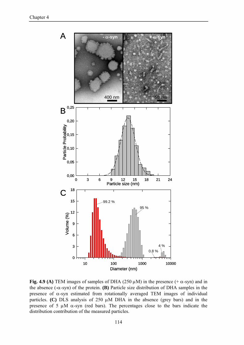
Chapter 4
Particle size (nm)0 3 6 9 12 15 18 21 24
Par
ticle
Pro
babi
lity
0,00
0,05
0,10
0,15
0,20
0,25
400 nm
- -syn
50 nm
+ -synA
C
B
Diameter (nm)10 100 1000 10000
Vol
ume
(%)
0
3
6
9
12
15
18
95 %
4 %
99.2 %
0.8 %
Particle size (nm)0 3 6 9 12 15 18 21 24
Par
ticle
Pro
babi
lity
0,00
0,05
0,10
0,15
0,20
0,25
400 nm
- -syn
50 nm
+ -syn
400 nm
- -syn
50 nm
+ -syn
50 nm
+ -synA
C
B
Diameter (nm)10 100 1000 10000
Vol
ume
(%)
0
3
6
9
12
15
18
95 %
4 %
99.2 %
0.8 %
Fig. 4.9 (A) TEM images of samples of DHA (250 M) in the presence (+ -syn) and in the absence (--syn) of the protein. (B) Particle size distribution of DHA samples in the presence of -syn estimated from rotationally averaged TEM images of individual particles. (C) DLS analysis of 250 M DHA in the absence (grey bars) and in the presence of 5 M -syn (red bars). The percentages close to the bars indicate the distribution contribution of the measured particles.
114

Chapter 4
4.3.3 Role of different regions of -synuclein in the interaction with DHA
Conformational analysis
In order to investigate the involvement of different regions of -syn in the
interaction with DHA, truncated forms of the protein were prepared (Fig. 4.10 A). Syn1-
99 lacks the acidic C-terminal region but retains all the seven imperfect repeats. Syn1-52
lacks also the NAC region (61-95) and includes four imperfect repeats. The central
polypeptide syn57-102 includes the NAC region and the last three imperfect repeats.
Finally, the peptide syn108-140 spans almost the whole acidic tail. At the N-terminal, the
polypeptides syn1-52 and syn57-102 have an extra-sequence constituted by GSH and
GSHM residues, respectively.
Far UV CD titration experiments of the several peptides in the presence of
increasing concentration of DHA were performed. In Fig. 4.10 B, the ellipticity of the
polypeptides and of -syn samples at 222 nm (one of the two typical minima of -helical
secondary structure), is shown as a function of the molar ratio between DHA and the
peptides. Increasing the amount of DHA, syn108-140 remains unfolded, while a
corresponding increment of the ellipticity is observed for syn1-99, syn1-52 and syn57-
102. The trends of the curves for syn1-99 and syn1-52 are quite similar to that of -syn
and the conformational transition involves the whole polypeptide chain; indeed syn1-99
has an helix content of ~87 % and syn1-52 of ~90 % (Rohl et al., 1996). Moreover, from
the plots it can be estimated that ~ 40 and 38 DHA molecules are required per syn1-99
and syn1-52 molecule for complete folding. On the contrary, syn57-102 needs an higher
amount of DHA to complete the transition and to reach the saturation of helix content that
is estimated to be ~ 67 %.
115

Chapter 4
NAC
1 99
1 52
NAC57 102
108 140
NAC1 140
-syn
syn1-99
syn1-52
syn57-102
syn108-140
[DHA]/[polypeptide]0 20 40 60 80 100 120
[] 2
22 n
m x
10-3
(de
g. cm
2 . dm
ol-1
)
-40
-30
-20
-10
0
A
B
NAC
1 99NACNAC
1 99
1 52
NAC57 102
108 140
NAC1 140
-syn
syn1-99
syn1-52
syn57-102
syn108-140
1 521 52
NAC57 102
NACNAC57 102
108 140108 140
NAC1 140
-synNACNAC
1 140
-syn
syn1-99
syn1-52
syn57-102
syn108-140
[DHA]/[polypeptide]0 20 40 60 80 100 120
[] 2
22 n
m x
10-3
(de
g. cm
2 . dm
ol-1
)
-40
-30
-20
-10
0
A
B
Fig. 4.10 (A) Schematic representation of truncated forms of -syn. The -syn species herewith studied correspond to the N-terminal fragments 1-99 (syn1-99, blue), 1-52 (syn1-52, pink), 57-102 (syn57-102, green) and the C-terminal fragment 108-140 (syn108-140, red). The N-terminal fragments are obtained by recombinant methods. At the N-terminal syn1-52 and syn57-102 have extra-sequence GSH and GSHM residues, respectively (see Materials & Methods). The C-terminal fragment has been obtained by chemical synthesis. (B) DHA-induced structural changes monitored by far-UV CD spectroscopy. CD signals at 222 nm are plotted against the molar ratio between DHA and polypeptides. The analysis was conducted in PBS pH 7.4, using a peptide concentration of 5 M.
116

Chapter 4
Proteolytic mapping
In order to verify the accessibility of the polypeptide chains of the different
truncated form of -syn once bound to DHA, we performed limited proteolysis
experiments. We have used proteinase K as protease, since it displays enough substrate
aspecificity and to have a comparison with the proteolysis conducted on -syn. The
HPLC analyses of the proteolytic mixture of syn1-99 and syn 1-52 with proteinase K
(Fig.4.11 A and B, respectively), in the absence (top) and in the presence (bottom) of 250
M DHA after 1 hour of incubation were reported. In the absence of DHA, both the
peptides are cleaved quite rapidly and proteinase K cleavage produces a large number of
peptides as indicated by the presence of numerous peaks in the chromatograms. The
identification of the peptide materials was obtained by mass spectrometry analysis and the
results are reported in Table 4.2. In the presence of the fatty acid, syn1-99 and syn1-52
show protease resistance and the peak relative to the intact peptide is the main species
present in the chromatogram after 1 hour of incubation. For syn1-99 there is only a
moderate proteolysis due to the free molecules in equilibrium; moreover, a peak relative
to the oxidized form of syn1-99 are isolated. In the case of syn1-52 a partial cleavage
occurs at the level of the peptide bond His50-Gly51 and fragment species 1-50 is
produced. Also a small amount of 1-49 is detectable and the calculated mass indicates
that partial oxidation of a Met residue is occurred (Table 4.2). We have also compared the
proteolytic maps of syn57-102 and syn108-140 by proteinase K obtained in the absence
and in the presence of 250 M DHA. The HPLC analyses relative to the proteolysis
mixtures of syn57-102 and syn108-40 are shown in Fig. 4.12 A and B, respectively, and
the peptides masses are reported in Table 4.3. As already observed for the N-terminal
fragments, in the absence of DHA syn 57-102 is cleaved by proteinase K generating
many fragments that span all the sequence of the peptide chain. In the presence of DHA,
only moderate fragmentation occurs, as assessed by the presence of small few peaks in
the chromatograms and by the peak of intact syn57-102. Far UV CD assessed that at a
molar ratio ([DHA]/[peptide]) of 50 not all the peptide molecules are bound to the lipid
and part is still free, so it can be that proteolysis occurs on free molecules.(Fig. 4.10 B,
green circles). At variance from the other syn species, syn108-140 results resistant to
proteolytic attack in the absence as well as in the presence of the fatty acid. The unusual
rigidity of the C-terminal tail of -syn to proteases was already seen for the interaction
between -syn and SDS micelles (Polverino de Laureto et al., 2006). This fact cannot be
117

Chapter 4
attributed to possible structural transitions of the C-terminal of the molecule, but rather to
a protection deriving from the unusual sequence (14 negative charges and 5 Pro residues).
118

Chapter 4
Retention Time (min)20 25 30 35
Rel
ativ
e A
bsor
banc
e at
226
nm
42-5243-52 31-50
5-18
19-5220-52
1-4*
9-525-42
1-17*1-18*
5-52
1-50*
1-52*
0 M DHA1-hr
250 M DHA1-hr
1-49ox*
syn1-52
B
Retention Time (min)20 25 30 35 40 45
Rel
ativ
e A
bsor
banc
e at
226
nm
0 M DHA1-hr
250 M DHA1-hr
1-9931-5033-50
1-99
1-99ox
57-7257-9973-99
syn1-99
A
Fig. 4.11 Proteolysis of syn1-99 (A) and syn1-52 (B) by proteinase K analyzed by RP-HPLC. The reactions were conducted at a protein concentration of 5 M in PBS buffer in the absence (top) or in the presence (bottom) of 250 M DHA, using an E/S ratio of 1:1000. RP-HPLC chromatograms correspond to the analysis of aliquots taken from the proteolysis mixtures after 1-hr of incubation with the protease.
119

Chapter 4
Retention Time (min)15 20 25 30
Rel
ativ
e A
bsor
banc
e at
226
nm
Retention Time (min)10 15 20 25
Rel
ativ
e A
bsor
banc
e at
226
nm
0 M DHA1-hr
0 M DHA1-hr
250 M DHA1-hr
250 M DHA1-hr
78-10279-10280-10278-9361-8962-86
57-81*
73-10275-102
57-89*57-90*
57-92*
57-102*
57-102ox*
67-78
93-10295-10292-10291-102
90-102
57-72*
60-80
65-8066-8057-71
57-79*76-102
57-102*
108-140
108-140
syn57-102 syn108-140
A B
Fig. 4.12 Proteolysis of syn57-102 (A) and syn108-140 (B) by proteinase K analyzed by RP-HPLC. The reactions were conducted at a protein concentration of 5 M in PBS buffer in the absence (top) or in the presence (bottom) of 250 M DHA, using an E/S ratio of 1:1000. RP-HPLC chromatograms correspond to the analysis of aliquots taken from the proteolysis mixtures after 1-hr of incubation with the protease.
120

Chapter 4
Table 4.2 Molecular masses of fragments obtained by proteolysis of syn1-99 and syn1-52 by proteinase K
Molecular mass (Da)
Polypeptide Fragmenta Foundb Calculatedc
syn1-99 57-72 1658.8 1659.8 33-50 1930.3 1931.2 31-50 2116.4 2116.4 73-99 2604.2 2604.9 57-99 4247.3 4246.7 1-99 9892.6 9891.4 syn1-52 1-4* 792.4 791.8 43-52 1053.7 1052.6 42-52 1140.8 1139.6 5-18 1388.0 1387.8 1-17* 2090.4 2091.5 31-50 2117.0 2116.5 1-18* 2162.5 2162.5 20-52 3385.8 3385.8 19-52 3456.8 3456.9 5-42 3792.6 3792.4 9-52 4397.9 4398.0 5-52 4827.6 4827.6 1-49ox* 5323.9 5324.1 1-50* 5445.6 5445.3 1-52* 5601.4 5601.4
aPeptides obtained by proteolysis of syn1-99 and syn1-52 by proteinase K. The peptides were purified by RP-HPLC (see Figure 4.11) and listed in the order of increasing molecular weight.
bExperimental molecular masses determined by ESI-MS. cMolecular masses calculated from the amino acid sequence of syn1-99 and syn1-52
*The peptides have an extra sequence (GSH) at the N-terminal.
121

Chapter 4
Table 4.3 Molecular masses of fragments obtained by proteolysis of syn57-102 and syn108-140 by proteinase K
aPeptides obtained by proteolysis of syn57-102 and syn108-140 by proteinase K. The peptides were purified by RP-HPLC (see Figure 4.12) and listed in the order of increasing molecular weight.
Molecular mass (Da)
Polypeptide Fragmenta Foundb Calculatedc
syn57-102 95-102 914.6 915.1 67-78 1000.9 1001.1 93-102 1118.7 1119.3 92-102 1219.7 1220.4 91-102 1290.8 1291.5 66-80 1355.7 1356.5 90-102 1361.8 1362.6 78-93 1432.8 1431.5 65-80 1468.9 1470.6 57-71* 1971.2 1971.2 60-80 2053.6 2056.3 57-72* 2071.5 2072.3 80-102 2276.0 2276.6 62-86 2312.5 2313.6 79-102 2404.4 2404.7 78-102 2476.3 2475.8 76-102 2645.8 2646.0 57-79* 2699.3 2699.0 61-89 2713.0 2714.0 75-102 2747.1 2747.1 73-102 2903.6 2903.3 57-81* 2928.4 2928.3 57-89* 3612.3 3613.0 57-90* 3684.5 3684.1 57-92* 3856.0 3856.3 57-102* 4957.5 4957.6 57-102ox* 4973.22 4973.6 syn108-140 108-140 3787.8 3787.9
bExperimental molecular masses determined by ESI-MS. cMolecular masses calculated from the amino acid sequence of syn57-102 and syn108-140
*The peptides have an extra sequence (GSHM) at the N-terminal.
122

Chapter 4
Effects on DHA aggregates
The aggregation properties of DHA were also analyzed in the presence of the
truncated forms of -syn. Turbidity measurements show that the presence of the peptides
(5 M) with the exception of syn108-140, prevents the formation of large fatty acid
aggregates (Fig 4.13), since there isn’t an increase in OD at 400 nm. The turbidity of
DHA samples was also measured in the presence of 2.5 µM syn57-102 (Fig. 4.13 C,
green triangles): there is no variation of the OD up to 242 ±49 M DHA. This value is
very similar to that obtained with 2.5 M -syn. Above this concentration, the turbidity
starts to increase as a function of DHA concentration, showing that the amount of syn57-
102 is not sufficient to prevent the formation of large lipid aggregates. In the presence of
syn108-140 (5 M) there is no appreciable variation of turbidity at 400 nm up to 75 ±9
M DHA. Increasing the concentration above this value, an increment of turbidity is
observed, but the OD values are lower than those obtained for DHA samples alone (Fig.
4.13 D).
The CAC of DHA was also evaluated by pyrene fluorescence assay. In Fig. 4.14
the I1/I3 pyrene ratio was plotted as a function of DHA concentration in the presence of
syn1-99 (A, blue circles), syn1-52 (B, pink circles), syn57-102 (C, green circles) and
syn108-140 (D, red circles). In the plots I1/I3 pyrene ratio for DHA alone was reported as
a control (black circles and line). For all the samples, increasing the DHA amount the I1/I3
ratio decreases. This fact suggests that a more hydrophobic micro-environment is
induced. The experimental data points fit with a sigmoidal curve so the CACs of the fatty
acid in the presence of the several peptides can be calculated at the inflection point of the
plots (Aguiar et al., 2003). In the presence of 5 M syn1-99 and syn1-52 , the I1/I3 ratio
shows a steeper decrease with an inflection point at 71 ±7 M and 69 ±22 M DHA,
respectively (Fig. 4.14 A, blue dash line and B, pink dash line). This indicates that syn1-
99 and syn1-52 exert a similar effect in reducing FA concentration for aggregate
formation, comparable to that of -syn (47 ±13 M). On the contrary, syn57-102 affects
in minor extent the aggregative properties of DHA, since in the presence of this
polypeptide CAC of DHA is 103 ±11 M (Fig. 4.14 C, green dash line). The addition of
syn108-140 does not interfere with pyrene fluorescence and it is possible to calculate a
CAC for DHA of 140 ±8 M (Fig. 4.14 D, red dash line).
A morphological analysis of DHA aggregate species were performed by TEM. In
Fig.4.15 TEM images relative to FA samples in the presence of the different polypeptides
123

Chapter 4
are reported. As controls, in panels A two representative pictures of DHA sample in PBS
pH 7.4 are shown. They indicate the presence of DHA aggregates with an heterogeneous
distribution of sizes. It is possible to see collapsed small spherical particles (red arrow)
and large particles with different degree of contrast due to a different exclusion of the
stain (green arrows). In panel B smaller spherical aggregates are formed in the presence
of -syn as already seen (§ 4.3.2). Also the presence of the other polypeptides with the
exception of syn108-140 causes a resizing of DHA assemblies (panel C, D, E, F). In the
presence of syn1-99 (panel C) and syn1-52 (panel D) DHA molecules form high contrast
spherical aggregates with diameters of ~ 20 and ~ 24 nm, respectively. On the other hand,
DHA assembles in smaller species also in the presence of syn57-102, but
morphologically these aggregates can be described as collapsed spheres (panel E). The
presence of syn108-140 does not modify DHA aggregates morphologies (panel F), TEM
picture shows large spherical particles comparable to those observed for DHA samples
(panel A). All these data obtained by different methods suggest that the N-terminal region
of -syn exerts a main role in modifying the aggregative properties of DHA.
124

Chapter 4
[DHA] (M)0 100 200 300 400 500
OD
400
nm
0,0
0,2
0,4
0,6
0,8
1,0
0 100 200 300 400 500
OD
400n
m
0,0
0,2
0,4
0,6
0,8
1,0
[DHA] (M) [DHA] (M)0 100 200 300 400 500
OD
400
nm
0,0
0,2
0,4
0,6
0,8
1,0
[DHA] (M)0 100 200 300 400 500
OD
400
nm
0,0
0,2
0,4
0,6
0,8
1,0
syn108-140syn57-102
syn1-99 syn1-52
A
C
B
D[DHA] (M)
0 100 200 300 400 500
OD
400
nm
0,0
0,2
0,4
0,6
0,8
1,0
0 100 200 300 400 500
OD
400n
m
0,0
0,2
0,4
0,6
0,8
1,0
[DHA] (M) [DHA] (M)0 100 200 300 400 500
OD
400
nm
0,0
0,2
0,4
0,6
0,8
1,0
[DHA] (M)0 100 200 300 400 500
OD
400
nm
0,0
0,2
0,4
0,6
0,8
1,0
syn108-140syn57-102
syn1-99 syn1-52
A
C
B
D
Fig. 4.13 Turbidity analysis at 400 nm of samples containing DHA up to 500 M, in the absence (black circles) and in the presence of syn1-99 (A, 5 M blue circles), syn1-52 (B, 5 M, pink circles), syn57-102 (C, 2.5 M, green triangles, 5 M green circles) and syn108-140 (D, red circles).
125

Chapter 4
[DHA] (M)0 100 200 300 400 500
I 1/I
3
1,0
1,2
1,4
1,6
1,8
[DHA] (M)0 100 200 300 400 500
I 1/I
3
1,0
1,2
1,4
1,6
1,8
[DHA] (M)0 100 200 300 400 500
I 1/I
3
1,0
1,2
1,4
1,6
1,8
[DHA] (M)0 100 200 300 400 500
I 1/I
3
1,0
1,2
1,4
1,6
1,8
syn108-140syn57-102
syn1-99 syn1-52
A
C
B
D[DHA] (M)
0 100 200 300 400 500
I 1/I
3
1,0
1,2
1,4
1,6
1,8
[DHA] (M)0 100 200 300 400 500
I 1/I
3
1,0
1,2
1,4
1,6
1,8
[DHA] (M)0 100 200 300 400 500
I 1/I
3
1,0
1,2
1,4
1,6
1,8
[DHA] (M)0 100 200 300 400 500
I 1/I
3
1,0
1,2
1,4
1,6
1,8
syn108-140syn57-102
syn1-99 syn1-52
A
C
B
D
Fig. 4.14 Pyrene fluorescence assay. Ratio between the first and the third vibronic band (I1/I3) of the pyrene emission spectrum as a function of DHA concentration (0-500 M) in the absence (black circles) and in the presence of -syn fragments. The analyses were conducted using 5 M peptide in PBS pH 7.4 and are reported as follows: A, syn1-99, B, syn1-52, C, syn57-102 and D, syn108-140.
126

Chapter 4
Fig. 4.15 TEM pictures of DHA samples in the absence (A) and in the presence of -syn (B) or its truncated forms (C-F).
127
Chapter 4
4.3.4 Aggregation process of -synuclein in the presence of DHA
Since several studies reported that -syn in the presence of PUFAs can form both
soluble oligomers that insoluble amyloid fibrils, the aggregation process of the protein
was analyzed by several biochemical and biophysical methods.
Firstly, aggregation of -syn samples (50 M) containing different concentrations
of DHA ([DHA]/[-syn] molar ratios of 10 and 50) was monitored by native
polyacrylamide gel electrophoresis (Fig. 4.16). This method is an excellent tool to reveal
the presence of covalent and non-covalent oligomers and aggregates. In the absence of
FA only one band relative to the -syn monomer is detected up to 118 hrs (Fig. 4.16 A).
On the contrary, in the presence of DHA in a molar ratio of 10, during incubation at 37°C
500 rpm, there are a decrease of the -syn monomer and an increase of material into the
wells. Also some smearing, which are indicative of a wide range of oligomeric species,
can be observed on the top of the separating gel (Fig. 4.16 B). In the presence of DHA in
a molar ratio of 50 the electrophoretic pattern is quite similar to those obtained with a
lower concentration of DHA but more intense smearing is detected into the gel (Fig. 4.16
C). Since DHA promotes -syn aggregation, a parameter such as the effect of lipid
concentration on protein aggregation was evaluated. Indeed, the molar ratio determines
the amount and type of the aggregate species of DHA in solution and the population of
free unbound protein molecules.
A conformation analysis of -syn at different stages of aggregation was carried out by
using CD measurements. During incubation (37°C and shaking at 500 rpm) -syn
acquires a -sheet conformation in the presence of DHA in a molar ratio of 10 (Fig. 4. 17
A). The initial spectrum of the protein reveals that 65 % of -syn molecules are unfolded
while the remaining population has an -helix structure. During the aggregation process
-syn undergoes a conformational rearrangement. After 74 hrs of incubation, the
spectrum is dominated by the band at 218 nm, evidencing a high content in -structure. It
is important to underline that, during the aggregation process triggered by saturating
concentration of DHA (molar ratio of 50), we did not observe any conformational
transition from -helix to -sheet during 142 hrs of incubation but only a partial reduction
of the intensity of the CD signals (data not shown). Moreover, in the absence of DHA no
conformational transition takes place and -syn remains unfolded in solution up to 142
hrs.
128

Chapter 4
In order to investigate the presence of amyloid-like fibrils in the samples, ThT
binding assay was performed. Only the aggregates formed in the presence of non-
saturating condition of DHA exhibit a common behaviour of amyloid like fibrils. Samples
containing the lipid in a molar ratio of 10 show an increase of ThT fluorescence at 485
nm. The lag phase seems to be very short since growth phase reaches a plateau after only
25 hrs of incubation (Fig. 4.17 B). These data was confirmed by TEM. After 142 hrs of
incubation in the absence of DHA and in the presence of saturating FA concentration
(Fig. 4.18 A, C), there are not fibrillar aggregates. On the contrary, DHA, in a molar ratio
[DHA]/[-syn] of 10 (Fig 4.18 B), promotes fibrils formation. After 142 hrs of
incubation, -syn forms aggregates with amyloid-fibrillar morphology, long, unbranched
with a diameter of 12-14 nm. In the presence of DHA, in a molar ratio [DHA]/[-syn] of
50, worm-like fibrils (left) and spherical oligomers (right) (diameter ~ 11 nm) are
detectable.
129

Chapter 4
Fig. 4.16 -Syn aggregation in the absence (A) and in the presence of DHA in a molar ratio [DHA]/[-syn] of 10 (B) or 50 (C) monitored by native-PAGE.
130

Chapter 4
Wavelength (nm)200 210 220 230 240 250
[] x
10-3
deg
. cm
2. d
mol
-1)
-20
-15
-10
-5
0
5
0 hrs
17 hrs
25 hrs
49 hrs
74 hrs94 hrs
Time (hrs)0 20 40 60 80 100 120
Flu
ores
cenc
e at
485
nm
(a.
u.)
0
100
200
300
400
500
B
A
Wavelength (nm)200 210 220 230 240 250
[] x
10-3
deg
. cm
2. d
mol
-1)
-20
-15
-10
-5
0
5
0 hrs
17 hrs
25 hrs
49 hrs
74 hrs94 hrs
Time (hrs)0 20 40 60 80 100 12
0
Flu
ores
cenc
e at
485
nm
(a.
u.)
0
100
200
300
400
500
B
A Fig. 4.17 (A) Evolution of the secondary structure of -syn monitored by far-UV CD during aggregation in the presence of DHA in a molar ratio of 10. (B) Time-course analysis of the aggregation process of -syn (PBS buffer, 37°C, shaking at 500 rpm) in absence (black circles) and in the presence of DHA in a molar ratio of 10 (red circles) or 50 (green circles) monitored by ThT fluorescence.
131

Chapter 4
50 nm100 nm
50 nm20 nm
C
BA Fig. 4.18 The effect of DHA on aggregation process of -syn was analyzed by TEM. TEM pictures relative to -syn aggregation samples after 142 hrs of incubation in the absence (A) and in the presence of DHA (B, molar ratio of 10; C, molar ratio of 50) were reported.
132
Chapter 4
4.4 DISCUSSION AND CONCLUSIONS
Many reports suggest that PUFAs are strictly connected to neurological disorders,
including depression, Alzheimer’s disease and Parkinson’s disease (Marszalek & Lodish,
2005, and references therein). Specifically, alterations in PUFAs levels have been found
related to aggregation of -syn (Necula et al., 2003; Broersen et al., 2006; Perrin et al.,
2001; Sharon et al., 2003; Assayag et al., 2007). It has been proposed that -syn could
interact with free FAs (Sharon et al., 2001; Broersen et al., 2006), or, alternatively, with
FAs in a micellar or bilayer state (Necula et al. 2003; Lücke et al., 2006). A recent study
also suggests that -syn normally interacts with PUFAs to carry out its physiological
functions, forming active forms of soluble oligomers (Ben Gedalya et al., 2008).
There is ample evidence that natively unfolded proteins, such as -syn, are not a
simple random coil, but rather form transient ordered structures that could be stabilized
by the interactions with natural ligands or cofactors, which are able to affect their net
charge or hydrophobicity (Uversky et al., 2007). In this study, we demonstrated that -
syn acquires an -helical conformation in the presence of DHA, and that the imperfect
repeats of -syn sequence play an important role in the -helix transition and thereby in
the interaction with DHA. -Syn strongly affects the self-association of the fatty acids
and its N-terminal region has a crucial role even in the modulation of this process. The
first 70 residues of the polypeptide chain of -syn seem to be strongly involved in the
interaction with DHA, while the NAC region results accessible to proteases. Moreover,
we demonstrated that the interaction with DHA has important implication for -syn
aggregation.
Upon binding to DHA, -syn adopts an -helix secondary structure in a simple
two state transition and the helix content is estimated to be ~70 %. The interaction
between -syn and micellar systems, especially those obtained using the detergent SDS,
has been intensively studied by many groups and topological models have been drawn on
the basis of NMR studies (Bisaglia et al., 2005; Ulmer et al., 2005; Bussel et al., 2004)
and limited proteolysis experiments (Polverino de Laureto et al., 2006). In our case, the
interaction involves an aggregated form of DHA since it does not allow a detailed NMR
characterization of the N-terminal region of -syn. On the other hand, the C-terminal
acidic region (residues 100-140) remains unfolded and freely tumbling in solution (Fig.
4.15 B). Moreover, the HSQC spectra of -syn in the presence of DHA are quite similar
133

Chapter 4
to those recorded in the presence of acidic small unilamellar vesicles (Eliezer et al.,
2001).
Proteolysis experiments coupled with mass spectrometry have proven useful to
define the region of -syn more directly interacting with DHA. A typical feature of a
natively unfolded protein, such as -syn, is to be largely sensitive to proteases because of
its low compactness and lack of secondary and tertiary structure (Uversky, 2002). As a
matter of fact, there are no regions with such persistent structure as to hinder protease’s
attack. Upon binding to DHA, -syn appears to be quite resistant to proteolysis, as
expected from the fact that the polypeptide has adopted an -helix conformation which
strictly involves the first 70 amino acid residues. The proteinase K-resistant product,
which contributes to the CD spectra during proteolysis, is still characterized by the
presence of -helical secondary structure (Fig. 4.7). A considerable accessibility of -syn
polypeptide chain to proteinase K and trypsin is confined only at the level of region 70-90
(Fig. 4.19), providing evidence that this segment is the most flexible and sufficiently
protruded to be protease-sensitive (Hubbard et al., 1994; Fontana et al., 1997). Of course,
we do not know to which specific residue this region can be extended, because there are
many potential cleavage sites along the sequence and to be effective a protease requires a
stretch of residues (Fontana et al., 1997; Schechter, 1965). However, it is quite cogent
that using two proteases with different specificity, proteolysis occurs at the level of the
same region.
In the presence of non-saturating concentrations of DHA (molar ratio
DHA/protein <50), the rate of proteolysis by proteinase K of -syn is slower than that in
the absence of DHA and with no accumulation of protected fragments. Above a molar
ratio of 50, all -syn molecules seem to be bound to the fatty acid as assessed by CD data
and the fragment encompassing the N-terminal region 1-72 is the main product in this
case. This clearly suggests that in the first case, there is the simultaneous presence of two
different populations of conformers of -syn, one free in solution and one bound to the
lipid, probably in rapid interchange between each other. Although largely unfolded, as
assessed by NMR, the C-terminal region hampers extensive proteolytic events in the case
of proteinase K. This fact cannot be attributed to possible structural transitions of this part
of the molecule, but rather to a protection deriving from the unusual sequence (14
negative charges and 5 Pro residues) (Polverino de Laureto et al., 2006).
134

Chapter 4
Fig. 4.19 Schematic representation of the main cleavage sites by trypsin (top) and proteinase K (bottom) in the polypeptide chain of -syn in the presence of saturating concentration of DHA.
The conformational analyses of the truncated species of -syn suggest that there is an
important action of the 7 imperfect repeats in the random-helix transition of the protein
induced by DHA. The 11-residue repeats of apolipoproteins are known to assume the
conformation of amphipathic α-helix, which is essential for the binding to lipids. As
predicted by the structural similarity to apolipoproteins, the N-terminal region of -syn is
shown to form an amphipathic α-helix that can interact with acidic phospholipids
(Davidson et al., 1998; § 3.4.2). Here we demonstrate that this N-terminal amphipathic -
helix of -syn is also required for its binding to DHA. Syn1-99, syn1-52 and syn57-102
have a similar isoelectric point, (9.4, 9.4 and 9.31) and they contain 7, 4 and 3 imperfect
repeats of the entire molecule, respectively. However, syn1-99 and syn1-52 exhibit a
similar conformational behavior in the presence of DHA. They completely undergo
helical structural transition and their maximum folding is achieved at a lipid/peptide
molar ratio (~ 40 and ~ 38, respectively) comparable to that required for -syn (~35). At
variance, syn57-102 reaches the maximum folding at a higher DHA/peptide molar ratio
(> 100) and shows lower helix content (67%). Since this last peptide lacks the first 4
structural repeats, these results may suggest that these segments in the N-terminal region
of -syn, are important to trigger the conformational transition of -syn in the presence of
DHA. We don’t have experimental evidences that competitive phenomena such as
aggregation are taking place, but this could give another possible explanation of the
inability of syn57-102 to completely convert its conformation in -helical structure.
135
Chapter 4
Furthermore syn108-140 remains unfolded in the presence of any kind or amount of lipid
(Eliezer et al., 2001; Ulmer et al., 2005). However, proteolysis data shows that also the C-
terminal region has a role in the -syn lipid-bound conformation, since the truncated
forms exhibit a different susceptibility to protease attack in comparison to the intact
molecule. The fragment species herewith investigated are resistant to proteolysis in the
presence of saturating concentrations of DHA. This gives an indication that, at variance
from -syn, all the peptide sequence is probably involved into the interaction and no
particular regions are exposed. For example, the proteolysis of syn1-99 and syn57-102 in
the presence of DHA do not produce any resistant peptides, like fragment 1-72 (Fig.4.5
D) that accumulates during the reaction with -syn. These results suggest that the C-
terminal tail could modulate the portion of -syn buried into the lipid compartment.
Phenomenon such as the electrostatic repulsion between the acidic residues of region 108-
140 and the deprotonated head groups of the FA molecules could be involved in this
process.
To define a unifying picture of the -syn/DHA interaction, we characterized the
species formed by DHA and the transition occurring in the presence of -syn by several
techniques. DHA can undergo self-aggregation phenomena strongly dependent on its
concentration, ionic strength and temperature. In particular, it has been shown that pH
defines precisely the species formed by DHA (Walde, 2006; Namani et al., 2007). The
presence of partly ionized DHA molecules is required to stabilize bilayers and a net
negative charge at the bilayer surface is generated. At room temperature and under neutral
conditions (pH 7.4), an increase in DHA concentration leads to the oil droplets formation
(Namani et al., 2007). When they are allowed to form spontaneously (Chen & Szostak,
2004), fatty acids aggregates are generally highly poly-dispersed and form an ensemble of
morphologically distinct species in dynamic equilibrium. Under the conditions used in
our experiments, DHA aggregates span a wide range of diameters from 50 to more than
600 nm and exhibit features compatible with the formation of lipid droplets, with a
critical aggregate concentration in the 80-130 M range (Fig. 4.8). Because the pH
conditions are at close to the edge for the formation of vesicles, also a small amount of bi-
layered structures can reasonably be formed. Indeed TEM pictures relative to DHA
samples (Fig. 4.15 A) show the presence of several aggregates with different degree of
contrast due to a different exclusion of the stain and also the presence of collapsed small
spherical particles, resembling vesicles. The addition of the protein causes a deep
136

Chapter 4
variation of both their size and properties. TEM observation and DLS show that the new
product of DHA self-assembly has a more regular shape and a smaller diameter (Fig. 4.9).
Moreover, pyrene fluorescence assay indicates that the final aggregation state is reached
at lower DHA concentrations and that these species have a reduced hydrophobic volume
(Fig. 4.8). Therefore, -syn seems to exert a double effect, inhibiting the formation of
giant aggregates and depressing the concentration for aggregate formation. Of interest is
that this phenomenon is correlated to the availability of the protein; since, when all the
protein molecules are bound to the fatty acid, the excess DHA is not prevented to form
aggregates with various sizes. The ability of proteins to interfere with lipid packing in
vesicles (Kamp & Beyer, 2006; Madine et al., 2006; Madine et al., 2008) as well as to
distort micelles has been already observed (Ulmer et al., 2005). Lipid aggregates are very
deformable because of the flexibility of the hydrocarbon chains. The shape and the
dimension of the aggregate can adjust to maximize the interaction with the protein. In
DHA, the presence of six double bonds confers high conformational flexibility to its self-
assembly products (Gawrish et al., 2003). Moreover, depression of the critical micelle
concentration for anionic detergents was regularly observed in the presence of cationic
electrolytes including protein such as tau (Chirita et al., 2003) and results from the
electrostatic interactions between positively charged amino acid side chains and
negatively charged FAs or detergent. -Syn is negatively charge at pH 7.4, but Necula
and coworkers suggested that for amphoteric polyelectrolytes such as proteins, depression
of anionic surfactant CMC is mediated by clustered charge rather than net charge (Necula
et al., 2003). This conclusion is verified by our results deriving from analysis with
truncated species of -syn. Syn108-140, corresponding to the acidic C-terminus, does not
alter the physical properties of DHA, while the syn1-99 and syn1-52 (the basic N-
terminus, with a net charge of +4.0 and +3.2 respectively) exert a effect similar to -syn
in the reduction of FA concentration for aggregate formation. Syn57-102 (net charge of
+2.1) has an ambiguous and weaker effect on DHA self-assembly properties, indeed it
prevents the turbidity of DHA samples but it depresses the DHA CAC in minor extent.
Probably both the short sequence and the low positive net charge contribute to this
peptide behavior.
In our experimental conditions, initially DHA forms oil droplets and the presence
of -syn could cause only a resizing into nano droplets or could alter the aggregative
properties of the FAs toward the formation of vesicles. Indeed, it is reasonable to assume
137

Chapter 4
that the positively charged amino acid side chains of -syn shield the negatively charged
fatty acid polar head groups, determining also a decrease of their pKa. At pH 7.4, this
causes a larger fraction of dissociated (ionized) fatty acid molecules to be present,
possibly inducing the formation of vesicles (bilayer structure) in the presence of -syn.
Moreover, the protein-DHA interface may experience a locally modified dielectric
constant, which further affects the ionization equilibrium. However, we don’t have the
clear cut evidence that the DHA species formed in the presence of the protein are
vesicles: cryo-electron microscopy and the use of soluble dye molecule could give
important information to verify the presence of lipid bilayers and of the interior aqueous
space.
A general consideration concerns the ability of DHA and probably of other long
chain PUFAs to induce oligomerization and fibrillation of -syn (Perrin et al., 2001;
Sharon et al., 2003; Broarsen et al., 2006) and also other amyloidogenic proteins (Wilson
& Binder, 1997; Ma & Westermark, 2002). We conducted all the experiments about the
study of -syn DHA interaction under conditions designed to avoid fast protein
aggregation, performing the measurements immediately after sample preparation and
using very low protein concentrations. We observed SDS-resistant -syn oligomers,
corresponding to dimer, trimer and tetramer only after 48 hours (data not shown). Since
fatty acids-induced protein oligomers are shown to be associated with decreased cell
viability (Assaying et al., 2007) it is of utmost importance to define the exact mechanism
responsible for PUFA-dependent multimerization and the specific role played by the
aggregative state of the fatty acid in modulating this process. From our studies we can
suggest a mechanism for -syn fibril formation in the presence of DHA.
The partial exposure of the NAC region evidenced by proteolysis mapping has a
deep effect on the aggregation propensity of -syn. Indeed, the NAC region is the most
hydrophobic and amyloidogenic segment of the -syn polypeptide chain (Giasson et al.,
2001; Miake et al., 2002). This segment that is not strictly involved into the interaction
with DHA aggregates could work as an anchorage for nucleating fibrils growth. To
develop regular fibrils, free protein molecules are required. In condition of a DHA/protein
molar ratio of 10, anionic aggregate species of DHA are already present and 65% of
protein is unbound. So protein fibrillogenesis is favored because the energy barrier of
nucleation is overcome and the protein molecules bound to DHA aggregates may function
as anchors for site directed fibril assembly. In the presence of saturating concentration of
138

Chapter 4
DHA (DHA/protein molar ratio ≥ 50), fibrils formation is slowed down and only
oligomeric species are formed. It is quite interesting that also SDS is able to induce -syn
fibril formation in a multiple structural forms (Ahmad et al., 2006, Ferreon and Deniz,
2007). Both of models are consistent with the existence of an ensemble of populations of
-syn and those with accessible hydrophobic surface and partial -helical contents favor
significantly protein aggregation. In our case, -syn aggregation process is modulated by
two contemporary factors. First, the interaction with DHA stabilizes a more
amyloidogenic conformation of -syn containing some -helical secondary structure
elements and that exposes part of the NAC region. Second, a particular ratio between
DHA and the protein is important to allow the presence of free protein molecules that will
be engaged in fibril growth.
In conclusion, our work may clarify the debated question on the type of interaction
between -syn and DHA, giving clear evidence for the formation of DHA aggregates in
the presence of the protein which are different from those formed by DHA alone. Fatty
acid aggregates can work as carriers for proteins, suggesting a more important role of
lipids in neurodegeneration. Furthermore, though -syn acquires concomitantly an -
helical conformation, proteolysis experiments showed that a region of the polypeptide
chain remains accessible to proteases. This means that this region is not characterized by
persistent structure, and it is conceivable that it could be involved in other interactions.
139

Chapter 4
4.5 REFERENCES
Adibhatla, R. M. & Hatcher, J. F. (2008). Altered Lipid Metabolism in Brain Injury and Disorders. Subcell. Biochem.48.
Aguiar, J., Carpena, P., Molina-Bolivar, J. A. & Ruiz C. C. (2003). On the
determination of the critical micelle concentration by pyrene 1:3 ratio method. J. Colloid Interface Sci. 258, 116–122.
Ahmad, M. F., Ramakrishna, T., Raman, B., Rao, Ch. M. (2006). Fibrillogenic
and non-fibrillogenic ensembles of SDS-bound human alpha-synuclein. J. Mol. Biol., 364, 10611072.
Assayag, K., Yakunin, E., Loeb, V., Selkoe, D. J. & Sharon, R. (2007).
Polyunsaturated fatty acids induce alpha-synuclein-related pathogenic changes in neuronal cells. Am. J. Pathol. 171, 2000–2011.
Atherton, E., and Sheppard, R. C. (1989) Solid Phase Peptide Synthesis, IRL
Press, Oxford, U.K Ben Gedalya, T., Loeb, V., Israeli, E., Altschuler, Y., Selkoe, D. J., Sharon, R.
(2009). Alpha-synuclein and polyunsaturated fatty acids promote clathrin-mediated endocytosis and synaptic vesicle recycling. Traffic, 10, 218234.
Bisaglia, M., Trolio, A., Tessari, I., Bubacco, L., Mammi, S., Bergantino, E.
(2005). Cloning, expression, purification, and spectroscopic analysis of the fragment 57-102 of human alpha-synuclein.. Protein Expr Purif., 39, 9096.
Bisaglia, M., Tessari, I., Pinato, L., Bellanda, M., Girando, S., Fasano, M.,
Bergantino, E., Bubacco, L. & Mammi, S. (2005). A topological model of the interaction between alpha-synuclein and sodium dodecyl sulfate micelles. Biochemistry, 44, 329–339.
Bousquet, M., Saint-Pierre, M., Julien, C., Salem, N. Jr., Cicchetti, F., Calon, F.
(2008). Beneficial effects of dietary omega-3 polyunsaturated fatty acid on toxin-induced neuronal degeneration in an animal model of Parkinson's disease. FASEB J., 22, 12131225.
Breckenridge, W. C., Gombos, G & Morgan, I. G. (1972). The lipid composition
of adult rat brain synaptosomal plasma membranes. Biochim. Biophys. Acta, 266, 695–707.
Broersen, K., van den Brink, D., Fraser, G., Goedert, M. & Davletov, B. (2006).
Alpha-synuclein adopts an alpha-helical conformation in the presence of polyunsaturated fatty acids to hinder micelle formation. Biochemistry, 45, 15610–15616.
Bussell, R. Jr. & Eliezer, D. (2004). Effects of Parkinson's disease-linked
mutations on the structure of lipid-associated alpha-synuclein. Biochemistry, 43, 4810–4818.
140

Chapter 4
Chandra, S., Chen, X., Rizo, J., Jahn, R. & Südhof, T. C. (2003). A broken alpha -helix in folded alpha -Synuclein. J. Biol. Chem. 278, 15313–15318.
Chen, I. A. & Szostak, J. W. (2004). A kinetic study of the growth of fatty acid
vesicles. Biophys. J. 87, 988–998. Chirita, C. N., Necula, M., Kuret, J. (2003). Anionic micelles and vesicles induce
tau fibrillization in vitro. J. Biol. Chem., 278, 2564425650. Croke, R. L., Sallum, C. O., Watson, E., Watt, E. D. & Alexandrescu, A. T.
(2008). Hydrogen exchange of monomeric alpha-synuclein shows unfolded structure persists at physiological temperature and is independent of molecular crowding in Escherichia coli. Protein Sci. 17, 1434–45.
Dalfó, E., Portero-Otín, M., Ayala, V., Martínez, A., Pamplona, R. & Ferrer, I.
(2005). Evidence of oxidative stress in the neocortex in incidental Lewy body disease. J. Neuropathol. Exp. Neurol,. 64, 816–830.
Davidson, W. S., Jonas, A., Clayton, D. F. & George, J. M. (1998). Stabilization
of alpha-synuclein secondary structure upon binding to synthetic membranes. J. Biol. Chem., 273, 9443–9449.
de Lau, L. M., Bornebroek, M., Witteman, J. C., Hofman, A., Koudstaal, P. J.,
Breteler, M. M. (2005). Dietary fatty acids and the risk of Parkinson disease: the Rotterdam study. Neurology, 64, 20402045.
Ebeling, W., Hennrich, N., Klockow, M., Meta, H., Orth, D., and Lang, H. (1974).
Proteinase K from Tritirachium album Limber. Eur. J. Biochem. 47, 91–97. Eliezer, D., Kutluay, E., Bussell, R. Jr & Browne, G. (2001). Conformational
properties of alpha-synuclein in its free and lipid-associated states. J. Mol. Biol. 307, 1061–1073.
Ferreon AC, Deniz AA. (2007). Alpha-synuclein multistate folding thermodynamics: implications for protein misfolding and aggregation. Biochemistry, 46, 44994509.
Fontana, A., Zambonin, M., Polverino de Laureto, P., De Filippis, V., Clementi, A. & Scaramella, E. (1997). Probing the conformational state of apomyoglobin by limited proteolysis. J. Mol. Biol. 266, 223–230.
Fontana, A., Polverino de Laureto, P., Spolaore ,B., Frare, E., Ricotti, P. &
Zambonin, M. (2004). Probing protein structure by limited proteolysis. Acta Biochim Pol. 51, 299–321.
Frank, J., Radermacher, M., Penczek, P., Zhu, J., Li, Y., Ladjadj, M., Leith, A. J.
(1996). SPIDER and WEB: processing and visualization of images in 3D electron microscopy and related fields. Struct. Biol. 116, 190–199.
141
Chapter 4
Gawrisch, K., Eldho, N. V. & Holte, L. L. (2003). The structure of DHA in phospholipid membranes. Lipids, 4, 445–452
Giasson, B. I., Murray, I. V., Trojanowski, J. Q., Lee, V. M. (2001). A
hydrophobic stretch of 12 amino acid residues in the middle of alpha-synuclein is essential for filament assembly. J. Biol. Chem., 276, 23802386.
Gill, S. G & von Hippel, P. H. (1989) Calculation of protein extinction
coefficients from amino acid sequence data. Anal. Biochem. 182, 319–326. Golovko, M. Y., Rosenberger, T. A., Faergeman, N. J., Feddersen, S., Cole, N. B.,
Pribill, I., Berger, J., Nussbaum, R. L. & Murphy, E. J. (2006). Acyl-CoA synthetase activity links wild-type but not mutant alpha-synuclein to brain arachidonate metabolism. Biochemistry, 45, 6956–6966.
Golovko, M. Y., Rosenberger, T. A., Feddersen, S., Faergeman, N. J., Murphy,
E.J. (2007). Alpha-synuclein gene ablation increases docosahexaenoic acid incorporation and turnover in brain phospholipids. J. Neurochem. 101, 201–211.
Hamilton, J. A. (1999). Transport of fatty acids across membranes by the diffusion
mechanism. Prostaglandins Leukot. Essent. Fatty Acids, 60, 291–297. Hamilton, J. A.; Kamp, F. (1999). How are free fatty acids transported in
membranes? Is it by proteins or by free diffusion through the lipids? Diabetes, 48, 2255–2269.
Huang, C., Ren, G., Zhou, H. & Wang, C. C. (2005). A new method for
purification of recombinant human alpha-synuclein in Escherichia coli. Protein Expr. Purif. 42, 173–177.
Hubbard, S. J., Eisenmenger, F.& Thornton, J. M. (1994). Modeling studies of the
change in conformation required for cleavage of limited proteolytic sites. Protein Sci. 3, 757–768.
Innis S. M. (2007). Dietary (n-3) fatty acids and brain development. J. Nutr.,137,
855–859. Julien, C., Berthiaume, L., Hadj-Tahar, A., Rajput, A. H., Bédard, P. J., Di Paolo,
T., Julien, P., Calon, F. (2006). Postmortem brain fatty acid profile of levodopa-treated Parkinson disease patients and parkinsonian monkeys. Neurochem. Int., 48, 404414.
Kalyanasundaram, K. & Thomas J. K. (1977). Environmental Effects on Vibronic
Band Intensities in Pyrene Monomer Fluorescence and Their Application in Studies of Micellar Systems. J. Am. Chem. Soc. 99, 2039−2044.
Kamp, F. & Beyer, K. (2006) Binding of -synuclein affects the lipid packing in
bilayers of small vesicles. J. Biol. Chem. 281, 9251–9259.
142
Chapter 4
Le Vine, H. (1993). Thioflavin T interaction with synthetic Alzheimer’s disease beta-amyloid peptides: Detection of amyloid aggregation in solution. Protein Sci., 2, 404410.
Lòpez-Diaz, D., Garcia-Mateos, I. & Velazqueez, M. M. (2005). Synergism in
mixtures of zwitterionic and ionic surfactants. Coll. Surf. A Physicochem. Eng. Aspects, 270, 153−162.
Ludtke, S. J., Baldwin, P. R. & Chiu, W. (1999). EMAN: semiautomated software
for high-resolution single-particle reconstructions. J. Struct. Biol. 128, 82–97. Lücke, C., Gantz, D. L., Klimtchuk, E. & Hamilton J. A. (2006). Interactions
between fatty acids and alpha-synuclein. J. Lipid Res. 47, 1714–1724. Lukiw, W. J., Bazan, N. G.. (2008). Docosahexaenoic acid and the aging brain. J.
Nutr., 138, 25102514. Ma, Z. & Westermark, G. T. (2002). Effects of free fatty acid on polymerization
of islet amyloid polypeptide (IAPP) in vitro and on amyloid fibril formation in cultivated isolated islets of transgenic mice overexpressing human IAPP. Mol. Med. 8, 863–868.
Madine, J., Doig, A. J., Middleton, D. A. (2006). A study of the regional effects of
alpha-synuclein on the organization and stability of phospholipid bilayers. Biochemistry, 45, 578392.
Madine, J., Hughes, E., Doig, A. J., Middleton, D. A. (2008). The effects of alpha-
synuclein on phospholipid vesicle integrity: a study using 31P NMR and electron microscopy. Mol. Membr. Biol., 25, 518527.
Marszalek, J. R. & Lodish, H. F. (2005). Docosahexaenoic acid, fatty acid-
interacting proteins, and neuronal function: breastmilk and fish are good for you. Annu. Rev. Cell. Dev. Biol. 21, 633–57.
McNulty, B. C., Tripathy, A., Young, G. B., Charlton, L. M., Orans, J. & Pielak,
G. J. (2006). Temperature-induced reversible conformational change in the first 100 residues of alpha-synuclein. Protein Sci. 15, 602–608.
Miake, H., Mizusawa, H., Iwatsubo, T., Hasegawa, M. (2002). Biochemical
characterization of the core structure of alpha-synuclein filaments. J. Biol. Chem., 277, 1921319219.
Namani, T. & Walde, P. (2005). From decanoate micelles to decanoic
acid/dodecylbenzenesulfonate vesicles. Langmuir, 21, 6210–6219. Namani, T., Ishikawa, T., Morigaki, K. & Walde, P. (2007). Vesicles from
docosahexaenoic acid. Coll. Surf. B Biointer. 54, 118–123. Necula, M., Chirita, C. N. & Kuret, J. (2003). Rapid anionic micelle-mediated
alpha-synuclein fibrillization in vitro. J. Biol. Chem. 278, 46674–46680.
143

Chapter 4
Perrin, R. J., Woods, W. S., Clayton, D. F. & George, J. M. (2001). Exposure to long chain polyunsaturated fatty acids triggers rapid multimerization of synucleins. J. Biol. Chem. 276, 41958–41962.
Polverino de Laureto, P., Tosatto, L., Frare, E., Marin, O., Uversky, V. N. &
Fontana, A. (2006). Conformational properties of the SDS-bound state of alpha-synuclein probed by limited proteolysis: unexpected rigidity of the acidic C-terminal tail. Biochemistry, 45, 11523–11531.
Rapoport, S. I., Rao, J. S., Igarashi, M. (2007). Brain metabolism of nutritionally
essential polyunsaturated fatty acids depends on both the diet and the liver. Prostaglandins Leukot. Essent. Fatty Acids, 77, 251–261
Richieri, G.. V., Ogata, R. T., Zimmerman, A. W. (2000). Fatty acid binding
proteins from different tissues show distinct patterns of fatty acid interactions. Biochemistry, 39, 7197–7204.
Rohl, C. A., Chakrabartty, A. & Baldwin, R. L. (1996). Helix propagation and N-
cap propensities of the amino acids measured in alanine-based peptides in 40 volume percent trifluoroethanol. Protein Sci. 5, 2623–37.
Sakai, T., Kaneko, Y., Tsujii, K. (2006). Premicellar aggregation of fatty acid N-
methylethanolamides in aqueous solutions. Langmuir, 22, 2039–2044. Schägger, H., & von Jagow, G. (1987) Tricine-sodium dodecyl sulfate-
polyacrylamide gel electrophoresis for the separation of proteins in the range from 1 to 100 kDa. Anal. Biochem. 166, 368–379.
Schechter, I. & Berger, A. (1967). On the size of the active site in proteases. I.
Papain. Biochem. and Bioph. Res. Comm. 27, 157. Scopes R. K. (1974). Measurement of protein by spectraphotometry at 205 nm.
Anal. Biochem., 59, 277282. Sharon, R., Goldberg, M. S., Bar-Josef, I., Betensky, R. A., Shen, J. & Selkoe, D.
J. (2001). Alpha-Synuclein occurs in lipid-rich high molecular weight complexes, binds fatty acids, and shows homology to the fatty acid-binding proteins. Proc. Natl. Acad. Sci. U S A, 98, 9110–9115.
Sharon, R., Bar-Joseph, I., Frosch, M. P., Walsh, D. M., Hamilton, J. A. & Selkoe,
D. J. (2003). The formation of highly soluble oligomers of alpha-synuclein is regulated by fatty acids and enhanced in Parkinson's disease. Neuron, 37, 583–595.
Sharon, R., Bar-Joseph, I., Mirick, G. E., Serhan, C. N. & Selkoe, D. J. (2003).
Altered fatty acid composition of dopaminergic neurons expressing alpha-synuclein and human brains with alpha-synucleinopathies. J. Biol. Chem., 278, 49874–49881.
Turro, N. J. & Kuo P. L. (1986). Pyrene Excimer Formation in Micelles of
Nonionc Detergents and of Water-Soluble Polymers. Langmuir, 2, 438–442.
144

Chapter 4
Uéda, K., Fukushima, H., Masliah, E., Xia, Y., Iwai, A., Yoshimoto, M., Otero, D. A., Kondo, J., Ihara, Y. & Saitoh, T. (1993). Molecular cloning of cDNA encoding an unrecognized component of amyloid in Alzheimer disease. Proc. Natl. Acad. Sci. U S A, 90, 11282–11286.
Ulmer, T. S., Bax, A., Cole, N. B. & Nussbaum, R. L. (2005). Structure and
dynamics of micelle-bound human alpha-synuclein. J. Biol. Chem. 280, 9595–9603. Uversky, V. N. (2002). Natively unfolded proteins: A point where biology waits
for physics. Protein Sci. 11, 739–756. Uversky, V. N. (2007) Neuropathology, biochemistry, and biophysics of -
synuclein aggregation. J. Neurochem. 103, 17–37. Walde, P. (2006). Surfactant assemblies and their various possible roles for the
origin(s) of life. Orig. Life Evol. Biosph. 36, 109–150. Wilson, D. M. & Binder, L. I. (1997). Free fatty acids stimulate the
polymerization of tau and amyloid beta peptides. In vitro evidence for a common effector of pathogenesis in Alzheimer's disease. Am. J. Pathol. 150, 2181–2195.
Zhai, L., Zhang, J., Shi, Q., Chen, W. & Zhao, M. (2005). Transition from micelle
to vesicle in aqueous mixtures of anionic/zwitterionic surfactants studied by fluorescence, conductivity, and turbidity methods. J. Colloid. Interface Sci. 284, 698−703.
145

Chapter 4
146

Appendix
APPENDIX
Main analytical techniques used
I. Circular Dichroism
Circular dichroism (CD) is the election spectroscopic technique used to determine
the presence and the type of secondary structure in peptides and proteins, to evaluate their
tertiary structure and to monitor structural transitions during unfolding and folding
processes (Woody, 1995; Kelly et al., 2005). CD refers to the differential adsorption of
the left and right circularly polarized components of plane-polarised radiation. This effect
will occur when a chromofore is chiral (optically active) either (a) intrinsically by reason
of its structure, or (b) by being covalently linked to a chiral centre, or (c) by being placed
in an asymmetric environment. In practice the plane polarized radiation is split into its
two circularly polarized components by passage through a modulator subjected to an
alternating (50 kHz) electric field. The modulator usually consists of a piezoelectric
quartz crystal and a thin plate of isotropic material (e.g. quartz) tightly coupled to the
crystal. The alternating electric field induced structural changes in the quartz crystal
which make the plate transmit circularly polarized light at the extremes of the field. If,
after the passage through the sample, the left and right circularly polarized components
are not absorbed (or are absorbed to the same extent), combination of the components
would regenerate radiation polarized in the original plane. However, if one of the
components is absorbed by the sample to a greater extent than the other, the resultant
radiation (combined components) would now be elliptically polarized, i.e. the resultant
would trace out an ellipse. In practice, the CD instrument (spectropolarimeter) does not
recombine the components, but detects the two components separately; it will then
display the dichroism at a given wavelength of radiation expressed as either difference in
absorbance of the two components (A = AL – AR) or as the ellipticity in degrees () ( =
tan-1 (b/a), where b and a are the minor and major axes of the resultant ellipse. There is a
simple numerical relationship between A and q (in degrees), i.e. = 33 (AL – AR).
A CD spectrum is obtained when the dichroism, i.e. the variation of θ expressed in
mdeg, is measured as a function of wavelength. Once that the spectrum has been
acquired, the measurements must be normalized in order to make them independent from
the protein concentration and the cuvette pathlength. This step is reached expressing the
ellipticity as mean residue ellipticity, []MRW, calculated dividing the ellipticity for the
147

Appendix
molar concentration (using as molecular weight the mean molecular weight for residue)
and for the pathlength:
[] = · MRW / 10 · c · d
Where is expressed in mdeg, the protein concentration c in mg/ml, the pathlength d in
cm and MRW is the mean residue molecular weight, obtained dividing the protein
molecular weight for the number of its amino acid residues. The value [] is expressed in
deg · cm2 · dmol-1.
Every chiral chromophore or belonging to a chiral molecule presents a
characteristic activity of circular dichroism, i.e. it is active at certain wavelength values.
As far as proteins are concerned, interesting information can be obtained evaluating the
region between 250 and 180 nm (far-UV CD). In this region the chromophore responsible
for the dichroic signal is the amidic bond that presents a different ellipticity pattern
depending on the conformation of the adjacent bond angles , i.e. depending on the kind of
secondary structure in which it is embedded (alpha-helix, beta-sheet or random coil).
Each kind of secondary structure possesses a typical spectrum, characterized by specific
signals. Alpha-helix is characterized by two intense negative bands at 222 and 208 nm
and one positive band at 192 nm. Beta-sheet structure presents a weak negative band at
218 nm and a positive band at 198 nm, while random coil structure shows a weak positive
band at 218 nm and an intense negative one at ~ 198 nm (Fig. I.1).
Fig. I.1. Far UV CD spectra associated with various types of secondary structure. Solid line, α-helix; long dashed line, anti-parallel β-sheet; dotted line, type I β-turn; cross dashed line, extended 31-helix or poly (Pro) II helix; short dashed line, irregular structure. (reprinted from Kelly et al., 2005)
148

Appendix
II. Fluorescence
Fluorescence emission is observed when an excited electron returns from the first
excited state back to the ground state. As some energy is always lost by non-radiative
processes, such as vibrational transitions, the energy of the emitted light is always less
than that of the absorbed light. Hence the fluorescence emission is shifted to longer
wavelengths compared with the absorption of the respective chromophore. Fluorescence
spectroscopy is one of the most powerful methods to study protein folding, dynamics,
assembly, and interactions, as well as membrane structure. It has been successfully
applied to investigate the complex mechanisms of protein aggregation including amyloid
fibril formation and the interaction of amyloidogenic proteins with membranes, as well as
their dynamic structures. Almost all proteins have natural fluorophores, tyrosine and
tryptophan residues, which allow study of changes in protein conformation. Also site-
specific labeling with external fluorophores is easily achievable by mutagenesis and
chemical modifications. Fluorescence spectroscopy requires a small amount of material
(pM–nM range) and has a high signal-to-noise ratio.
Thioflavin T assay
ThT is a fluorescent dye used as a non-covalent extrinsic fluorescent probe in
studies of amyloid fibril formation (LeVine, 1993). It has a two-ring structure: the
conjugated benzothiazol and aminobenzol rings are arranged in an almost planar
orientation ( 30°) in the minimum energy conformation (Fig. II.1) (Munishkina & Fink,
2007). During excitation, the rings rotate in order to obtain the most stable exited state
conformation ( 90°) which has low fluorescence efficiency. ThT may bind between the
betasheets of the fibril; however, no experimental data exist to support this assumption.
ThT has two excitation (~335 nm and 430 nm) and two corresponding emission peaks
(425–455 nm and 483 nm). Excitation at ~350 nm results in emission at ~438 nm,
whereas excitation at 440 nm leads to emission at ~483 nm. The binding to amyloid
fibrils induces a blue shift of the ThT λmax from 483 nm to ~478 nm and probably
stabilizes the planar form of the molecule and leads to a 10–500-fold increase in ThT
fluorescence intensity.
149

Appendix
Fig. II.1. Ball-and-stick model of ThT: the rotational angle is around 30 °. Carbons are dark gray, hydrogens are cyan, nitrogens are blue, and sulfur is yellow. (Reprinted from Munishkina & Fink, 2007). Pyrene Fluorescence
Pyrene is a polycyclic aromatic hydrocarbon (Fig. II.2, A) consisting of 4 fused
benzene rings, resulting in a large, flat aromatic system. Pyrene is a valuable molecular
probe for fluorescence spectroscopy, having a high quantum yield and lifetime (0.65 and
410 nanosecond, respectively, in ethanol at 293K). Its fluorescence emission spectrum is
very sensitive to the solvent's polarity, so pyrene has been used as a probe to determine
solvent environments. This is due to its excited state having a different, non-planar
structure than the ground state. Certain emission bands are unaffected, but others vary in
intensity due to the strength of interaction with a solvent. Indeed, the pyrene fluorescence
fine structure presents five peaks (Fig. II.2, B). It is well established that the ratio between
the intensities of the first (~ 373 nm) and the third (~ 384 nm) vibration bands of the
pyrene environment (Kalyanasundaram & Thomas, 1977; Agiuar et al., 2003). Low
values of the I1/I3 ratio correspond to a non polar environment. The change of I1/I3 value
as a function of surfactant concentration is commonly used to determine the critical
micelle concentration of surfactant solution, the critical aggregate concentration for
polymer-surfactant complexes and also to have information on molecular dissolution of
oil droplets dispersed in water. Pyrene can also adopt a dimeric structure, with molecules
arranged in sandwiched pairs in order to form lattice units. This results in a high degree of
symmetry, belonging to the C2h symmetry group. In solution, dimers exhibit high
wavelength fluorescence (red-shift) and also excimer (excited state dimer of pyrene)
formation (Fig. II.2, B). Pyrene excimer formation is a well-known concentration-
dependent phenomenon in organic solutions. However, because of the low solubility of
pyrene in water, pyrene excimer formation is not observed in pure water solutions. The
ratio between the maximum emission intensity of the excimer and the I1 band of pyrene
represents the probability of excimer formation for pyrene molecules, in relation to the
150

Appendix
local concentration in the environment (Turro et al., 1986). Thus, this ratio gives a good
insight into the hydrophobic microdomains formed by surfactants in solution. Pyrene
excimer formation is also used to study lateral diffusion of lipids and proteins (pyrene-
labeled) in the lipid bilayer (Munishkina & Fink, 2007; Somerharju, 2002).
A B I1
I3 Iex
Fig. II.2. (A) Molecular structure of pyrene. (B) Typical fluorescence emission spectra of pyrene.
III. Proteolysis
Proteolysis of a protein substrate can occur only if the polypeptide chain can bind
and adapt to specific stereochemistry of the proteases active site (Fontana et al., 2004).
However, since the active site of proteases have not been designed by nature to fit the
specific sequence and fixed stereochemistry of a stretch of at least 8-10 amino acid
residues of a particular globular protein, an induced-fit mechanism of adaptation of the
protein substrate to the active site of the protease is required for binding and formation of
the transition state of the hydrolytic reaction. Therefore, the native rigid structure of a
globular protein cannot act as a substrate for a protease, as documented by the fact that
folded proteins under physiological conditions are rather resistant to proteolysis. This is
no longer the case when the fully unfolded state (U) of a globular protein exists at
equilibrium with the native state (N). However, the N↔U equilibrium is much shifted
towards the native state under physiological conditions, according to the Boltzmann
relationship DG= -RTln [U]/[N], where DG is 5-15 kcal/mol. Therefore, only a tiny
fraction (10-6-10-9) of protein molecules is in the U state that is suitable for proteolysis.
Consequently, native globular proteins are rather resistant to proteolytic degradation, as a
151

Appendix
result of the fact that the N↔U equilibrium actually dictates and regulates the rate of
proteolysis.
Nevertheless, even native globular proteins can be attacked by a protease and, in a
number of cases, it has been shown that the peptide bond fission occurs only at one (or a
few) peptide bond(s). This behavior results from the fact that a globular protein is not a
static entity as can be inferred by a picture of its crystallographic determined 3D-
structure, but instead is a dynamic system capable of fluctuations around its average
native state at the level of both side chains and polypeptide backbone. Indeed,
crystallographers analyze this protein mobility in terms of B-factor for both side chains
and Cα-backbone. The main chain B-factor is a measure of average displacements of a
polypeptide chain from its native structure, so that it can experience displacements
leading to some local unfolding, it can be envisaged that these higher energy, locally
unfolded states are those required for a native protein to be attacked by a proteolytic
enzyme. Evidence for this mechanism of local unfolding requires for limited proteolysis
has been provided by demonstrating a close correspondence between sites of limited
proteolysis and sites of higher backbone displacements in the 316-residue polypeptide
chain of thermolysin. It is plausible to suggest that limited proteolysis derives also from
the fact that a specific chain segment of the folded protein substrate is sufficiently
exposed to bind at the active site of the protease. However, the notion of
exposure/protrusion/accessibility is a required property, but clearly not at all sufficient to
explain the selective hydrolysis of just one peptide bond, since it is evident that even in a
small globular protein there are many exposed sites (the all protein surface) which could
be targets of proteolysis. Instead, enhanced chain flexibility (segmental mobility) appears
to be the key feature of the site(s) of limited proteolysis.
The results obtained with thermolysin are in line with those derived from limited
proteolysis experiments conducted on a variety of other proteins of known (3D) structure.
In many cases, limited proteolysis was observed to occur at sites of the polypeptide chain
displaying high segmental mobility or poorly resolved in the electron density map,
implying significant static/dynamic disorder. Therefore, it was concluded that limited
proteolysis of a globular protein occurs at flexible loops and, in particular, that chain
segments in a regular secondary structure (such as helices) are not sites of limited
proteolysis. Indeed Hubbard et al. (1998) conducted modeling studies of the
conformational changes required for proteolytic cleavages and concluded that the sites of
152

Appendix
limited proteolysis require a large conformational change (local unfolding) of a chain
segment of up to 12 residues.
Fig. III.1 A segment of local unfolding is a suitable site of proteolysis
A possible explanation of the fact that helices and, in general, elements of regular
secondary structure are not easily hydrolyzed by proteolytic enzymes can be given also
on the basis of energetic considerations. If proteolysis is occurring at the centre of the
helical segment, likely the helix is fully destroyed by end-effects and consequently all
hydrogen bonds, which cooperatively stabilize it, are broken. On the other hand, a peptide
bond fission at a disordered flexible site likely does not change much the energetic of that
site, since the peptide hydrolysis can easily be compensated by some hydrogen bonds
with water. Therefore, it can be proposed that proteolysis of rigid elements of secondary
structure is thermodynamically very disadvantageous.
The limited proteolysis approach fro probing protein conformation implies that the
proteolytic event should be dictated by the stereochemistry and flexibility of the protein
substrate and not by the specificity of the attacking protease (Fontana et al., 1999;
Hubbard, 1998). To this aim, the most suitable proteases are those displaying broad
substrate specificity, such as subtilysin, thermolysin, proteinase K and pepsin (Bond,
1990). These endopeptidases display a moderate preference for hydrophobic or neutral
amino acid residues, but often cleavages occur at other residues as well. The
recommended approach is to perform trial experiments of proteolysis of the protein of
interest in order to find out the most useful protease, the optimal protein
substrate:protease (E:S) ratio and the effect of temperature and time of incubation
(Fontana et al., 1999). Possible ways to control proteolysis is by using a low
concentration of protease, short reaction times and low temperature. It is not easy to
predict in advance the most useful experimental conditions for conducting a limited
proteolysis experiment, since these depend upon the structure, dynamics, stability/rigidity
153

Appendix
properties of the protein substrate and from the actual aim of the experiment, i.e.,
identification of the sites of protein flexibility, isolation of the rigid core of the protein or
preparation of a nicked protein. In typical experiments of limited proteolysis, it has been
found that an E:S ratio of 1:100 (by weight) is recommended, but occasionally both 1:20
or 1:5000 can be used. This results from the fact that there is a great variation in the rate
of the selective peptide fission in a globular protein, requiring seconds or days for the
limited proteolysis event. Moreover, if isolation of the nicked protein resulting from the
initial proteolysis is desired, both the time and temperature of reaction should be properly
controlled, since the nicked species may be present only transiently in the protein
mixture. Indeed, a nicked protein is usually much more flexible and unstable than the
native one and easily unfolds to a protein substrate that is finally degraded to small
peptides
IV. Mass Spectrometry
Thanks to recent technological progresses, mass spectrometry has become one of
the most powerful tool in protein analysis (Aebersold & Mann, 2003). A further incentive
to the development of such techniques has come also from the huge amount of data
deriving from the sequencing of entire genomes. Indeed, mass spectrometry (MS),
allowing the identification of thousands of proteins starting from complex mixtures, has
given significant insights into biological and medical matters. MS can be exploited in
many fields, ranging from chemistry and biology to pharmaceutical analysis. It can be
used to measure the accurate molecular weight of peptides or proteins, both chemically
synthesized or produced by recombinant methods. Other applications are the evaluation
of the purity of a protein sample, the identification of post-translational modifications, the
monitoring of chemical/enzymatic reactions, the sequencing of proteins or
oligonucleotides. Lately, MS has been used also to study the folding and unfolding
processes of proteins, or the formation of sovramolecular complexes and the
determination of macromolecular structures.
Analysis of peptides and proteins using MS has been allowed by the development
of ionization techniques not inducing protein fragmentation. The instrument used in the
work presented in this Thesis is an ESI-Q-TOF (Electrospray-Quadrupole-Time of
Flight) from Micromass (Manchester, UK), an instrument (see the schematic below)
having an electrospray sorgent and an analyzer system composed of two quadrupoles and
154

Appendix
a time of flight, in series. Between the two analyzers there is a collision chamber, used,
when needed, for ions fragmentation.
In a generic MS experiment, a peptide/protein sample, dissolved in a solvent
mixture containing H2O/Acetonitrile/formic acid, is injected in the electrospray (ESI) ion
source through a very small, charged and usually metal capillary. In electrospray
ionization, a liquid is pushed though the capillary, with the analyte, dissolved in a large
amount of solvent, which is usually much more volatile than the analyte. Volatile acids,
bases or buffers are often added to this solution too. The analyte exists as an ion in
solution either in its anion or cation form. Because like charges repel, the liquid pushes
itself out of the capillary and forms an aerosol, a mist of small droplets about 10 μm
across. An uncharged carrier gas such as nitrogen is sometimes used to help to nebulize
the liquid and to help evaporate the neutral solvent in the droplets. As the solvent
evaporates, the analyte molecules are forced closer together, repel each other and break
up the droplets. This process is called Coulombic fission because it is driven by repulsive
Coulombic forces between charged molecules. When the analyte is an ion free of solvent,
it moves to the mass analyzer.
In the following phase the ion produced is analyzed, in the instrument, by a Q-Tof
Micro (Micromass, Manchester), that connects an ESI source with two combined
analyzers quadrupole Tof (time of flight). Mass analyzers separate ions according to their
mass-to-charge ratio, following the dynamic properties of charged particles in electric and
magnetic fields in vacuum. The quadrupole mass analyzer uses oscillating electrical fields
to selectively stabilize or destabilize ions passing through a radio frequency (RF)
quadrupole field, acting as a mass selective filter. The time-of-flight (Tof) analyzer uses
an electric field to accelerate the ions through the same potential, and then measures the
155

Appendix
time they take to reach the detector. If the particles all have the same charge, then their
kinetic energies will be identical, and their speed will depend only on their masses.
Lighter ions will reach the detector first.
The data produced are represented in a mass chromatogram of total ion current
(TIC), measured in the ion source. The instrument acquires a mass spectrum of the
injected peptide/protein, based on an intensity vs. m/z (mass-to-charge ratio) plot.
Afterwards, the instrument can determine the sequence of the peptides though a Tandem
mass spectrometry analysis.
V. Transmission Electron Microscopy
Negative staining has been a useful specimen preparation technique for biological
and medical electron microscopists for almost 50 years, following its introduction as an
established procedure by Robert (Bob) Horne (Brenner et al., 1959).
Preparation of carbon supports
A solution of 0.5% w/v butvar was prepared in a 9% v/v glycerol–91% v/v
chloroform emulsion. This emulsion is stable at room temperature and needs only to be
vigorously shaken before use. Clean glass microscope slides were inserted into the
glycerol–chloroform emulsion to approx. two thirds of their length and withdrawn
vertically. Excess emulsion was removed by touching the end onto a tissue paper and one
side of the slide/mica was wiped to remove the surface film. The slide was then
positioned horizontally and the fluid film on the upper glass surface allowed to dry. The
small glycerol–water droplets penetrate the drying film of butvar, thereby creating small
holes. After wiping the edges of the slide with a tissue, the perforated butvar film was
floated onto a clean water surface. An evenly opaque appearance indicates the presence of
a suitable array of small holes. EM 400 mesh copper grids (shiny side up) were then
placed on the floating film, and a piece of white paper over-layered without moving the
grids. The paper slowly became completely wet and was then removed with forceps,
along with the attached grids and perforated butvar film, and dried in a dust-free
environment. The sheets of paper+grids and perforated butvar film were then carbon-
coated. Before use, the butvar was dissolved by spraying the grids with chloroform and
grids were briefly glow-discharge treated (20 s) immediately before use. The hole size
ranges from ca.1 to 10 μm.
156

Appendix
Preparation of negatively stained specimens on holey carbon supports
To prepare the specimens using carbon support films we used the single-droplet
Parafilm procedure. Specimens were individually negatively stained with 1 % w/v uranyl
acetate. Briefly, 25 μl sample droplets and 20 μl droplets of negative stain were placed in
rows on a clean Parafilm surface. The sample was applied to the holey carbon support
film by touching a grid to the droplet surface and most of the fluid removed by touching
to the edge of a filter paper wedge. Then, depending upon the salt concentration of the
sample solution, the negative stain solution was applied and removed as a single or
multiple droplets (e.g. ×2 or ×3), with the intention of sequentially washing away the
buffer and other salts or solutes that may interfere with the production of an amorphous
stain-sugar film. At the final stage, maximal removal of negative stain was necessary, so
the filter paper was held in contact with the grid edge for ca. 10–20 s, thereby leaving
only a thin film of aqueous negative stain+sample. If this final precaution is not adhered
to, the stain film will often tend to be too thick. Specimen grids, still held by fine forceps,
were then positioned horizontally and the sample+stain allowed to air-dry at room
temperature (22°C).
157

Appendix
VI. Dynamic Light Scattering
DLS is based on the scattering fluctuation on a small volume in the time-scale of
the molecular diffusion (typically microseconds), due to Brownian motion of the particles
(due to random collision of molecules). The light scattered by particles is shifted
according to Doppler Effect on their random motion. As the molecules diffuse randomly
in the illuminated volume of the sample, they tend to locally cluster and separate from
each other randomly. As the frequency shift, through Doppler Effect, is negligible for
low-velocity molecules, the diffusion coefficient of the particle is obtained from the
decay rate (Ã) of the intensity autocorrelation function using the relation à = Dq2 (ref).
Finally, using the Stokes–Einstein equation, the value of the hydrodynamic radius (RH)
can be determined from D (ref).
where ç is the dispersant viscosity, ê the Boltzmann constant and T the absolute
temperature. Depending on the equipments and the quality of the sample preparation, the
range of detection of the RH varies from 0.6 nm to 6 µm.
The intensity-weighted diffusion coefficient (D) is calculated using several
methods, such as Cumulants (Koppel, 1972; Frisken, 2001) or NNLS (non-negative least
squares) or CONTIN (Provencher, 1982). They are algorithms used in DLS experiments
to extract size information from the measured correlogram. Cumulants uses a
monoexponential correlogram fit to get information about an average D. This analysis
assumes a single particle family and represents the distribution as a simple Gaussian, with
the Z-Average being the mean value and the PdI (polydispersity index) being the relative
variance of the hypothetical Gaussian. Note, that for a Gaussian particle size distribution
of width (ó) and Z average (X) it may be shown that the %Polydispersity from a
Cumulant fit is ó/X and the PdI is ó2/X2. NNLS and CONTIN algorithms on the other
hand, use a multiexponential correlogram fit to assess D distribution in a solution and
attempt to extract more information. They model the correlogram as an intensity
contribution for each of a number of size bands. These intensities are represented in the
displayed size distribution as peaks, each with a characteristic width. In conclusion, while
the Cumulant algorithm and the Z average are useful for describing general solution
characteristics, for multimodal solutions consisting of multiple particle size groups, the Z
average can be misleading. For multimodal solutions, it is more appropriate to fit the
158

Appendix
correlation curve to a multiple exponential form, using common algorithms such as
CONTIN or NNLS.
The area under each peak in the DLS measured intensity particle size distribution
is proportional to the relative scattering intensity of each particle family. The scattering
intensity is proportional to the square of the molecular weight (or R6), and as such, the
intensity distribution will tend to be skewed towards larger particle sizes. A
transformation of the intensity to a volume or mass distribution can be accomplished
using Mie theory, wherein the optical properties of the analyte are used to normalize the
effects of the R6 dependence of the scattering intensity.
Fig. VI.1. Schematic representation of a DLS measurement.
159

Appendix
VII. References
Aebersold, R. and Mann, M. (2003). Mass spectrometry-based proteomics. Nature, 422, 198207.
Aguiar, J., Carpena, P., Molina-Bolivar, J. A. & Ruiz C. C. (2003). On the
determination of the critical micelle concentration by pyrene 1:3 ratio method. J. Colloid Interface Sci., 258, 116–122.
Brenner, S., Horne, R. W. (1959). A negative staining method for high resolution
electron microscopy of viruses. Biochim. Biophys. Acta, 34, 103110. Creighton, T. E. (1997). Protein Structure: A Practical Approach (Practical
Approach Series). Fontana, A., Polverino de Laureto, P., De Filippis, V., Scaramella, E., Zambonin,
M. (1999). Limited proteolysis in the study of protein conformation. In Proteolytic Enzymes: Tools and Targets. Sterchi EE, Stocker W, eds, 257284. Springer Verlag, Heidelberg.
Fontana, A., Polverino de Laureto, P., Spolaore, B., Frare, E., Picotti, P.,
Zambonin, M. (2004). Probing protein structure by limited proteolysis. Acta Biochim. Pol., 51, 299321.
Frisken, B. J. (2001). Revisiting the method of cumulants for the analysis of
dynamic light-scattering data. Appl. Opt., 40, 4087–4091. Hubbard, S. J. (1998). The structural aspects of limited proteolysis of native
proteins. Biochim. Biophys. Acta, 1382, 191206. Kalyanasundaram, K. and Thomas J. K. (1977). Environmental Effects on
Vibronic Band Intensities in Pyrene Monomer Fluorescence and Their Application in Studies of Micellar Systems. J. Am. Chem. Soc., 99, 2039−2044.
Kelly, S. M., Jess, T. J., Price, N. C. (2005). How to study proteins by circular
dichroism. Biochim. Biophys. Acta, 1751, 119139. Koppel, D. E. (1972). Analysis of macromolecular polidispersity in intensity
correlation spectroscopy: the method of cumulants. J. Chem. Phys., 57, 4814–4820. LeVine, H. (1993). Thioflavine T interaction with synthetic Alzheimer's disease
beta-amyloid peptides: detection of amyloid aggregation in solution. Protein Sci., 2, 404410.
Munishkina, L. A. and Fink, A.L. (2007). Fluorescence as a method to reveal
structures and membrane-interactions of amyloidogenic proteins. Biochim. Biophys. Acta, 1768, 1862–1885.
160

Appendix
Provencher, S. W. (1982). Contin – a general-purpose constrained regularization program for inverting noisy linear algebraic and integral equations. Comput. Phys. Commun., 27, 229–242.
Somerharju, P. (2002). Pyrene-labeled lipids as tools in membrane biophysics and
cell biology. Chem. Phys. Lipids, 116, 5774. Turro, N. J. and Kuo P. L. (1986). Pyrene Excimer Formation in Micelles of
Nonionc Detergents and of Water-Soluble Polymers. Langmuir, 2, 438–442. Woody, R. W. (1995). Circular dichroism. Methods Enzymol., 246, 3471.
161

Appendix
162