UNIVERSIDADE DE LISBOA FACULDADE DE MEDICINA DENTÁRIA...
57
UNIVERSIDADE DE LISBOA FACULDADE DE MEDICINA DENTÁRIA COMPARATIVE ANALYSIS OF ROOT CANAL ANATOMY AFTER DIFFERENT MECHANICAL PREPARATION FILIPA DOS SANTOS NETO DISSERTAÇÃO MESTRADO INTEGRADO EM MEDICINA DENTÁRIA 2015
Transcript of UNIVERSIDADE DE LISBOA FACULDADE DE MEDICINA DENTÁRIA...

UNIVERSIDADE DE LISBOA
FACULDADE DE MEDICINA DENTÁRIA
COMPARATIVE ANALYSIS OF ROOT CANAL ANATOMY AFTER DIFFERENT MECHANICAL
PREPARATION
FILIPA DOS SANTOS NETO
DISSERTAÇÃO
MESTRADO INTEGRADO EM MEDICINA DENTÁRIA
2015

UNIVERSIDADE DE LISBOA
FACULDADE DE MEDICINA DENTÁRIA
COMPARATIVE ANALYSIS OF ROOT CANAL ANATOMY AFTER DIFFERENT
MECHANICAL PREPARATION
Filipa dos Santos Neto
Dissertação orientada
Pelo Prof. Doutor António Ginjeira
MESTRADO INTEGRADO EM MEDICINA DENTÁRIA
2015

Aos meus pais, por aquilo que sou e conquistei.

INDEX Agradecimentos -----------------------------------------------------------------------------------iii Resumo --------------------------------------------------------------------------------------------- v Palavras-chaves----------------------------------------------------------------------------------- xi Abstract -------------------------------------------------------------------------------------------xiii Keywords -----------------------------------------------------------------------------------------xiii
1. Introduction
1.1Endodontics – definition ------------------------------------------------------------------1
1.2Endodontics aims --------------------------------------------------------------------------1
1.3Advances in mechanical rotary instruments -------------------------------------------2
1.3.1NiTi alloys ---------------------------------------------------------------------------3
1.3.2M-Wire NiTi alloy ------------------------------------------------------------------4
1.4 Rotary instruments -----------------------------------------------------------------------4
1.4.1 ProGliderTM --------------------------------------------------------------------4
1.4.2 ProTaper UneversalTM --------------------------------------------------------5
1.4.3 ProTaper NextTM --------------------------------------------------------------6
1.4.4 ProTaper GoldTM --------------------------------------------------------------7
2. Aims --------------------------------------------------------------------------------------------8
3. Materials and methods------------------------------------------------------------------------9
3.1 Canal instrumentation --------------------------------------------------------------------9
3.2 Image analysis ---------------------------------------------------------------------------12
3.3 Statistical analysis -----------------------------------------------------------------------16
4. Results -----------------------------------------------------------------------------------------17
4.1 Quantitative results----------------------------------------------------------------------17
4.2 Qualitative analysis ---------------------------------------------------------------------20
5. Discussion ------------------------------------------------------------------------------------25
6. Conclusions ----------------------------------------------------------------------------------27
References --------------------------------------------------------------------------------------- -xv Appendix -----------------------------------------------------------------------------------------xxi
Abbreviations ---------------------------------------------------------------------------xxi Symbols ----------------------------------------------------------------------------------xxi Units --------------------------------------------------------------------------------------xxi Figure index ----------------------------------------------------------------------------xxiii Table’s index ---------------------------------------------------------------------------xxv Chart’s index --------------------------------------------------------------------------xxvii

ii

AGRADECIMENTOS
Porque somos um conjunto de vivências, experiências, emoções, sentimentos,
conhecimentos e conversas, sem pessoas ao nosso lado não existíamos.
Ao Professor António Ginjeira, pela orientação deste trabalho que marca o fim de um
percurso marcante que para sempre irei recordar. Pela simpatia, dedicação, partilha e
vontade de ensinar. Por aquilo que com ele aprendi.
Ao Professor Mário Bernardo e ao Professor Pires Lopes pela disponibilidade em me
receber sempre que necessário.
Ao Professor Vasconcelos Tavares pela simpatia e confiança.
À Carlota pela dupla que sempre foi desde o primeiro dia.
À AAMDL e à minha direção que me acompanhou durante dois anos de trabalho,
preocupações e bons momentos, em especial à Fátima Pereira, Rúben Machacás e Nuno
Souto também pelas pessoas que são. À Dona Fátima pelo compromisso e amizade.
Aos meus amigos da faculdade, sem esquecer a Rita Rodrigues, Marialice Fernandes,
Inês Henriques, Mariana Vaz, Tiago Sousa e Diogo Monteiro pelos conselhos, apoio e
ajuda.
Ao João pela sua dedicação e presença todos os dias.
Ao Caeiro por ser quem é.
À Mariana pela amizade.
Ao Didi e ao André pela companhia.
À Patrícia Ferreira pela ajuda neste trabalho.
E por fim e mais importante, aos meus pais pela vida que me deram e pela pessoa que
sou hoje, ao meu irmão e aos meus avós por serem a melhor família que podia desejar.
iii

iv

RESUMO
INTRODUÇÃO: A Sociedade Europeia de Endodontia, define esta área da Me-
dicina Dentária como a ciência que estuda a forma, função, saúde, lesões e doenças da
polpa dentária e região perir radicular, a sua prevenção e tratamento.
Os objetivos da preparação de um canal radicular passam pela remoção do tecido
pulpar remanescente, pela eliminação dos microorganismos, remoção de detritos e con-
formação cónica do canal com constrição apical mantida. A manutenção da anatomia
original do canal é um fator de extrema importância para se alcançar os objetivos acima
citados, sabendo-se que a existência de curvaturas acentuadas condiciona os resultados.
Na última década, têm-se assistido ao desenvolvimento de inúmeros modelos de
instrumentos rotatórios de NiTi com diferentes configurações e desenhos com o objetivo
de reduzir o tempo de preparação e simplificar o procedimento de preparação canalar. As
técnicas de instrumentação mecanizada têm igualmente vindo a desenvolver-se mas, in-
dependentemente da técnica de instrumentação e material utilizado, a limpeza e modela-
gem do canal são procedimentos que, invariavelmente, levam à remoção de dentina das
paredes dos canais, remoção esta que não deve ser excessiva num só sentido no interior
do canal, mas sim em todas as direções, equidistante do eixo do canal, mantendo desta
forma a anatomia inicial do mesmo.
OBJETIVO: O objetivo deste estudo passou pela avaliação da manutenção da
anatomia original do canal após diferentes preparações mecânicas: ProTaper UniversalTM,
ProTaper NextTM e Protaper GoldTM.
MATERIAIS E MÉTODOS: A amostra era constituída por 36 canais com curva-
tura em forma de S. A partir destas 36 amostras foram constituídos três grupos de 12
canais, cada um preparado até um calibre de 0.25 mm e a um comprimento de 16 mm.
Cada grupo de 12 foi instrumentado por um sistema rotatório diferente: Grupo A - Pro-
Taper UniversalTM ; Grupo B - ProTaper NextTM; Grupo C - Protaper GoldTM.
Para se proceder a uma análise quantitativa, foram tiradas a cada amostra uma
foto antes e após a preparação mecânica que foram depois sobrepostas e tratadas no pro-
grama Rhinoceros Software. Neste programa de tratamento de imagem, foi determinado
o eixo médio do canal e identificados os pontos de medição correspondentes às curvaturas
coronais e apical, através da interceção de duas retas tangentes de cada curva
v

vi

A largura do canal provocada pela instrumentação foi medida através da distância
entre a margem do canal pré-instrumentado e a margem do canal pós-instrumentado atra-
vés de uma aplicação de dimensões existente no programa. Estes valores, obtidos à escala
real, permitem fazer a avaliação quantitativa da modificação da anatomia inicial do canal.
A segunda parte do estudo baseia-se numa avaliação qualitativa da existência ou
não de retificação nas curvaturas coronal e apical e na presença ou não de transporte apical
e se este é muito ou pouco significativo. Foram escolhidos nove examinadores, dois es-
pecialistas em endodontia, dois não especialistas e dois alunos para fazer a avaliação de
nove imagens escolhidas de forma aleatória, três para cada grupo de instrumentos.
A análise estatística foi feita com ajuda do programa SPSS IBM®, com recurso
ao teste Kolmogorov-Smirnov para identificação da distribuição normal dos resultados,
tendo-se usado o teste não paramétrico Mann-Whitney post hoc U test de comparações
múltiplas para a análise dos resultados com distribuição não normal e o teste paramétrico
One-Way ANOVA para análise dos valores com distribuição normal, considerando os
valores estatisticamente significantes com p<0,05. A análise descritiva dos resultados
também foi feita com médias e desvio padrão.
RESULTADOS: As diferenças na preparação que os três sistemas de limas fazem
na porção convexa da curvatura coronal não são estatisticamente significativas, contudo,
na porção côncava do canal estas já apresentam uma diferença estatisticamente significa-
tiva, sendo que a ProTaper UniversalTM é o sistema que introduz um maior alargamento
enquanto a ProTaper GoldTM regista os menores valores.
A diferença na preparação que os três sistemas de limas fazem na porção côncava
da curvatura apical é estatisticamente significante, sendo que a ProTaper UniversalTM é
novamente o sistema que introduz um maior alargamento, enquanto a ProTaper GoldTM
regista um menor valor. Na porção convexa da curvatura apical, as diferenças entre a
preparação do sistema ProTaper UniversalTM, ProTaper GoldTM e ProTaper NextTM são
estatisticamente significantes, contudo, não há diferenças estatisticamente significantes
entre a preparação executada por uma ProTaper GoldTM e uma ProTaper NextTM .
O sistema ProTaper UniversalTM causa um alargamento significativamente maior
no canal em comparação com os outros dois grupos, especialmente nos lados internos da
curvatura apical e coronal, provocando a retificação do canal. O sistema ProTaper GoldTM
os menores valores.
vii

viii

DISCUSSÃO: Para comparar a anatomia do canal após diferentes preparações
mecânicas e para avaliar a manutenção da sua forma original, foram usados neste estudo
canais simulados, normalizando as condições experimentais. Apesar de os blocos de re-
sina nem sempre refletirem a verdadeira ação dos instrumentos em canais radiculares de
dentes reais, os canais em forma de S, possivelmente por resultarem no aumento da difi-
culdade de instrumentação, têm sido relatados como sendo um bom instrumento para a
avaliação das diferenças no desempenho dos instrumentos.
Para esta análise quantitativa é necessário ter em conta a possível introdução de
vieses, tendo em consideração: o grau de incerteza do Software Rhinoceros, considerando
0,006; os dados dependentes da precisão do operador durante o procedimento experimen-
tal: manutenção do comprimento de trabalho exata; estabilização do bloco de resina du-
rante a preparação mecânica.
ProTaper GoldTM originou significativamente uma menor modificação da curva-
tura coronal e apical em comparação ProTaper UniversalTM. No que diz respeito ao sis-
tema ProTaper NextTM e comparando-o com o sistema ProTaper UniversalTM , este novo
sistema apresenta uma modificação significativamente menor apenas na curvatura apical,
apesar do facto de promover no geral menos alargamento do canal. Estes resultados são
consistentes com as conclusões do estudo de Shori et ai. 2.015, diz que a ProTaper NextTM
é capaz de induzir menos defeitos dentinários do que o sistema ProTaper UniversalTM.
Portanto, nas condições deste estudo, pode-se afirmar que ProTaper GoldTM é o sistema
rotativo que tem mais respeito pela anatomia inicial do canal. Uma maior flexibilidade
pode ser responsável pela manutenção da anatomia original do canal. Apesar da arquite-
tura e modo de utilização idênticos, os sistemas ProTaper GoldTM e ProTaper UniversalTM
diferem no que toca à flexibilidade, resistência à fadiga e à torção. Isto deve-se possivel-
mente aos diferentes processos de fabricação dos instrumentos, conferindo melhores pro-
priedades aos sistemas ProTaper GoldTM .
A segunda etapa do estudo compreendeu uma análise qualitativa que avaliou a
presença ou não de retificações nas curvaturas coronal e apical assim como a presença ou
não de transporte apical. Na análise destes parâmetros participaram dois endodontistas,
médicos dentistas não especialistas e alunos. As diferenças registadas entre estes três gru-
pos pode dever-se a diferente experiência clínica e a diferentes níveis de
ix

x

conhecimento na área da endodontia. A discrepância mais significativa registpu-
se na avaliação do transporte apical em canais instrumentados por ProTaper GoldTM,
onde a sua manutenção foi confirmada apenas pela totalidade dos endodontistas. A reti-
ficação da curvatura apical em canais instrumentados por ProTaper UniversalTM e a ma-
nutenção da anatomia inicial de canais instrumentados com ProTaper GoldTM são consis-
tentes com os resultados quantitativos e entre os diferentes examinadores cegos. ProTaper
UniversalTM e ProTaper NextTM foram responsáveis por algumas irregularidades apicais
CONCLUSÃO: De acordo com as limitações deste estudo, ProTaper GoldTM foi
o sistema rotativo que melhor manteve a anatomia original do canal em forma de S, com
menos modificação das curvaturas coronal e apicais, revelando mais flexibilidade em re-
lação aos sistemas ProTaper NextTM e ProTaper Universal TM .
Por sua vez, ProTaper Universal TM foi o sistema que originou a maior modifica-
ção do canal original, apresentando uma tendência significativa para a retificação da cur-
vatura apical.
Durante a prática clínica, os médicos devem estar cientes das propriedades mecâ-
nicas dos instrumentos escolhidos para melhor adaptar um sistema rotativo para um caso
específico. É importante respeitar a anatomia do canal original e evitar o transporte apical
de modo que o tratamento endodôntico não seja comprometido.
Palavras-passe: ProTaper Next; ProTaper Universal; ProTaper Gold; instrumen-
tos rotatórios; instrumentação canalar; endodontia
xi

xii

ABSTRACT
INTRODUCTION: Endodontology is concerned with the study of the form,
function and health of, injuries to and diseases of the dental pulp and periradicular region,
their prevention and treatment. To ensure the success of the endodontic tratment, is
important to consider the respect by original root canal anatomy.
AIM: Evaluate the maintenance of the original canal anatomy when comparing
three different rotary systems, ProTaper UniversalTM, ProTaper NextTM and Protaper
GoldTM.
MATHERIALS AND METHODS: A quantitative analysis was made by
measuring the canal with of 36 samples, distributed by three groups of twelve samples
each (Group A -ProTaper UniversalTM, Group B - ProTaper NextTM , Group C - Protaper
GoldTM, by superimposed images of pre and post instrumentation using Rhinoceros
Software. In the qualitative analysis, blinded examiners evaluated three images from each
group and refer the presence or absence of rectifications in the coronal and apical
curvatures, as well as the presence of significant apical transportation. The statistical
analysis was obtained using Kolmogorov-Smirnov test, Mann-Whitney post hoc multiple
comparisons U test and One-Way ANOVA, with a significance of p<0,05.
RESULTS: Considering the inner side of both curvatures, differences between
files are statistically significant (p<0,05), where the ProTaper UniversalTM system is
responsible for a bigger widening, while ProTaper GoldTM presents the smaller mean
value.
DISCUSSION AND CONCLUSION: It might be assumed that ProTaper GoldTM
was the rotary system that has more respect for original canal anatomy. Higher flexibility
might be the predominant propriety responsible by these results. ProTaper Universal TM
was the system that originated the greatest modification of the original canal, presenting
a significant tendency to straightened apical curvature.
KEYWORDS: ProTaper Next; ProTaper Universal; ProTaper Gold; rotary in-
struments; root canal shaping; endodontics
xiii

xiv

COMPARATIVE ANALYSIS OF ROOT CANAL ANATOMY AFTER DIFFERENT MECHANICAL PREPARATION
2015
Filipa Neto
1. INTRODUCTION
1.1 Endodontics - definition
Endodontology is concerned with the study of the form, function and health of,
injuries to and diseases of the dental pulp and periradicular region, their prevention and
treatment; the principle disease being apical periodontitis, caused by infection. (Europea
Society of Endodontology 2006). This definition stated by the European Society of En-
dodontology is very similar to the definition that is brought to us by the American As-
sociation of Endodontics – Endodontics is the branch of dentistry concerned with the
morphology, physiology and pathology of the human dental pulp and periradicular tis-
sues; its study and practice encompass the basic and clinical sciences including the biol-
ogy of the normal pulp and the etiology, diagnosis, prevention and treatment of diseases
and injuries of the pulp and associated periradicular conditions. These differences in
definition reflect a different combination of words with the same content.
1.2 Endodontics aims
Root canal treatment is carried out when the pulp is non vital or has been re-
moved to prevent or treat apical periodontitis. The objectives of preparation are to: re-
move remaining pulp tissue, eliminate microorganisms, remove debris and shape the
root canal(s) so that the root canal system can be cleaned and filled. (European Society
of Endodontology 2006; Vaudt et al. 2009). The desired canal configuration for pre-
pared root canals should be a conical tapered canal with the smallest diameter and a
marked stop at the apical constriction. Such adequate canal preparation becomes more
difficult as root canal curvature increases. (Schäfer et al. 1996) Moreover, it has been
suggested that canal geometry might also influence rotary instrument performance in
terms of shaping outcomes. (Peters et al. 2003)
Schilder has stated that the final root canal preparation should be in
conformation with the general shape and direction of the original canal may be the most
neglected phase of endodontic treatment and that the greatest problems lie in attempting
to maintain the canal curvatures in the apical regions. (Esposito et al. 1995)
1
COMPARATIVE ANALYSIS OF ROOT CANAL ANATOMY AFTER DIFFERENT MECHANICAL PREPARATION
2015
Filipa Neto
1.3 Advances in mechanical rotary instruments
In the last decade, several rotary nickel–titanium (NiTi) instruments with
different configurations and designs have been developed with the aim to reduce the
preparation time and to simplify the preparation procedure. (Vaudt et al. 2009; Esposito
et al. 1995) NiTi-alloy has the advantages of super elasticity and the shape memory
effect, which can maintain the original canal curvature and create a tapered root canal
shape. (Ding-ming et al. 2007; Yoshimine et al. 2005) The introduction of nickel-
titanium (NiTi) instruments allowed a safer and easier preparation of canals with
complex anatomic characteristics.
The rotary techniques of instrumentation significantly improved during the last
few years, but regardless of the instrumentation technique, cleaning and shaping
procedures invariably lead to dentin removal from the canal walls. Excessive dentin
removal in a single direction within the canal rather than in all directions equidistantly
from the main tooth axis causes what is known as canal transportation. (Hartmann et al.
2007) Many of these systems have been investigated with regard to their shaping and
cleaning ability, handling safety, and working time. These studies have shown that NiTi
instruments can effectively prepare continuously tapered and centered root canal forms
exhibiting only minor deviations from the main axis of the root canal. (Vaudt et al.
2009; Thompson et al. 2000; Schäfer et al. 2001)
However, in clinical practice these instruments carry a risk of fracture, mainly as
a result of flexural (fatigue fracture) and torsional (shear failure) stresses. Canal
curvature is suspected to be the predominant risk factor for instrument failure caused by
flexural stresses. This risk might be reduced by performing coronal enlargement and
manual preflaring to create a glide path before using NiTi rotary instrumentation. Thus
the root canal diameter should be bigger than or at least the same size as the tip of the
first rotary instrument used. (Berutti et al.2009). It is commonly advocated to explore
and shape a root canal with a #15 or #20 hand instrument before using a rotary NiTi
instrument to full working length to create a glide path for the safe advancement of the
rotary instrument tip. The creation of a rotary glide path has shown advantages
compared to traditional hand file preparation: better preservation of the canal anatomy
and fewer aberrations, and less incidence of postoperative pain. (Arias et al. 2015)
Considering this, in this study, mechanical preparation of the S-shaped canals
was preceded by a ProGliderTM.
2

COMPARATIVE ANALYSIS OF ROOT CANAL ANATOMY AFTER DIFFERENT MECHANICAL PREPARATION
2015
Filipa Neto
Improved flexibility of endodontic files is another factor that might reduce
iatrogenic errors resulting from canal transportation, and the efficiency and safety of
root canal treatment, increasing it. The geometry and composition of the metal and its
thermomechanical improvements affect the flexibility of NiTi rotary files. (Uygun et al.
2015)
1.3.1 NiTi alloys
In the early 1960s, a nickel–titanium alloy was developed by W. F. Buehler, a
metallurgist investigating nonmagnetic, salt resisting, waterproof alloys for the space
program at the Naval Ordnance Laboratory in Silver Springs, Maryland, USA
The alloy was named Nitinol, an acronym for the elements from which the
material was composed; ni for nickel, ti for titanium and nol from the Naval Ordnance
Laboratory. Nitinol is the name given to a family of intermetallic alloys of nickel and
titanium which have been found to have unique properties of shape memory and super-
elasticity.
The super-elastic behavior of Nitinol wires means that on unloading they return
to their original shape before deformation. As the alloy has greater strength and a lower
modulus of elasticity compared with stainless steel, there may be an advantage in the
use of NiTi instruments during the preparation of curved root canals, because the files
will not be permanently deformed as easily as it would happen with traditional alloys.
The nickel–titanium alloys used in root canal treatment contain approximately
56% (wt) nickel and 44% (wt) titanium. They have a nearly equiatomic ratio of nickel
and titanium and can exist in various crystallographic forms. Their properties are
intimately connected with their inherent ability to alter their type of atomic bonding
with temperature and stress, which causes unique and significant changes in the
mechanical properties and crystallographic arrangement.
Because of their super-elasticity, nickel–titanium alloys are being used
increasingly in the construction of endodontic instruments. (Thompson 2000)
Tulsa Dental introduced nickel-titanium to endodontics with the ProFile® Series
29® rotary file in 1994. (DENTSPLY Tulsa Dental Specialties)
3
COMPARATIVE ANALYSIS OF ROOT CANAL ANATOMY AFTER DIFFERENT MECHANICAL PREPARATION
2015
Filipa Neto
1.3.2 M-Wire NiTi alloy
Thermomechanical processing is frequently used to optimize the microstructure
and transformation behavior of NiTi alloys, which in turn has greater influence on the
mechanical properties of NiTi files. (Hieawy et al. 2015)
One of many promising solutions to improve fatigue resistance of rotary
instruments is to optimize the microstructure of NiTi alloys through novel
thermomechanical processing or new manufacturing technologies. Tulsa Dental
Specialties introduced the M-Wire NiTi technology in 2007. This new NiTi wire has
been developed through a proprietary thermomechanical processing procedure and
showed significantly improved cyclic fatigue resistance on endodontic rotary instrument
products in comparison with those made of conventional super elastic NiTi alloys. M-
Wire contains all 3 crystalline phases, including deformed and microtwinned
martensite, R-phase, and austenite, being a more flexible alloy. (Ye et al. 2012; Arias et
al. 2015; DENTSPLY Tulsa Dental Specialties)
1.4 Rotary instruments
The purpose of this study was to consider the maintenance of a canal anatomy
and the incidence of canal transportation when comparing three different rotary systems,
ProTaper UniversalTM, ProTaper NextTM and Protaper GoldTM, with the glide path
established by ProGliderTM .
1.4.1 ProGliderTM
ProGliderTM is a single file glide path instrument made of M-Wire alloy that
features a variable progressive taper of 2–8.5 % with a tip size 16.02. (Figure 1) The
manufacturer advocates that it creates a glide path faster than hand files or any other
alternative rotary glide path solutions. Its usage parameters are established in
manufacturer’s recommendations which are 300 rpm and torque preset between 2 and
2,5 N cm. (Arias et al. 2015; DENTSPLY Tulsa Dental Specialties)
4

COMPARATIVE ANALYSIS OF ROOT CANAL ANATOMY AFTER DIFFERENT MECHANICAL PREPARATION
2015
Filipa Neto
Figure 1 – ProGliderTM file, a single file glide path instrument made of M-Wire alloy (Dentsply
Maillefer
1.4.2 ProTaper UneversalTM
ProTaper UniversalTM is made of conventional Ni-Ti wire and has been widely
used in root canal treatment for the past decade. (Wu et al. 2015).
The fully set of files is represented in Figure 2 and it is composed by too shaping
files, S1 and S2, responsible for shaping the coronal and mesial portion of the canal
with brushing movements and the finishing files, F1, F2, F3, F4 and F5, which prepare
the apical portion of the canal an only can be used until they reach the full working
length, without brushing movements, all in different lengths (21, 25 and 31 mm). These
files have, in sequence, purple (S1), white (S2), yellow (F1), red (F2), blue (F3), double
black (F4) and double yellow (F5) identification rings corresponding to sizes 18/02,
20/04, 20/07, 25/08, 30/09, 40/06 and 50/05. Sx shaper file is used to improve the canal
access, size 19/04. Sx, S1, S2, F1 and F2 have a convex triangular cross section that is
responsible for giving them resistance. (Figure 3). F3, F4 and F5, present a different
section, this time a concave triangular cross section, giving them some flexibility. (Fig-
ure 4) These files, manufactured with a variable taper over the length of the cutting
blades, with noncutting tips, have a rotation center coinciding with their mass center.
(Hieawy et al; Dentsply Maillefer)
Figure 2 – ProTaper UniversalTM system composed by the shaping and finishing files
(Dentisply Maillefer)
5

COMPARATIVE ANALYSIS OF ROOT CANAL ANATOMY AFTER DIFFERENT MECHANICAL PREPARATION
2015
Filipa Neto
Figure 3 and Figure 4 – Sx, S1, S2, F1 and F2 convex triangular cross section and F3, F4 and
F5 concave triangular cross section, respectively
1.4.3 ProTaper NextTM
The ProTaper NextTM rotary file system (Figure 5) had its market debut on April
2013, and, according to the manufacturers, these files are the convergence of three
significant design features: progressive percentage tapers on a single file, M-wire®
technology and the off-set configuration. This system is composed by five files, X1, X2,
X3, X4 and X5, all in different lengths (21, 25 and 31 mm) and with the same
rectangular cross section, used with brushing movements (Figure 6). These files have, in
sequence, yellow, red, blue, double black and double yellow identification rings
corresponding to sizes 17/04, 25/06, 30/07, 40/06 and 50/06 respectively.
The rectangular cross section along with the non-coincidence between the
rotation center and the mass center of the file, results in a limited contact of the cutting
blades with the dentin wall, where only two points of the rectangular cross section are
responsible for cutting. The rotation movement is this way asymmetric. The lack of
contact between the cutting blade and the dentin wall creates a space inside the canal
which allows a better derby removal, a reduction in the screw effect and the unwanted
taper lock. (Shori et al. 2015; Dentsply Maillefer)
6

COMPARATIVE ANALYSIS OF ROOT CANAL ANATOMY AFTER DIFFERENT MECHANICAL PREPARATION
2015
Filipa Neto
Figure 5 – ProTaper NextTM system composed by X1, X2, X3, X4 and X5. (Dentsply Maillefer)
Figure 6 – ProTaper NextTM rectangular cross section (Dentsply Maillefer)
1.4.4 ProTaper GoldTM
ProTaper GoldTM, from all the above mechanical systems, is the one which
entered the market most recently. The ProTaper GoldTM files have a design that features
identical geometries as ProTaper UniversalTM have been developed with proprietary
advanced metallurgy. The manufacturer claims that these instruments have more
flexibility and a fatigue resistance superior to ProTaper UniversalTM. However, most of
the properties of this system have not been examined. (Hieawy et al. 2015)
The set of files is represented in Figure 7 and it is composed by two shaping
files, S1 and S2, responsible for shaping the coronal and mesial portion of the canal
with brushing movements and the finishing files, F1, F2, F3, F4 and F5, which prepare
the apical portion of the canal an only can be used until they reach the full working
length, without brushing movements, all in different lengths (21, 25 and 31 mm). These
files have, in sequence, purple (S1), white (S2), yellow (F1), red (F2), blue (F3), double
black (F4) and double yellow (F5) identification rings corresponding to sizes 18/02,
7

COMPARATIVE ANALYSIS OF ROOT CANAL ANATOMY AFTER DIFFERENT MECHANICAL PREPARATION
2015
Filipa Neto
20/04, 20/07, 25/08, 30/09, 40/06 and 50/05, the same features as ProTaper
UniversalTM. Sx shaper file is used to improve the canal access, size 19/04. Sx, S1, S2,
F1 and F2 have a convex triangular cross section and F3, F4 and F5, present a concave
triangular cross section. The only difference between ProTaper GoldTM and ProTaper
UniversalTM is comprised by the different size of the handle, being smaller in this new
system, eleven millimeters compared to the thirteen from the original system.
According to the manufacture, this smaller handle allows improved accessibility to
teeth.
Figure 7 - ProTaper GoldTM system composed by the shaping and finishing files (Dentisply
Tulsa Dental Specialties)
2. AIMS
The purpose of this study was to compare the morphological characteristics of
prepared canals with an S-shaped curvature in clear resin blocks by the use of three ro-
tary files: ProTaper UniversalTM, ProTaper NextTM and Protaper GoldTM
In a double curve canal, is important to understand whether the shaping effect
is bigger in the inner or outer portion of the curvature and whether the shaping effect is
more significant in the coronal or apical curvature.
8

COMPARATIVE ANALYSIS OF ROOT CANAL ANATOMY AFTER DIFFERENT MECHANICAL PREPARATION
2015
Filipa Neto
3. MATERIALS AND METHODS
3.1 Canal instrumentation
A total of 36 simulated canal with an S-shaped curvature in clear resin blocks
(ISO 15, Endo-Training-Bloc-S .02 Taper; Dentsply-Maillefer, Ballaigues, Switzerlan)
(Figure 8) were prepared by three different Ni-Ti rotary files system, using the tech-
nique recommended by the manufacturer: ProTaper Universal TM (Dentsply Maillefer);
ProTaper Next TM (Dentsply Maillefer); ProTaper Gold TM (Dentsply Tulsa Dental Spe-
cialties)
Figure 8 - S-shaped curvature in clear resin blocks (ISO 15, Endo-Training-Bloc-S .02
Taper; Dentsply Maillefer, Ballaigues, Switzerlan)
Out of the 36 simulated canal resin blocks, three groups of 12 resin blocks were
made, each one prepared by one of the rotary system files above: Group A – 12 simulat-
ed canal resin blocks, prepared with ProTaper Universal TM (Dentsply Maillefer) (Fig-
ure 9); Group B – 12 simulated canal resin blocks, prepared with ProTaper Next TM
(Dentsply Maillefer) (Figure 10); Group C – 12 simulated canal resin blocks, prepared
with ProTaper Gold TM (Dentsply Tulsa Dental Specialties) (Figure 11).
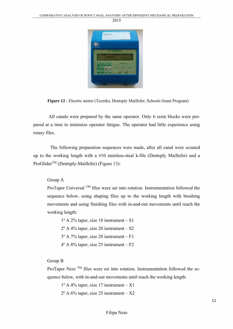
Each simulated canal was prepared to a working length of 16 millimeters, at a
speed of 300 rpm and a torque-control level of 40, as the suggested settings, using a
reduction hand-piece powered by an electric motor (Tecnika, Dentsply Maillefer,
Schools Grant Program) (Figure 12). The final apical preparation in Group A was set to
F2, in Group B set to X2 and in Group C was set to F2. Copious irrigation with water
was performed after the use of each file, using a disposable syringe and 27 gauge irriga-
tion needle.
9

COMPARATIVE ANALYSIS OF ROOT CANAL ANATOMY AFTER DIFFERENT MECHANICAL PREPARATION
2015
Filipa Neto
Figure 9 – Sterilized ProTaper UniversalTM Kit, Sx-F3, 25mm (Dentsply Maillefer)
Figure 10 – Sterilized ProTaper NextTM Kit, X1-X3, 25mm (Dentsply Maillefer)
Figure 11 – Sterilized ProTaper GoldTM Kit, Sx-F3, 25mm (Dentsply Tulsa Dental
Specialties 10
COMPARATIVE ANALYSIS OF ROOT CANAL ANATOMY AFTER DIFFERENT MECHANICAL PREPARATION
2015
Filipa Neto
Figure 12 - Electric motor (Tecnika, Dentsply Maillefer, Schools Grant Program)
All canals were prepared by the same operator. Only 6 resin blocks were pre-
pared at a time to minimize operator fatigue. The operator had little experience using
rotary files.
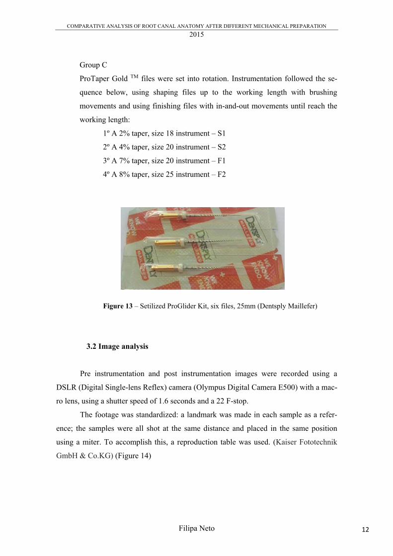
The following preparation sequences were made, after all canal were scouted
up to the working length with a #10 stainless-steal k-file (Dentsply Maillefer) and a
ProGliderTM (Dentsply-Maillefer) (Figure 13):
Group A
ProTaper Universal TM files were set into rotation. Instrumentation followed the
sequence below, using shaping files up to the working length with brushing
movements and using finishing files with in-and-out movements until reach the
working length:
1º A 2% taper, size 18 instrument – S1
2º A 4% taper, size 20 instrument – S2
3º A 7% taper, size 20 instrument – F1
4º A 8% taper, size 25 instrument – F2
Group B
ProTaper Next TM files were set into rotation. Instrumentation followed the se-
quence below, with in-and-out movements until reach the working length:
1º A 4% taper, size 17 instrument – X1
2º A 6% taper, size 25 instrument – X2
11
COMPARATIVE ANALYSIS OF ROOT CANAL ANATOMY AFTER DIFFERENT MECHANICAL PREPARATION
2015
Filipa Neto
Group C
ProTaper Gold TM files were set into rotation. Instrumentation followed the se-
quence below, using shaping files up to the working length with brushing
movements and using finishing files with in-and-out movements until reach the
working length:
1º A 2% taper, size 18 instrument – S1
2º A 4% taper, size 20 instrument – S2
3º A 7% taper, size 20 instrument – F1
4º A 8% taper, size 25 instrument – F2
Figure 13 – Setilized ProGlider Kit, six files, 25mm (Dentsply Maillefer)
3.2 Image analysis
Pre instrumentation and post instrumentation images were recorded using a
DSLR (Digital Single-lens Reflex) camera (Olympus Digital Camera E500) with a mac-
ro lens, using a shutter speed of 1.6 seconds and a 22 F-stop.
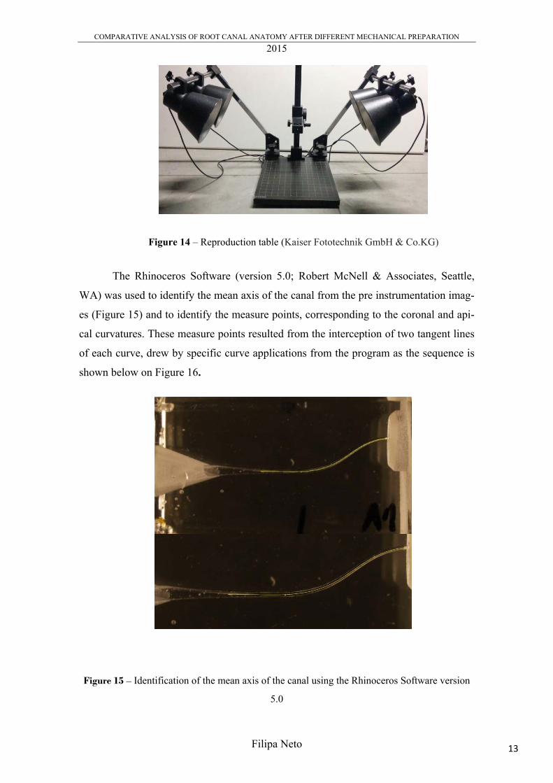
The footage was standardized: a landmark was made in each sample as a refer-
ence; the samples were all shot at the same distance and placed in the same position
using a miter. To accomplish this, a reproduction table was used. (Kaiser Fototechnik
GmbH & Co.KG) (Figure 14)
12
COMPARATIVE ANALYSIS OF ROOT CANAL ANATOMY AFTER DIFFERENT MECHANICAL PREPARATION
2015
Filipa Neto
Figure 14 – Reproduction table (Kaiser Fototechnik GmbH & Co.KG)
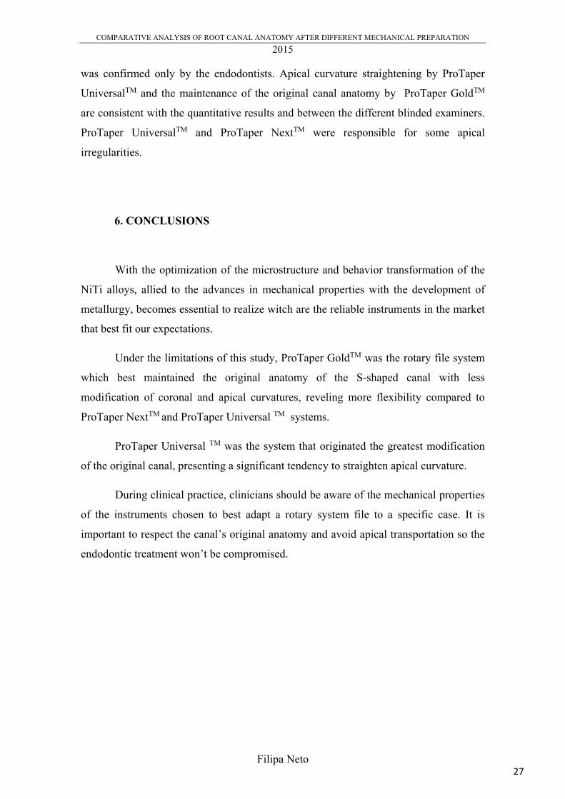
The Rhinoceros Software (version 5.0; Robert McNell & Associates, Seattle,
WA) was used to identify the mean axis of the canal from the pre instrumentation imag-
es (Figure 15) and to identify the measure points, corresponding to the coronal and api-
cal curvatures. These measure points resulted from the interception of two tangent lines
of each curve, drew by specific curve applications from the program as the sequence is
shown below on Figure 16.
Figure 15 – Identification of the mean axis of the canal using the Rhinoceros Software version
5.0
13

COMPARATIVE ANALYSIS OF ROOT CANAL ANATOMY AFTER DIFFERENT MECHANICAL PREPARATION
2015
Filipa Neto
Figure 16 – This six pictures show the sequence made in the Rhinoceros Software to define the
measure point of the coronal and apical curvature of the S-shape canal. Two tangent of each
curve were trace and intercepted: first coronal curvature tangent; second coronal curvature
tangent; interception of the two coronal curve tangent – measure point; first apical curvature
tangent; second apical curvature tangent; interception of the two apical curve tangent – measure
point.
The post instrumentation digital images were superimposed over the pre
instrumentation images. This was accomplished by reducing the opacity of the post
instrumentation images, done in a digital imaging software (Adobe Photoshop, version
CS6; Adobe Systems Inc, San Jose, Ca) (Figure 17)
14

COMPARATIVE ANALYSIS OF ROOT CANAL ANATOMY AFTER DIFFERENT MECHANICAL PREPARATION
2015
Filipa Neto
Figure 17 - Post instrumentation digital image superimposed over the pre instrumentation
image (Adobe Photoshop, version CS6; Adobe Systems Inc, San Jose, Ca)
The canal with was assessed by measuring the distance from the center of the
canal to the inner and outer margins of the prepared curve canal with specific dimension
displays of the program. The distance between the margin of the pre instrumentation
canal and de the margin of the prepared canal were also registered. Rhinoceros Software
allowed to get real measures. These paired images and the measures obtained give a
quantitative evaluation of the incidence of canal transportation after mechanical
preparation.
To proceed with qualitative analysis, a compilation of questions were made to
three different groups of blinded examiners:
Group 1: two endodontics specialists
Group 2: two inexpert clinicians
Group 3: two pre-graduated students.
The examiners evaluated three images, randomly chosen, from each group. The
images were set by a random sequence and the blinded examiners had to refer the
presence or absence of rectifications in the coronal and apical curvatures, as well as the
presence of significant apical transportation.
15

COMPARATIVE ANALYSIS OF ROOT CANAL ANATOMY AFTER DIFFERENT MECHANICAL PREPARATION
2015
Filipa Neto
3.3 Statistical analysis
The statistical analysis was obtained using the IBM ® SPSS ® Statistics version
22.0.0 software. Descriptive statistical analysis was performed to each group (A, B and
C). In each experimental group mean and standard deviation were calculated for the
inner and outer coronal and apical curvatures values.
The Kolmogorov-Smirnov test for was used to evaluate the data normality. The
null hypothesis were the following, set for inner and outer coronal and apical curvatures
values:
1. H0: The distribution of ICC is normal with mean 0,324 and standard
deviation 0,04
2. H0: The distribution of OCC is normal with mean 0,101 and standard
deviation 0,03
3. H0: The distribution of IAC is normal with mean 0,232 and standard
deviation 0,07
4. H0: The distribution of OAC is normal with mean 0,040 and standard
deviation 0,03
The data corresponding to the inner coronal curvature and to the outer apical
curvature didn’t follow a normal distribution, therefore, the first and last null hypothesis
were rejected. The Mann-Whitney post hoc multiple comparisons U test, a non-
parametrical test, was use to evaluate the differences among the inner coronal curve and
outer apical curve data while the One-Way ANOVA, a parametric test, evaluated the
differences among the other two groups. Differences were considered statistically
significant when p<0,05.
16

COMPARATIVE ANALYSIS OF ROOT CANAL ANATOMY AFTER DIFFERENT MECHANICAL PREPARATION
2015
Filipa Neto
4. RESULTS
4.1 Quantitative results
The results of the experimental procedure regarding the distance between the
margin of the pre instrumentation canal and de the margin of the prepared canal and the
distance from the mean axis of the canal to the inner and outer margins of the prepared
curve canal are displayed in the following tables. The experimental procedure was
repeated for the three groups: A (Table 1), B (Table 2) and C (Table 3).
Group A
Coronal curve Apical curve Inner (mm) Outer (mm) Inner (mm) Outer (mm)
A1 0,36 0,48 0, 09 0,21 0,34 0,44 0,01 0,11 A2 0,36 0,48 0,15 0,26 0,35 0,46 0,00 0,10 A3 0,40 0,51 0,08 0,20 0,31 0,40 0,06 0,14 A4 0,39 0,51 0,08 0,19 0,31 0,40 0,05 0,13 A5 0,36 0,48 0,06 0,21 0,24 0,35 0,00 0,09 A6 0,37 0,49 0,09 0,22 0,23 0,31 0,04 0,13 A7 0,38 0,49 0,10 0,22 0,32 0,41 0,05 0,15 A8 0,45 0,55 0,11 0,23 0,39 0,48 0,00 0,10 A9 0,35 0,46 0,12 0,24 0,29 0,39 0,00 0,11
A10 0,27 0,40 0,15 0,28 0,36 0,40 0,00 0,10 A11 0,35 O,47 0,14 0,27 0,32 0,42 0,00 0,11
A12 0,36 0,49 0,12 0,24 0,29 0,38 0,04 0,13
Table 1 - Group A – ProTaper UniversalTM. Measures obtained with the Rhinoceros
Software. Every left column of the inner and outer variables regard the distance between the
margin of the pre instrumentation canal and the margin of the prepared canal and very right
column regard the distance from the center of the canal to the inner and outer margins of the
prepared curve canal .
17

COMPARATIVE ANALYSIS OF ROOT CANAL ANATOMY AFTER DIFFERENT MECHANICAL PREPARATION
2015
Filipa Neto
Group B
Coronal curve Apical curve Inner (mm) Outer (mm) Inner (mm) Outer (mm)
B1 0,37 0,49 0,05 0,17 0,19 0,31 0,05 0,17 B2 0,32 0,43 0,05 0,20 0,14 0,23 0,08 0,17 B3 0,30 0,41 0,08 0,22 0,23 0,36 0,00 0,11 B4 0,31 0,41 0,11 0,23 0,21 0,32 0,05 0,16 B5 0,34 0,45 0,09 0,21 0,26 0,38 0,03 0,14 B6 0,32 0,45 0,10 0,22 0,24 0,32 0,01 0,16 B7 0,29 0,41 0,07 0,17 0,20 0,29 0,06 0,15 B8 0,28 0,40 0,10 0,22 0,22 0,34 0,06 0,16 B9 0,30 0,42 0,14 0,23 0,24 0,34 0,03 0,13
B10 0,32 0,44 0,07 0,21 0,16 0,26 0,08 0,19 B11 0,32 0,44 0,11 0,23 0,28 0,40 0,04 0,17 B12 0,33 0,48 0,09 0,21 0,23 0,32 0,07 0,18
Table 2 - Group B – ProTaper NextTM. Measures obtained with the Rhinoceros
Software. Every left column of the inner and outer variables regard the distance between the
margin of the pre instrumentation canal and de the margin of the prepared canal and very right
column regard the distance from the center of the canal to the inner and outer margins of the
prepared curve canal.
Group C
Coronal curve Apical curve Inner (mm) Outer (mm) Inner (mm) Outer (mm)
C1 0,32 0,43 0,12 0,22 0,19 0,28 0,04 0,13 C2 0,28 0,40 0,14 0,27 0,20 0,30 0,04 0,13 C3 0,28 0,42 0,11 0,23 0,14 0,24 0,04 0,15 C4 0,32 0,45 0,09 0,20 0,18 0,29 0,07 0,16 C5 0,31 0,41 0,12 0,22 0,17 0,25 0,08 0,17 C6 0,28 0,40 0,13 0,27 0,20 0,30 0,04 0,14 C7 0,30 0,41 0,12 0,23 0,17 0,27 0,06 0,17 C8 0,28 0,38 0,12 0,23 0,15 0,22 0,06 0,16 C9 0,26 0,36 0,09 0,20 0,13 0,22 0,04 0,16
C10 0,29 0,41 0,10 0,23 0,18 0,27 0,06 0,16 C11 0,29 0,38 0,12 0,23 0,15 0,25 0,04 0,18 C12 0,25 0,39 0,11 0,24 0,15 0,24 0,07 0,17
Table 3 - Group C – ProTaper GoldTM. Measures obtained with the Rhinoceros
Software. Every left column of the inner and outer variables regard the distance between the
margin of the pre instrumentation canal and de the margin of the prepared canal and very right
column regard the distance from the center of the canal to the inner and outer margins of the
prepared curve canal.
18

COMPARATIVE ANALYSIS OF ROOT CANAL ANATOMY AFTER DIFFERENT MECHANICAL PREPARATION
2015
Filipa Neto
Mean ratio A Mean ratio B Mean ratio C Total
Coronal Apical Coronal Apical Coronal Apical Coronal Apical
0,238333 0,52 0,215278 0,48 0,211389 0,4175 0,221667 0,4725
Table 4 – The distance from the mean axis of the canal to the inner and outer margins
of the prepared canal were summed, resulting in the total width of the post instrumentation
canal. This total width was divided by the correspondent diameter of the file, giving us a ratio.
This table presents the mean ratio for each group. Inside each group, by directly comparing
coronal and apical ratio, is possible to understand which curvature lost more material.
Descriptive statistics of the four variables was done. The mean width and
standard deviation for each experimental group are displayed in table 5.
Coronal Curvature Apical Curvature
Inner (mm) Outer (mm) Inner (mm) Outer (mm)
A – ProTaper Universal 0,37 ± 0,04 0,10 ± 0,04 0,31 ± 0,05 0,02 ± 0,03
B – ProTaper Next 0,32 ± 0,02 0,09 ± 0,03 0,22 ± 0,04 0,0 5 ± 0,03
C – ProTaper Gold 0,29 ± 0,02 0,11 ± 0,02 0,17 ± 0,02 0,0 5 ± 0,02
Table 5 – Canal width in the measure points of the coronal and apical curvatures.
Differences between the three files systems canal preparations on the outer side
of the coronal curvature are not statistically significant, however, in its inner side, these
differences are statistically significant, where the ProTaper UniversalTM system is
responsible for a bigger widening, while ProTaper GoldTM presents the smaller mean
value.
Concerning the inner side of the apical curvature, the differences between the
three files systems canal preparations is statistically significant, where the ProTaper
UniversalTM is, once more, responsible for a bigger widening, while ProTaper GoldTM
presents the smaller mean value. In the outer side of the apical curvature, differences in
the canal preparation between ProTaper UniversalTM, ProTaper NextTM and ProTaper
19

COMPARATIVE ANALYSIS OF ROOT CANAL ANATOMY AFTER DIFFERENT MECHANICAL PREPARATION
2015
Filipa Neto
0
1
2
3
4
5
6
7
8
9
10
Curvature maintenance Less SignificantStraightning
Significative straitning
Students
Inexpert clinicians
Endondotists
GoldTM are statistically significant, however, differences between ProTaper NextTM and
ProTaper GoldTM are not statistically significant.
The ProTaper UniversalTM system caused significantly greater widening of
canals than the other two groups, especially at the inner sides of both curved regions,
tending toward the straightening of the canal. The ProTaper GoldTM showed the lowest
widening on both regions.
Additionally, every rotary file system removed more resin wall in the apical
curve compared to the coronal curve.
4.2 Qualitative analysis
Considering each blinded examiners evaluation, the following graphics shows
what was their evaluation concerning the presence or absence of rectifications in the
coronal and apical curvatures, as well as the presence of significant apical
transportation, for each system file.
Graphic 1 – Evaluation of the coronal curvature prepared by ProTaper UniversalTM
20

COMPARATIVE ANALYSIS OF ROOT CANAL ANATOMY AFTER DIFFERENT MECHANICAL PREPARATION
2015
Filipa Neto
0
1
2
3
4
5
6
7
8
9
10
Curvature maintenance Less SignificantStraightning
Significative straitning
Students
Inexpert clinicians
Endodontics
0
2
4
6
8
10
12
14
16
Curvature maintenance Less SignificantStraightning
Significative straitning
Students
Inexpert clinicians
Endodontists
Graphic 2 – Evaluation of the coronal curvature prepared by ProTaper NextTM
Graphic 3 – Evaluation of the coronal curvature prepared by ProTaper GoldTM
21

COMPARATIVE ANALYSIS OF ROOT CANAL ANATOMY AFTER DIFFERENT MECHANICAL PREPARATION
2015
Filipa Neto
0
2
4
6
8
10
12
14
Curvature maitenance Less SignificantStraightning
Significative straitning
Students
Inexpert clinicians
Endodontists
Graphic 4 – Evaluation of the apical curvature prepared by ProTaper UniversalTM
Graphic 5 – Evaluation of the apical curvature prepared by ProTaper NextTM
0
2
4
6
8
10
12
14
Curvature maintenance Less SignificantStraightning
Significative straitning
Students
Inexpert clinicians
Endodontists
22

COMPARATIVE ANALYSIS OF ROOT CANAL ANATOMY AFTER DIFFERENT MECHANICAL PREPARATION
2015
Filipa Neto
0
2
4
6
8
10
12
14
16
18
Curvature maintenance Less SignificantStraightning
Significative straitning
Students
Inexpert clinicians
Endodontists
0
2
4
6
8
10
12
No apical transport Low significancy apicaltransport
High significancy apicaltransport
Students
Inexpert clinicians
Endodontists
Graphic 6 – Evaluation of the apical curvature prepared by ProTaper GoldTM
Graphic 7 – Evaluation of the apical transport, prepared by ProTaper UniversalTM
23

COMPARATIVE ANALYSIS OF ROOT CANAL ANATOMY AFTER DIFFERENT MECHANICAL PREPARATION
2015
Filipa Neto
0
2
4
6
8
10
12
14
16
No apical transport Low significancy apicaltransport
High significancy apicaltransport
Students
Inexpert clinicians
Endodontists
0
2
4
6
8
10
12
No apical transport Low significancy apicaltransport
High significancy apicaltransport
Students
Inexpert clinicians
Endodontists
Graphic 8 – Evaluation of the apical transport, prepared by ProTaper NextTM
Graphic 9 – Evaluation of the apical transport, prepared by ProTaper GoldTM
24

COMPARATIVE ANALYSIS OF ROOT CANAL ANATOMY AFTER DIFFERENT MECHANICAL PREPARATION
2015
Filipa Neto
The clinician’s expertise (endodontist versus inexpert clinicians versus students)
did not appear to have a significant impact in the following parameters:
1. Maintenance of the original shape of the canal by ProTaper GoldTM
2. Maintenance of the original shape of the coronal curvature by ProTaper
NextTM and ProTaper GoldTM
3. Straitening of the apical curvature by ProTaper UniversalTM
4. Apical transportation by ProTaper NextTM and ProTaper UniversalTM
A significant difference is present when considering the apical transportation by
ProTaper GoldTM, where just endodontist evaluate as it is maintained.
5. DISCUSSION
Analysis of modifications in canal curvature after instrumentation has been
widely used to evaluate the tendency of a technique, or of the mechanical properties of
an instrument, to maintain the original canal anatomy or to straighten the curves (Berutti
et al. 2009). The disrespect of the original anatomy can lead the clinician to miss
preparation objectives: remove remaining pulp tissue, eliminate microorganisms,
remove debris and shape the root canal(s) so that the root canal system can be cleaned
and filled. . (European Society of Endodontology 2006)
To compare the canal anatomy after different mechanical preparations and to
evaluate the maintenance of its original shape, simulated canals were used to
standardize experimental conditions, but always regarding the fact that this method only
gives 2D dimensions. Despite the fact that resin blocks may not always reflect the
action of the instruments in root canals of real teeth because of the many different
configurations, the S-shape canal used, has been reported to be of use in pointing up
differences in performance of instruments, possibly as a result of the increased difficulty
of instrumentation. (Berutti et al. 2009; Yoshimine et al. 2005).
The first stage of the study comprised a quantitative analysis through observa-
tion of changes between pre instrumentation and post instrumentation curvature fol-
25

COMPARATIVE ANALYSIS OF ROOT CANAL ANATOMY AFTER DIFFERENT MECHANICAL PREPARATION
2015
Filipa Neto
lowed by a qualitative observation of any canal aberrations concerning the presence of
straightening curves and apical transportation. The experimental method used appeared
to be reliable in representing changes in canal curvature and for extrapolating the re-
sults, however, this analysis may not be completely accurate taking in consideration:
1. The uncertainty degree of the Rhinoceros Software, considering 0,006;
2. Data dependent on operator’s accuracy on prosecuting the experimental pro-
cedure:
2.1 Maintenance of the exact working length;
2.2 Stabilization of the resin block during the mechanical preparation
ProTaper GoldTM produced significantly less modification in coronal and apical
canal curvature compared ProTaper UniversalTM. When comparing ProTaper NextTM
and ProTaper UniversalTM, the results of the first system show less modification in
coronal and apical canal curvature compared to the second one. This results are
consistent with Shori et al. 2015 conclusions, which says that ProTaper NextTM can
induce less dentinal defects than ProTaper UniversalTM. Therefore, under the study
conditions, it might be assumed that ProTaper GoldTM is the rotary system that has more
respect for original canal anatomy. Higher flexibility might be the predominant property
responsible by the system’s facility to maintain the canal’s original anatomy. Despite
the identical architecture and operation of the ProTaper GoldTM and ProTaper
UniversalTM systems, the different manufacturing processes of the instruments clearly
affect their flexibility, stress-strain distribution patterns and fatigue resistance behavior.
(Hieawy et al. 2015).
Every system file, proportionally, removes more resin in the apical curve than in
the coronal.
No macroscopic deformations or fractures of any instrument, mechanical or
manual, occurred during the experiment.
The second stage of the study comprised a qualitative analysis where
endodontists, inexpert clinicians and students evaluated coronal apical curvatures
rectification and apical transport. The differences registered are due to clinical
experience and different levels of endodontic knowledge. The more significant result
was the evaluation of apical transportation by ProTaper GoldTM, where its maintenance
24 26
COMPARATIVE ANALYSIS OF ROOT CANAL ANATOMY AFTER DIFFERENT MECHANICAL PREPARATION
2015
Filipa Neto
was confirmed only by the endodontists. Apical curvature straightening by ProTaper
UniversalTM and the maintenance of the original canal anatomy by ProTaper GoldTM
are consistent with the quantitative results and between the different blinded examiners.
ProTaper UniversalTM and ProTaper NextTM were responsible for some apical
irregularities.
6. CONCLUSIONS
With the optimization of the microstructure and behavior transformation of the
NiTi alloys, allied to the advances in mechanical properties with the development of
metallurgy, becomes essential to realize witch are the reliable instruments in the market
that best fit our expectations.
Under the limitations of this study, ProTaper GoldTM was the rotary file system
which best maintained the original anatomy of the S-shaped canal with less
modification of coronal and apical curvatures, reveling more flexibility compared to
ProTaper NextTM and ProTaper Universal TM systems.
ProTaper Universal TM was the system that originated the greatest modification
of the original canal, presenting a significant tendency to straighten apical curvature.
During clinical practice, clinicians should be aware of the mechanical properties
of the instruments chosen to best adapt a rotary system file to a specific case. It is
important to respect the canal’s original anatomy and avoid apical transportation so the
endodontic treatment won’t be compromised.
27

REFERENCES
Arias A. et al. 2015. Differences in torsional performance of single- and multiple instrument rotary systems for glide path preparation. Odontology DOI10.1007/s10266-015-0199-0. Available at: http://www.ncbi.nlm.nih.gov/pubmed/?term=Odontology+DOI+10.1007 %2Fs10266-015-0199-0
Berutti E. et al. 2009. Use of Nickel-Titanium Rotary PathFile to Create the Glide
Path: Comparison With Manual Preflaring in Simulated Root Canals. J En-dod. 2009 Mar;35(3):408-12. doi: 10.1016/j.joen.2008.11.021. Available at: http://www.ncbi.nlm.nih.gov/pubmed/?term=Berutti+E.+et+al.+2009
Blum J-Y et al. 2003. Analysis of Mechanical Preparations in Extracted Teeth Using ProTaper Rotary Instruments: Value of the Safety Quotient. OURNAL OF EN-DODONTICS VOL. 29, NO. 9, SEPTEMBER 2003. Available at: http://www.ncbi.nlm.nih.gov/pubmed/?term=Analysis+of+Mechanical+Prepara-tions+in+Extracted+Teeth+Using+ProTaper+Rotary+Instru-ments%3A+Value+of+the+Safety+Quotient
Brochure or ProTaper Gold. Available at:
http://www.tulsadentalspecialties.com/Libraries/Tab_Content_-_Endo_Ac-cess_Shaping/Brochure_for_ProTaper_Gold.sflb.ashx
Ding-Ming et al. 2007. Study of the Progressive Changes in Canal Shape After
Using Different Instruments by Hand in Simulated S Shaped Canals. J En-dod. 2007 Aug;33(8):986-9. Available at: http://www.ncbi.nlm.nih.gov/pub-med/?term=J+Endod.+2007+Aug%3B33(8)%3A986-9
European Society of Endodontology, 2006. Quality guidelines for endodontic treat
ment: consensus report of the European Society of Endodontology. International Endodontic Journal, 39(12), pp.921–930. Available at: http://doi.wiley.com/10.1111/j.1365-2591.2006.01180.x [Accessed February 28, 2014].
Esposito P. et al. 2005. A Comparison of Canal Preparation with Nickel- Titanium and
Stainless Steel Instruments. J Endod. 1995 Apr;21(4):173-6. Vailable at: http://www.ncbi.nlm.nih.gov/pubmed/7673815
Hartmann M. et al. 2007 Canal Transportation after Root Canal Instrumentation: A
Comparative Study with Computed Tomography. J Endod. 2007 Aug;33(8):962-5. Epub 2007 May 3. Available at: http://www.ncbi.nlm.nih.gov/pub-med/17878083
xv

xvi

Hieawy A. et al. 2015. Phase Transformation Behavior and Resistance to Bending and Cyclic Fatigue of ProTaper Gold and ProTaper Universal Instruments. J En-dod. 2015 Jul;41(7):1134-8. Avaiable at: http://www.ncbi.nlm.nih.gov/pub-med/?term=J+Endod.+2015+Jul%3B41(7)%3A1134-8.
Jardine S. et al. 1999 An in vitro comparison of canal preparation using two automated rotary nickel–titanium instrumentation techniques. Int Endod J. 2000 Jul;33(4):381-91. Available at: http://www.ncbi.nlm.nih.gov/pub-med/?term=Int+Endod+J.+2000+Jul%3B33(4)%3A381-91.
M-Wire Brochure Merret S. et al. 2006 Comparison of the Shaping Ability of RaCe and FlexMaster Ro
tary Nickel-Titanium Systems in Simulated Canals. J Endod. 2006 Oct;32(10):960-2.. Available at: http://www.ncbi.nlm.nih.gov/pub-med/?term=J+Endod.+2006+Oct%3B32(10)%3A960-2.
Peters O. et al. 2001. ProTaper rotary root canal preparation: effects of canal anatomy on final shape analysed by micro CT. Int Endod J. 2003 Feb;36(2):86-92.. Vail able at: http://www.ncbi.nlm.nih.gov/pubmed/?term=Int+En-dod+J.+2003+Feb%3B36(2 )%3A86-92.
Peters O. et al. 2003. ProTaper rotary root canal preparation: assessment of torque and force in relation to canal anatomy. Int Endod J. 2003 Feb;36(2):93-9. Vailable at: http://www.ncbi.nlm.nih.gov/pubmed/?term=Int+En-dod+J.+2003+Feb%3B36(2)%3A93-9.
Scäfer E. 1996 Effects of Four Instrumentation Techniques on Curved Canals: A Com parison Study. J Endod. 1996 Dec;22(12):685-9. Vailable at: http://www.ncbi.nlm.nih.gov/pubmed/?term=J+En-dod.+1996+Dec%3B22(12)%3A685-9
Schäfer E. 2001 Shaping ability of Hero 642 rotary nickel-titanium instruments and stainless steel hand K-Flexofiles in simulated curved root canals. Oral Surg Oral al Radiol Endod. 2001 Aug;92(2):215-20. Vailable at: http://www.ncbi.nlm.nih.gov/pub-med/?term=Oral+Surg+Oral+Med+Oral+Pathol+Oral+Radiol+En-dod.+2001+Aug%3B92(2)%3A215-20.
xvii

xviii

Shori et al. 2015. Stereomicroscopic evaluation of dentinal defects induced by new
rotary system: “ProTaper NEXT”. J Conserv Dent. 2015 May-Jun; 18(3): 210–
213. Available at:
http://www.ncbi.nlm.nih.gov/pubmed/?term=J+Conserv+Dent.+2015+May-
Jun%3B+18(3)%3A+210%E2%80%93213.http://www.ncbi.nlm.nih.gov/pubme
d/?term=J+Conserv+Dent.+2015+May-
Jun%3B+18(3)%3A+210%E2%80%93213.
Thompson S. et al. 2000 An overview of nickel–titanium alloys used in
dentistry. Int Endod J. 2000 Jul;33(4):297-310. Available at: http://www.ncbi.nlm.nih.gov/pubmed/11307203
Uygun et.al 2015 Variations in cyclic fatigue resistance among ProTaper Gold, ProTa per Next and ProTaper Universal instruments at different levels. Int Endod J. ble at: http://www.ncbi.nlm.nih.gov/pubmed/?term=Variations+in+cyclic+fa-tigue+resistance+among+ProTaper+Gold%2C+ProTaper+Next+and+ProTa-per+Universal+instruments+at+different+levels
Vaudt J. et al. 2008 Ex vivo study on root canal instrumentation of two rotary nickel–
titanium systems in comparison to stainless steel hand instruments. Int Endod J. 2009 Jan;42(1):22-33. Vailable at: m.nih.gov/pubmed/?term=Int+En-dod+J.+2009+Jan%3B42(1)%3A22-33.
Wu H. et al. 2015 Shaping ability of ProTaper Universal, WaveOne and ProTaper
next in simulated L-shaped and S-shaped root canals. BMC Oral Health. 2015 Mar 1;15:27. Vailable at: http://www.ncbi.nlm.nih.gov/pubmed/?term=Shaping+ability+of+ProTaper+Universal%2C+WaveOne+and+ProTaper+Next+in+simulated+L-shaped+and+S-shaped+root+canals
Ye J. et al. 2012 Metallurgical Characterization of M-Wire Nickel-Titanium Shape
Memory Alloy Used for Endodontic Rotary Instruments during Low-cycle Faigue. J Endod. 2012 Jan;38(1):105-7. Vailable at: http://www.ncbi.nlm.nih.gov/pubmed/?term=J+En-dod.+2012+Jan%3B38(1)%3A105-7.
Yoshimine Y. et al. 2005 The Shaping Effects of Three Nickel-Titanium Rotary Instruments in Simulated S-Shaped Canals. J Endod. 2005 May;31(5):373-5. Vailable at: http://www.ncbi.nlm.nih.gov/pubmed/?term=J+En-dod.+2005+May%3B31(5)%3A373-5
xix

xx

APPENDIX Abbreviations 2D – two dimensional
NiTi – Nickel-Titanium
ICC – inner coronal curvature
OCC – outer coronal curvature
IAC – inner apical curvature
OAC – outer apical curvature
Symbols
% - percentage
p - significance
® - registered trademark
TM - unregistered trademark
Units
mm - millimeters
N cm - Newton centimeter
rpm - rotations per minute
xxi

xxii
FIGURE INDEX Figure 1 – ProGliderTM file, a single file glide path instrument made of M-Wire alloy
(Dentsply Maillefer, page 5
Figure 2 – ProTaper UniversalTM system composed by the shaping and finishing files
(Dentisply Maillefer) , page 5
Figure 3 and Figure 4 – Sx, S1, S2, F1 and F2 convex triangular cross section and F3,
F4 and F5 concave triangular cross section, respectively, page 6
Figure 5 – ProTaper NextTM system composed by X1, X2, X3, X4 and X5. (Dentsply
Maillefer) 7
Figure 6 – ProTaper NextTM rectangular cross section (Dentsply Maillefer) , page 7
Figure 7 - ProTaper GoldTM system composed by the shaping and finishing files
(Dentisply Tulsa Dental Specialties) , page 8
Figure 8 - S-shaped curvature in clear resin blocks (ISO 15, Endo-Training-Bloc-S .02
Taper; Dentsply Maillefer, Ballaigues, Switzerlan) , page 9
Figure 9 – Sterilized ProTaper UniversalTM Kit, Sx-F3, 25mm (Dentsply Maillefer) ,
page 10
Figure 10 – Sterilized ProTaper NextTM Kit, X1-X3, 25mm (Dentsply Maillefer) , page
10
Figure 11 – Sterilized ProTaper GoldTM Kit, Sx-F3, 25mm (Dentsply Tulsa Dental
Specialties, page 10
Figure 12 - Electric motor (Tecnika, Dentsply Maillefer, Schools Grant Program) , page
11
Figure 13 – Setilized ProGlider Kit, six files, 25mm (Dentsply Maillefer) , page 12
Figure 14 – Reproduction table (Kaiser Fototechnik GmbH & Co.KG) , page 13
Figure 15 – Identification of the mean axis of the canal using the Rhinoceros Software
version 5.0, page 13
Figure 16 – This six pictures show the sequence made in the Rhinoceros Software to
define the measures point , page 14
Figure 17 - Post instrumentation digital image superimposed over the pre
instrumentation image (Adobe Photoshop, version CS6; Adobe Systems Inc, San Jose,
Ca) , page 15
xxi xxiii

xxiv

TABLES INDEX
Table 1 - Group A – ProTaper UniversalTM. Measures obtained with the Rhinoceros Software,
page 17
Table 2 - Group B – ProTaper NextTM. Measures obtained with the Rhinoceros Software, page
18
Table 3 - Group C – ProTaper GoldTM. Measures obtained with the Rhinoceros Software, page
18
Table 4 - This table presents the mean ratio for each group, page 19
Table 5 – Canal width in the measure points of the coronal and apical curvatures, page 19
xxv

xxvi

CHART INDEX Graphic 1 – Evaluation of the coronal curvature prepared by ProTaper UniversalTM, page 20
Graphic 2 – Evaluation of the coronal curvature prepared by ProTaper NextTM , page 21
Graphic 4 – Evaluation of the apical curvature prepared by ProTaper UniversalTM , page 22
Graphic 5 – Evaluation of the apical curvature prepared by ProTaper NextTM , page 22
Graphic 6 – Evaluation of the apical curvature prepared by ProTaper GoldTM , page 23
Graphic 7 – Evaluation of the apical transport, prepared by ProTaper UniversalTM, page 23
Graphic 8 – Evaluation of the apical transport, prepared by ProTaper NextTM, page 24
Graphic 9 – Evaluation of the apical transport, prepared by ProTaper GoldTM , page 24
xxvii