Understanding Posterior Reversible Encephalopathy Syndrome
24
Understanding Posterior Reversible Encephalopathy Syndrome S. Legriel, F. Pico, and E. Azoulay J.-L. Vincent (ed.), Annual Update in Intensive Care and Emergency Medicine 2011 DOI 10.1007/978-3-642-18081-1, Springer Science+Business Media LLC 2011 Introduction Posterior reversible encephalopathy syndrome (PRES) [1, 2] is a clinicoradiological entity that was well described by Hinchey et al. [3] in 1996 based on 15 cases, shortly after two other small case-series were published [4, 5]. This condition has been designated by a variety of names (reversible posterior leukoencephalopathy syndrome, reversible posterior cerebral edema syndrome, and reversible occipital parietal encephalopathy). PRES is now the accepted term [1, 2, 6] but has been challenged recently based on the risk of neurological impairment and up to 15 % mortality rate [7, 8]. PRES is characterized by variable associations of seizure activ- ity, consciousness impairment, headaches, visual abnormalities, nausea/vomiting, and focal neurological signs. The cerebral imaging abnormalities are often sym- metric and predominate in the posterior white matter (Fig. 1). Recognition of PRES has evolved with increasing availability of magnetic resonance imaging (MRI). PRES can develop in association with a vast array of conditions. However, regardless of the underlying cause, the main abnormality is cerebral vasogenic edema, the pathogenesis of which is still under debate [1, 2]. PRES is typically reversible once the cause is removed. However, patients with severe manifesta- tions of PRES, such as coma and/or status epilepticus, may require admission to the intensive care unit (ICU) [9, 10]. Moreover, permanent neurological impair- ment or death occurs in a minority of patients [5, 7, 8]. The objective of this chapter is to provide clinicians with guidance for diag- nosing and treating patients with PRES. The diagnostic criteria are described in detail and management recommendations are given with an algorithm. Epidemiology The global incidence of PRES is unknown. The only epidemiological data come from retrospective studies of patients seen between 1988 and 2008 [3, 6–8, 10–13]. PRES has been reported in patients aged 4 to 90 years, although most cases occur in young to middle-aged adults, the mean age ranging across case- series from 39 to 47 years. There is a marked female predominance that may reflect some of the causes. Many patients with PRES have comorbidities, which may be severe conditions, such as bone marrow or solid organ transplantation, chronic renal failure, and chronic hypertension. Mechanical ventilation is required in 35 % to 40 % of patients with PRES, for 3 to 7 days [7, 8]. Status epilepticus may require ICU admission [10]. No epidemi- 631 XV
Transcript of Understanding Posterior Reversible Encephalopathy Syndrome

Understanding Posterior ReversibleEncephalopathy Syndrome
S. Legriel, F. Pico, and E. Azoulay
J.-L. Vincent (ed.), Annual Update in Intensive Care and Emergency Medicine 2011DOI 10.1007/978-3-642-18081-1, ˇ Springer Science+Business Media LLC 2011
Introduction
Posterior reversible encephalopathy syndrome (PRES) [1, 2] is a clinicoradiologicalentity that was well described by Hinchey et al. [3] in 1996 based on 15 cases,shortly after two other small case-series were published [4, 5]. This condition hasbeen designated by a variety of names (reversible posterior leukoencephalopathysyndrome, reversible posterior cerebral edema syndrome, and reversible occipitalparietal encephalopathy). PRES is now the accepted term [1, 2, 6] but has beenchallenged recently based on the risk of neurological impairment and up to 15 %mortality rate [7, 8]. PRES is characterized by variable associations of seizure activ-ity, consciousness impairment, headaches, visual abnormalities, nausea/vomiting,and focal neurological signs. The cerebral imaging abnormalities are often sym-metric and predominate in the posterior white matter (Fig. 1). Recognition of PREShas evolved with increasing availability of magnetic resonance imaging (MRI).
PRES can develop in association with a vast array of conditions. However,regardless of the underlying cause, the main abnormality is cerebral vasogenicedema, the pathogenesis of which is still under debate [1, 2]. PRES is typicallyreversible once the cause is removed. However, patients with severe manifesta-tions of PRES, such as coma and/or status epilepticus, may require admission tothe intensive care unit (ICU) [9, 10]. Moreover, permanent neurological impair-ment or death occurs in a minority of patients [5, 7, 8].
The objective of this chapter is to provide clinicians with guidance for diag-nosing and treating patients with PRES. The diagnostic criteria are described indetail and management recommendations are given with an algorithm.
Epidemiology
The global incidence of PRES is unknown. The only epidemiological data comefrom retrospective studies of patients seen between 1988 and 2008 [3, 6–8,10– 13]. PRES has been reported in patients aged 4 to 90 years, although mostcases occur in young to middle-aged adults, the mean age ranging across case-series from 39 to 47 years. There is a marked female predominance that mayreflect some of the causes. Many patients with PRES have comorbidities, whichmay be severe conditions, such as bone marrow or solid organ transplantation,chronic renal failure, and chronic hypertension.
Mechanical ventilation is required in 35 % to 40 % of patients with PRES, for3 to 7 days [7, 8]. Status epilepticus may require ICU admission [10]. No epidemi-
631
XV

Fig. 1. Cerebral magnetic resonance imaging in a patient with posterior reversible encephalopathy syn-drome (PRES). Fluid-attenuated inversion recovery (FLAIR) sequence showing bilateral high-signal fociin the cerebellum, basal ganglia, and occipital, parietal, frontal, and temporal lobes
ological data are available on the subgroup of patients with PRES requiring ICUadmission. Mean hospital length of stay was 20 days [7, 8].
632 S. Legriel, F. Pico, and E. Azoulay
XV

Diagnosis
PRES is a clinicoradiological entity. The intensity and severity of clinical manifesta-tions vary and may require ICU admission. Imaging findings also vary in severity.Thorough familiarity with the imaging criteria is crucial to the diagnosis. The com-bination of suggestive clinical manifestations and radiological criteria establishesthe diagnosis of PRES. In doubtful cases, the clinical and radiological improvementthat occurs once appropriate treatment is given confirms the diagnosis. Neverthe-less, there are no consensual guidelines to validate diagnosis of PRES.
Clinical Manifestations of PRES
Typical presenting manifestationsThe typical features of PRES consist of consciousness impairment, seizure activ-ity, headaches, visual abnormalities, nausea/vomiting, and focal neurologicalsigns (Table 1). Consciousness impairment may range in severity from confusion,somnolence, and lethargy to encephalopathy or coma. Consciousness impairmenthas been reported in 13 % [14] to 90 % [8] of cases. Seizure activity occurs in upto 92 % of cases [7]. The seizures are rarely isolated (23 %-28 %) [7, 8]. Secondarygeneralized seizures are common (53–62 %) [3, 8]. Status epilepticus, defined as
Table 1. Topographic distribution of clinical and radiological features in cohort studies of posteriorreversible encephalopathy syndrome (PRES)
Hinchey1996 [3]n = 15
Casey2000 [6]n = 16
Bartynski2007 [11]n = 136
McKinney2007[14]n = 76
Lee2008 [7]n = 36
Burnett2010 [8]n = 79
Clinical Features of PRESConsciousnessimpairment
10 (67 %) NR 39 (26 %) 10 (13 %) 34 (94 %) 76 (90 %)
Seizure activity 11 (73 %) NR 97 (71 %) 58 (76 %) 33 (92 %) 56 (70 %)Headaches 8 (53 %) NR 39 (26 %) 3 (4 %) 19 (53 %) 26 (31 %)Visual abnormalities 10 (67 %) NR 39 (26 %) 3 (4 %) 13 (36 %) 24 (29 %)Nausea/vomiting 8 (53 %) NR 39 (26 %) NR NR NRFocal neurological signs NR NR NR 2 (3 %) 1 (3 %) 14 (17 %)Acute hypertension 12 (80 %) NR 91 (67 %) NR NR 62 (78 %)
Radiological Features of PRESBilateral 15 (100 %) 11 (69 %) > 98 (> 72 %) NR 36
(100 %)NR
Asymmetric 10 (67 %) NR 21 (15 %) 2 (3 %) NR NRConfluent NR 2 (13 %) 31 (23 %) 44 (58 %) 2 (13 %) 12 (16 %)Gray matter 4 (27 %) NR NR 22 (29 %) 16 (44 %) NRPosterior > anterior 14 (93 %) 15 (94 %) 30 (22 %) NR NR NROccipital 14 (93 %) NR 134 (99 %) 75 (99 %) NR NRParietal 13 (87 %) 8 (50 %) 134 (99 %) 75 (99 %) NR 50 (67 %)Frontal 7 (47 %) 14 (88 %) 93 (68 %) 60 (89 %) 22 (61 %) 61 (81 %)Temporal 9 (60 %) 16 (100 %) 55 (40 %) 52 (68 %) NR 62 (83 %)Brainstem 2 (13 %) NR 17 (13 %) 14 (18 %) 21 (58 %) NRCerebellum 1 (7 %) NR 41 (30 %) 26 (34 %) 21 (58 %) NRBasal ganglia 1 (7 %) 3 (19 %) 19 (14 %) 9 (12 %) NR NR
NR: None reported
Understanding Posterior Reversible Encephalopathy Syndrome 633
XV

ongoing continuous seizure activity for at least 5 minutes (continuous) or as morethan two motor seizures without full recovery of consciousness in the interval(intermittent), has been described in 3 % [7] to 13 % [10] of patients. Visualabnormalities were found in 26 % [11] to 67 % [3] of patients and consisted ofblurred vision (7 % [3]-18 % [8]), visual neglect (4 % [8]-27 % [3]), homonymoushemianopsia (4 % [8]-20 % [3]), visual hallucinations (3 % [7]-5 % [8]), and cor-tical blindness (8 % [8]-33 % [3]). Headaches and nausea/vomiting were reportedin 26 % [11] to 53 % [3] of patients. Focal neurological signs were either not men-tioned at all or reported in only 3 % [7, 14] to 17 % [8] of cases.
A frequently associated sign: Acute hypertensionAcute hypertension is not usually described among the main signs of PRES. How-ever, hypertension has been reported in most studies [3–5, 7, 8, 11, 12], in 67 %[11] to 80 % [3] of patients. In one study, mean systolic blood pressure reached187 (80–240) mm Hg [7]. It is worth noting that mean blood pressure defined as[systolic blood pressure +2(diastolic blood pressure)/3] reflects cerebral autoreg-ulation of blood flow. Acute hypertensive emergency was not significantly associ-ated with the intensity of the clinical or radiological manifestations of PRES [11].Therefore, high mean blood pressure is often observed in PRES but its level is notcorrelated to the severity of PRES.
Radiological Characteristics of PRES
The topographic distribution of radiological features reported in cohort studies isgiven in Table 1 [3, 6–8, 11, 14].
The four radiological patterns of PRES [11]Until recently, PRES was believed to consistently produce bilateral and symmetricregions of edema typically located in the white matter and predominating in theposterior parietal and occipital lobes. Occasionally, edema has been described inthe frontal lobes, temporal, basal ganglia or cerebellum and brainstem in the pos-terior fossa, and cortical gray matter.
In a study of 136 patients, however, this pattern was found in only 26 % ofcases [11]: Three radiological patterns were found in 99 patients, and incompleteforms of these three patterns in the remaining 37 patients (Fig. 2):
a. Holohemispheric watershed pattern (23 %) (Fig. 2a)A swath of confluent vasogenic edema extends through the frontal, parietal, andoccipital lobes. Involvement of the temporal lobes is less marked. This topogra-phy matches the watershed zone between the anterior and posterior cerebralarteries, on the one hand, and the middle cerebral artery, on the other.
b. Superior frontal sulcus pattern (27 %) (Figure 2b)Patchy edema predominates in the frontal lobes along the superior frontal sulci.The parietal and occipital lobes are variably involved.
c. Dominant parietal-occipital pattern (22 %) (Fig. 2c)In this pattern previously thought to be typical of PRES, the posterior part of theparietal and occipital lobes is predominantly involved. The edema varies in sever-ity from mild to extensive.
634 S. Legriel, F. Pico, and E. Azoulay
XV

Fig. 2. Four main magnetic resonance imaging patterns of posterior reversible encephalopathy syn-drome (PRES).2a. Holohemispheric watershed pattern. Fluid-attenuated inversion recovery (FLAIR) sequences show-ing bilateral vasogenic edema in a linear pattern involving the white matter of the cerebellum, brainstem,and occipital, parietal, frontal, and temporal lobes. This pattern was found in 23 % of patients.2b. Superior frontal sulcus pattern. Fluid-attenuated inversion recovery (FLAIR) sequences showingbilateral vasogenic edema in a non-confluent pattern involving the frontal sulcus area and, to a lesserdegree, the white matter of the parietal, occipital, and temporal lobes. This pattern was found in 27 %of patients.2c. Dominant parietal-occipital pattern. Fluid-attenuated inversion recovery (FLAIR) sequences show-ing bilateral vasogenic edema in the white matter of the occipital and parietal lobes. This so-called ‘clas-sic’ pattern was found in 22 % of patients.2d. Partial expression of the three primary patterns. Fluid-attenuated inversion recovery (FLAIR)sequences showing bilateral vasogenic edema in the white matter of the parietal and frontal lobes butnot in the occipital lobes. This pattern can also be asymmetric. Partial and/or asymmetric forms of thethree primary patterns were found in 28 % of patients.
Understanding Posterior Reversible Encephalopathy Syndrome 635
XV

d. Partial or asymmetric expression of the primary patterns (28 %) (Fig. 2d)The partial form is defined as bilateral absence of edema in either the parietal orthe occipital lobes. The frontal lobes are often involved. The asymmetric form ischaracterized by unilateral absence of edema in either a parietal or an occipitallobe. Finally, in the partial and asymmetric form, there is both absence ofinvolvement of either the parietal or the occipital lobes and asymmetric abnor-malities in the affected parietal or occipital lobes.
Roles for computed tomography and magnetic resonance imaging in thediagnosis of PRES
) Computed tomography (CT): In retrospective reviews, CT scans were avail-able for review in 65 % [11] to 100 % [7] of cases. CT findings are often nor-mal or nonspecific [11]. Hypodensities in a suggestive topographic distribu-tion suggest PRES (Fig. 3).) MRI: Cerebral MRI is the key investigation for the diagnosis of PRES.
Proton-density and T2-weighted images show regions of high signal indicat-ing edema. Fluid-attenuated inversion recovery (FLAIR) sequences also visu-alize the lesions. The use of FLAIR has been shown to improve the diagnosisof PRES and the detection of subcortical and cortical lesions in PRES [6].T1-weighted images show low-intensity foci. Diffusion-weighted imaging(DWI) is normal but the apparent diffusion coefficient is increased [13].Finally, enhancement is seen in about half the cases [11].
MRI is superior to CT for the diagnosis of PRES. Of 67 patients who had both CTand MRI at least 2 days after the clinical onset of PRES [11], 22 had both investi-gations on the same day and of these, only 7 (32 %) had contributive CT findings.Interestingly, the proportion of patients with contributive CT findings was 74 %on day 2, suggesting that repeated CT scanning may be helpful when MRI isunavailable. MRI has been performed in 84 % [11] to 100 % [7] of patients withPRES and both MRI and CT in 49 % [11] to 87 % [6].
Complications diagnosed radiologically at presentation of PRES
) Cerebral ischemia: Cerebral infarction is seen as high DWI signal intensitywith a decrease in the apparent diffusion coefficient below 20 %. Cerebralinfarction is among the early signs of non-reversible damage associated withadverse outcomes [13]. This complication was present at the acute phase ofPRES in 9 (10 %) of the 82 patients with available DWI in one study [11]and in 5 (23 %) of 22 patients in another [13]. In this setting, every effortmust be taken to exclude a reversible cerebral vasoconstriction syndromedefined as at least two narrowings per artery on two different cerebral arter-ies at brain magnetic resonance angiography (MRA) or at conventional angi-ography [15]. Ducros et al. reported a 9 % incidence of PRES in reversiblecerebral vasoconstriction syndrome [15].) Cerebral hemorrhage: Cerebral hemorrhage is uncommon in PRES (5 % [7]
to 17 % [14] of patients). Reported cases were about evenly distributed inthree categories, parenchymal hematoma, subarachnoid hemorrhage, andfocal intraparenchymal hemorrhage measuring less than 5 mm in diameter[14, 16]. Cerebral hemorrhage may be more common among patients withallogeneic bone marrow transplantation or anticoagulant treatment, whereas
636 S. Legriel, F. Pico, and E. Azoulay
XV

Fig. 3. Cerebral computed tomography in a patient with occipital, parietal, frontal, and temporal abnor-malities suggesting posterior reversible encephalopathy syndrome (PRES). Several low-density areas arevisible in the occipital, parietal, frontal and temporal lobes (white arrows).
blood pressure levels may have no influence on the bleeding risk [16]. A statis-tically significant association has been reported between edema severity onFLAIR sequences and bleeding risk [14].In the presence of cerebral or subarachnoid hemorrhage, vascular imagingmust rule out cerebral aneurysm and reversible cerebral vasoconstriction syn-drome.) Cerebral herniation: Posterior edema, particularly when located in the cere-
bellum and brainstem, may cause transtentorial cerebral herniation [17].
Understanding Posterior Reversible Encephalopathy Syndrome 637
XV

Retrospective Diagnosis of PRES after Regression of the Initial Clinical andRadiological Abnormalities
In some cases, the diagnosis of PRES remains in doubt. In this situation, regres-sion of the clinical and radiological abnormalities with appropriate treatmentsupports the diagnosis. Thus, repeated brain imaging is helpful.
Differential Diagnosis
The non-specific clinical manifestations and multiplicity of radiological patternsraise diagnostic challenges. Many conditions may resemble PRES, including ictalor post-ictal state (with or without status epilepticus), progressive multifocal leu-koencephalopathy (PML), severe leukoaraiosis, cerebral autosomal dominantarteriopathy with subcortical infarcts and leukoencephalopathy (CADASIL),infectious encephalitis, acute disseminated encephalomyelitis, mitochondrialmyopathy encephalopathy lactacidosis and stroke-like episodes syndrome(MELAS), vasculitis, Creutzfeld-Jakob disease, cerebral venous sinus thrombosis,and ischemic stroke (watershed or posterior cerebral artery territory) [18, 19].The MRI characteristics of these conditions are reported in Table 2.
Pathophysiology
The pathophysiology of PRES remains controversial. The two main hypothesescontradict each other. One involves impaired cerebral autoregulation responsiblefor an increase in cerebral blood flow (CBF), whereas the other involves endothe-lial dysfunction with cerebral hypoperfusion. This hypoperfusion hypothesis maybe most relevant to cases of PRES associated with cytotoxic therapy. Under bothhypotheses, the result of the cerebral blood perfusion abnormalities is blood-brain barrier dysfunction with cerebral vasogenic edema [2] (Fig. 4).
Cerebral Hyperperfusion Results in Vasogenic Edema by Exceeding the Capacity forAutoregulation of Perfusion Pressure
When mean arterial blood pressure (MAP) is within the 60– 120 mmHg range,cerebral autoregulation via variations in vasoconstriction and vasodilatationkeeps the CBF at about 50 ml/100 g/min in healthy individuals. To overcome thisautoregulation mechanism, MAP must exceed 170 mmHg (systolic/diastolic bloodpressure of 220/110 mmHg). However, a smaller MAP increase of only 50 mm Hg(systolic/diastolic blood pressure of 160/100 mmHg) in a patient with de novohypertension is sufficient to trigger severe vasoconstriction [31].
Cerebral hyperperfusion leads to the release of the vasodilators nitric oxide(NO) and prostacyclin under the influence of endothelial agonists such as acetyl-choline, norepinephrine, and substance P. Concomitantly, there is overproductionof catecholamines, vasopressin, thromboxane, and endothelin 1. These substancesincrease vasoreactivity and activate the renin-angiotensin-aldosterone system.Angiotensin II activates the gene expression of pro-inflammatory cytokines suchas interleukin (IL)-6 and the transcription of nuclear factor-kappa B (NF-κB),leading to direct cytotoxic effects on the blood vessel wall. This damage to the
638 S. Legriel, F. Pico, and E. Azoulay
XV

Tabl
e2.
Diffe
rent
ial
diag
nosis
ofce
rebr
alm
agne
ticre
sona
nce
imag
ing
(MRI
)fin
ding
sin
patie
nts
with
whi
tem
atte
rab
norm
aliti
esm
imic
king
post
erio
rre
vers
ible
ence
phal
opat
hysy
ndro
me
(PRE
S) T1T2
FLAI
RDW
IAD
CGd
GMD/
WM
DOt
her
char
acte
ristic
s
PRES
[1–
5,11
]r
‹‹
‹or=
ror=
0or
+W
MD
>>
>GM
DTy
pica
llybi
late
rala
ndsy
mm
etric
,loc
ated
inth
ew
hite
mat
ter
ofth
epo
ster
ior
parie
tal-
occi
pita
llob
es;b
utal
soin
volv
esth
efro
ntal
lobe
s,te
mpo
ralp
oste
rior
foss
a,or
brai
nste
m
Icta
l/Pos
t-ic
tals
tate
[20]
r‹‹
‹r
+GM
D>
>>
WM
DTh
eore
tical
ly,co
mpl
ete
reso
lutio
nof
MRI
chan
ges
inth
ear
eain
volv
edin
the
seiz
ure
activ
ity
Prog
ress
ive
mul
tifoc
alle
uko-
ence
phal
opat
hy(P
ML)
[21]
r‹‹
‹(n
ewer
orac
tive
lesio
n)r
(old
erle
sion)
r(n
ewer
orac
tive
lesio
n)‹
(old
erle
sion)
0or
+W
MD
+co
rtic
alGM
D–
WM
Dju
nc-
tion
(Ufib
ers)
Mul
tifoc
alle
sions
Seve
rele
ukoa
raio
sis[2
2]r
‹‹
‹‹
0W
MD
Diffu
se,c
onflu
ent
whi
tem
atte
rab
norm
ality
arou
ndth
efro
ntal
horn
and/
orth
epo
ster
ior
part
ofth
ela
tera
lven
tric
les
(par
ieto
-occ
ipita
l)an
din
the
cent
rum
sem
iova
le
Cere
bral
auto
som
aldo
min
ant
arte
riopa
thy
with
subc
ortic
alin
farc
tsan
dle
ukoe
ncep
halo
-pa
thy
(CAD
ASIL
)[2
3]
r‹‹
‹r
0W
MD
Sym
met
ricin
volv
emen
tof
whi
tem
atte
ran
dba
salg
angl
ia,d
ilate
dpe
rivas
cula
rsp
aces
Infe
ctio
usen
ceph
aliti
s[2
4]r
‹‹
‹or=
ror=
+W
MD
and/
orGM
DDe
pend
onth
est
age
ofth
edi
seas
ean
dty
peof
mic
roor
gani
sm
Acut
edi
ssem
inat
eden
ceph
a-lo
mye
litis
(ADE
M)
[25]
ror =‹‹
=‹
+W
MD
Asym
met
ricm
ultif
ocal
lesio
nsof
less
than
5cm
,con
fluen
tm
ultif
ocal
lesio
nsof
mor
eth
an5
cm,a
ndm
ultif
ocal
lesio
nsin
volv
ing
the
basa
lgan
glia
Understanding Posterior Reversible Encephalopathy Syndrome 639
XV

Tabl
e2.
(con
tinue
d)
T1T2
FLAI
RDW
IAD
CGd
GMD/
WM
DOt
her
char
acte
ristic
s
Mito
chon
dria
lmyo
path
yen
ceph
alop
athy
lact
acid
osis
and
stro
ke-li
keep
isode
ssy
n-dr
ome
(MEL
AS)
[26]
‹rr
=orr
=or‹
0or
+W
MD
+GM
DLe
sions
inpa
rieto
-occ
ipita
lreg
ions
and
cort
exof
the
cere
brum
,cer
ebel
lum
,and
adja
cent
whi
tem
atte
r
CNS
vasc
uliti
s[2
7]r
‹‹
r‹
+W
MD
+GM
DDe
pend
onth
eun
derly
ing
dise
ase.
Usef
ulne
ssof
brai
npe
rfus
ion
MRI
.
Creu
tzfe
ld-J
akob
dise
ase
[28]
=‹‹
‹r
0GM
DLe
sions
ofba
salg
angl
ia,c
auda
tenu
cleu
s,st
riatu
m,a
nd/o
rth
alam
us
Cere
bral
veno
ussin
usth
rom
-bo
sis[2
9]‹
‹‹
‹or=
orr
depe
nds
onas
soci
atio
nw
ithce
rebr
alisc
hem
ia
‹orr
depe
nds
onas
soci
atio
nw
ithce
rebr
alisc
hem
ia
0GM
D+
/-W
MD
Veno
usth
rom
bus
isse
enon
T2ec
hogr
adie
ntas
hypo
T2in
the
first
day
and
ashy
perT
2an
dhy
perT
1sig
nali
nve
nous
sinus
betw
een
day
3an
dda
y30
Acut
eisc
hem
icst
roke
(1–
7da
ys)
[30]
r‹‹
‹r
0W
MD
+GM
D
Suba
cute
ische
mic
stro
ke(1
–4
wee
ks)
[30]
r‹‹
‹or=
‹+
WM
D+
GMD
Old
ische
mic
stro
ke(>
1m
onth
)[3
0]r
‹‹
‹or=
‹0
WM
D+
GMD
T1:T
1-w
eigh
ted
imag
ing;
T2:T
2-w
eigh
ted
imag
ing;
FLAI
R:flu
idat
tenu
ated
inve
rsio
nre
cove
ry;D
WI:
diffu
sion-
wei
ghte
dim
agin
g;Gd
:gad
olin
ium
enha
ncem
ent;
GMD:
gray
mat
ter
dise
ase;
WM
D:w
hite
mat
ter
dise
ase;
CNS,
cent
raln
ervo
ussy
stem
;=
:iso
signa
l;‹:
hype
rsig
nal;r:
hypo
signa
l
640 S. Legriel, F. Pico, and E. Azoulay
XV
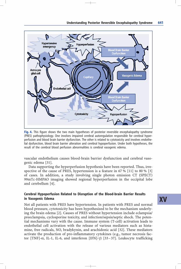
Fig. 4. This figure shows the two main hypotheses of posterior reversible encephalopathy syndrome(PRES) pathophysiology. One involves impaired cerebral autoregulation responsible for cerebral hyper-perfusion and blood brain barrier dysfunction. The other is related to cytotoxicity and involves endothe-lial dysfunction, blood brain barrier alteration and cerebral hypoperfusion. Under both hypotheses, theresult of the cerebral blood perfusion abnormalities is cerebral vasogenic edema.
vascular endothelium causes blood-brain barrier dysfunction and cerebral vaso-genic edema [31].
Data supporting the hyperperfusion hypothesis have been reported. Thus, irre-spective of the cause of PRES, hypertension is a feature in 67 % [11] to 80 % [3]of cases. In addition, a study involving single photon emission CT (SPECT)99mTc-HMPAO imaging showed regional hyperperfusion in the occipital lobeand cerebellum [4].
Cerebral Hypoperfusion Related to Disruption of the Blood-brain Barrier Resultsin Vasogenic Edema
Not all patients with PRES have hypertension. In patients with PRES and normalblood pressure, cytotoxicity has been hypothesized to be the mechanism underly-ing the brain edema [2]. Causes of PRES without hypertension include eclampsia/preeclampsia, cyclosporine toxicity, and infection/sepsis/septic shock. The poten-tial mechanisms vary with the cause. Immune system (T-cell) activation leads toendothelial cell activation with the release of various mediators such as hista-mine, free radicals, NO, bradykynin, and arachidonic acid [32]. These mediatorsactivate the production of pro-inflammatory cytokines (e.g., tumor necrosis fac-tor [TNF]-α, IL-1, IL-6, and interferon [IFN]-γ) [33–37]. Leukocyte trafficking
Understanding Posterior Reversible Encephalopathy Syndrome 641
XV

increases via the release of adhesion molecules (e.g., intercellular adhesion mole-cule [ICAM]-1, P-selectin, E-selectin, and cell adhesion molecule [CAM]-1) [2,38]. Upregulation of endothelial surface antigens and the release of endothelinaffect the local vascular tone [39]. All these changes result in vascular instabilitywith vasoconstriction and downstream hypoperfusion. Blood-brain barrier dys-function occurs, leading to vasogenic cerebral edema [2, 40].
This hypothesis is supported by studies involving catheter angiography, MRA,and MR perfusion imaging, which show cerebral hypoperfusion [41–43].
Pathophysiology of Complications of PRES: Cerebral Ischemia and CerebralHemorrhage
Ischemia following vasogenic edema may involve conversion to cytotoxic edema andmay result from longer exposure to the initial source of toxicity [40]. However, thedistinction between vasogenic and cytotoxic edema may be somewhat artificial, asboth forms of edema probably co-exist in many conditions. Cytotoxic edema isdefined as a pre-morbid cellular process characterized by induction of swelling of allcellular elements of the brain (neurons, glia, astrocytes, and endothelial cells) [44].The swelling is indirectly related to ATP depletion with failure of the ATP-dependentNa+/K+ channel and diffusion of extracellular water according to the osmotic gradi-ent into the intracellular sector. Cells in both the white and the gray matter areaffected, and swelling is more severe in the astrocytes than in the neurons [44]. Com-pensatory mechanisms induce calcium overload and activation of proteases (cathep-sin B, calpain, serine proteases), nucleases, and phospholipases (cytosolic Ca2+-dependent phospholipase A2), leading to necrosis and apoptosis [45]. These phe-nomena are potentiated by mitochondrial damage related to ATP depletion [45].
Bleeding related to reperfusion injury is another potential complication ofblood-brain-barrier dysfunction and cerebral edema. Oxidative stress with over-production of reactive oxygen species (ROS) and oxidative damage to lipid mem-branes in the blood-brain-barrier causes vessels within ischemic foci to leak orrupture [44]. Leukocyte trafficking with endothelial cell adhesion and activationleads to proteolysis of catenin, a component of the endothelial cell-cell junction[46]. The resulting damage to microvascular endothelial cells causes edema andbleeding [44]. Proteolysis by matrix metalloproteinases and proteases secreted byactivated leukocytes may cause bleeding after reperfusion injury [44].
Conditions Most Commonly Associated With PRES
The list of conditions associated with PRES is increasing steadily.
Toxic Agents
Exposure to toxic agents is the most common condition associated with PRES, in11 % [7] to 61 % [14] of cases. Box 1 shows an exhaustive list of toxic agentsknown to be associated with PRES. This etiology has been the focus of specificstudies. In a study of cyclosporine neurotoxicity in 16 patients, exposure durationat symptom onset ranged from 6 days to 5 years and the plasma cyclosporine lev-els were within the therapeutic range at diagnosis [5]. All patients had hyperten-sion. Symptomatic treatment and cyclosporine withdrawal was followed by a full
642 S. Legriel, F. Pico, and E. Azoulay
XV

Box 1. List of toxic agents known to be associated with posterior reversible encephalopathy syndrome(PRES)
Cancer chemotherapy agents (in combination) [8, 12, 14]Cytotoxic agents
Alkylating agentsCisplatin [48]Oxaliplatin [49]Carboplatin [50]
Anti-metabolitesGemcitabine [48]Cytarabine [51]Methotrexate [52]
Mitotic inhibitorsVincristine [53]Irinotecan hydrochloride [54]
OthersL-asparaginase [14]
Anti-angiogenic agentsBevacizumab [54]Sunitinib [55]RAF kinase inhibitor BAY 43 – 9006 [56]
Immunomodulatory cytokinesInterferon-alpha [3, 57]Interleukin-2 [58]
Monoclonal antibodiesRituximab (anti-CD20) [59]Infliximab (anti-TNF-α) [60]
Intravenous immunoglobulins [61]Anti-TNF-α protein
Etanercept [62]Anti-lymphocyte globulin [63]Immunosuppressive agents
Anticalcineurin agents [8]Cyclosporine A [3, 5, 10, 12, 14]Tacrolimus (FK 506) [3, 12, 14]
Sirolimus [64]High-dose corticosteroid therapy (e.g., dexamethasone and methylprednisolone) [14]
Blood transfusion [65]Other agents
Granulocyte-stimulating factor [66]Antiretroviral agents [67]Linezolid [68]Erythropoeitin [69]Cocaine [14]Ephedra sinica (traditional Chinese remedy) [70]Intravenous contrast agents [14]Lysergic acid amide [19]Carbamazepine [71]Intravenous caffeine [72]
TNF: tumor necrosis factor
Understanding Posterior Reversible Encephalopathy Syndrome 643
XV

recovery in 14 patients. One patient with an occipital lobe hemorrhage had per-manent visual field impairment and another had severe bleeding with transtento-rial herniation and died shortly after diagnosis.
Transplantation patients are at risk for PRES, as they are exposed to cancerchemotherapy and/or immunosuppressive therapy. PRES has been reported afterbone marrow or stem cell transplantation and after solid organ transplantation.In a study of 27 patients with PRES after liver or kidney transplantation, the timefrom transplantation to PRES diagnosis was 2 months in liver transplant recipi-ents and 1 year in kidney transplant recipients [47]. Common concomitants ofPRES were cytomegalovirus or bacterial infection and moderately severe trans-plant rejection. Hypertension was a feature and was more severe in the kidneytransplant group than in the liver transplant group.
Hypertension
Hypertension is the second most common condition associated with PRES, beingpresent in 6 % [12] to 72 % [7] of cases. The first case-series, published in 1992,had 14 patients, all of whom recovered fully within 2 weeks after blood pressurecontrol was achieved [4]. The main cause of hypertension was acute or chronickidney failure. Cases associated with hypertension due to autoimmune disease ortoxic exposures have been reported [19].
Infection/Sepsis/Septic Shock
Infections have been reported in 8 % [14] to 24 % of cases [12]. The most com-mon situation was PRES onset within 2 weeks after a Gram-positive bloodstreaminfection, often with hypertension at diagnosis [12]. PRES has also been reportedin patients with Escherichia coli bloodstream infection [73].
Preeclampsia/Eclampsia
Pre-eclampsia/eclampsia [3, 7, 8, 12, 14, 74] was present in 7 % [14] to 20 % [3]of patients with PRES. The outcome was usually favorable. Hypertension was aprominent feature at presentation. PRES onset occurred from 28 weeks’ gesta-tional age [74] to day 13 postpartum [3].
Autoimmune Disease
Autoimmune disease has been encountered in 8 % [14] to 10 % [12] of cases.PRES has been reported in patients with systemic lupus erythematosus [75, 76],systemic sclerosis [76], polyarteritis nodosa [77], Wegener’s granulomatosis [78],thrombotic microangiopathy [79], polyangiitis [80], Takayasu arteritis [81], Has-himoto encephalopathy [82], and Crohn’s disease [83].
Other Conditions
There are myriad conditions associated with PRES, sometimes only anecdotally.They include sickle cell disease [12], Guillain-Barre syndrome [84], hypomagne-semia [3], hypercalcemia [85], tumor lysis syndrome [86], porphyria [87], pheo-chromocytoma [88], and Cushing syndrome [89].
644 S. Legriel, F. Pico, and E. Azoulay
XV

Outcomes
As indicated by the name of this syndrome, appropriate treatment is expected toensure a full recovery. However, permanent complications and fatal cases havebeen reported, leading some authors to suggest that a better name may be“potentially reversible encephalopathy syndrome” [90]. Clinical findings returnedto baseline in 35 % [8] to 100 % [3] of patients. Radiological recovery is more dif-ficult to document, as all published studies were retrospective and repeated brainimaging was performed in only 44 % [8] to 87 % [11] of cases. Among patientswith follow-up imaging studies, 49 % [6] to 75 % [3] had resolution of the initialabnormalities within 5 days [7] to 17 months [3]. Permanent neurological abnor-malities are related to ischemia and/or bleeding. Recurrences have been reportedin 6 % of cases [8].
Limited data are available on functional outcomes. In one study, the medianmodified Rankin Scale score was 2.5 at discharge, indicating mild-to-moderatedisability [8]. Mild disability is defined as being capable of handling one’s ownaffairs without help but not of carrying out all previous activities; and moderatedisability is defined as requiring help for some activities but being able to walkunassisted.
Death has been reported in up to 15 % [7, 8] of patients. However, the relativecontributions of PRES and of associated factors to the fatal outcomes are unclear.
Management of PRES
PRES must be diagnosed early and investigations must be performed to identifythe causative factors. Symptomatic treatment should be given immediately andthe causative factors corrected without delay. ICU admission and life-supportingtreatments may be required [9, 10].
Diagnostic Strategy
The diagnostic strategy for PRES is fairly well standardized (Fig. 5). After a carefulhistory and thorough physical examination, investigations should be performed asappropriate, starting with the simplest and moving to the more sophisticated.
CT may be easier to obtain first. However, MRI must be performed, either asthe first or as the second imaging study. MRI is considerably better than CT forthe diagnosis of PRES and can provide information regarding many of the causesof PRES [1–3, 6, 7, 11, 13]. MRA must be added to MRI to identify an associatedcerebral reversible vasoconstriction syndrome.
Electroencephalography (EEG) should be performed routinely to look for non-convulsive status epilepticus. Patients most likely to have non-convulsive statusepilepticus are those in a deep coma or prolonged post-ictal state [10]. Lumbarpuncture findings are not specific in PRES [7]. However, the cerebrospinal fluid(CSF) must be examined in patients with a fever or clinical suspicion of meningi-tis and when deemed appropriate by the attending physicians. Laboratory testsshould be obtained routinely. Plasma anticonvulsant drug assays (including mag-nesemia dosage) and qualitative tests for toxic agents or medications associatedwith seizures and other symptoms of PRES should be performed at the discretionof the attending physicians.
Understanding Posterior Reversible Encephalopathy Syndrome 645
XV

Variable combination of suggestive clinical manifestations:seizure activity, consciousness impairment, headaches,
visual abnormalities, nausea/vomiting and focal neurological signs
CTwith contrast
MRI+/- MRA
White matterhypodensitiesof topography
suggestiveof PRES
Normal
FLAIR and T2-weighted sequences reveal high-signalfoci in the white matter,
which may or not be bilateral/symmetric.Variable involvement of the parietal, occipital,
frontal and temporal lobes,cerebellum and brainstem. The cortical gray matter
may be affected.
Normal
MRI+/- MRA
DEFINITE DIAGNOSISOF PRES
Negativediagnosis of
PRES
PRES ruled out:Per/post-ictal state (seizure or status epilepticus)
Progressive Multifocal Leukoencephalopathy (PML)Severe Leukoaraiosis
Cerebral Autosomal Dominant Arteriopathy with Subcortical Infarcts and Leukoencephalopathy (CADASIL)Infectious encephalitis, Acute Disseminated EncephaloMyelitis (ADEM)
Mitochondrial myopathy Encephalopathy Lactacidosis and Stroke-like episodes syndrome (MELAS)CNS vasculitis
Creutzfeld-Jakob diseaseCerebral venous sinus thrombosis
Ischemic stroke
Repeatif < Day 3
from onsetof clinical
manifestations
Fig. 5. Diagnostic strategy for posterior reversible encephalopathy syndrome (PRES). MRI: magnetic res-onance imagery; MRA: magnetic resonance angiography; CT: computed tomography
A neurosurgical biopsy should be performed in patients who fail to respond to appro-priate treatment and whose cerebral imaging studies show focal lesions of unknownor doubtful nature. Brain biopsy may show non-specific white matter changes consis-tent with vasogenic edema (activated astrocytes, scattered macrophages, and rarelymphocytes). Findings at a later stage include demyelination and anoxic neuronalalterations, sometimes with bleeding in the white and gray matter [91].
646 S. Legriel, F. Pico, and E. Azoulay
XV

DEFINITE DIAGNOSISOF PRES
Evaluationof associatedorgan failures
Look forPRES-associated
conditions
Early correction ofunderlying cause of PRES
Intravenousantihypertensive therapy
Anticonvulsant
Variable combination ofsuggestive clinical manifestations
CTwith contrast
MRI +/- MRA
Electroencephalogram (EEG)Consider lumbar puncture
Laboratory tests (Magnesemia +++)Consider toxicologic investigationsConsider Oro-
Tracheal Intubation
Symptomatic management
LabetololNicardipine
Fenoldopam if avalaibleUrapidil
Benzodiazepines
Phenobarbital(fos)phenytoin
MidazolamPropol
Thiopental
If indicated:
Blood pressure controlToxic agent withdrawal
Cesarean sectionDialysis
NimodipineMetabolic disturbances
...
Correction of hypomagnesemia if indicated
Treatment
The treatment strategy associates general measures with correction of the under-lying cause of PRES (Fig. 6).
Fig. 6. Treatment of posterior reversible encephalopathy syndrome (PRES). MRI: magnetic resonanceimaging; MRA: magnetic resonance angiography; CT: computed tomography
Understanding Posterior Reversible Encephalopathy Syndrome 647
XV
General measuresPatients with PRES require the symptomatic measures usually taken in the ICU.Although most patients have stable hemodynamics, catecholamines are requiredoccasionally. The need for upper airway protection should be evaluated continu-ously in patients with marked consciousness impairment or seizure activity. Ifendotracheal intubation is performed, rapid-sequence induction with etomidateand succinylcholine can be used, provided there is no evidence of hyperkalemia.Propofol or thiopental are also good choices, since they have anticonvulsanteffects. Neuromuscular blocking agents may transiently mask seizures.
Hypoglycemia should be looked for routinely and corrected. If glucose isgiven, 100 mg of thiamine should be administered concomitantly, most notablywhen there is evidence of vitamin B1 deficiency. Patients should be routinelyevaluated for hyperthermia and metabolic disturbances, in particular hypomag-nesemia, which require prompt correction. Aspiration pneumonia may compli-cate the initial consciousness disorders.
Antiepileptic treatment, appropriate for the electrical and clinical pattern inthe patient, should be initiated on an emergency basis and according to currentguidelines. Patients with persistent seizure activity at ICU admission should begiven intravenous benzodiazepines (clonazepam 1 mg or diazepam 10 mg) eitherbefore ICU admission or in the ICU. The dose can be repeated up to three timesif necessary. Patients with continuing seizure activity despite intravenous benzo-diazepines should receive standard complementary intravenous anticonvulsantdrugs (phenobarbital 10 to 15 mg/kg, phenytoin 18 mg/kg, or equivalent dose offosphenytoin). Patients with refractory status epilepticus need midazolam, propo-fol, or thiopental in titrated doses until remission of the clinical seizure activity.When the EEG reveals electrical status epilepticus, these anesthetic drugs aregiven in titrated doses to induce EEG burst suppression then as a continuousinfusion for at least 12 hours [92].
Control of hypertensive emergency, if present, is an important part of thesymptomatic management. The aim is not to normalize the blood pressure butrather to decrease the MAP by 20– 25 % within the first 2 hours and to bring theblood pressure down to 160/100 mmHg within the first 6 hours [31, 93]. Morerapid blood pressure reduction is not recommended since it can aggravate thecerebral perfusion pressure alterations and promote ischemia [94]. Intravenousantihypertensive drugs are necessary. Appropriate choices include labetolol,nicardipine, or fenoldopam if available [31, 94]. Urapidil has been suggested as asecond-line agent, perhaps in combination with another agent [95].
Correction of the underlying cause of PRESAn early etiologic diagnosis allows prompt correction of the cause of PRES.Patients may require blood pressure control, withdrawal of cancer chemotherapyor immunosuppressive agents, Cesarean section, dialysis, or other interventions.Prompt correction of the cause is crucial to decrease the risk of ischemia orbleeding and therefore to avoid permanent disability or death [3].
Conclusion
This review highlights recent advances in the diagnosis, pathophysiologicalunderstanding, and management of PRES. Although the clinical presentation is
648 S. Legriel, F. Pico, and E. Azoulay
XV

non-specific, most patients have a suggestive combination of symptoms. MRI iscrucial for diagnosing PRES, monitoring the course, and assessing treatmenteffectiveness. MRA performed during MRI can be useful to identify associatedcerebral vasoconstriction. Repeated cerebral imaging helps to support the diag-nosis and identifies complications potentially responsible for permanent impair-ments. The pathophysiology of PRES remains controversial. However, the list ofconditions known to be associated with PRES is increasing steadily. Early recog-nition and resolution of the underlying cause is the keystone of management.Persistence of the cause carries a risk of ischemia, bleeding, and death. Finally,studies are needed to identify factors of adverse prognostic significance and todevelop neuroprotective strategies.
References
1. Bartynski WS (2008) Posterior reversible encephalopathy syndrome, part 1: fundamentalimaging and clinical features. AJNR Am J Neuroradiol 29: 1036– 1042
2. Bartynski WS (2008) Posterior reversible encephalopathy syndrome, part 2: controversiessurrounding pathophysiology of vasogenic edema. AJNR Am J Neuroradiol 29: 1043– 1049
3. Hinchey J, Chaves C, Appignani B, et al (1996) A reversible posterior leukoencephalopathysyndrome. N Engl J Med 334: 494– 500
4. Schwartz RB, Jones KM, Kalina P, et al (1992) Hypertensive encephalopathy: findings onCT, MR imaging, and SPECT imaging in 14 cases. AJR Am J Roentgenol 159: 379–383
5. Schwartz RB, Bravo SM, Klufas RA, et al (1995) Cyclosporine neurotoxicity and its rela-tionship to hypertensive encephalopathy: CT and MR findings in 16 cases. AJR Am JRoentgenol 165: 627– 631
6. Casey SO, Sampaio RC, Michel E, Truwit CL (2000) Posterior reversible encephalopathysyndrome: utility of fluid-attenuated inversion recovery MR imaging in the detection ofcortical and subcortical lesions. AJNR Am J Neuroradiol 21: 1199– 1206
7. Lee VH, Wijdicks EF, Manno EM, Rabinstein AA (2008) Clinical spectrum of reversibleposterior leukoencephalopathy syndrome. Arch Neurol 65: 205– 210
8. Burnett MM, Hess CP, Roberts JP, Bass NM, Douglas VC, Josephson SA (2010) Presenta-tion of reversible posterior leukoencephalopathy syndrome in patients on calcineurininhibitors. Clin Neurol Neurosurg 112: 886– 889
9. Servillo G, Striano P, Striano S, et al (2003) Posterior reversible encephalopathy syndrome(PRES) in critically ill obstetric patients. Intensive Care Med 29: 2323–2326
10. Kozak OS, Wijdicks EF, Manno EM, Miley JT, Rabinstein AA (2007) Status epilepticus asinitial manifestation of posterior reversible encephalopathy syndrome. Neurology 69:894– 897
11. Bartynski WS, Boardman JF (2007) Distinct imaging patterns and lesion distribution inposterior reversible encephalopathy syndrome. AJNR Am J Neuroradiol 28: 1320– 1327
12. Bartynski WS, Boardman JF, Zeigler ZR, Shadduck RK, Lister J (2006) Posterior reversibleencephalopathy syndrome in infection, sepsis, and shock. AJNR Am J Neuroradiol 27:2179–2190
13. Covarrubias DJ, Luetmer PH, Campeau NG (2002) Posterior reversible encephalopathysyndrome: prognostic utility of quantitative diffusion-weighted MR images. AJNR Am JNeuroradiol 23: 1038– 1048
14. McKinney AM, Short J, Truwit CL, et al (2007) Posterior reversible encephalopathy syn-drome: incidence of atypical regions of involvement and imaging findings. AJR Am JRoentgenol 189: 904– 912
15. Ducros A, Boukobza M, Porcher R, Sarov M, Valade D, Bousser MG (2007) The clinicaland radiological spectrum of reversible cerebral vasoconstriction syndrome. A prospec-tive series of 67 patients. Brain 130: 3091– 3101
16. Hefzy HM, Bartynski WS, Boardman JF, Lacomis D (2009) Hemorrhage in posteriorreversible encephalopathy syndrome: imaging and clinical features. AJNR Am J Neurora-diol 30: 1371–1379
Understanding Posterior Reversible Encephalopathy Syndrome 649
XV

17. Belogolovkin V, Levine SR, Fields MC, Stone JL (2006) Postpartum eclampsia complicatedby reversible cerebral herniation. Obstet Gynecol 107: 442–445
18. Lamy C, Mas JL (2001) [Reversible posterior leukoencephalopathy. A new syndrome or anew name for an old syndrome?]. Presse Med 30: 915–920
19. Legriel S, Bruneel F, Spreux-Varoquaux O, et al (2008) Lysergic acid amide-induced poste-rior reversible encephalopathy syndrome with status epilepticus. Neurocrit Care 9:247– 252
20. Cole AJ (2004) Status epilepticus and periictal imaging. Epilepsia 45 (Suppl 4): 72–7721. Thurnher MM, Post MJ, Rieger A, Kleibl-Popov C, Loewe C, Schindler E (2001) Initial and
follow-up MR imaging findings in AIDS-related progressive multifocal leukoencephalopa-thy treated with highly active antiretroviral therapy. AJNR Am J Neuroradiol 22: 977–984
22. O’Sullivan M (2008) Leukoaraiosis. Pract Neurol 8: 26–3823. Chabriat H, Joutel A, Dichgans M, Tournier-Lasserve E, Bousser MG (2009) Cadasil. Lan-
cet Neurol 8: 643– 65324. Gilman S (1998) Imaging the brain. First of two parts. N Engl J Med 338: 812– 82025. Sonneville R, Klein IF, Wolff M (2010) Update on investigation and management of post-
infectious encephalitis. Curr Opin Neurol 23: 300–30426. Matthews PM, Tampieri D, Berkovic SF, et al (1991) Magnetic resonance imaging shows
specific abnormalities in the MELAS syndrome. Neurology 41: 1043– 104627. Pomper MG, Miller TJ, Stone JH, Tidmore WC, Hellmann DB (1999) CNS vasculitis in
autoimmune disease: MR imaging findings and correlation with angiography. AJNR AmJ Neuroradiol 20: 75– 85
28. Kallenberg K, Schulz-Schaeffer WJ, Jastrow U, et al (2006) Creutzfeldt-Jakob disease: com-parative analysis of MR imaging sequences. AJNR Am J Neuroradiol 27: 1459–1462
29. Stam J (2005) Thrombosis of the cerebral veins and sinuses. N Engl J Med 352: 1791–179830. Culebras A, Kase CS, Masdeu JC, et al (1997) Practice guidelines for the use of imaging in
transient ischemic attacks and acute stroke. A report of the Stroke Council, AmericanHeart Association. Stroke 28: 1480– 1497
31. Vaughan CJ, Delanty N (2000) Hypertensive emergencies. Lancet 356: 411–41732. Baethmann A, Maier-Hauff K, Kempski O, Unterberg A, Wahl M, Schurer L (1988) Media-
tors of brain edema and secondary brain damage. Crit Care Med 16: 972–97833. Ferrara JL (2000) Pathogenesis of acute graft-versus-host disease: cytokines and cellular
effectors. J Hematother Stem Cell Res 9: 299– 30634. Antin JH, Ferrara JL (1992) Cytokine dysregulation and acute graft-versus-host disease.
Blood 80: 2964– 296835. Holler E, Kolb HJ, Moller A, et al (1990) Increased serum levels of tumor necrosis factor
alpha precede major complications of bone marrow transplantation. Blood 75: 1011–101636. Schots R, Kaufman L, Van Riet I, et al (2003) Proinflammatory cytokines and their role in
the development of major transplant-related complications in the early phase after alloge-neic bone marrow transplantation. Leukemia 17: 1150–1156
37. de Vries HE, Kuiper J, de Boer AG, Van Berkel TJ, Breimer DD (1997) The blood-brainbarrier in neuroinflammatory diseases. Pharmacol Rev 49: 143– 155
38. Stanimirovic D, Satoh K (2000) Inflammatory mediators of cerebral endothelium: a role inischemic brain inflammation. Brain Pathol 10: 113–126
39. Narushima I, Kita T, Kubo K, et al (2003) Highly enhanced permeability of blood-brainbarrier induced by repeated administration of endothelin-1 in dogs and rats. PharmacolToxicol 92: 21– 26
40. Wijdicks EF (2001) Neurotoxicity of immunosuppressive drugs. Liver Transpl 7: 937–94241. Bartynski WS, Boardman JF (2008) Catheter angiography, MR angiography, and MR per-
fusion in posterior reversible encephalopathy syndrome. AJNR Am J Neuroradiol 29:447– 455
42. Brubaker LM, Smith JK, Lee YZ, Lin W, Castillo M (2005) Hemodynamic and permeabil-ity changes in posterior reversible encephalopathy syndrome measured by dynamic sus-ceptibility perfusion-weighted MR imaging. AJNR Am J Neuroradiol 26: 825– 830
43. Engelter ST, Petrella JR, Alberts MJ, Provenzale JM (1999) Assessment of cerebral micro-circulation in a patient with hypertensive encephalopathy using MR perfusion imaging.AJR Am J Roentgenol 173: 1491–1493
650 S. Legriel, F. Pico, and E. Azoulay
XV

44. Wang X, Lo EH (2003) Triggers and mediators of hemorrhagic transformation in cerebralischemia. Mol Neurobiol 28: 229–244
45. Szabo C (2005) Mechanisms of cell necrosis. Crit Care Med 33 (12 Suppl): S530– 53446. Allport JR, Ding H, Collins T, Gerritsen ME, Luscinskas FW (1997) Endothelial-dependent
mechanisms regulate leukocyte transmigration: a process involving the proteasome anddisruption of the vascular endothelial-cadherin complex at endothelial cell-to-cell junc-tions. J Exp Med 186: 517–527
47. Bartynski WS, Tan HP, Boardman JF, Shapiro R, Marsh JW (2008) Posterior reversibleencephalopathy syndrome after solid organ transplantation. AJNR Am J Neuroradiol 29:924– 930
48. Kwon EJ, Kim SW, Kim KK, Seo HS, Kim do Y (2009) A case of gemcitabine and cisplatinassociated posterior reversible encephalopathy syndrome. Cancer Res Treat 41: 53– 55
49. Nagata Y, Omuro Y, Shimoyama T, et al (2009) [A case of colon cancer with reversible pos-terior leukoencephalopathy syndrome following 5-FU and oxaliplatin (FOLFOX regime)].Gan To Kagaku Ryoho 36: 1163– 1166
50. Vieillot S, Pouessel D, de Champfleur NM, Becht C, Culine S (2007) Reversible posteriorleukoencephalopathy syndrome after carboplatin therapy. Ann Oncol 18: 608– 609
51. Saito B, Nakamaki T, Nakashima H, et al (2007) Reversible posterior leukoencephalopathysyndrome after repeat intermediate-dose cytarabine chemotherapy in a patient with acutemyeloid leukemia. Am J Hematol 82: 304– 306
52. Dicuonzo F, Salvati A, Palma M, et al (2009) Posterior reversible encephalopathy syn-drome associated with methotrexate neurotoxicity: conventional magnetic resonance anddiffusion-weighted imaging findings. J Child Neurol 24: 1013–1018
53. Hualde Olascoaga J, Molins Castiella T, Souto Hernandez S, et al (2008) [Reversible poste-rior leukoencephalopathy: report of two cases after vincristine treatment]. An Pediatr(Barc) 68: 282–285
54. Allen JA, Adlakha A, Bergethon PR (2006) Reversible posterior leukoencephalopathy syn-drome after bevacizumab/FOLFIRI regimen for metastatic colon cancer. Arch Neurol 63:1475–1478
55. Cumurciuc R, Martinez-Almoyna L, Henry C, Husson H, de Broucker T (2008) Posteriorreversible encephalopathy syndrome during sunitinib therapy. Rev Neurol (Paris) 164:605– 607
56. Govindarajan R, Adusumilli J, Baxter DL, El-Khoueiry A, Harik SI (2006) Reversible pos-terior leukoencephalopathy syndrome induced by RAF kinase inhibitor BAY 43– 9006. JClin Oncol 24: e48
57. Kamar N, Kany M, Bories P, et al (2001) Reversible posterior leukoencephalopathy syn-drome in hepatitis C virus-positive long-term hemodialysis patients. Am J Kidney Dis 37:E29
58. Karp BI, Yang JC, Khorsand M, Wood R, Merigan TC (1996) Multiple cerebral lesionscomplicating therapy with interleukin-2. Neurology 47: 417– 424
59. Zito JA, Lee CC, Johnson S, Singer A, Vacirca J (2010) Reversible posterior leukoencepha-lopathy syndrome after rituximab. Am J Emerg Med 28: 537
60. Zamvar V, Sugarman ID, Tawfik RF, Macmullen-Price J, Puntis JW (2009) Posterior revers-ible encephalopathy syndrome following infliximab infusion. J Pediatr Gastroenterol Nutr48: 102–105
61. Belmouaz S, Desport E, Leroy F, et al (2008) Posterior reversible encephalopathy inducedby intravenous immunoglobulin. Nephrol Dial Transplant 23: 417– 419
62. Kastrup O, Diener HC (2008) TNF-antagonist etanercept induced reversible posterior leu-koencephalopathy syndrome. J Neurol 255: 452–453
63. Greaves P, Oakervee H, Kon SS, Jones R, Farah N (2006) Posterior reversible encephalopa-thy syndrome following anti-lymphocyte globulin treatment for severe aplastic anaemia.Br J Haematol 134: 251
64. Bodkin CL, Eidelman BH (2007) Sirolimus-induced posterior reversible encephalopathy.Neurology 68: 2039–2040
65. Huang YC, Tsai PL, Yeh JH, Chen WH (2008) Reversible posterior leukoencephalopathysyndrome caused by blood transfusion: a case report. Acta Neurol Taiwan 17: 258–262
66. Leniger T, Kastrup O, Diener HC (2000) Reversible posterior leukencephalopathy syn-
Understanding Posterior Reversible Encephalopathy Syndrome 651
XV

drome induced by granulocyte stimulating factor filgrastim. J Neurol Neurosurg Psychia-try 69: 280– 281
67. Ridolfo AL, Resta F, Milazzo L, et al (2008) Reversible posterior leukoencephalopathy syn-drome in 2 HIV-infected patients receiving antiretroviral therapy. Clin Infect Dis46:e19–22
68. Nagel S, Kohrmann M, Huttner HB, Storch-Hagenlocher B, Schwab S (2007) Linezolid-induced posterior reversible leukoencephalopathy syndrome. Arch Neurol 64: 746– 748
69. Delanty N, Vaughan C, Frucht S, Stubgen P (1997) Erythropoietin-associated hypertensiveposterior leukoencephalopathy. Neurology 49: 686– 689
70. Moawad FJ, Hartzell JD, Biega TJ, Lettieri CJ (2006) Transient blindness due to posteriorreversible encephalopathy syndrome following ephedra overdose. South Med J 99:511– 514
71. Furuta N, Fujita Y, Sekine A, Ikeda M, Okamoto K (2009) [Reversible posterior leukoence-phalopathy syndrome associated with carbamazepine-induced hypertension]. RinshoShinkeigaku 49: 191–193
72. Ortiz GA, Bianchi NA, Tiede MP, Bhatia RG (2009) Posterior reversible encephalopathysyndrome after intravenous caffeine for post-lumbar puncture headaches. AJNR Am JNeuroradiol 30: 586–587
73. Fabbian F, Pala M, Fallica E, et al (2010) Posterior reversible encephalopathy syndrome inan 87-year-old woman with Escherichia coli bloodstream infection. Clin Exp Nephrol 14:176– 179
74. Servillo G, Apicella E, Striano P (2008) Posterior reversible encephalopathy syndrome(PRES) in the parturient with preeclampsia after inadvertent dural puncture. Int J ObstetAnesth 17: 88–89
75. Baizabal-Carvallo JF, Barragan-Campos HM, Padilla-Aranda HJ, et al (2009) Posteriorreversible encephalopathy syndrome as a complication of acute lupus activity. Clin NeurolNeurosurg 111: 359– 363
76. Yong PF, Hamour SM, Burns A (2003) Reversible posterior leukoencephalopathy in apatient with systemic sclerosis/systemic lupus erythematosus overlap syndrome. NephrolDial Transplant 18: 2660–2662
77. Stanzani L, Fusi L, Gomitoni A, Roncoroni M, Villa P, Grampa G (2008) A case of poste-rior reversible encephalopathy during polyarteritis nodosa vasculitis. Neurol Sci 29:163– 167
78. Nishio M, Yoshioka K, Yamagami K, et al (2008) Reversible posterior leukoencephalopathysyndrome: a possible manifestation of Wegener’s granulomatosis-mediated endothelialinjury. Mod Rheumatol 18: 309–314
79. Burrus TM, Mandrekar J, Wijdicks EF, Rabinstein AA (2010) Renal failure and posteriorreversible encephalopathy syndrome in patients with thrombotic thrombocytopenic pur-pura. Arch Neurol 67: 831–834
80. Kamimura H, Hirose S, Tazaki K, Suzuki Y, Satou M (2007) [Reversible posterior leukoen-cephalopathy syndrome with microscopic polyangiitis]. Nippon Naika Gakkai Zasshi 96:2532–2535
81. Fujita M, Komatsu K, Hatachi S, Yagita M (2008) Reversible posterior leukoencephalopa-thy syndrome in a patient with Takayasu arteritis. Mod Rheumatol 18: 623–629
82. Yildiz OK, Segmen H, Oztoprak I, Bolayir E, Topaktas S (2010) Posterior reversibleencephalopathy and alexia without agraphia in a patient with Hashimoto’s encephalopa-thy. Neurol Sci 31: 523– 525
83. Zipper SG, Tischendorf M, Westphal K (2006) [Postoperative occurrence of reversible pos-terior encephalopathy in a patient with Crohn’s disease]. Anaesthesist 55: 1064– 1067
84. Fugate JE, Wijdicks EF, Kumar G, Rabinstein AA (2009) One Thing Leads to Another: GBSComplicated by PRES and Takotsubo Cardiomyopathy. Neurocrit Care 11: 395– 397
85. Ma ES, Chiu EK, Fong GC, Li FK, Wong CL (2009) Burkitt lymphoma presenting as poste-rior reversible encephalopathy syndrome secondary to hypercalcaemia. Br J Haematol146: 584
86. Greenwood MJ, Dodds AJ, Garricik R, Rodriguez M (2003) Posterior leukoencephalopathyin association with the tumour lysis syndrome in acute lymphoblastic leukaemia--a casewith clinicopathological correlation. Leuk Lymphoma 44: 719–721
652 S. Legriel, F. Pico, and E. Azoulay
XV

87. Kang SY, Kang JH, Choi JC, Lee JS (2010) Posterior reversible encephalopathy syndromein a patient with acute intermittent porphyria. J Neurol 257: 663– 664
88. Rodriguez-Uranga JJ, Franco-Macias E, Bernal Sanchez-Arjona M, Villalobos-Chavez F(2003) [Posterior reversible leukoencephalopathy syndrome, pheochromocytoma and vonHippel-Lindau disease]. Rev Neurol 37: 797– 798
89. Lodish M, Patronas NJ, Stratakis CA (2010) Reversible posterior encephalopathy syn-drome associated with micronodular adrenocortical disease and Cushing syndrome. EurJ Pediatr 169: 125– 126
90. Narbone MC, Musolino R, Granata F, Mazzu I, Abbate M, Ferlazzo E (2006) PRES: poste-rior or potentially reversible encephalopathy syndrome? Neurol Sci 27: 187–189
91. Schiff D, Lopes MB (2005) Neuropathological correlates of reversible posterior leukoence-phalopathy. Neurocrit Care 2: 303–305
92. Meierkord H, Boon P, Engelsen B, et al (2010) EFNS guideline on the management of sta-tus epilepticus in adults. Eur J Neurol 17: 348–355
93. Ramsay LE, Williams B, Johnston GD, et al (1999) British Hypertension Society guidelinesfor hypertension management 1999: summary. BMJ 319: 630– 635
94. Varon J, Marik PE (2003) Clinical review: the management of hypertensive crises. CritCare 7: 374–384
95. Buch J (2010) Urapidil, a dual-acting antihypertensive agent: Current usage consider-ations. Adv Ther 27: 426–443
Understanding Posterior Reversible Encephalopathy Syndrome 653
XV

http://www.springer.com/978-3-642-18080-4