Comparative Anatomical Studies on the Fetal … the urachus, while in camel, it contained the same...
10
IOSR Journal of Agriculture and Veterinary Science (IOSR-JAVS) e-ISSN: 2319-2380, p-ISSN: 2319-2372. Volume 11, Issue 5 Ver. I (May 2018), PP 12-21 www.iosrjournals.org DOI: 10.9790/2380-1105011221 www.iosrjournals.org 12 | Page Comparative Anatomical Studies on the Fetal Remnants in Donkey (Asinus Equus) And Camel (Camelus Dromedarius) Nora, A. shaker and Samar M. EL Gammal Department of Anatomy and Embryology, Faculty of Veterinary Medicine, Cairo University Abstract Aim: The present study described the anatomical structures of the umbilical cord, fetal circulation and their remnants in the adult. This work will help veterinary practitioners in diagnosis of the abnormalities of these umbilical structures in newly born foals and camel calves. Material and methods: it was applied on five specimens from donkeys and camels; three aborted feti and two apparently healthy adults. Fetal specimens were injected with the colored latex in the umbilical veins and arteries then preserved in a 10% formalin solution for two days before dissection. The structures of the umbilical cord and the embryonic structures were identified and photographed. The adult specimens were treated by ordinary routine method of preservation; mixture of 10%formalin, 4%phenol and 1% glycerin for two days then dissected to identify the fetal remnants. Result: The umbilical cord contained four structures in donkey; single umbilical vein, two umbilical arteries and the urachus, while in camel, it contained the same structures but two umbilical veins instead of one. In the adult, these fetal tubes were occluded as the round ligament of liver, round ligaments of urinary bladder and cicatrix on the bladder apex. The other fetal structures were; the foramen ovale and the ductus arteriosus and the paired gubernaculua related to the testes in males and the ovaries in females. The camel had the ductus venosus but not occured in the donkey's fetus. Conclusion: The use of the precise normal anatomical structure of the umbilical cord, it contents and other embryonic structures will lead to accurate diagnosis and proper surgical interference for treatment of the congenital abnormalities. Key words: umbilical cord, fetal remnant, Anatomy, donkey, camel -------------------------------------------------------------------------------------------------------------------------------------- Date of Submission: 18-04-2018 Date of acceptance: 05-05-2018 --------------------------------------------------------------------------------------------------------------------------------------- I. Introduction The umbilical cord is considered as the pathway for blood transport from the placenta to the fetus and vice versa. The umbilical cord in the horse consists of two part; the proximal amniotic portion, which is covered by amnion and is attached to the fetus at the umbilicus, and the distal allantoic portion covered by allantois that is attached to the allantochorion. The amniotic portion of the cord contained two umbilical arteries, one umbilical vein and the urachus. (Schlafer 2004; Whitwell and Jeffcott 1975). In ruminants, there are two arteries and two veins along the whole length of the cord but in other species, such as equines and carnivores, the amniotic portion end of the cord has two arteries but only one vein. The testis is suspended from the dorsal abdominal wall, by a broad thin mesorchium and is connected to the deep inguinal ring by the gubernaculum. Under hormonal effect, the gubernaculum gets shortened and stretched through the inguinal canal, allowing the descent of the testis into the scrotum. The present study helps understand the pathologic conditions associated with these embryonic structures such as several urachal anomalies reported in different animal species including horses, calves and cats; a congenital persistent urachal remnant as cyst and fistula, bladder diverticulum (vesicourachal) and urachal adenocarcinoma, (Lojszczyk,Szczepaniak et al, 2010; Wilsher et al., 2009; Laverty and Salisbury 2002; Sadler 1995; Adams and Fessler 1987 and Baxter et al., 1987). The congenital cardiac defects in horses were as patent ductus arteriosus (Morgan-Hughes et al., 2003) and patent foramen ovale. Another congenital abnormalities like the cryptorchidism. II. Material and methods: The current study was carried out on five specimens from donkeys and camels; three aborted feti at late stage of trimester (males in donkey and females in camel) and two apparently healthy adults were used from each animal. Camel specimens were obtained from slaughter houses while donkey specimens were collected from the faculty of veterinary medicine, Cairo University. Fetal specimens were washed carefully by normal saline injection in the umbilical vein then injected with the colored latex through the insertion of a catheter in
Transcript of Comparative Anatomical Studies on the Fetal … the urachus, while in camel, it contained the same...

IOSR Journal of Agriculture and Veterinary Science (IOSR-JAVS)
e-ISSN: 2319-2380, p-ISSN: 2319-2372. Volume 11, Issue 5 Ver. I (May 2018), PP 12-21
www.iosrjournals.org
DOI: 10.9790/2380-1105011221 www.iosrjournals.org 12 | Page
Comparative Anatomical Studies on the Fetal Remnants in
Donkey (Asinus Equus) And Camel (Camelus Dromedarius)
Nora, A. shaker and Samar M. EL Gammal Department of Anatomy and Embryology, Faculty of Veterinary Medicine, Cairo University
Abstract Aim: The present study described the anatomical structures of the umbilical cord, fetal circulation and their
remnants in the adult. This work will help veterinary practitioners in diagnosis of the abnormalities of these
umbilical structures in newly born foals and camel calves.
Material and methods: it was applied on five specimens from donkeys and camels; three aborted feti and two
apparently healthy adults. Fetal specimens were injected with the colored latex in the umbilical veins and
arteries then preserved in a 10% formalin solution for two days before dissection. The structures of the
umbilical cord and the embryonic structures were identified and photographed. The adult specimens were
treated by ordinary routine method of preservation; mixture of 10%formalin, 4%phenol and 1% glycerin for
two days then dissected to identify the fetal remnants.
Result: The umbilical cord contained four structures in donkey; single umbilical vein, two umbilical arteries
and the urachus, while in camel, it contained the same structures but two umbilical veins instead of one. In the
adult, these fetal tubes were occluded as the round ligament of liver, round ligaments of urinary bladder and
cicatrix on the bladder apex. The other fetal structures were; the foramen ovale and the ductus arteriosus and
the paired gubernaculua related to the testes in males and the ovaries in females. The camel had the ductus
venosus but not occured in the donkey's fetus.
Conclusion: The use of the precise normal anatomical structure of the umbilical cord, it contents and other
embryonic structures will lead to accurate diagnosis and proper surgical interference for treatment of the
congenital abnormalities.
Key words: umbilical cord, fetal remnant, Anatomy, donkey, camel
----------------------------------------------------------------------------------------------------------------------------- ---------
Date of Submission: 18-04-2018 Date of acceptance: 05-05-2018
----------------------------------------------------------------------------------------------------------------------------- ----------
I. Introduction The umbilical cord is considered as the pathway for blood transport from the placenta to the fetus and
vice versa. The umbilical cord in the horse consists of two part; the proximal amniotic portion, which is covered
by amnion and is attached to the fetus at the umbilicus, and the distal allantoic portion covered by allantois that
is attached to the allantochorion. The amniotic portion of the cord contained two umbilical arteries, one
umbilical vein and the urachus. (Schlafer 2004; Whitwell and Jeffcott 1975). In ruminants, there are two arteries
and two veins along the whole length of the cord but in other species, such as equines and carnivores, the
amniotic portion end of the cord has two arteries but only one vein.
The testis is suspended from the dorsal abdominal wall, by a broad thin mesorchium and is connected
to the deep inguinal ring by the gubernaculum. Under hormonal effect, the gubernaculum gets shortened and
stretched through the inguinal canal, allowing the descent of the testis into the scrotum.
The present study helps understand the pathologic conditions associated with these embryonic
structures such as several urachal anomalies reported in different animal species including horses, calves and
cats; a congenital persistent urachal remnant as cyst and fistula, bladder diverticulum (vesicourachal) and
urachal adenocarcinoma, (Lojszczyk,Szczepaniak et al, 2010; Wilsher et al., 2009; Laverty and
Salisbury 2002; Sadler 1995; Adams and Fessler 1987 and Baxter et al., 1987). The congenital
cardiac defects in horses were as patent ductus arteriosus (Morgan-Hughes et al., 2003) and patent foramen
ovale. Another congenital abnormalities like the cryptorchidism.
II. Material and methods: The current study was carried out on five specimens from donkeys and camels; three aborted feti at late
stage of trimester (males in donkey and females in camel) and two apparently healthy adults were used from
each animal. Camel specimens were obtained from slaughter houses while donkey specimens were collected
from the faculty of veterinary medicine, Cairo University. Fetal specimens were washed carefully by normal
saline injection in the umbilical vein then injected with the colored latex through the insertion of a catheter in

Comparative Anatomical Studies on the Fetal Remnants in Donkey (Asinus Equus) And Camel ..
DOI: 10.9790/2380-1105011221 www.iosrjournals.org 13 | Page
the umbilical vein and the umbilical artery then kept in 10% formalin solution for two days. The structures of
the umbilical cord were dissected and photographed. The adult donkeys were anaesthetized by chloral hydrate
(0.5- 1 mg/kg), I.V. injection then exsanguinated by cutting the common carotid artery. The adult specimens
were preserved in the mixture of 10%formalin, 4%phenol and 1% glycerin for two days then dissected to
identify the fetal remnants. All photographs from the feti were compared with those of their corresponding
adults in each animal.
III. Result The amniotic part of the umbilical cord (fig.1and 2/1) was the cylindrical helical structure connected
between the fetus and the placenta. It contained four structures in donkey; the umbilical vein, two umbilical
arteries and the urachus. While in camel, it contained the urachus in between two umbilical arteries and
accompanied by two umbilical veins. These structures were embedded in Wharton’s jelly. At late stage of
pregnancy, the large umbilical vein together with the large umbilical artery proceeded toward the left side of the
cord while in right side, the small vein accompanied by the small artery. After birth, the umbilical cords became
degenerated and remained as raised area on the abdomen at the site of the attachment of this cord termed the
umbilicus (fig. 3/2). These fetal tubes were obliterated and became cords as the round ligament of liver, round
ligaments of urinary bladder and cicatrix on the bladder apex.
Another two fetal structures were found related to the heart of the donkey and camel. The first, was the
foramen ovale that connected between the right and left atria. The second was the ductus arteriosus which
shunted between the pulmonary trunk and the descending aorta.
The camel possessed the ductus venosus; the fetal channel between the umbilical veins and the caudal
vena cava. No ductus venosus was noticed in the donkey's fetus.
The paired gubernacula were embryonic structures related to the testes in males and the ovaries in
females. These remnants will be discussed in the order of the course of the fetal blood circulation from the
placenta to the fetus.
The umbilical vein in fetus donkey (fig.2, 3, 4 and 8/ 3) was a single slender vessel, emerged from
the amniotic part of the umbilical cord, coursed along the floor of the abdomen and directed cranioventrally to
the visceral surface of the fetal liver. Inside the fetal liver, the umbilical vein gave off four branches. The first
one (fig. 4/3a) supplied the hepatic quadrate lobe. The second (fig. 4/3b) supplied the left lateral lobe. The third
and fourth aroused by a common trunk. The largest branch (fig. 4/3c) of the trunk divided into several fine
branches, which was ramified to the right, quadrate and caudate lobes of the liver (fig. 4/3c1, 3c2, 3c3). While
the other branches (fig. 4/3d) were distributed to the left medial and lateral lobes of the liver (fig. 4/3d1, 3d2).
The two umbilical veins in fetus camel (fig. 1 and 5/3) were merged together near the abdominal
wall forming an abdominal venous sinus (fig. 5 and 6/ 4), about 8cm of the cord from the umbilicus. This
venous sinus was distributed into several branches upon its entry into the different hepatic lobes. The venous
sinus terminated as the ductus venosus (fig. 6/ 5), which was represented as a direct shunt, in the form of an
acute angle, between the venous sinus and the caudal vena cava. The duct passed between the right lateral lobe
of the liver and the papillary process of the caudate lobe and joined the caudal vena cava at its ventral surface
beside the right hepatic vein then obliterated in the adult as ligamentum venosum.
The abdominal venous sinus (fig. 5 and 6/ 4) gave off the following five branches in order; the first
branch (fig. 6/ 4a) was distributed to the quadrate lobe, while the second one (fig. 6/ 4b) supplied the right
medial hepatic lobe. The third (fig. 6/ 4c) was a relatively larger vessel compared to the preceding ones, divided
into two fine branches. One branch coursed to the left medial and the other proceeded to the left lateral hepatic
lobes. The fourth branch (fig. 6/ 4d) was a very small vessel, supplied the papillary hepatic lobe. The last branch
(fig. 6/ 4e) was distributed to the right lateral and caudate lobes of the liver.
After birth, the umbilical vein was completely occluded by fibrous tissue to become the round ligament of the
liver (ligamentum teres hepatis) (fig. 3/ 6) in donkey. It appeared as cord structure in the free margin of the
falciform ligament and it considered as an important landmark of the inner surface of the anterior abdominal
wall.
The Oval foramen (Foramen ovale) (fig.7/7) in fetal donkey was an inter-atrial communication between the
right and the left atria. In adult, a thin fibrous tissue that covered the foramen resulted in formation of the fossa
ovalis (fig. 7/8) in donkey that appeared as an oval depression at the inter-atrial septum, in the right atrium of
the heart.
The ductus arteriosus (fig. 7/9) in fetal donkey was a normal fetal structure represented as a large vessel
connecting between the dorsal aspect of the fetal pulmonary trunk to the ventral aspect of the initial part of
descending thoracic aorta. After birth, the duct was occluded with fibrous tissue and remained as the arterial
ligament (ligamentum arteriosum) (fig. 7/10) in donkey.
The umbilical arteries were paired vessels (fig. 2 and 8/ 11) in fetal donkey and (fig. 9 and 11/11) in fetus
camel, originated from the abdominal aorta beyond the origin of the external iliac arteries in donkey and camel

Comparative Anatomical Studies on the Fetal Remnants in Donkey (Asinus Equus) And Camel ..
DOI: 10.9790/2380-1105011221 www.iosrjournals.org 14 | Page
then passed along the both sides of the urinary bladder then directed cranioventrally on the floor of the
abdominal wall to the umbilical cord. Postnatal closure of the umbilical arteries was obliterated and became as
two round ligaments of the urinary bladder (fig. 8/12) in donkey was accompanied by two lateral long folds of
parietal peritoneum (lateral vesicoumbilical ligaments).
The urachus (fig. 2 and 8/13) in fetal donkey and (fig. 1,9 and 11 /13) in fetal camel was a single duct
connected between the vertex of the fetal urinary bladder and the allantois. It coursed in between the two
umbilical arteries, after parturition, it became atrophied and the lumen was obliterated forming a cicatrix on the
bladder apex (fig. /14).
The paired gubernacula were attached the caudal end of the gonads (testes in males and ovaries in
females) with the peritoneal vaginal ring. Before birth, in male; it began to undergo fibrosis and shrinkage due
to the hormonal changes that applied traction on the testicle to pull it through the inguinal canal into the
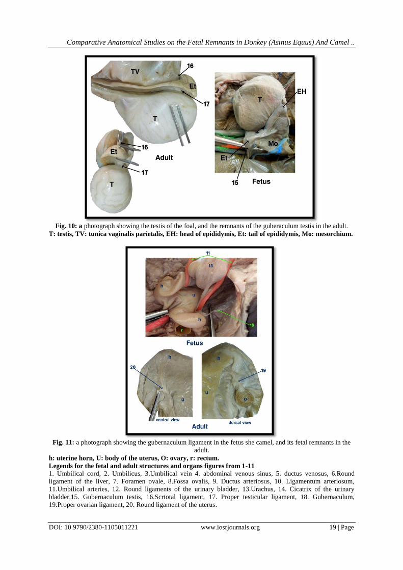
scrotum. The gubernaculum testis in fetus donkey (fig. 10/15) persisted as the scrotal ligament (fig. 10/16) and
proper testicular ligament (fig. 10/17). These ligaments secured the testis to the most inferior portion of the
scrotum. The scrotal ligament was attached at one end to the caudal end of the epididymis and at its other end to
the bottom of the scrotum. The proper testicular ligament connected between the caudal end of the testis and the
tail of epididymis. While in females, the gubernaculum in fetus she camel (fig. 11/18) had two vestigial
remnants, the proper ovarian ligament and the round ligament of the uterus (ligamentum teres uteri) (fig. 11/19,
20). The proper ovarian was connected between the uterine extremity of the ovary with cranial end of the
uterine horn. The round ligament appeared as round appendix and extended between the cranial extremity of
the uterine horn and the abdominal inguinal ring.
Fig. 1: a photograph showing the contents of the umbilical cord of the camel calf.

Comparative Anatomical Studies on the Fetal Remnants in Donkey (Asinus Equus) And Camel ..
DOI: 10.9790/2380-1105011221 www.iosrjournals.org 15 | Page
Fig. 2: a photograph showing the contents of the umbilical cord in the foal (left side).
Fig. 3: a photograph showing the umbilical vein in the foal and its remnant in the adult, ventral view.

Comparative Anatomical Studies on the Fetal Remnants in Donkey (Asinus Equus) And Camel ..
DOI: 10.9790/2380-1105011221 www.iosrjournals.org 16 | Page
Fig.4: a photograph showing the visceral surface of the fetal liver and the distribution of the umbilical vein in
foal, visceral view.
Fig. 5: a photograph showing the umbilical veins and the abdominal venous sinus upon entering the visceral
surface of liver in the camel calf, (visceral side).

Comparative Anatomical Studies on the Fetal Remnants in Donkey (Asinus Equus) And Camel ..
DOI: 10.9790/2380-1105011221 www.iosrjournals.org 17 | Page
Fig. 6: a photograph showing the distribution of the abdominal venous sinus inside the liver of the camel calf
and the site of the ductus venosus, (visceral view).
Fig. 7: a photograph showing the ductus arteriosus and foramen ovale in foal and their fetal remnants in the
adult.

Comparative Anatomical Studies on the Fetal Remnants in Donkey (Asinus Equus) And Camel ..
DOI: 10.9790/2380-1105011221 www.iosrjournals.org 18 | Page
Fig. 8: a photograph showing the umbilical arteries and the urachus in the pelvic cavity of the foal and their fetal
remnants in the adult.
Fig. 9: a photograph showing the origin of the umbilical arteries directly from the abdominal aortal in the camel
calf, ventral view.
Comparative Anatomical Studies on the Fetal Remnants in Donkey (Asinus Equus) And Camel ..
DOI: 10.9790/2380-1105011221 www.iosrjournals.org 19 | Page
Fig. 10: a photograph showing the testis of the foal, and the remnants of the guberaculum testis in the adult.
T: testis, TV: tunica vaginalis parietalis, EH: head of epididymis, Et: tail of epididymis, Mo: mesorchium.
Fig. 11: a photograph showing the gubernaculum ligament in the fetus she camel, and its fetal remnants in the
adult.
h: uterine horn, U: body of the uterus, O: ovary, r: rectum.
Legends for the fetal and adult structures and organs figures from 1-11
1. Umbilical cord, 2. Umbilicus, 3.Umbilical vein 4. abdominal venous sinus, 5. ductus venosus, 6.Round
ligament of the liver, 7. Foramen ovale, 8.Fossa ovalis, 9. Ductus arteriosus, 10. Ligamentum arteriosum,
11.Umbilical arteries, 12. Round ligaments of the urinary bladder, 13.Urachus, 14. Cicatrix of the urinary
bladder,15. Gubernaculum testis, 16.Scrtotal ligament, 17. Proper testicular ligament, 18. Gubernaculum,
19.Proper ovarian ligament, 20. Round ligament of the uterus.

Comparative Anatomical Studies on the Fetal Remnants in Donkey (Asinus Equus) And Camel ..
DOI: 10.9790/2380-1105011221 www.iosrjournals.org 20 | Page
K: kidneys, T: testis Mo: mesorchium, S: stomach , SP: spleen C: caudate lobe of liver , R: right lobe of liver
(donkey), RL: right lateral lobe of liver (camel) , RM: right medial lobe of liver (camel) , Q: quadrate lobe of
liver, LL: left lateral lobe of liver , LM: left medial lobe of liver. P: papillary process, Ca: caudal vena cava,
HV: hepatic veins, DA: descending aorta. Pt: pulmonary trunk. UB: urinary bladder.
IV. Discussion The present study recorded that the umbilical cord was a cylindrical helical structure connected
between the fetus and the placenta. In accordance to (Spurway, et al. 2012; Hong et al., 1993; Whitwell, 1975)
in horse, the current work showed that it contained four structures in donkey; the umbilical vein, two umbilical
arteries and the urachus. While in camel, it contained the urachus in between two umbilical arteries and
accompanied by two umbilical veins. This was similar to that reported by Bashir, 2010 in dromedary camel,
Tibary (1997) in the Bactrian camel and Benirschke and Miller (1982) in some wild animals like the African
lion and Speke’s Gazalle.
In the present findings, the fetal circulation begins from the placenta and enters the fetus via the
umbilical vein that located in the umbilical cord. The blood in umbilical vein passed to the fetal liver. Most of
blood in camel continues via the ductus venosus into the caudal vena cava and finally into the right atrium, then
blood was returned to the placenta through two large umbilical arteries, in agreement with (Bashir, 2010 in
camel; Kent and Carr, (2001) in horse and Getty, (1975) in donkey.
Two umbilical veins pass through most of the length of the umbilical cord in mammals. Our
observations revealed that it was a single, slender vessel in donkey, formed by the fusion of two veins within the
amniotic part of the cord as that reported by (McGeady et al., 2006 and Sisson, 1953) in the horse and pig.
Moreover, the current findings noted in camel that the two umbilical veins were merged together near the
abdominal wall forming an abdominal venous sinus as those reported by (Bashir, 2010; Mohammed
2008;Carlson, 1981and Getty, 1975). However, (Noden and de Lahunta, 1985) in carnivores and ruminants,
stated that two umbilical veins were joined to form the left umbilical vein before entering the body of the
embryo.
In agreement with (Bashir, 2010; Carlson, 1981 and Getty, 1975), the present study reported that the
venous sinus terminated as the ductus venosus which was represented as a direct shunt in the form of an acute
angle, between the venous sinus and the caudal vena cava, located between the right lateral liver lobe and the
papillary process of the caudate lobe. The fetal camel, ox and dog had the ductus venosus; but no
ductus venosus was noticed in the donkey's fetus.
The present investigation was in accordance with (McGeady et al., 2006, Merkle and Gilkeson, 2005;
Carlson,1981), that the remnant of the umbilical vein in adult donkey was a cord structure termed the round
ligament of the liver. The latter ligament was accompanied by the falciform ligament, similar to that given by
(Getty, 1975). While in the adult ox, sheep and goat, the round ligaments were absent due to complete
degeneration (Getty, 1975; Nickel et al., 1973). In camel, our results explained that the round ligament of the
liver persisted. In addition, the ductus venosus was closed, after birth as ligamentum venosum, similar to that
presented by (Bashir, 2010 ; Merkle and Gilkeson, 2005 and Carlson 1981).
Regarding the foramen ovale and ductus arteriosus, the former was interatrial foramen connected
between the right and left atria. The latter shunted between the pulmonary trunk and the dorsal aorta. In adult,
these structures remained as the fossa ovalis and ligamentum arteriosum, respectively. Our statement was in line
with (Kiserud 2005; Merkle and Gilkeson, 2005; Zahaka and Patel, 2002, Kent and Carr, 2001, Carlson, 1981
and Sisson, 1953).
In the present result, the umbilical arteries were paired vessels in fetus donkey and camel, originated
from the abdominal aorta after the origin of the external iliac arteries, these finding was confirmed by (Bashir,
2010 and Smuts and Hout, 1987) but it a likely than that recorded by (Getty, 1975) in equine, bovine, sheep that
derived from the internal iliac artery.
In agreement to the current work, Postnatal, the umbilical arteries were obliterated and became the two
round ligaments of the urinary bladder that accompanied by two lateral long folds of parietal peritoneum
(lateral umbilical ligaments), these results are in agreement with (Bashir, 2010 ; McGeady, et al., 2006; Noden
and de Lahunta, 1985; Carlson, 1981; Getty,1975 and (Sisson, 1953).
The urachus was a single duct connected between the vertex of the fetal urinary bladder and
the allantois. It coursed in between the two umbilical arteries, after parturition, it became atrophied and the
lumen was obliterated forming a cicatrix on the bladder apex. These result were confirmed by (Sadler 1995 and
Getty, 1975).
According to our investigation, The paired gubernacula were fibrous cord extend from the tail of testis
to the peritoneal vaginal ring. While in females, the gubernaculum in fetus had two vestigial remnants, the
proper ovarian ligament and the round ligament of the uterus.A finding was like that by (Sadler 1995 and Getty,
1975).

Comparative Anatomical Studies on the Fetal Remnants in Donkey (Asinus Equus) And Camel ..
DOI: 10.9790/2380-1105011221 www.iosrjournals.org 21 | Page
V. Conclusion The use of the precise normal anatomical structure of the umbilical cord, it contents and the
understanding of the different fetal structures which undergo atrophy or regression in adult, will lead to
accurate diagnosis and proper surgical interference for treatment of the congenital abnormalities that occur after
birth.
Reference [1]. Adams S.B., Fessler J.F. (1987): Umbilical cord remnant infections in foals: 16 cases (1975-1985). J Am Vet Med Assoc. ,
190(3):316-8. [2]. Bashir S.O., (2010): Foetal circulation and ancillary sense organs of the Dromedary Camel: Architecture, Histogenesis and
Histochemical observations. M.V.SC. Degree, Department Of Anatomy, Faculty Of Veterinary Medicine, University Of Khartoum.
[3]. Baxter GM, Darien BJ And Wallace CE (1987): Persistent Urachal remnant causing intestinal strangulation in a cow. Journal Of the American Veterinary Medical Association 191, 555-558.
[4]. Benirschke, k. and Miller, C. J. (1982): Anatomical and functional differences in the placenta of primates. Biology of Reproduction
[5]. Carlson, B.M. (1981): The circulatory system in: Patten’s foundation of embryology. Fourth edition. Mcgraw-Hill Company. [6]. Getty, R. (1975): Sisson and Grossman’s Anatomy of the Domestic Animals. 5th edition. Volume І and П. W.B. Saunders
Company Philadelphia- London Toronto. [7]. Hong, C.B., Donahue, J.M., Giles, R.C, Petrites- Murphy, M.B., Poonacha, K.B., Tramontin, R.P., Tuttle, P.A. and Swercze, T.W.
(1993): Adenomatous hyperplasia of equine allantoic epithelium. Veterinary Patholology. 30:171-175.
[8]. Kent, G. C and Carr, R.K. (2001): Comparative Anatomy of the Vertebrates 9th edit. Page: 315-344 [9]. Kiserud T. (2005): Physiology of fetal circulation. Semin Fetal Neonatal Med., 10: 493–503.
[10]. Laverty PH And Salisbury SK (2002): Surgical Management of true patent urachus in a cat. Journal Of Small Animal Practice 43,
227- 229. [11]. Lojszczyk, Szczepaniak A, Smiech A And Wojnowski T (2010): Congenital Urachal diverticulum in dogs: a case report. Medycyna
Weterynaryjna 66(6),421- 424.
[12]. McGeady, T.A., Quinn, P.J., FitzPatrick, E.S. and Ryan, M.T. (2006): Cardiovascular system. In: Veterinary Embryology, Ed: T.A. McGeady, Blackwell Publishing, Oxford. pp 126-127.
[13]. Merkle and Gilkeson, (2005): Remnants of Fetal Circulation: Appearance on MDCT in Adults AJR, 185, (2): 545-549.
[14]. Morgan-Hughes GJ, Marshall AJ, Roobottom C.( 2003): Morphologic assessment of patent ductus arteriosus in adults using retrospectively ECG-gated multidetector CT. AJR ; 181:749–754
[15]. Mohammed, E. I. E. (2008): Morphology and histochemistry of the foetal membranes and placenta of the dromedary camel
(Camelus dromedarius). PhD. Thesis, University of Khartoum [16]. Nickel, R, Schummer, A. and Seiferl, E. (1973): The viscera of the domestic mammals. Translation and revision by W.O. Sack.
Verlag Paul Parey. Berlin, Hamburg.
[17]. Noden, D.M and de Lahunta, A (1985): Developmental mechanisms and malformations. In: The Embryology of Domestic Animals.
Williams and Wilkins. Baltimore. London. Los Angeles.
[18]. Sadler, T .W .(1995): Langman’s Medical Embryology 7th edition. Williams and Wilkins. A Waverly Company
[19]. Schlafer, D.H. (2004): The umbilical cord – lifeline to the outside world: structure, function, and pathology of the equine umbilical cord. In: Proceedings of A Workshop on the Equine Placenta, Eds: D. Powell D. Furry and G. Hale, Kentucky Agricultural
Experimental Station, Lexington. pp 92-99.
[20]. Sisson, S. (1953): The Anatomy of the Domestic Animals. Fourth edition. Philadelphia and London W. B. SAUNDERS COMPANY.
[21]. Smuts, M.S., and Benzuiden Hout , A. J. (1987). Anatomy of the Dromedary camel. Clarendon Press .Oxford.
[22]. Spurway, J., Logan, p. and Pak, S. (2012): The development, structure and blood flow within the umbilical cord with particular reference to the venous system AJUM 15 (3): 97-102
[23]. Tibary, A. (1997): Theriogenology in Camelidae . Anatomy, Physiology, Pathology Article Breeding. Abu Dhabi printing and
publishing company, Mina, United Arab Emirates. [24]. Whitwell, K.E. (1975): Morphology and pathology of the equine umbilical cord. Journal of Reproduction and Fertility. Supplement.
23: 599-603.
[25]. Whitwell, K.E. and Jeffcott, L.B. (1975): Morphological studies on the fetal membranes of the normal singleton foal at term. Res. vet. Sci. 19, 44-55.
[26]. Wilsher, S., Ousey, J.C. and Allen, W.R. (2009): Abnormal umbilical cord attachment sites in the mare: A review illustrated by
three case reports. Equine vet. J. 41, 930-939. [27]. Zahaka, KG and Patel, CR. (2002): Congenital defects. Fanaroff, AA and Martin, R.J (eds). Newnatal-perinatal Medicine. In:
Diseases of the Fetus and Infant. 7th ed. 1120-1139.St. Louis: Mosby.
Nora, A. shaker "Comparative Anatomical Studies on the Fetal Remnants in Donkey (Asinus
Equus) And Camel (Camelus Dromedarius)." IOSR Journal of Agriculture and Veterinary
Science (IOSR-JAVS) 11.5 (2018): 12-21.


![1 CHAPTER 1 The fetal circulation - John Wiley & Sons...in fetal sheep [5]. Umbilical venous blood passing through the ductus venosus into the inferior vena cava is preferentially](https://static.fdocuments.in/doc/165x107/5e54dd7197391d1eec3463a2/1-chapter-1-the-fetal-circulation-john-wiley-sons-in-fetal-sheep-5.jpg)